Vol 21 No. 1
Vol 21 No. 1
Vol 21 No. 1

Create successful ePaper yourself
Turn your PDF publications into a flip-book with our unique Google optimized e-Paper software.
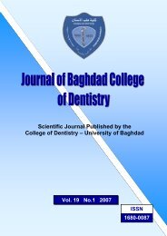
Scientific Journal Published by the<br />
College of Dentistry – University of Baghdad<br />
<strong>Vol</strong>. <strong>21</strong> <strong>No</strong>.1 2009<br />
ISSN<br />
1680-0087
A quarterly peer reviewed published scientific journal of the College of Dentistry,<br />
University of Baghdad.<br />
Editor in chief: Prof. Dr. Ali Hussain AlKhafaji B.D.S., M.Sc. D (UK)<br />
Vice editor in chief: Prof. Dr. Hussain Faisal Al-Huwaizi M.Sc., PhD<br />
Editorial Board:<br />
National Members<br />
Prof. Dr. Khalid Mirza<br />
Prof. Dr. Fakhri Al-Fatlawi MSc<br />
Prof. Dr. Athraa Yahiai MSc, PhD<br />
International Members<br />
Prof. J. L. Gutmann D.D.S., Ph.D.(USA)<br />
Prof. Dr. M. Goldberg PhD (France)<br />
Prof. Dr. Adel Farhan Ibraheem M.Sc.<br />
Prof. Dr. Khulood Al-Safi M.Sc. Ph.D.<br />
Assist. Prof. Dr. Amer Maki MSc<br />
Assist. Prof. Dr. Sabah Nema Ph.D.<br />
Assist. Prof. Dr. Wesal Al-Obaidi MSc<br />
Dr. Jamal Abid MSc<br />
Dr. Aeda Zaki MSc<br />
Board of editorial consultants:<br />
1- Prof. Dr. Wael Al-Aloosi MSc 9- Prof. Dr. Waleed Al-Hashemi MSc<br />
2- Prof. Sulafa Al-Samaria MSc, PhD 10- Prof. Abdullatif Al-Jubory PhD<br />
3- Prof. Dr. Ausama Al-Mulla PhD 11- Prof. Dr. Widad Al-Naqash MSc<br />
4- Prof. Dr. Raad Muhi AlDeen Helmi MSc 12- Prof. Dr. Ahlam Hameed MSc<br />
5- Prof. Nidhal Hussein MSc 13- Assist. Prof. Akram Faisal Al-Huwaizi MSc, PhD<br />
6- Prof. Nabeel Abdulfatah MSc 14- Assist. Prof. Jamal Aziz MSc<br />
7- Prof. Dr. Zainab Al-Dahan 15- Assist. Prof. Dr. Bashar Hamed MSc, PhD<br />
8- Prof. Riyad Al-Qaisi MSc, PhD 16- Assist. Prof. Kadim Al-Soudani MSc<br />
Computer executives: Lecturer Dr. Abdalbasit Ahmed<br />
Linguistic referee: Prof. Dr. Hussain Faisal Al-Huwaizi<br />
Administrative secretary: Hadeel Abdul Wahab.<br />
For consultation, please contact:<br />
Website: www.baghdentistry.com<br />
E-mail: baghdad_dentistry@yahoo.com<br />
Telephone: (+9641)4169375 Fax: (+9641)4140738<br />
i
Contents<br />
i<br />
ii<br />
vi<br />
Editor and Editorial Board<br />
Contents<br />
Instructions for the Authors<br />
Restorative Dentistry<br />
1<br />
5<br />
9<br />
15<br />
18<br />
24<br />
28<br />
33<br />
38<br />
41<br />
46<br />
The influence of posterior composite type and application technique on the fracture resistance of maxillary<br />
premolar teeth (an in vitro study). Shamma`a A. Sahib Al – Ansari, Ali H. Alkhafaji,<br />
Depth of cure evaluation of four different light-activated composites using different curing modes. Ali A.<br />
Razooki Al- Shekhli<br />
The effect of using different impression techniques and materials on vertical tissue displacement in free end<br />
extension ridges. (Dental survey and clinical study). Rawia. N. AL- Dafaii,, Amir. M. Khamas<br />
The effectiveness of carbide fissure bur in cutting dentin with light, moderate and heavy work load. Angham G.<br />
AL-Hashimi<br />
Effect of ozonated water on adherent Mutans Streptococci (In vitro study). Hasanain M Habeeb, Abbas S Al-<br />
Mizraqchi, Adel Farhan Ibraheem.<br />
The effect of dowel length on the retention of two different endodontic posts. Lamis A. Al- Taie,<br />
Assessment of consistency and compressive strength of manufactured dental base materials from enamel<br />
powder and synthetic hydroxyapatite with or without CO 2 laser treatment. Mohammed R. Al-Jabouri, Haitham<br />
J.Al- Aazawi, Hussein A.<br />
The effect of amalgam condensation techniques on the tensile bond strength using different dentin adhesives (in<br />
vitro study). Abdul Munaim S. AL-Khafaji<br />
The visible portion of upper anterior teeth at rest. Reem A. Al Obaidy<br />
Reliability of fovea palatinea in determining the posterior palatal seal. Yasmen Taha AL – Alousi<br />
Study the Microleakage of class II Composite Using Different Etching Techniques. Zainab M. Abdul-Ameer<br />
ii
Oral Diagnosis<br />
49<br />
Distribution and localization of ground substance of carbohydrate group in an inflammatory and phenytion<br />
induced gingival enlargement using histochemical method. Athraa Al Hijazi, Saif S. Saliem, Ali A. Abdulkareem<br />
53<br />
57<br />
60<br />
66<br />
70<br />
Oral findings and health status among elderly Iraqi patients, (aged 65 and above). Fawaz Al-Aswad<br />
Evaluation of Oral Hygiene, gingival health and dental knowledge among 4-12 years-old children attending<br />
the dental hospital. Eman K. Chaloob<br />
Serum and Salivary levels of proinflammatory cytokines as potential biomarkers in the diagnosis of oral<br />
squamous cell carcinoma. Nazar G. Al Talabani, Shanaz Mohammad Gaphor, Abdul-Wahab R. Hamad<br />
Assessment of Magnesium and Calcium Status in Oral Cancer Patients. Seta A.Sarkis, Suad AL-Ani,<br />
Marwan Al-Nimr<br />
The role of lipid peroxidation in the inducation and progression of chronic periodontitis. Taghreed F.<br />
Zaidan<br />
74<br />
Clinical observation of recurrent aphthous stomatitis in Sulaimania. Shanaz M. Gaphor, Shokhan A. Hussien<br />
Oral and Maxillofacial Surgery and Periodontology<br />
80<br />
Prevalence of dentine hypersensitivity in different age groups. Abdul-Karim Abd Ali Al- Muhammadawi<br />
84<br />
88<br />
91<br />
Prevalence and distribution of gingival recession and root caries in a group of dental patients in Ramadi city,<br />
Iraq. Raad S. Al– Ani, Ahmed M. Abdul- Razzak<br />
The effect of locally applied ciprofloxacin on the incidence rate of dry socket. Emad A. Salman, Jabbar J.<br />
Sabur<br />
Closed reduction for comminuted mandibular fractures. Thaer Abdul Lateef<br />
Orthodontics, Pedodontic, and Preventive Dentistry<br />
98<br />
104<br />
107<br />
Hypodontia in Down’s syndrome patients. Nidhal H. Ghaib, Mustafa M. Al-Khatieeb, Dheaa H. Abd Awn<br />
Malocclusion of Primary Dentition among kindergarten Children in Zayona Part of Baghdad City. Shahbaa<br />
A. M. Al- Ajwadi<br />
Mandibular antegonial notch depth distribution and its relationship with craniofacial morphology in<br />
different skeletal patterns. Yassir A. Yassir Ausama A. Al-Mulla<br />
iii
Instruction for the Authors<br />
The Journal of the College of Dentistry accepts manuscripts that address all topics related to<br />
dentistry. Manuscripts should be prepared in the following manner:<br />
Typescript. Type the manuscript on A4 white paper, with page setup of 2.5 cm margins. Type the<br />
manuscript with English language font Times New Roman and the sizes are as follows:<br />
1) Font size 18 and Bold for the title of the manuscript.<br />
2) Font size 14, Bold and capital letters for the headings as ABSTARCT, INTRODUCTION,<br />
MATERIALS AND METHODS, RESULTS and REFERENCES.<br />
3) Font size 12 Bold and italic for the names and addresses of the authors ex. Ahmed G. Husam<br />
4) Font size 11 for the legends of the tables and figures.<br />
5) Font size 10.5 for the text in the manuscript.<br />
6) Font size 10 for the text inside the tables.<br />
7) Font size 9 for the references at the end of the manuscript.<br />
Use single spacing throughout the manuscript and numbering of the pages should be in the lower<br />
right hand corner.<br />
Title of the manuscript:<br />
The title should be written with a capital letter for the first word as (Effect of the retention and<br />
stability….etc).<br />
Abstract and key words. The abstract should contain no more than 250 words. The abstract should be<br />
divided to the following categories: Background: (It contains a brief explanation about the problem<br />
for which the research was done as well as the aim of the study), Materials and methods:, Results:,<br />
and Conclusion:. Below the abstract, write 3-5 key words that refer as close as possible to the article.<br />
The abstract should be written by the font Century Gothic size 8.<br />
Text. The body of the manuscript should be divided into sections preceded by the appropriate major<br />
headings (INTRODUCTION, MATERIALS AND METHODS, RESULTS and REFERENCES)<br />
which are written in bold and capital. Minor headings should be typed in bold and subheadings should<br />
be not bold but underlined.<br />
References. References are placed in the text using the Vancouver system (Numbering system).<br />
Number references consecutively in the order in which they are first mentioned in the text. Identify<br />
references in the text, tables, and figures by Arabic numerals, and place them in parentheses within the<br />
sentence as superscription ex. (2) .<br />
Use the style of the examples given below in listing the references at the end of the manuscript :<br />
Book<br />
1. Hickey JC, Zarb GA, Bolender CL. Boucher’s prosthodontic treatment for edentulous patients. 9 th<br />
ed. St. Louis: CV Mosby; 1985. p.312-23.<br />
Journal article<br />
4. Jones ER, Smith IM, Doe JQ. Occlusion. J Prosthet Dent 1985; 53:120-9.<br />
Tables. All tables must have a title placed above the table. Identify tables with Arabic numbers (e.g.<br />
Table 1). The tables should be done with a width of no more than 8 cm.<br />
Figures and illustrations. All figures must have a title placed below the figure. Identify figures with<br />
Arabic numbers (e.g. Figure 1). The figures should be done with a width of no more than 8 cm.<br />
The article should not exceed 7 pages. The author should submit three copies of the article (one<br />
original and two copies) and a (CD) containing the article.<br />
iv
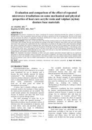
J Bagh College Dentistry <strong>Vol</strong>. <strong>21</strong>(1), 2009 The influence of posterior …..<br />
The influence of posterior composite type and application<br />
technique on the fracture resistance of maxillary premolar<br />
teeth (an in vitro study)<br />
Shamma`a A. Sahib Al - Ansari, BDS (1)<br />
Ali H. Alkhafaji, BDS. MSc. D (UK) (2)<br />
ABSTRACT<br />
Background: A restorative material with the potential to increase resistance to cuspal fracture is available to dentists<br />
and the packable composite is one of them. This in vitro study was conducted to evaluate and compare the cuspal<br />
fracture resistance of weakened maxillary premolar teeth with MOD preparations restored with different composite<br />
materials and techniques.<br />
Materials and Methods: fifty maxillary premolar teeth were divided into five groups (n=10). Class II MOD cavities were<br />
prepared in forty specimens. Group A, were sound. Group B were prepared but not restored. Group C and D were<br />
restored with successive cusp build up using Z250 microhybrid for group C and P60 packable for group D. Finally,<br />
Group E was restored with bulk using P60 packable. A 5 mm diameter steel sphere contacted the buccal and lingual<br />
cusps of the tested teeth until fracture occurred. The values obtained in this study were subjected to Analysis of<br />
Variance (ANOVA) and student t – test was carried out between the two types of posterior composite materials.<br />
Results: There's a high significant improvement of the fracture resistance of restored teeth using posterior composite<br />
as compared to the unrestored ones, but; there's no difference of the type of the posterior composite material used,<br />
or the type of placement technique used, while the sound teeth remained the strongest teeth compared with all the<br />
other groups.<br />
Conclusions: The study concluded that Posterior composite resin restoration whatever type or technique used,<br />
showed a great improvement in the resistance to cuspal fracture.<br />
Keywords: composite resin, successive cusp build up, fracture resistance. J Bagh Coll Dentistry 2009; <strong>21</strong>(1):1-4)<br />
INTRODUCTION<br />
Attention has focused on the strength of teeth<br />
after preparation for restorative treatment as it<br />
relates directly to their long – term longevity in<br />
the oral environment. Actually the use of adhesive<br />
materials to reinforce weakened teeth and support<br />
undermined enamel has been widely supported<br />
and many studies have shown that the weakening<br />
effect of cavity preparation can be alleviated with<br />
the use of such materials; the fracture resistance<br />
of teeth restored with adhesive materials is<br />
increased by 80 – 362% (!) . Packable composites<br />
are promoted for stress – bearing posterior<br />
restorations with improved handling properties<br />
and possible bulk curing of the restorations are<br />
some of the advantages (2) .<br />
One way to reduce the effect of contraction<br />
stress is the incremental layering of resin – based<br />
composites during placement to minimize<br />
bridging between cavity walls and to reduce<br />
shrinkage stresses through the sequential use of<br />
small volumes of material. But, the benefit of the<br />
incremental technique for reducing<br />
polymerization contraction stresses is somewhat<br />
controversial (3)<br />
(1) MSc Student, department of conservative dentistry, university<br />
of Baghdad.<br />
(2) Dean of the College of Dentistry. Baghdad University<br />
The aim of this study was to evaluate and<br />
compare the cuspal fracture resistance of<br />
weakened maxillary premolar teeth with MOD<br />
preparations restored with different posterior<br />
composite materials and techniques.<br />
MATERIALS AND METHODS<br />
Fifty maxillary premolar teeth removed for<br />
orthodontic purposes were collected immediately<br />
after extraction and placed in distilled water at<br />
room temperature before being evaluated for use<br />
in this study. All of the teeth selected were intact,<br />
noncarious, and unrestored. They were cleaned<br />
with pumice and examined under a magnifying<br />
lens to detect any pre-existing defects.<br />
To simulate the periodontium, root surfaces<br />
were dipped into melted sticky wax to a depth of<br />
2 mm below the facial CEJ junction to produce a<br />
0.2 to 0.3 mm layer approximately equal to the<br />
average thickness of the periodontal ligament.<br />
Teeth were then mounted in cold cure acrylic<br />
resin confined in a casting metal ring. Each tooth<br />
was removed from the resin when the<br />
polymerization was observed. The wax spacer<br />
was removed from the root surface and from the<br />
alveolus of the acrylic resin. Polyether (Impregum,<br />
ESPE, Germany) was delivered into the acrylic<br />
resin alveolus. The tooth was then reinserted into<br />
the test block, and the polyether material was<br />
allowed to set. Excess polyether material was<br />
removed to provide a flat surface 2 mm below the<br />
Restorative Dentistry 1
J Bagh College Dentistry <strong>Vol</strong>. <strong>21</strong>(1), 2009 The influence of posterior …..<br />
facial CEJ of each tooth. The thin layer of<br />
polyether material simulated the periodontal<br />
(4,<br />
ligament<br />
5) . Care was taken to prevent<br />
dehydration of the specimens. They were then<br />
stored in distilled water.<br />
The distance from the buccal cusp tip to the<br />
CEJ and the intercuspal distance on the occlusal<br />
surface of each tooth were measured using a<br />
vernier to standardize the cavity preparations.<br />
Class II MOD cavities were prepared in all<br />
specimens with parallel walls and no proximal<br />
boxes, except group A. The resulting isthmus<br />
width was 1/3 the intercuspal distance and 5mm<br />
from tip of facial cusp depth of the cavity. After<br />
preparation, the teeth were randomly divided into<br />
five groups (n=10). The teeth in group A were<br />
sound not prepared. Teeth in group B were<br />
prepared not restored. With exception of the<br />
composite type and placement technique,<br />
specimens in group C, D, and E were restored<br />
using the Adper Single Bond2 adhesive system<br />
and Z250 composite resin for group C and P60<br />
composite resin for group D and E (3M dental<br />
product-ESPE, USA) following the<br />
manufacturer’s recommendations. Ivory no.8<br />
retainer and a metal matrix band were placed on<br />
each specimen. Specimens in group C and D were<br />
restored with successive cusp build up (Figure 1),<br />
in this technique, the first composite increment is<br />
applied to a single dentin surface without<br />
contacting the opposing cavity walls and the<br />
restoration is build up by placing a series of<br />
wedge – shaped 1.5-mm, triangular apicoocclusal<br />
layers of uncured composite that are condensed<br />
and sculpted directly in the preparation using a<br />
composite instrument. Specimens in group E were<br />
restored in bulk technique (Figure 2) using P60<br />
packable, and Z350 flowable as initial layer. The<br />
specimens were stored for one week in distilled<br />
water in 100%relative humidity at 37°C, and the<br />
fracture test was conducted in an compressive<br />
testing machine (Leybold Harris, England 36110),<br />
A 5 mm diameter steel sphere contacted the<br />
buccal and lingual cusps of the tested teeth until<br />
fracture occurred. The fractured specimens in<br />
group C, D, and E were stained with 1%<br />
methylene – blue dye for 24 hours<br />
(6) .the<br />
specimens examined by stereo microscope ×40 so<br />
that the type of failure could be evaluated.<br />
RESULTS<br />
Data obtained by the fracture test for each of<br />
the studied treatments were submitted to ANOVA<br />
for a totally random design. The estimated F value<br />
was 17.83, showing a statistical significant<br />
difference (P < 0.01) among five estimations of<br />
means value (Table 1 and Figure 3). Student t –<br />
test was used to show comparison between the<br />
means of forces using one type of adhesive<br />
bonding agent (Adper Single Bond2) with two<br />
types of restorative composite materials, Z250<br />
microhybrid and P60 packable (considering group<br />
D and group E as one group). The results are<br />
presented in Table (2) which showed that there<br />
was a non significant difference (t = 0.30 P ><br />
0.05). The ANOVA test revealed that between<br />
group A (sound teeth) and group B (unrestored)<br />
there was a highly significant difference (P <<br />
0.01). Among group A and groups: C (Z250 /<br />
incremental), D (P60 / incremental), E (P60 /<br />
bulk), there was a significant differences (P <<br />
0.05). Moreover; among group B and groups: C,<br />
D, E, there was a highly significant differences (P<br />
0.05). The mode of failure<br />
observed after testing the specimens in<br />
compressive testing machine, using the<br />
stereomicroscope; are observed in the Table (3)<br />
for the fractured restored groups C, D, and E.<br />
DISCUSSION<br />
The restorative material is not only restoring<br />
the lost tooth structure, but also to strengthen the<br />
tooth and provide an effective seal between the<br />
restoration and the tooth. Comparing the results of<br />
fracture resistance to cusps statistically of this<br />
study revealed that the force required to fracture<br />
the cusps of group B (unrestored) (force<br />
mean=79.03Kgf) represents the lowest value<br />
among the five groups, while the restorative<br />
groups C (force mean=158.6Kgf), D (force<br />
mean=158.1Kgf) and E (force mean=167.7Kgf);<br />
their improvement in fracture resistance was<br />
statistically high significant. This result concluded<br />
that tooth reinforcement is another benefit of<br />
posterior bonded composite resin restorations.<br />
This high significant differences in fracture<br />
resistance between the unrestored and restored<br />
groups may be due to that the micro-mechanical<br />
bonding between bonding system and tooth<br />
structure tend to bind the walls of the cusps<br />
together and strengthen the remaining tooth<br />
structure and to distribute the forces more evenly<br />
among the various interfaces in composite<br />
restorative material that have been bonded to<br />
enamel and dentin by adhesive bonding agent.<br />
This reduction in localized forces offer greater<br />
opportunity for reinforcing tooth structure and<br />
increases the fracture resistance of the cusps. This<br />
result was in an agreement with (5, 7). The present<br />
study the statistical analysis of differences among<br />
group D (P60 packable / incremental) and group E<br />
Restorative Dentistry 2
J Bagh College Dentistry <strong>Vol</strong>. <strong>21</strong>(1), 2009 The influence of posterior …..<br />
(P60 packable / bulk) was statistically non<br />
significant, despite that there's an improvement in<br />
the fracture resistance force mean for group E(P60<br />
/ Bulk) , in contrast to that of group D (P60 /<br />
incremental) , even such improvement was not<br />
significant. Which revealed that the mode of<br />
application technique employed of the packable<br />
composite has no difference on the cuspal<br />
reinforcement of the weakened teeth to a cavity<br />
depth of 5mm? The presence of white lines<br />
between successive layers of resin composite or<br />
inside layers is an indication for improper<br />
adaptation of the layers with each other, which<br />
also affects physical properties. In Bulk build up a<br />
whole single increment was adapted properly to<br />
the floor and cavity walls, which eliminates the<br />
possible previous defects correlated to the<br />
composite successive increments. This<br />
explanation could be more accepted for the<br />
packable composite (10, 11). In our study it’s seemed<br />
that Z250 and P60 are sharing many of their<br />
physical properties and have closure values for<br />
each other including the modulus of elasticity (12) .<br />
Also using of the same bonding system, these<br />
factors for both posterior composite types are<br />
approximate from each other which may explain<br />
the comparable values of both materials of the<br />
cuspal deflection and therefore on the cuspal<br />
fracture resistance. The majority of cohesive<br />
failure for group D supported the explanation; that<br />
successive incremental technique with packable<br />
composite resulted in weakening of the flexural<br />
strength of the material itself and reduced the<br />
surface hardness as a result of dry spots and voids<br />
in between the resin layers coming from the<br />
improper adaptation of the heavy filled material.<br />
Table 1: Descriptive statistics of values of<br />
five groups.<br />
GA GB GC GD GE<br />
Mean (Kg f) 204.5 79.03 158.6 158.1 167.7<br />
S.D 39.01 35.51 42.48 27.82 22.86<br />
S.E 12.34 11.23 13.44 8.799 7.229<br />
C.V% 19.08 44.93 26.79 17.6 13.63<br />
Table 2: Mode of failure observed in the<br />
fractured restored specimens of groups C, D,<br />
and E.<br />
Groups Cohesive Adhesive Mixed<br />
Group C 1 3 6<br />
Group D 7 _ 3<br />
Group E 1 6 3<br />
A<br />
Figure 2: Bulk technique using P60 packable<br />
composite with flowable one<br />
250<br />
204,5<br />
200<br />
158,6 158,1<br />
167,7<br />
150<br />
Mean<br />
100<br />
79,03<br />
Figure 1: Successive cusp builds up<br />
technique; A group C using microhybrid<br />
Z250composite, B group D using packable<br />
P60 composite with flowable composite<br />
B<br />
50<br />
0<br />
GA GB GC GD GE<br />
Figure 3: Means of fracture forces values in<br />
(Kg f) for the five groups<br />
Restorative Dentistry 3
J Bagh College Dentistry <strong>Vol</strong>. <strong>21</strong>(1), 2009 The influence of posterior …..<br />
REFERENCES<br />
1. Mackenzie DF. The reinforcing effect of MOD acid–<br />
etch composite restoration on weakened posterior teeth.<br />
Br Dent J 1984; 161: 410–4.<br />
2. Fortin D, Vargas MA. The spectrum of composites: new<br />
technique and new materials. J Am Dent Assoc 2000;<br />
1(131): 26S – 30S.<br />
3. Ghavamnasiri M, Moosavi H, Tahvildarnejad N. Effect<br />
of centripetal and incremental methods in Class II<br />
composite resin restorations on gingival microleakage. J<br />
Contemp Dent Pract 2007; 2(8): 113–9.<br />
4. Sirimai S, Riis DN, Morgano SM. An in vitro study of<br />
the fracture resistance and the incidence of vertical root<br />
fracture of pulpless teeth restored with post and core<br />
systems. J Prosthet Dent 1999; 81(3): 262–9.<br />
5. Franca FG, Worschech CC, Paulillo AM, Martins LR,<br />
Lovadino JR. Fracture resistance of premolar teeth<br />
restored with different filling techniques. J Contemp<br />
Dent Pract 2005; 3(6): 85– 92.<br />
6. Gorgul G, Alacam T, Kivanc B.H, Uzun O, Tinaz C.<br />
Microleakage of packable composites used in post<br />
spaces condensed using different methods. J Contemp<br />
Dent Pract 2002; 2(3): 23–30.<br />
7. Salih M.M. An evaluation of the cuspal fracture<br />
resistance using different restorative materials and<br />
techniques with reinforcing effect of adhesive bonding<br />
system. A master thesis, Conservative Department,<br />
University of Baghdad, 2000.<br />
8. Giachetti L, Russo DS, Bambi C, Grandini R. A review<br />
of polymerization shrinkage stress: Current techniques<br />
for posterior direct resin restorations. J Contemp Dent<br />
Pract 2006; 4(7): 79– 87.<br />
9. Fano V, Ortalli I, Pozela K. Porosity in composite<br />
resins. J Am Dent Assoc 1995; 16: 1291– 5.<br />
10. Huysmans M, Varst P, Van de Lautenschlager EP,<br />
Monaghan P. The influence of simulated clinical<br />
handling on the flexural and compressive strength of<br />
posterior composite restorative materials. J Dent Mater<br />
1996; 12: 116–20.<br />
11. 3M Dental products; Filtek P60 technical manual.<br />
Minneapolis: 1999; 5–33.<br />
Restorative Dentistry 4
J Bagh College Dentistry <strong>Vol</strong>. <strong>21</strong>(1), 2009 Depth of cure evaluation…..<br />
Depth of cure evaluation of four different light-activated<br />
composites using different curing modes<br />
Ali A. Razooki Al- Shekhli B.D.S., M.Sc., Ph.D. (1)<br />
ABSTRACT<br />
Background: As light passes through the bulk of the composite material, the light intensity is greatly reduced due to<br />
light absorption and scattering by resin composites, thus, decreasing the effectiveness of cure through the depth of<br />
the composite layer. This study investigated the influence of different new curing modes (conventional and<br />
experimental) and composite formulations on depth of cure using the ISO scraping test.<br />
Materials and methods: This study investigated the depth of cure (ISO scraping method) of four conventional<br />
composites after exposure to different curing modes. A two-piece aluminum mold with a diameter of 4mm and a<br />
height of 8mm was used as a mold for the composite specimens.VIP light curing unit was used to deliver all the<br />
curing modes for photo-curing of all the composite specimens in this study. Parameters included six curing modes:<br />
Control (C), Pulse Delay I (PDI), Pulse Delay II (PDII), Soft-start (SS), Pulse Cure I (PCI), and Pulse Cure II (PCII) plus three<br />
experimental curing modes of higher energy density: Prolonged low-intensity pulse cure mode (PLPC), Prolonged<br />
moderate-intensity pulse cure mode (PMPC) and Rapid high-intensity continues cure mode (RHCC) for each of the<br />
four different light-activated composite materials (Tetric Ceram, Heliomolar, Herculite XRV and Degufill Mineral). The<br />
height of the cylinder of cured material was measured with a micrometer to an accuracy of 0.01 mm. This value was<br />
divided by two (in compliance with ISO CD4049: 2000), and recorded as the depth of cure in mm for that specific<br />
specimen.<br />
Results: Statistical analysis of the data by using the one-way analysis of variance revealed that, there is statistically<br />
very highly significant difference for all the depth of cures between the curing modes and composite types.<br />
Conclusion: This study indicated that, although, both curing mode and composite type significantly affect depth of<br />
cure but the effect of composite composition on the depth of cure is more predominant than that of curing mode.<br />
Key words: Resin composite, light curing modes, composite depth of cure. J Bagh Coll Dentistry 2009; <strong>21</strong>(1):5-8)<br />
INTRODUCTION<br />
A common problem associated with<br />
photocuring is that the amount of light available<br />
to excite the photoinitiator, dramatically decreases<br />
from the top surface inward as a result of light<br />
absorption and scattering (1) . This decrease in light<br />
intensity (attenuation) results in what is referred<br />
to as the “depth of cure” problem. Knowing the<br />
depth of cure of a particular shade of lightactivated<br />
composite material would guide dentists<br />
in regard to the thickness of a composite layer that<br />
could be adequately cured clinically and provide<br />
them with a valuable baseline information about<br />
the specific depth of cure of different lightactivated<br />
composite materials used by dentists.<br />
The ISO depth of cure (scraping) test ensured<br />
adequate polymerization of most resin-based<br />
composites (2) . The International Standardization<br />
Organization, or ISO (3) , standard for polymerbased<br />
filling materials requires resin-based<br />
composites to have a minimum depth of cure of<br />
1.5 millimeters when irradiated for the<br />
manufacturer’s recommended time. “Depth of<br />
cure” is defined in the specification as 50% of the<br />
length of the cured composite sample after the<br />
soft, uncured portion has been scraped away<br />
manually.<br />
(1) Assistant Professor, Department of Conservative Dentistry,<br />
Faculty of Dentistry, Ajman University of Science and<br />
Technology Network, UAE.<br />
The length of the cured portion is measured<br />
with a micrometer to an accuracy of 0.1 mm, this<br />
value is divided by two (in compliance with ISO<br />
CD4049: 2000), and recorded as the depth of<br />
cure, Morrow et al., (4) and Manhart et al., (5) used<br />
the same procedure. The objective of this research<br />
was to investigate the influence of different curing<br />
modes plus three experimental curing modes of<br />
high energy density on the depth of cure of four<br />
different light-activated composites.<br />
MATERIALS AND METHODS<br />
A light-curing unit with programmable time<br />
and intensity (variable intensity polymerizer)<br />
(VIP Light, Bisco Inc., Schaumburg, Ill.;<br />
Spectrum 800, Dentsply/Caulk, Milford, Del.)<br />
was used as the light curing unit for all curing<br />
procedures later on. A digital light meter<br />
(Coltolux) (Coltène/Whaledent.com, France) was<br />
used to measure the light intensity delivered from<br />
the curing tip. Four different light-activated resin<br />
composite materials of A2 Vita shade were<br />
selected: Tetric Ceram (Ivoclar, Vivadent AG FL-<br />
9494 Schaan/Liechtenstein.Lot: E58102),<br />
Heliomolar (Ivoclar, Vivadent AG FL-9494<br />
Schaan/Liechtenstein.Lot: C37535), Herculite<br />
XRV (sds Kerr, 1717 West Collins Orange, CA<br />
92867,<br />
Restorative Dentistry 15
J Bagh College Dentistry <strong>Vol</strong>. <strong>21</strong>(1), 2009 Depth of cure evaluation…..<br />
U.S.A. Lot: 205466.Item <strong>No</strong>.: 22860) and<br />
Degufill Mineral (Degussa-Hüls AG, Degussa<br />
Dental GmbH & Co. KG, Postfach 1364. D-<br />
63403 Hanau, Germany.Lot: 0885).<br />
For the preparation of a cylindrical specimen,<br />
a two-piece aluminum mold with a diameter of<br />
4mm and a height of 8mm (Iraqi construction)<br />
was used as a mold for the composite specimens.<br />
A Transparent celluloid strip band (Hawe-Neos<br />
Dental, CH-6925 Gentilino, Switzerland) was<br />
placed on a flat glass slide (Blue star glass<br />
industries, Delhi, India) (Blue star glass<br />
industries, Delhi, India) on top of a white filter<br />
paper (England) then, the mold was placed over<br />
the transparent celluloid-strip and slightly<br />
overfilled it in one increment with the composite<br />
materials being tested then, a second transparent<br />
celluloid-strip was placed on top of the mold and<br />
overlaid it with a cover slide, then a finger<br />
pressure was applied to the cover slide to extrude<br />
excess material. The exit window of the curing<br />
light was placed over the cover slide (the light tip<br />
in contact with the cover slide) and each<br />
composite material was irradiated, through the<br />
transparent celluloid strip and the cover slide,<br />
with the nine different curing modes (Table 1 &<br />
2).<br />
One hour after completing irradiation, the<br />
composite specimen was removed from the mold<br />
and the uncured material at the bottom of the<br />
sample, was removed by scraping it away<br />
manually with a plastic spatula. The height of the<br />
cylinder of cured material was measured with a<br />
micrometer (Hommel Werke, England) to an<br />
accuracy of 0.01 mm (Figure 1). This value was<br />
divided by two (in compliance with ISO CD4049:<br />
2000), and recorded it as the depth of cure.<br />
Mean and standard deviation were calculated<br />
for each specific depth of cure. The results were<br />
analyzed with one-way ANOVA and Least<br />
significant difference (LSD)-test at significance<br />
level 0.05.<br />
RESULTS<br />
Mean depth of cure in mm and standard<br />
deviation of the four different light-activated<br />
composites cured with the nine-different curing<br />
modes are listed in Table 3. Figure 2 summarizes<br />
mean depth of cure of the four different lightactivated<br />
composites cured with the nine-different<br />
curing modes. Statistical analysis of the data by<br />
using the one-way analysis of variance revealed<br />
that, there is statistically very highly significant<br />
difference for all the depth of cures between the<br />
curing modes and composite types. LSD-test of<br />
the depth of cure according to the composite type<br />
is summarized in Table 4.<br />
Table 1: The conventional light- curing<br />
Light-curing<br />
mode<br />
Control (CC)<br />
Pulse Delay I<br />
(PDI)<br />
Pulse Delay II<br />
(PDII)<br />
Soft-start (SS)<br />
Pulse Cure I<br />
(PCI)<br />
Pulse Cure II<br />
(PCII)<br />
modes (6) .<br />
Regimen<br />
400mW/cm2<br />
(40 seconds)<br />
100mW/cm2→Delay→<br />
500mW/cm2 (3 seconds)<br />
(3 minutes) (30 seconds)<br />
200mW/cm2→Delay→<br />
500mW/cm2 (20 seconds)<br />
(3 minutes) (30 seconds)<br />
200mW/cm2 → 600mW/cm2<br />
(10 seconds) (30 seconds)<br />
400 mW/cm2→Delay→ 400<br />
mW/cm2→<br />
Delay→ 400 mW/cm2 (10<br />
seconds)<br />
(10 seconds) (10 seconds)<br />
(10 seconds) (20 seconds)<br />
400 mW/cm2→Delay→ 400<br />
mW/cm2<br />
(20 seconds) (20 seconds) (20<br />
seconds)<br />
Table 2: The experimental light-curing<br />
modes used in this study (7) .<br />
Light-curing mode<br />
Prolonged lowintensity<br />
pulse cure mode<br />
(PLPC)<br />
Prolonged moderateintensity<br />
pulse cure mode<br />
(PMPC)<br />
Rapid high-intensity<br />
continues cure mode<br />
(RHCC)<br />
Regimen<br />
100mW/cm2 → Delay →<br />
300 mW/cm2<br />
(20 seconds)(10<br />
seconds)(120 seconds)<br />
100mW/cm2 → Delay →<br />
400 mW/cm2<br />
(20 seconds)(10 seconds)<br />
(90 seconds)<br />
600mW/cm2<br />
(60 seconds)<br />
Figure 1: The micrometer devise for<br />
measuring the depth of cure.<br />
Restorative Dentistry 6
J Bagh College Dentistry <strong>Vol</strong>. <strong>21</strong>(1), 2009 Depth of cure evaluation…..<br />
Table 3: Mean depth of cure in mm and standard<br />
deviation of the four different light-activated<br />
composites cured with the nine-different<br />
curing modes.<br />
Depth of cure in mm<br />
Curing<br />
Mode<br />
Tetric<br />
Ceram<br />
Helio<br />
molar<br />
Herculite<br />
XRV<br />
Degufill<br />
Mineral<br />
Control<br />
2.36 1.75 2.87<br />
(0.01) (0.02) (0.05)<br />
1.95 (0.03)<br />
PDI<br />
2.29 1.72 2.86 1.975<br />
(0.04) (0.04) (0.02) (0.03)<br />
PDII<br />
2.46<br />
2.85<br />
1.9 (0.04)<br />
(0.02)<br />
(0.04)<br />
2.01 (0.04)<br />
SS<br />
2.39 1.84 2.98<br />
(0.05) (0.06) (0.04)<br />
2.11 (0.05)<br />
PCI<br />
2.34 1.83 2.89<br />
(0.03) (0.06) (0.05)<br />
1.98 (0.06)<br />
PCII<br />
2.37 1.81 2.89<br />
(0.04) (0.06) (0.05)<br />
2.06 (0.03)<br />
PLPC<br />
2.47 1.87<br />
(0.05) (0.04)<br />
3.2 (0.05) 1.95 (0.04)<br />
PMPC<br />
2.52<br />
3.25<br />
1.9 (0.03)<br />
(0.03)<br />
(0.05)<br />
2.08 (0.04)<br />
RHCC<br />
2.28 1.77 3.<strong>21</strong><br />
(0.03) (0.03) (0.07)<br />
1.9 (0.06)<br />
Standard deviation in parentheses.<br />
DISCUSSION<br />
In this study, although both curing mode and<br />
composite type significantly affect depth of cure but<br />
Figure 4 demonstrated that, the effect of composite<br />
composition on the depth of cure is much more than<br />
that of curing mode and this is due to the fact that, the<br />
most important factors affecting the polymerization<br />
depth are the composition and the physical<br />
properties of the composite resins and not the<br />
energy density and this finding is in agreement<br />
with DeBacker & Dermaut (8) . Herculite XRV<br />
light-activated composite exhibited the highest<br />
depth of cure values for all the nine curing modes<br />
(Figure 2) followed by Tetric Ceram, Degufill<br />
Mineral, and Heliomolar, which exhibited the<br />
lowest depth of cure values. Depth of cure of light<br />
activated resin-based composites is a function of<br />
the material’s filler composition and resin<br />
chemistry, its shade and translucency, the intensity<br />
of the light source, and the length of the radiation<br />
exposure (9) . The data of this study indicated that,<br />
microhybrid resin-based composite had the<br />
greatest depth of cure because of their high filler<br />
loading (79% by weight) and relatively large<br />
Depth of cure in mm<br />
3.5<br />
3<br />
2.5<br />
2<br />
1.5<br />
Tetric Heliomolar Herculite Degufill<br />
Composite type<br />
Control<br />
PDI<br />
PDII<br />
SS<br />
PCI<br />
PCII<br />
PLPC<br />
PMPC<br />
RHCC<br />
Figure 2: Mean depth of cure in mm of the<br />
four different light-activated composites cured<br />
with the nine-different curing modes<br />
according to the composite type.<br />
Table 4: LSD-test of the depth of cure<br />
according to the composite type.<br />
TetricTetric Tetric Helio Helio<br />
molar molar<br />
Herculite<br />
Helio<br />
HerculiteDegufillHerculite Degufill Degufill<br />
molar<br />
C *** *** *** *** *** ***<br />
PDI *** *** *** *** *** ***<br />
PDII *** *** *** *** *** ***<br />
SS *** *** *** *** *** ***<br />
PCI *** *** *** *** *** ***<br />
PCII *** *** *** *** *** ***<br />
PLPC*** *** *** *** ** ***<br />
PMPC*** *** *** *** *** ***<br />
RHCC*** *** *** *** *** ***<br />
** : Highly significant difference<br />
*** : Very highly significant difference<br />
average particle size (0.6-0.7 micron) while for the<br />
microfills (66.7% by weight) for the filler loading<br />
and (0.04 micron) for the average particle size and<br />
in this study, Herculite XRV and Tetric Ceram<br />
composites are micro-hybrids and both of them<br />
exhibited high depth of cure values in comparison<br />
with Heliomolar composite which is a microfilled<br />
composite and this finding is in agreement with the<br />
findings of Jain & Pershing (10) . The findings of this<br />
study, is not in agreement with the findings of Jain<br />
& Pershing (10) in that, greater irradiance (energy<br />
density) or longer exposure times are needed to cure<br />
small particle resin-based composites in an attempt<br />
to increase their depth of cure because in this study,<br />
the experimental curing modes of high energy<br />
density did not greatly increase the depth of cure<br />
especially with Heliomolar microfilled lightactivated<br />
composite (Figure 2).<br />
Restorative Dentistry 7
J Bagh College Dentistry <strong>Vol</strong>. <strong>21</strong>(1), 2009 Depth of cure evaluation…..<br />
REFERENCES<br />
1. Myers ML, Caughman WF, Rueggeberg FA.<br />
Effect of restoration composition, shade, and thickness on<br />
the cure of a photoactivated resin cement. J Prosthodont<br />
1994; 3:149-57.<br />
2. Cook WD, Standish PM. Cure of resin based<br />
restorative materials, II: white light photopolymerized<br />
resins. Aust Dent J 1983; 28: 307-11.<br />
3. International Organization for Standardization:<br />
ISO 4049:2000: Dentistry-polymer-based filling,<br />
restorative and luting materials. 3 rd ed. Geneva,<br />
Switzerland: International Organization for<br />
Standardization; 2000.<br />
4. Morrow L, Wilson NH, Setcos JC. Single-use,<br />
disposable, presterilized light activation probe: the future?<br />
Quintessence Int 1998; 29(12): 781-5.<br />
5. Manhart J, Chen HY, Hickel R. The suitability of<br />
packable resin-based composites for posterior<br />
restorations. J Am Dent Assoc 2001: 132(5): 639-45.<br />
6. Yap AUJ, Soh MS, Siow KS. Effectiveness of<br />
composite cure with pulse activation and soft-start<br />
polymerization. Oper Dent 2002; 27:44-9.<br />
7. Al-Shekhli AA, Al- Azzawi HJ, Al-Aubi IA.<br />
Effctiveness of four different light-activated composites<br />
cure with different light energy densities. Mustansiria<br />
Dental Journal 2006; 3(3):224-9.<br />
8. DeBacker J, Dermaut L. Visible light sources and<br />
posterior visible light cured resins: a particle mixture.<br />
Quintessence Int 1986; 17: 635-41.<br />
9. Rueggeberg FA, Caughman WF, Curtis JW &<br />
Davis HC. A predictive model for the polymerization of<br />
photo-activated resin composites. Int J Prosthodont 1994;<br />
7: 159-66.<br />
10. Jain P, Pershing A. Depth of cure and<br />
microleakage with high-intensity and ramped resin-based<br />
composite curing lights. J Am Dent Assoc 2003; 134 (9):<br />
1<strong>21</strong>5-23.<br />
Restorative Dentistry 8
J Bagh College Dentistry <strong>Vol</strong>. <strong>21</strong>(1), 2009 The effect of using different …..<br />
The effect of using different impression techniques and<br />
materials on vertical tissue displacement in free end<br />
extension ridges. (Dental survey and clinical study)<br />
Rawia. N. AL- Dafaii, B.D.S., MSc. (1)<br />
Amir. M. Khamas, B.D.S., M.S.D. ( 2)<br />
ABSTRACT<br />
Background: Various impression techniques used in the construction of free- end extension partial denture are based<br />
on theories relative to the compressibility and behaviour of the soft tissue during impression making and during<br />
function, the aim of the study was to measure vertical tissue displacement during impression making for free- and<br />
extension using different impression technique and materials.<br />
Material and methods: the study consisted of two parts (question are and clinical) the questionnaire was circulated<br />
among (90) dentists at different working places, to see which impression technique they use in free- and extension. In<br />
the clinical part (24) mandibular distal extension impression were made using three impression techniques and<br />
materials (special tray with alginate, special tray with polyether and double tray with zinc- oxide eugenol impression<br />
paste). The amount of vertical tissue displacement was measured on stone cast using (dial indicator dimension).<br />
Results: The results of the survey showed that (44%) of the dentist tend to use special tray with alginate, and (38.1%) of<br />
prosthodontist use double tray impression, the clinical part showed that there was significant difference between the<br />
impression techniques.<br />
Conclusions: The most popular impression technique used is special tray with alginate, which showed more tissue<br />
displacement in the clinical part of the study, while the double tray impression technique showed the least<br />
displacement.<br />
Keyword: Tissue displacement, free- end extension, impression technique. J Bagh Coll Dentistry 2009; <strong>21</strong>(1):9-14)<br />
INTRODUCITON<br />
The major problem that may face the dentist<br />
during making impression is the soft tissue<br />
displacement, since the oral tissue is of varying<br />
degree of displacability according to their health<br />
and support. To provide physiologically and<br />
mechanically acceptable function, it is<br />
fundamental that the soft tissue must be copied<br />
with out distortion to prevent impingement and<br />
injury by the prosthesis (1) . In the impressionmaking<br />
procedure for free-end extension<br />
removable partial denture, the pressure produced<br />
at eh interface of he soft mucosal tissue and the<br />
impression is the most important factor for the<br />
stability of the saddle under functional load (2) . To<br />
achieve an impression with minimum tissue<br />
displacement, soft tissue displacement,<br />
impression concepts, impression techniques, and<br />
impression materials are to be taken in<br />
consideration, studies were carried out to<br />
evaluate and measure tissue displacement of freeend<br />
extension during impression making (3-5) . One<br />
study showed that the space between the inner<br />
surface of the tray and mucosal tissue, the flow<br />
of the impression material, and the size of the<br />
tray are interrelated with each other (6) .<br />
(1) Private practice<br />
(2) Assistant professor, Department of Prosthodontics, College<br />
of Dentistry, University of Baghdad.<br />
The tissue displacement can be under control<br />
according to the impression theory used<br />
(mucostatic mucofuncational, selective<br />
pressure) (7-9) . The application of these theories is<br />
though single impression, sectional impression<br />
and special try impression technique (10,11) . In the<br />
presence of all these factors (impression theories,<br />
impression materials, and impression<br />
techniques), this study was designed to find out<br />
which impression techniques and material is<br />
mostly used by dentist as first part, and the<br />
second part of the study was to measure the<br />
amount of vertical tissue displacement of distally<br />
extended ridges made by three impression<br />
techniques and materials and correlate it with the<br />
survey results, to reach a conclusion and suggest<br />
an impression technique, that cause least amount<br />
of distortion.<br />
MATERIAL AND METHODS<br />
Survey part: A questionnaire was distributed<br />
among 90 dentist (general, specialist and<br />
prosthodontist) at different working place<br />
(ministry of health, dental college, and private<br />
clinic), to find out which impression technique<br />
and material they use for free- extension lower<br />
partial denture. The results were analyzed to be<br />
correlated with the results of the clinical part.<br />
Restorative Dentistry 9
J Bagh College Dentistry <strong>Vol</strong>. <strong>21</strong>(1), 2009 The effect of using different …..<br />
Clinical part: The clinical part of the study was<br />
carried out on 15 patients with total of 24<br />
mandibular free-end extension ridges with age<br />
range (24-45) years, attending the prosthodontic<br />
clinic, college of dentistry, university of<br />
Baghdad. All patients have healthy natural teeth,<br />
free from periodontal involvement with last<br />
abutment as first or second premolar; the saddle<br />
area was healthy with well attached mucosa and<br />
no previous prosthesis. Three impressions were<br />
made for each patient; the impression technique<br />
and material was special tray with alginate,<br />
special tray with polyether and double tray<br />
impression (zinc oxide-eugenol paste and<br />
alginate). The special tray with alginate<br />
impression was secured with a special perforated<br />
tray constructed from autopolymersing acrylic<br />
resin form preliminary cast with wax spacer and<br />
stopper over the anterior teeth and residual ridge<br />
for proper seating of the tray and to control the<br />
pressure during the impression. The alginate<br />
(cavex-Holland) was mixed according to<br />
manufacture instruction, the impression obtained<br />
was wax boxed a poured figure 1. As for the<br />
double tray impression technique, an auto<br />
polymerising custom tray with one layer of base<br />
plate wax as spacer was constructed over the<br />
edentulous area excluding the teeth, but with<br />
occlusal stopper on last abutment to control the<br />
pressure from a preliminary impression. Border<br />
molding was made in the usual manner and<br />
impression for the edentulous area was made<br />
using zinc oxide- eugenol (S.S white group<br />
England), after setting the impression was<br />
removed and inspected, then it was reseated in<br />
patient mouth with a suitable size stock tray<br />
loaded with alginate an overall impression was<br />
made for the teeth and removed picking with it<br />
the zinc oxide- eugenol impression tray, so that<br />
the final impression is made two sections, the<br />
teeth with alginate and the edentulous area with<br />
zinc oxide- eugenol paste as shown in figure 2.<br />
Finally an impression with polyether impression<br />
material (medium viscosity, impregum,<br />
Germany), using perforated spaced special tray<br />
as shown in figure 3.<br />
Measurement and data collection: To measure<br />
and compare the amount of vertical tissue<br />
displacement of three casts for each patient, a<br />
measuring machine with measuring accuracies<br />
(0.001mm), (Mitutoyo corporation, Tokyo-<br />
Japan), was used. To ensure the same relation<br />
parallism of each cast to the measuring machine,<br />
an autopolymerising acrylic plate was<br />
constructed to the height of the occlusal surface<br />
and incisal edge of the remaining teeth. The cast<br />
and palate was placed on surveyor table with the<br />
indicator of the measuring machine touching on<br />
three selected points on the acrylic plate as<br />
shown in figure 4. Measurements of vertical<br />
tissue displacement were made on the cast at<br />
three selected points (A, B, C) which represent<br />
approximately the area of missing (first<br />
premolar, first and second molar). The occlusal<br />
surface of the last abutment was used as<br />
reference point, as shown in figures 5, 6.<br />
RESULTS<br />
Survey results: The statistical analysis of the<br />
survey results showed that a high percentage of<br />
dentists (44.44%) used special tray with alginate<br />
impression material, while 15.56% prefer the use<br />
of double tray impression as shown in table 1.<br />
According to working places, dentist in ministry<br />
of health showed high percentage (50%) in favor<br />
of special tray with alginate and 12.5% used<br />
double tray impression, while in university<br />
35.71% used special tray with alginate and<br />
14.29% used double tray impression. Finally<br />
private clinic dentist 46.67% used special tray<br />
with alginate, and 20% double tray impression as<br />
shown in table 2. According to specialty 50% of<br />
the general dentists prefer to use special tray with<br />
alginate, while 16.67% used double tray<br />
impression, other specialists (51.28%) used<br />
special tray with alginate, and 2.56% sued double<br />
tray impression, as for prosthodontists 23.8%<br />
used special tray with alginate and 38.1% used<br />
double tray impression as shown in table 3.<br />
Clinical results: The mean, standard deviation,<br />
standard error, and coefficient of variation for<br />
different impression techniques are shown in<br />
table 3 for the points (A,B,C), it revealed that the<br />
use of polyether impression material with special<br />
tray gave the least amount of displacement<br />
compared with double tray impression and<br />
special tray with alginate. Table 5 show that the<br />
special tray with alginate. The compression<br />
between double tray impression and single tray<br />
impression are shown in table 6, there were no<br />
significant differences in the amount of tissue<br />
displacement at points (A,B,C) between double<br />
tray impression and special tray with alginate.<br />
The compression of different impression<br />
technique and materials at each selected points<br />
are shown in table 7, which revealed that there<br />
was no significant difference in the amount of<br />
tissue displacement at points (A,B) a significant<br />
difference existed at point ©, and special tray<br />
with polyether and double tray causes less<br />
displacement that special tray with alginate.<br />
Restorative Dentistry 10
J Bagh College Dentistry <strong>Vol</strong>. <strong>21</strong>(1), 2009 The effect of using different …..<br />
DISCUSSION<br />
The results of the survey showed that dentist<br />
in working places used different impression<br />
techniques. Most dentist in the ministry of health<br />
and private practice used the special tray with<br />
alginate impression material, this may be because<br />
of its easy manipulation, relativity inexpensive,<br />
time saving and comfortable to the patient, while<br />
he result of the clinical part of the study showed<br />
that the sue of special tray with alginate<br />
impression material caused more soft tissue<br />
displacement when compared with the polyether<br />
with special tray and double tray techniques.<br />
Dentist in the dental school used all types of<br />
impression techniques; this may be due to that<br />
the dental school as teaching institute is<br />
responsible of teaching student all concepts and<br />
impression techniques.<br />
According to dentist specialty, half of the<br />
general dentist used the special tray with alginate<br />
and the vest uses the other techniques, while<br />
dentist with other specialties other than<br />
prosthodontist use special tray with alginate, in<br />
addition to the use of polyether with special tray,<br />
which indicate that specialist tend to use<br />
techniques that causes less tissue displacement<br />
prosthodontist prefer the use of double tray<br />
impression which according to the clinical results<br />
showed that least tissue displacement equally<br />
with the polyether, this is because prosthodontist<br />
are deeply involved in their filed in problems of<br />
support, retention, resorption and tissue<br />
displacement. There was as significant difference<br />
in the amount of tissue displacement between<br />
different impression techniques and materials at<br />
point © which represent the area of the first<br />
molar, may be extracted at different time during<br />
life resulting in variable amount of bone loss at<br />
that area. The results also showed that there was<br />
significant difference between double tray<br />
impression (less displacement at point ©) when<br />
com pared with special tray with alginate, this<br />
may be explained that the flow of impression<br />
material influence tissue displacement and the<br />
use of easy flowing zinc oxide- eugenol<br />
impression paste recorded the soft tissue at time<br />
of set gave better results when compared with<br />
alginate which is effected by may factors such as<br />
water/ powder ratio, humidity, temperature,<br />
mixing time, in addition to the syneresis and<br />
imbibitions that may cause poor dimensional<br />
stability. These results agreed with Leupold (3) ,<br />
James (12) , and Holmes (13) , who all demonstrated<br />
that alginate impression with special tray causes<br />
more tissue displacement, but disagree with<br />
Wang (14) who believed that it is comparable to<br />
double tray. As for the points (B and C) the<br />
results showed that there was significant<br />
difference between the special tray with alginate<br />
and special tray with polyether and the later<br />
showed least amount of tissue displacement. The<br />
results of tissue displacement at points (A,B,C)<br />
showed no significant difference between special<br />
tray with polyether and double tray techniques,<br />
this results agreed with EL- Shilich and adbdel<br />
hakim (15) , AL- Judy, AL-Obaidi (17) , and Frant (18) .<br />
.<br />
Figure 1: Special tray with alginate impression<br />
material<br />
Figure 2: Double tray impression (zinc oxideeugenol<br />
paste and alginate impression<br />
material)<br />
Restorative Dentistry 11
J Bagh College Dentistry <strong>Vol</strong>. <strong>21</strong>(1), 2009 The effect of using different …..<br />
Figure 3: Special tray with polyether<br />
impression material<br />
Figure 4: Ensure the parallism between the<br />
cast and measuring apparatus<br />
Figure 5: A vertical sliding pointed tool<br />
descending on the occlusal surface of the last<br />
tooth to be used as a reference point<br />
Figure 6: Selection of three measurement<br />
points (A, B and C)<br />
Table 1: Questionnaire results according to the preference of the dentists.<br />
Impression techniques <strong>No</strong>. of dentist %<br />
Stock tray with alginate 16 17.78<br />
Special tray with alginate 40 44.44<br />
Double tray 14 15.56<br />
Special tray with rubber base 20 22.22<br />
Table 2: Number and percentage % of dentists in different working places with their choices of<br />
impression techniques.<br />
Impression techniques<br />
Ministry of health (32) University (28) Private clinic(30)<br />
<strong>No</strong>. of dentist % <strong>No</strong>. of dentist % <strong>No</strong>. of dentist %<br />
Stock tray with alginate 6 18.75 5 17.86 5 16.67<br />
Special tray with alginate 16 50 10 35.71 14 46.67<br />
Double tray 4 12.5 4 14.29 6 20<br />
Special tray with rubber base 6 18.75 9 32.14 5 16.67<br />
Restorative Dentistry 12
J Bagh College Dentistry <strong>Vol</strong>. <strong>21</strong>(1), 2009 The effect of using different …..<br />
Table 3: Number and percentage % of dentists with different specialties and their choices of<br />
impression techniques.<br />
Impression techniques<br />
General dentist (30) Specialist (39) Prosthodontist(<strong>21</strong>)<br />
<strong>No</strong>. of dentist % <strong>No</strong>. of dentist % <strong>No</strong>. of dentist %<br />
Stock tray with alginate 5 16.67 7 17.95 3 14.29<br />
Special tray with alginate 15 50 20 51.28 5 23.81<br />
Double tray 5 16.67 1 2.56 8 38.1<br />
Special tray with rubber base 5 16.67 11 28.<strong>21</strong> 5 23.81<br />
Table 4: Mean, standard deviation, standard errors and coefficient of variation of displacement<br />
for the different impression techniques and materials at each measured points.<br />
Impression techniques and materials<br />
Statistic<br />
Special tray with alginate<br />
impression material<br />
Double tray<br />
impression<br />
Special tray with polyether<br />
impression material<br />
Point A Point B Point C<br />
Point Point Point<br />
A B C<br />
Point A Point B Point C<br />
Mean 8.311 9.077 8.491 8.063 8.766 8.025 8.132 8.674 7.885<br />
SD 1.786 1.706 1.627 1.814 1.653 1.581 2.010 2.042 2.124<br />
SE 0.364 0.348 0.332 0.370 0.337 0.322 0.410 0.416 0.433<br />
CV% <strong>21</strong>.49 18.795 19.161 22.498 18.857 19.7 24.7 23.5 26.94<br />
Table 5: t-test of displacement for two impression materials [special tray with alginate and<br />
special tray with polyether]<br />
Special tray with alginate and<br />
Point Statistical t-test P-value Sig.<br />
special tray with polyether<br />
A<br />
MD 0.179<br />
SD 0.643<br />
1.361 0.187 NS<br />
B<br />
MD 0.403<br />
SD 0.142<br />
2.832 0.009 S<br />
C<br />
MD 0.605<br />
SD 0.189<br />
3.336 0.003 S<br />
*P>0.05 <strong>No</strong>n significant<br />
**P0.05 <strong>No</strong>n significant<br />
**P0.05 NS<br />
B 2.604 0.084 P>0.05 NS<br />
C 3.388 0.042 P0.05 <strong>No</strong>n significant<br />
**P
J Bagh College Dentistry <strong>Vol</strong>. <strong>21</strong>(1), 2009 The effect of using different …..<br />
REFERENCES<br />
1. Denen HE. Impression for dentures. J Prosthet Dent<br />
1952; 2:737.<br />
2. Geng Q. Influence of various impression procedures<br />
for removable partial denture on displacement of soft<br />
mucosal tissue. J Prosthet Dent 1993; 60(1): 35-53.<br />
3. Leupold RJ. Comparison of vertical movement<br />
occurring during loading of distal- extension<br />
removable portical denture bases made by there<br />
impression techniques. J Prosthet Dent 1992; 68(2):<br />
290-9.<br />
4. Vahidi F. Vertical displacement of distal extensions<br />
ridges by different impression techniques. J Prosthet<br />
Dent 1978; (4):374-7.<br />
5. Sato M. Influence of various impression procedures<br />
on displacement of residual ridges. Kokubyo Gakkai<br />
Zasshi 1996; (4): 374-7.<br />
6. Sato M. Influence of various impression procedures<br />
for mandibular distal extension removable partial<br />
denture on displacement of residual ridges. Kokubyo<br />
Galkkai Zasshi 1996; 63(1): 88-107.<br />
7. Khamas AH. Impression material as related to tissue<br />
distortion. Thesis College of dentistry Alabama U.S.A<br />
1979.<br />
8. Jacobson TS, Krol AJ. A contemporary review of<br />
the factor involved in complete denture. Part III<br />
support. J Prosthet Dent 1983; 49(3): 306-13.<br />
9. Duncan JP, Raghavendas, Tylor T. A selective<br />
pressure impression technique for edentulous maxilla.<br />
J Prosthet Dent 2004; 92(3): 224.<br />
10. Rapuano JA. Single try dual- impression technique<br />
for distal extension partial dentures. J Prosthet Dent<br />
1970; 24(1): 41-5.<br />
11. Frank RP, Brudvik JS, <strong>No</strong>onan CJ. Clinical outcome<br />
of the altered cast impression procedure compared with<br />
the issue of a one piece cast. J Prosthet Dent 2004; 91:<br />
468-76.<br />
12. James JS. A simplified alternative to the alteredcast<br />
impression technique for removable partial<br />
denture. J Prosthet Dent 1985; 53(4): 598.<br />
13. Holmes J.B: Influence of impression procedures and<br />
occlusal loading on partial denture movement. J<br />
Prosthet Dent 2001; 84(4): 335-41.<br />
14. Wong H, Luy Shiauy, Tsou D. Vertical distortion in<br />
distal extension ridges and palatal area of casts made<br />
by different techniques. J Prosthet Dent 1996; 75(3):<br />
302-8.<br />
15. El-Sheikh, Abdel-Hakim AM. Sectional impression<br />
for mandibular distal extension removable partial<br />
dentures. J Prosthet Dent 1998; 80(2): <strong>21</strong>6-9.<br />
16. AL- Judy HJ. Measurement of the extension ridges<br />
tissue displacement on the cast obtained from various<br />
impression techniques. M.Sc. Thesis, College of<br />
Dentistry, University of Baghdad, 2001.<br />
17. AL-Obaidi MS. Comparison of different impression<br />
techniques in determining soft tissue displacement in<br />
distally extended removable partial denture. M.Sc.<br />
Thesis, College of Dentistry, University of Baghdad<br />
2004.<br />
18. Fran RP, Brudvik JS, <strong>No</strong>onan CJ. Clinical outcome<br />
of the altered- cast impression procedures compared<br />
with the use of one- piece cast. J Prosthet Dent 2004;<br />
91: 468-76.<br />
Restorative Dentistry 14
J Bagh College Dentistry <strong>Vol</strong>. <strong>21</strong>(1), 2009 The effectiveness of carbide ….<br />
The effectiveness of carbide fissure bur in cutting dentin<br />
with light, moderate and heavy work load.<br />
Angham G. AL-Hashimi,B.D.S., M.Sc. (1)<br />
ABSTRACT<br />
Background: The dentist believes that pressing harder on the tooth enhances the cutting effectiveness of the bur<br />
performance. The aim of the study: was to evaluate the effectiveness of carbide fissure bur in cutting dentin with<br />
light, moderate and heavy work load.<br />
Materials and Methods: The cutting of carbide bur under different work load was evaluated on dentin specimens<br />
mounted in acrylic blocks. Group I: Cutting performed with light work load (=25g), Group II: Cutting performed with<br />
moderate work load (=100g), Group III: Cutting performed with heavy work load (=175g). Ten cuts were performed<br />
with each work load and atotal of 30 cutting rates or CRs (mm/sec.) were recorded and were statistically analyzed<br />
using analysis of variance (ANOVA) test, student t-test.<br />
Results: A significant difference (P< 0.5) appeared between CRs of group I and III, and between group II and III.<br />
Conclusion: The effectiveness of carbide fissure bur in cutting dentin markedly reduced with heavy work load.<br />
Keywords: Effectiveness, Carbide bur, Work load. J Bagh Coll Dentistry 2009; <strong>21</strong>(1):15-17)<br />
INTERODUCTION<br />
Powered cutting equipment can be seen as a<br />
search for improved sources of energy and means<br />
of holding and controlling the cutting instrument.<br />
This culminated in the use of replaceable bladed<br />
or abrasive instrument held in a rotary hand<br />
pieces usually powered by compressed air. (1-4)<br />
Three speed ranges are generally recognized:<br />
low or slow speed (bellow 12,000 rpm), medium<br />
or intermediate speeds (12,000-200,000 rpm) and<br />
high or ultra-high speeds (above 200,000 rpm).<br />
Most useful instrument are rotated at either low<br />
or high speed. (1)<br />
Although most current air-turbine hand pieces<br />
have free running speeds of approximately<br />
300,000 rpm. The speed can drop to 200,000 rpm<br />
or less with work load during cutting. (4)<br />
Although intact tooth structure can be<br />
removed by an instrument rotating at low speeds,<br />
it is a traumatic experience for both the patient<br />
and the dentist. Low-speed cutting is in effective,<br />
time consuming, and requires a relatively heavy<br />
force application. This results in heat production<br />
at the operating site and produces vibration of<br />
low frequency and high amplitude. Heat and<br />
vibration are the main sources of patient<br />
discomfort. Furthermore, at low speeds burs have<br />
a tendency to roll out of the cavity preparation<br />
and mar the proximal margin or tooth surface. (5-<br />
10)<br />
In addition, carbide burs do not last long<br />
because their brittle blades as easily broken at<br />
low speeds. (10) therefore , this study was done to<br />
evaluate the effectiveness of carbide fissure bar<br />
in cutting dentin with light, moderate and heavy<br />
work load.<br />
(1) Lecturer, Department of Conservative Dentistry, College of<br />
Dentistry University of Baghdad.<br />
MATERIAL AND METHODS<br />
A controlled test regimen was performed<br />
using KaVo high speed hand piece mounted on a<br />
surveyor with a coolant flow rate of 25 milliliter<br />
per minute. (7-9)<br />
The specimens were prepared using extracted<br />
teeth (molars) that had been stored in deionized<br />
distilled water. The roots were removed and the<br />
occlusal and axial surfaces of each tooth were<br />
ground flat until all enamel was removed with a<br />
high-speed diamond stones using air/water spray.<br />
(11)<br />
The occlusal surface of each tooth was placed<br />
on glass slab and fixed by a sticky wax, then cold<br />
cure acrylic resin loaded into a metal mold<br />
(25x25x10 mm) on the tooth so that the crown<br />
will be imbedded in acrylic resin totally except<br />
it's occlusal surface that faces the glass.<br />
Cutting was performed with carbide fissure<br />
bur (Depha Carb FG 014), The bur was placed<br />
into a high-speed hand piece under different<br />
loading, and the cutting rates or CRs (mm/sec)<br />
were recorded as the time in second it took the<br />
carbide fissure bur to cut a straight channel (5mm<br />
length and 2 mm depth) in dentin. The bur and<br />
the tooth were painted with colored marks to the<br />
desired length and width of the straight channel<br />
(figure 1).<br />
The specimens were divided into three groups<br />
(each one of 10 specimens) according to the<br />
working load of carbide fissure bur:<br />
Group I cutting performed with carbide fissure<br />
burs with light work load (=25 g).<br />
Group II cutting performed with carbide fissure<br />
bur with moderate work load (=100g).<br />
Group III cutting performed with carbide fissure<br />
bur with heavy work load (=175g).<br />
Ten cuts were performed with carbide fissure<br />
bur began with cut 1 up to cut 10 for each work<br />
Restorative Dentistry 15
J Bagh College Dentistry <strong>Vol</strong>. <strong>21</strong>(1), 2009 The effectiveness of carbide ….<br />
load, a total of 30 CRs measurement were<br />
recorded. The CRs data was statistically analyzed<br />
using analysis of variance (ANOVA) test; the<br />
mean CRs for all ten cuts were statistically<br />
analyzed using student t-test.<br />
RESULTS<br />
The mean and standard deviation of burs CRs<br />
(mm/sec) of dentin for the ten cuts are<br />
summarized in table 1 and figure 2.<br />
It is clearly obvious that CRs decreased with<br />
increase work load, group I (light work load)<br />
showed higher CRs, while group III (heavy work<br />
load) showed the lowest CRs.<br />
The statistical analysis of data using ANOVA<br />
test showed a statistical significant difference<br />
(P>0.5). Further analysis using student t-test<br />
showed a significant difference between:<br />
• group I vs. group III<br />
• group II vs. group III<br />
In addition to that, there was no significant<br />
difference (P
J Bagh College Dentistry <strong>Vol</strong>. <strong>21</strong>(1), 2009 The effectiveness of carbide ….<br />
turbine resulted in slight reduction in the cutting<br />
performance of carbide fissure bur since the<br />
peripheral speed of the bur will reduced at the<br />
lower speed thus reduce the cutting effectiveness<br />
of the blades, however such a reduction was non<br />
significant.<br />
Group III: The heavy work load (=175) markedly<br />
drop the rotational speed to a lower speed range<br />
(from high moderate speed) as measured by<br />
Siegel et al 2000 (13), Therefore the cutting<br />
effectiveness of carbide fissure bur markedly<br />
reduced and a significant difference began to<br />
appear.<br />
REFERENCES<br />
1. Sturdevant CM, Roberson TM, Heymann HO.<br />
Sturdevent JR, the art and science of operative<br />
dentistry, 3 rd ed. St. Louis: Mosby 1995.<br />
2. Morrant GA. Burs and rotary instruments:<br />
introduction of a new standard numbering system,<br />
Brit Dent J 1979; 147 (4):97-8.<br />
3. Nelson RJ, Pelander CE, Kumpula JW. Hydraulic<br />
turbine contra angle hand piece, J Dent Assoc 1953;<br />
47:329.<br />
4. Taylor DF, Perkius RR, Kumpula JW.<br />
Characteristics of some air turbine hand pieces. J Am<br />
Dent Assoc 1962; 64:794-805.<br />
5. Peyton FA. Temperature rise in teeth developed by<br />
rotating instrument. J Am Dent Assoc 1955; 50: 629-<br />
30.<br />
6. Eames WB, Nale JL. A comparison of cutting<br />
efficiency of air -driven fissure burs. J Am Dent<br />
Assoc 1973; 86:412-5.<br />
7. Von- Frauhofer JA, Siegel SC. Enhanced dental<br />
cutting through chemomechanical effects. J Am Dent<br />
Assoc 2000; Oct; 131 (10) : 1465-9.<br />
8. Von-Fraunhofer JA, Siegel SC. Hand piece coolant<br />
flow rates and dental cutting. Oper Dent 2000; <strong>No</strong>v-<br />
Dec;25(5)544-8.<br />
9. Peyton FA. Effectiveness of water coolant with rotary<br />
cutting instrument. J Am Dent Assoc 1985; 56: 664-<br />
75.<br />
10. Sockwell CL. Dental hand pieces and rotary cutting<br />
instrument, Dent Clin <strong>No</strong>rth Am 1971; 15(1): <strong>21</strong>9-44.<br />
11. Lambert RL,Lambert RF. Variation in the design of<br />
3 330 dental burs. Oper Dent 1989;14: 73-6.<br />
12. Henry EE, Peyton FA. The relationship between<br />
design and cutting efficiency of dental burs. J Dent<br />
Res 1954; 33:281-2.<br />
13. Siegel SC. Cutting efficiency of three diamond bur<br />
grit sizes. J Am Dent Assoc 2000; 131:12.<br />
14. Craig RG, Marcus L. Restorative Dental Materials,<br />
10 th ed. St. Louis. Mosby 1997. Ch4: 56-103.<br />
Restorative Dentistry 17
J Bagh College Dentistry <strong>Vol</strong>.<strong>21</strong>(1), 2009 Effect o ozonated…<br />
Effect of ozonated water on adherent Mutans Streptococci<br />
(In vitro study)<br />
Hasanain M HabeeB, B.D.S., H.D.D., M.SC. (1)<br />
Abbas S AL-Mizraqchi, B.B.S., M.SC., PhD. (2)<br />
Adel Farhan Ibraheem, B.D.S., M.SC. (3)<br />
ABSTRACT<br />
Background: The aim was to evaluate the antibacterial efficiency of ozonated water against adherent bacteria (in<br />
vitro).<br />
Materials and Methods: Ten dentin samples per group were inoculated with bacterial suspension and treated as<br />
follows: (I) untreated served as –ve control, (II) sterile distilled water for 10 seconds served as +ve control, (III) 5.25%<br />
sodium hypochlorite (Sultan-USA) for 10 seconds, (IV) 0.2% chlorhexidine (Corsodyle ® , England) for 10 seconds, and<br />
(V) 4mg/L ozonated water (Ozonesolution-Enaly, USA) for 10 seconds. Swab taken from all samples and an inoculum<br />
was spread on the selective medium MSB (HiMedia, India). Count of bacteria was recorded expressed in colony<br />
forming unit (CFU) taking in consideration the dilution factor.<br />
Results: There was high significant reduction in viable count of adherent M.S treated with ozonatd water 4mg/L<br />
compared with the other groups at P
J Bagh College Dentistry <strong>Vol</strong>.<strong>21</strong>(1), 2009 Effect o ozonated…<br />
All teeth were embedded in a mold of<br />
2×2×3cm dimensions filled with autopolymerizing<br />
acrylic resin in such a way that the<br />
teeth were parallel to the long axis of the mold<br />
leaving 2 mm of the crown uncovered, then the<br />
blocks were left to polymerize under 6 bar<br />
pressure and 50°C for 10 minutes in the Ivomet<br />
(Japan) (7) , then a loop of acrylic was added at<br />
center base of the blocks, this will aid as an<br />
attachment mean for the hangers.<br />
The teeth samples were then sectioned<br />
horizontally using diamond cutting disc and low<br />
speed hand piece (MF-Tectorque, W&H,<br />
Austeria) to expose dentin surface. Each disc was<br />
used for 10 specimens, then the dentin surfaces<br />
were grounded using fine stone bur under running<br />
water to gain flat surface (12) .<br />
All teeth samples were then abraded using<br />
Rotofix machine with silicon carbide abrasive<br />
paper of a grit size of P600A under water<br />
irrigation, to create a standard surface i.e. a<br />
uniform smear layer (12) , the teeth were then<br />
inspected under dissecting microscope at 2X<br />
magnification to exclude any tooth containing<br />
perforations into the pulp (Figure 1). All abraded<br />
teeth were then placed in distilled water at 37°C<br />
in the incubator (11) .<br />
Figure 1: Complete tooth preparation (2X<br />
under dissecting microscope).<br />
Ozonated water was prepared using ozone<br />
generator based on corona discharge which<br />
installed according to the manufacturer instruction<br />
and the bubbling stone of the device was<br />
immersed into the bottom of 500 ml measuring<br />
cylinder filled with cooled distilled water. In this<br />
current study we used 4mg/L.<br />
Since the concentration of ozonated water<br />
decreases with time (13,8) and to insure it is<br />
correct, and keep it constant during the work, a<br />
chemical diagnostic test by specialized kit<br />
(CHEMets ® Kit) was used (Figure 2).<br />
Figure 2: Ozone generating device<br />
with tubing and stone and the ozone<br />
CHEMets®Kit.<br />
Saliva samples were collected from young-age<br />
volunteer and dispersed with vortex for 2 minutes.<br />
Ten folds dilutions were prepared (10 -1 -10 -4 )<br />
using sterile normal saline solution then 0.1 ml<br />
from 10 -1 -10 -4 was taken and spread in duplicate<br />
on MSB. Plates were then incubated anaerobically<br />
using a gas pack (bio Mériux, France) for 48<br />
hours at 37 °C then aerobically for 24 hours at<br />
room temperature (14,15) .<br />
The colonies of Mutans Streptococci were then<br />
identified first by colony morphology under<br />
dissecting microscope 20×magnification<br />
(Hamilton, ALTAY), second by gram's stain and<br />
investigated under light microscope (Olympus-<br />
XSZ-N107, Japan) with 1000×magnification (16)<br />
and last by biochemical test using Cystine<br />
Trypticase agar media (CTA) (17) .<br />
A glass trough of 4.5×7.5×5.5 cm dimensions<br />
was modified to be used as adhesion model for<br />
samples (Figure 3) filled with 50 ml Sucrose-<br />
Brain Heart Infusion Broth (SBHI-B) and<br />
inoculated with 1 ml activated bacterial inoculum<br />
then sterilized together at 15 Ibs pressure and<br />
1<strong>21</strong>ºC for 15 minutes.<br />
Figure 3: Glass trough and teeth attached<br />
to the suspenders (20 suspenders receiving<br />
20 sample).<br />
All dentin surfaces were rinsed by immersion<br />
the samples in sterile distilled water (25ml) for 10<br />
seconds to get rid of bacterial by-products,<br />
followed by gentle drying with filter paper. An<br />
adhesive tape with 4 mm hole was affixed on<br />
dentin surface to achieve swab area<br />
standardization, 10 samples for each group were<br />
randomly selected (n=10) and treated as follows:<br />
(I) untreated (negative control), (II) sterile<br />
Restorative Dentistry 19
J Bagh College Dentistry <strong>Vol</strong>.<strong>21</strong>(1), 2009 Effect o ozonated…<br />
distilled water for 10 seconds served as positive<br />
control, (III) 5.25% sodium hypochlorite (Sultan-<br />
USA) for 10 seconds, (IV) 0.2 % chlorhexidine<br />
(Corsodyle, England) for 10 seconds, and (V) 4<br />
gm/L ozonated water for 10 seconds. Immediately<br />
swab by scraping dentin surfaces using absorbent<br />
paper point #140 (Roeko-Geramny) and<br />
transferred to 10 ml sterile normal saline<br />
(Fresenius Kabi, Germany). Serial dilutions in<br />
saline were performed (10 -1 -10 -4 ). 0.1 ml<br />
inoculum from each dilution was cultured onto<br />
MSB agar plates using a sterile microbiological<br />
spreader and incubated at 37°C for 48 hours<br />
anaerobically followed by 24 hours aerobically.<br />
The colony-forming units grown were then<br />
counted in each plate and then recorded taking in<br />
consideration the dilutions factor (no. of colonies<br />
× reverse the dilution) and expressed as colony<br />
forming unit (CFU/sample) of M.S. The final<br />
result for the mean number of M.S binding to the<br />
dentin was obtained by calculating the mean<br />
scores of cultures from each dilution.<br />
RESULTS<br />
Two forms of colonies were revealed,<br />
spherical or ovoid in shape with raised or convex<br />
surface, light blue in color about 1-2 mm in<br />
diameter (Smooth type), the other form of<br />
colonies appeared as irregular with rough or<br />
frosted glass surface (Rough type). Most of M.S.<br />
colonies had a depression at the middle with a<br />
drop of polysaccharide in it, or sometimes the<br />
whole colony submerged in a pool of<br />
polysaccharide. Both types of M.S. colonies were<br />
adhered well to the agar surface (Figures 4,5).<br />
Figure 4: Smooth form of M.S. colonies<br />
(white arrow) submerged in polysaccharide<br />
(black arrow) (20×).<br />
The count of Mutans streptococci bacteria<br />
adhered on the dentin surface was expressed as<br />
colony forming unit (CFU x10 4 ). The number of<br />
colonies recorded multiplied by reverse of the<br />
dilution factor. The differences in M.S growth on<br />
MSB agar that were cultured from dentin surfaces<br />
of all groups are shown in figure (6).<br />
Figure 5: Rough form of M.S (20×).<br />
Figure 6: M.S grown on MSB from<br />
dentin Surfaces of each group. a)<br />
untreated, b) distilled water, c) Naocl, d)<br />
CHX, e) ozonated water.<br />
Table 1: Viable count of adherent M.S (values expressed in CFU x 10 4 ).<br />
Group N Viable adherent MS<br />
Mean ± SD*<br />
I 10 11.91 ± 2.2<br />
II 10 6.47 ± 0.68<br />
III 10 4.25 ± 0.48<br />
IV 10 3.02 ± 0.49<br />
V 10 0.65 ± 0.19<br />
* Highly significant difference at level P
J Bagh College Dentistry <strong>Vol</strong>.<strong>21</strong>(1), 2009 Effect o ozonated…<br />
Dentin surfaces treated with 4mg/L ozonated<br />
water exhibited the least viable count of adherent<br />
M.S, followed by the dentin surfaces rinsed with<br />
0.2% CHX, then the dentin surfaces rinsed with<br />
5.25% NaOCl, next the dentin surfaces treated<br />
with SDW (Table 1). This is clearly shown in<br />
figure (7).<br />
Figure 7: Mean of viable count of adherent<br />
M.S (CFU x 10 4 )<br />
Ozonated water exhibited high significant<br />
correlation at P
J Bagh College Dentistry <strong>Vol</strong>.<strong>21</strong>(1), 2009 Effect o ozonated…<br />
precipitation of cytoplasmic proteins, alters<br />
cellular osmotic balance, interferes with<br />
metabolism, growth, cell division, inhibits the<br />
membrane ATPase and inhibits the anaerobic (29) .<br />
To reach the maximum effect of the agents<br />
mentioned before, one minute and up exposure<br />
time needed based on several studies (27,30,31) . The<br />
exposure time set in this study was 10 seconds, to<br />
find if these agents able to eliminate adherent M.S<br />
properly.<br />
The results of study showed that the sensitivity<br />
of M.S to ozonated water 4mg/L was the highest<br />
among the other groups, chlorhexidine and<br />
sodium hypochlorite in other hand exhibit a<br />
sensitivity toward M.S too (4.25 ±0.48, 3.02<br />
±0.49) respectively when compared with –ve<br />
control and +ve control (11.91 ±2.27, 6.470<br />
±0.68) respectively as seen in figure 7, but when<br />
compared with ozonated water using ANOVA,<br />
there was a high significance correlation (P
J Bagh College Dentistry <strong>Vol</strong>.<strong>21</strong>(1), 2009 Effect o ozonated…<br />
engineering. Am Wat Works Assoc Res Found. Boca<br />
Raton: Lewis Publ, 1991.<br />
14. Ellen R, Banting D, Fillery E. Streptococcus mutans<br />
and Lactobacilli detection in the assessment of dental<br />
root surface caries risk. J Dent Res 1985; 64(10),<br />
1254-9.<br />
15. Holbrook W, Beighton D. Streptococcus mutans<br />
levels in saliva and distribution of serotypes among<br />
9-year-old Icelandic children. Scan J Dent Res 1986;<br />
95, 37-42<br />
16. Koneman EW, Schreckenberge PC, Allens SD,<br />
Janada WM. Diagnostic microbiology, 4th edn.<br />
Philadelphia: Lippincott, 1998.<br />
17. Fingold S, Baron E. Methods for identification of<br />
aetiologic agents of infectious disease: Diagnostic<br />
microbiology, 7th edn. St. Louis: Mosby, 1986.<br />
18. Van Houte J. Role of microorganisms in caries<br />
etiology. J Dent Res 1994; 73, 672-81.<br />
19. Love RM, Jenkinson HF. Invasion of dentinal tubules<br />
by oral bacteria. Crit Rev Oral Biol Med 2002; 13(2),<br />
171-83.<br />
20. Thylstrup A, Fejerskov O. Textbook of clinical<br />
cariology, 2nd edn. Copenhagen: Munksgaard, 1994.<br />
<strong>21</strong>. Ikeda M, Matin K, Nikaido T, Foxton RM, Tagami J.<br />
Effect of surface characteristics on adherence of<br />
Streptococcus mutans biofilms to indirect resin<br />
composites. Dent Mater J 2007; 26(6), 915-23.<br />
22. Drobni M. Adhesion-related interactions of<br />
Actinomyces and Streptococcus biofilm bacteria, M<br />
Sc thesis, Umeå Univ, Umeå, 2006.<br />
23. Ökte E, Sultan N, Dogan B, Asikainen S. Bacterial<br />
adhesion of Actinobacillus actionmycetemcomitans<br />
serotypes to titanium implants: Scanning electron<br />
microscopic study evaluation: A preliminary report. J<br />
Periodontol 1999; 70, 1376-81.<br />
24. Tenuta LMA, Ricomini FAP, Del Bel Cury AA,<br />
Cury JA. Effect of sucrose on the selection of mutans<br />
streptococci and lactobacilli in dental biofilm formed<br />
in situ. Caries Res 2006; 40, 546-9.<br />
25. Bocangel JS, Kraul AOE, Vargas AG, Demar FF,<br />
Matson E. Influence of disinfectant solutions on the<br />
tensile bond strength of a fourth generation dentin<br />
bonding agent. Pesq Odont Bras 2000; 14(2), 107-11.<br />
26. Brannstrom M. The cause of postrestorative<br />
sensitivity and its prevention. J Endod 1986; 10(4),<br />
475-81.<br />
27. Sim TPC, Knowles JC, Ng YL, Shelton J, Gulabivala<br />
K. Effect of sodium hypochlorite on mechanical<br />
properties of dentine and tooth surface strain. Int<br />
Endod J 2001; 34, 120-32.<br />
28. Gamage B. A guide to selection and use of<br />
disinfectants. Brit Col Center Disease Control, 2003.<br />
29. Estrela C, Ribeiro RG, Estrela CRA, Sousa-Netomd<br />
MD. Antimicrobial effect of 2% sodium hypochlorite<br />
and 2% chlorhexidine tested by different methods.<br />
Braz Dent J 2003; 14(1), 58-62.<br />
30. Radcliffe CE, Potouridou L, Qureshi R, Habahbeh N,<br />
Qualtrough A, Worthington H, Drucker DB.<br />
Antimicrobial activity of varying concentrations of<br />
sodium hypochlorite on the endodontic<br />
microorganisms Actinomyces israelii, A. naeslundii,<br />
Candida albicans and Enterococcus faecalis. Int<br />
Endod J 2004; 37, 438-46.<br />
31. Yang SE, Cha JH, Kim ES, Kum KY, Lee CY, Jung<br />
IY. Effect of smear layer and chlorhexidine treatment<br />
on the adhesion of Enterococcus faecalis to bovine<br />
dentin. J Endod 2006; 32(7), 663-7.<br />
32. Nagayoshi M, Kitamura C, Fukuizumi T, Nishihara<br />
T, Terashita M. Antimicrobial effect of ozonated<br />
water on bacteria invading dentinal tubules. J Endod<br />
2004; 30(11), 778-81.<br />
33. Restaino L, Frampton EW, Hemphill JB, Palnikar P.<br />
Efficacyof ozonated water against various foodrelated<br />
microorganisms. Appl Environ Microbiol<br />
1995; 61: 3471–5.<br />
34. Goldstein BD, McDonagh EM. Effect of ozone on<br />
cell membrane protein fluorescence I: in vitro studies<br />
utilizing the red cell membrane. Environ Res 1975;<br />
(9), 179-86.<br />
Restorative Dentistry 23
J Bagh College Dentistry <strong>Vol</strong>.<strong>21</strong>(1), 2009 The effect of dowel …<br />
The effect of dowel length on the retention of two different<br />
endodontic posts<br />
Lamis A. Al- Taie, B.D.S, M.Sc.<br />
ABSTRACT<br />
Background: Several factors govern the retentiveness of endodontic posts, the shape of the post and its length are<br />
among the essential factors.<br />
This invitro study formulated to compare the retentive values of stainless steel, parallel sided, serrated posts (Euro<br />
post) to fiber reinforced, tapered ,smooth posts (Easy post) at two embedment depth (5and 10 mm).<br />
Materials and methods: Thirty two intact human mandibular second premolars were selected for this study. These<br />
samples were endodonticlly treated, and randomly divided in to four groups of eight teeth each:<br />
Group Ι: teeth restored with Easy post at embedment depth 10mm. Group ΙΙ: teeth restored with Easy post at<br />
embedment depth 5mm. Group ΙΙΙ: teeth restored with Euro post at embedment depth 10mm. Group ΙV: teeth<br />
restored with Euro post at embedment depth 5mm. Tensile failure loads were measured in the absence of crowns<br />
using an instron test machine at across head speed of 5 mm/min .Values were recorded in Newton Mean values and<br />
standard deviations were analyzed with one way ANOVA test and the least significant differences test.<br />
Results: retentive failure loads were recorded for all test specimens; the means and standard deviation for each<br />
group were as follows: group Ι: 65.75±23.6 ; group ΙΙ: 64.25 ±22.2, group: 189.5±25.9,and group ΙV: 2<strong>21</strong>.1±27.<br />
Conclusion: Endodontically treated teeth restored with Euro post (stainless steel/parallel sided /serrated posts)<br />
showed significantly greater retentive values than Easy post (fiber reinforced composite /tapered/smooth posts), also<br />
there was no significant difference in retentive values are achieved with both systems at 5mm and 10mm post<br />
length.<br />
Key words: Euro post, fiber reinforced posts, post length, retention. J Bagh Coll Dentistry 2009; <strong>21</strong>(1): 24-27)<br />
INTRODUCTION<br />
Endodontically treated teeth regularly receive<br />
posts and cores to provide predictable<br />
replacement for lost tooth structure and to<br />
facilitate crown support and retention.<br />
Prefabricated and cast metal posts are traditionally<br />
used; they are as well as the novel all ceramic<br />
posts rigid in nature. The rigidity may pose a risk<br />
for root fracture. Recently, fiber reinforced<br />
composite root canal posts have been introduced<br />
as an alternative to more conventional materials<br />
(1) . The bio- mechanical properties of fiber<br />
reinforced composite posts have been reported to<br />
be close to those of dentin. Fiber reinforced<br />
composite posts exhibits high fatigue and tensile<br />
strength, its chemical nature is compatible with<br />
the BIS-GMA resins in the adhesive resin<br />
cements which effectively transmit stresses<br />
between the post and the root structure (2) . The<br />
introduction of new resinous cement provides<br />
dentistry with the opportunity to improve the<br />
success rate of post endodontic restorations (3) .<br />
Numerous studied of retentive properties have<br />
been conducted relative to the shape, diameter,<br />
length and surface configuration of the posts.<br />
Most invitro post and core experiments have been<br />
accomplished by evaluating the tensile force to<br />
remove the posts from the root canals, this rarely<br />
occurs clinically .<br />
(1) Lecturer, department of conservative dentistry, college of dentistry,<br />
university of Baghdad.<br />
As a result of the test specimens' geometry in<br />
the pullout test, a major portion of retention was<br />
created not only by the adhesive bonding agent<br />
but also through micro retention from the surface<br />
roughness and macro retention from the frictional<br />
fit between two surfaces (4) .<br />
MATERIALS AND METHODS<br />
Thirty two lower second premolars recently<br />
extracted of comparable sizes and shapes were<br />
selected for experimentation. All teeth were<br />
cleaned from soft tissue debris and stored in<br />
physiologic saline solution at room temperature<br />
from the time of extraction to the time of testing.<br />
Access opening for endodontic therapy was<br />
established for all teeth with a fissure carbide bur<br />
(<strong>No</strong>.<strong>21</strong> R/12; Komet, Germany) rotating at high<br />
speed under constant water spray the pulpal tissue<br />
was removed with a barbed broach (Produits<br />
Dentaire S.A Vevey , Switzerland), and a <strong>No</strong>.10<br />
file was inserted until its tip just appeared at the<br />
apex. The working length was recorded as 1 mm<br />
shorter than that length. Instrumentation of the<br />
canal was continued up to size 45 K-file<br />
(Dentsply, Switzerland), with the use of step back<br />
technique(filing action) under full sodium<br />
hypochlorite irrigation (NaOCl 0.25%),then dried<br />
with paper points. Zinc oxide based sealer cement<br />
(Dorifill, Dorident, Austria) was mixed according<br />
to manufacture's instructions, and a size 40 file<br />
was used to carry the sealer inside the canal and<br />
coat the walls of the canal. The canal was then<br />
obturated with gutta percha in conjunction with<br />
endodontic sealer using lateral cold condensation<br />
Restorative Dentistry<br />
24
J Bagh College Dentistry <strong>Vol</strong>.<strong>21</strong>(1), 2009 The effect of dowel …<br />
technique. The coronal portion of the teeth were<br />
removed at the level of 1mm coronal to the<br />
cementoenamel junction with a diamond disk<br />
(Komet, Germany) and a full water spray coolant,<br />
ending with root length of 14mm. Then the<br />
samples were randomly divided in to four groups<br />
of eight teeth each:<br />
Group Ι: teeth restored with fiber reinforced,<br />
tapered, smooth posts (Easy post) at embedment<br />
depth 10mm.<br />
Group ΙΙ: teeth restored with fiber reinforced,<br />
tapered, smooth posts (Easy post) at embedment<br />
depth 5mm.<br />
Group ΙΙΙ: teeth restored with prefabricated<br />
stainless steel, parallel sided, serrated posts (Euro<br />
post) at embedment depth 10mm.<br />
Group ΙV: teeth restored with prefabricated<br />
stainless steel, parallel sided, serrated posts (Euro<br />
post) at embedment depth 5mm.<br />
The most similar sizes available among the post<br />
systems used in the study were chosen. Each post<br />
in group Ι (Easy post) was marketed at a distance<br />
14mm from its apical end while posts in group ΙΙ<br />
were marketed at a distance 9 mm. A line was<br />
drawn around the post at this level, and all these<br />
posts were sectioned horizontally with a water<br />
cooled diamond fissure bur. This would<br />
standardize the post lengths with established<br />
similarity between post diameters of the different<br />
designs, all post designs used in this study had<br />
shown 1.8 mm diameter at the coronal and middle<br />
level, considering that the apical part varies<br />
according to post design which was left for<br />
comparison. The gutta percha was removed from<br />
the root canals of teeth with peeso drills<br />
(Dentsply, Switzerland), to a depth 10 mm<br />
measured from the coronal end of the root in<br />
group Ι and ΙΙΙ , and to a depth 5mm in group ΙΙ<br />
and ΙV then the post spaces were prepared with<br />
the special preparation drills of each system. Easy<br />
post (Fiber reinforced posts, tapered design, C<br />
0602, Dentsply, Switzerland) were used in group Ι<br />
and ΙΙ, posts <strong>No</strong>. 2 (C 0600, Dentsply,<br />
Switzerland) were selected. The special<br />
preparation drill of the system(C 0601 <strong>No</strong>.2) was<br />
used to prepare the post spaces under full water<br />
irrigation.<br />
Euro post (stainless steel /parallel sided/serrated<br />
posts), (Set ref 5320 Anthogyr, France) were used<br />
in group ΙΙΙ and ΙV, posts <strong>No</strong>.3 (long) (3L 5312<br />
Anthogyr, France) were selected for group ΙΙΙ,<br />
while posts <strong>No</strong>.3 (short) (3S 5312 Anthogyr,<br />
France)were selected for teeth in group ΙV. The<br />
special drill of the system (<strong>No</strong>.5313 Anthogyr,<br />
France) was used to prepare post spaces under full<br />
water irrigation. All teeth were marked 3mm<br />
below their coronal level and a line was drown<br />
around the root at this level with right angle to the<br />
long axis of the tooth, then teeth were embedded<br />
in individual blocks of acrylic resin to the depth<br />
identified by the circumferential line. All posts<br />
were first tried inside the canals with out cement<br />
to the full prepared length, then cementing using<br />
VariolinkΙΙ (Dual cure adhesive resin cement,<br />
Ivoclar Vivadent Ag Fl-94 Schan/Liechtenstein).<br />
Following manufacturer's directions, one drop of<br />
37% phosphoric acid gel (Alpha-Dent, Dental<br />
Technologies, USA) was applied to post spaces<br />
for 15 seconds then thoroughly rinsed with water<br />
for 30 seconds, dried with paper point and air<br />
blower. One drop of each base and catalyst of the<br />
bonding resin (Alpha-Dent, Dental Technologies,<br />
USA) was dispensed on a mixing dish and mixed<br />
for 4 seconds then applied homogenously on<br />
dentin on post spaces as well as on the surface of<br />
the posts and allowed to set for 60 seconds, then<br />
carefully dried with a faint air jet. The excess<br />
bonding in the root canal was removed with paper<br />
points. According to manufacture's instructions<br />
equal parts of Variolink ΙΙ resin cement paste<br />
were applied on a mixing pad and were gently<br />
mixed for 20 seconds with a plastic spatula until<br />
creamy consistency with a uniform color was<br />
obtained (the working time is 3.5minutes), the<br />
mixed cement was inserted in the prepared canal<br />
with a lentulo spiral (Produits Dentaires, S.A.<br />
Vevey Switzerland) and the post was uniformly<br />
coated with the cement and fully seated in to the<br />
canal to the prepared length(with finger pressure<br />
for posts in groups Ι and ΙΙ, and screwing action<br />
for posts in group ΙΙΙ and ΙV) . Excess cement was<br />
removed immediately after the post was seated by<br />
using a disposable brush. The light curing device<br />
was applied at the margin to initiate curing<br />
mechanism of the adhesive resin cement. Then the<br />
samples were stored in normal saline until the<br />
time of testing. The tensile forces required to<br />
dislodge the posts from their cannels were<br />
measured by a Zwick (Universal testing machine),<br />
the mounted teeth were grasped by the lower jaw<br />
of the testing machine. The posts were held by a<br />
special holder specially made for this purpose that<br />
assure loading along the long axis of the teeth,<br />
this holder was mounted in to the upper jaw of<br />
the testing machine. A constantly increasing<br />
tensile force was applied at a cross head speed of<br />
5mm/min until the post was displaced from the<br />
root canal which determined by a sudden release<br />
of load on the post as seen on recording graph.<br />
Peak tensile force (in Newton) required to remove<br />
posts were recorded on a strip chart.<br />
Restorative Dentistry<br />
25
J Bagh College Dentistry <strong>Vol</strong>.<strong>21</strong>(1), 2009 The effect of dowel …<br />
RESULTS<br />
Retentive failure was recorded as point of<br />
deflection on Instron recording sheet. Mean<br />
failure loads (in Newton) and standard deviation<br />
(±SD) were calculated for all tested groups (Table<br />
1), and illustrated graphically in Figure 1.<br />
Table 1: The data of tensile forces (in Newton)<br />
for all test specimens with Mean and Standard<br />
deviation of each group.<br />
Mean<br />
250<br />
200<br />
150<br />
100<br />
50<br />
0<br />
Mean ± SD<br />
Group1 65.75 23.6<br />
Group II 64.25 22.2<br />
Group III 189.5 25.9<br />
Group IIII 2<strong>21</strong>.1 27.0<br />
65.75 64.25<br />
189.5<br />
Group1 Group II Group III Group IIII<br />
2<strong>21</strong>.1<br />
Figure 1: Bar chart graph to compare the<br />
mean retentive values for the experimental<br />
groups.<br />
At an embedment depth of 5mm Euro post<br />
(stainless steel, parallel sided, serrated posts)<br />
(group ΙV) tended to be the most retentive post<br />
among tested groups with mean retentive force of<br />
2<strong>21</strong>.1±27N.The next more retentive configuration<br />
was Euro post (group ΙΙΙ) at embedment depth of<br />
10mm,which was retained at 189.5 ±25.9N. Easy<br />
post (fiber reinforced, tapered, smooth posts) at<br />
embedment depth 10mm (group Ι) exhibited mean<br />
retentive ability of 65.75±23.6 N while Easy post<br />
at embedment depth of 5mm( group ΙΙ) was the<br />
least retentive posts at 64.25 ±22.2 N.<br />
Statistical analysis of data using analysis of<br />
variance "ANOVA" revealed that there was a<br />
statistically highly significant difference (p<<br />
0.001) between the mean retentive values among<br />
the four groups tested as shown in Table 2.<br />
Further investigation using LSD (Least<br />
Significant differences) test showed that there was<br />
a statistically highly significant difference (P<<br />
0.001) between group Ι and group ΙV, also<br />
between group ΙI and group ΙV. There was<br />
statistically significant difference (P< 0.05)<br />
between group Ι and group ΙΙΙ, also between<br />
group ΙΙ and group III, while there was no<br />
significant difference (P>0.05) between group Ι<br />
and group ΙΙ, also between group ΙΙΙ and group ΙV<br />
(Table 3).<br />
Table 2: Analysis of variance (ANOVA) test<br />
for the four groups.<br />
F-value P-value<br />
Between<br />
groups<br />
29.40<br />
0.000<br />
HS<br />
Table 3: Least significant difference LSD test<br />
to compare the mean retentive values for the<br />
experimental groups.<br />
P-value Sig<br />
Group I & Group II 0.90 NS<br />
Group I & Group III 0.0014 S<br />
Group I & group IV 0.000 HS<br />
Group II & Group III 0.0012 S<br />
Group II & Group IV 0.000 HS<br />
Group III & Group IV 0.28 NS<br />
DISCUSSION<br />
The retention of a post in the root canal is<br />
critical for the successful restoration of<br />
endodontically treated teeth. The retention of<br />
various types of posts has been the subject of<br />
much experimentation; numerous studies of<br />
retentive properties have been conducted relative<br />
to the form, shape, diameter, length and surface<br />
configuration of the posts. Although every effort<br />
has been made to select specimens of comparable<br />
characteristics and to standardize the experimental<br />
procedure accurately, a range of retention values<br />
with in each group couldn't be avoided. The<br />
variability of the physical properties of human<br />
teeth may be a reason for such data range, dentin<br />
is a heterogeneous tissue, its structure, and degree<br />
of calcification can vary from tooth to another.<br />
Under the condition of the present study the<br />
results showed that endodontically treated teeth<br />
restored with Euro post (Stainless steel /parallel<br />
sided /serrated posts) recorded significantly<br />
higher retentive values than those restored with<br />
Easy post (fiber reinforced /tapered /smooth<br />
posts). Dowel design is very important variable<br />
affecting both retention and protection potential<br />
for the dowel. Dowels that are tapered and<br />
cemented in their channels are least retentive and<br />
act as wedges causing coronal stress<br />
concentrations. Parallel sided, serrated dowels act<br />
as intermediate retainers and distribute stress<br />
evenly through remaining root structure. Surface<br />
configuration of the dowel is another important<br />
variable in retention; the marked increase of<br />
Restorative Dentistry<br />
26
J Bagh College Dentistry <strong>Vol</strong>.<strong>21</strong>(1), 2009 The effect of dowel …<br />
retentive values was also attributed to the serrated<br />
surface of the Euro post. The superior retentive<br />
abilities of parallel sided design of the post over<br />
tapered design in resisting tensile, shear, and<br />
torque forces, and the serrations at the surface of<br />
the post as well as the use of adhesive resin<br />
cement for luting the post, all these factors will<br />
greatly improved the retention of the post inside<br />
the root canal. This finding is in agreements with<br />
that obtained by Colley et al (5) , Johnson and<br />
Sakamura (6) , Love and Purton (7) , Purton et al (8) ,<br />
and Cohen et al (9) , but disagrees with the findings<br />
of Stockton and Williams (10) who revealed that<br />
the serrated C-post (parallel design) required<br />
significantly more tensile force to dislodge them<br />
than Para post (Plus post). The serrations at the<br />
surface of the fiber post would significantly<br />
increase the retention of the post inside the canal,<br />
also with the findings of Drummond (11) who<br />
found that there was no significance difference in<br />
the retention of the fiber post versus stainless steel<br />
Para post. All the tapered dowels could easily be<br />
removed after the cement seal was broken, and<br />
they came out cleanly with no cement attached.<br />
The bond between the cement and the dowel was<br />
the failure site, while in the parallel sided design<br />
posts could not be removed from the tooth after<br />
the fracture of the cement bond. Tensile forces<br />
greater than the forces required to break the<br />
cement bond were required to remove the parallelsided<br />
dowels, once removed. The parallel sided<br />
dowels were coated with cement as were the<br />
dentinal walls of the prepared canals, thus failure<br />
site with in the cement. The serrations on the<br />
parallel sided dowels served as a mechanical<br />
locking device for the cement and prevent failure<br />
of the cement dowel bond.<br />
Post length has a pronounced effect on post<br />
retention .In this study although the retentive<br />
values of group Ι (Easy post 10mm depth) were<br />
higher than that of group ΙΙ (Easy post 5mm<br />
depth) but still its statistically not significant, this<br />
may be due to the fact that an increase in the<br />
bonded post surface area 50% most likely<br />
influenced the increased in the retentive strength.<br />
There was no significant difference in the<br />
retentive values of group ΙΙΙ (Euro post 10mm<br />
depth) and group ΙV (Euro post 5mm depth) this<br />
may due to that nearly the same number of<br />
retentive spirals engaged the canal wall. The<br />
results of this study are in agreement with that of<br />
Rovatti et al (12) , Purton and Payne (13) and Borer<br />
et al (14) , but disagree with that of Standlee et al<br />
(15) , who claimed that the more deeply the dowel<br />
were placed in their dentin canals the more<br />
retentive they became. The result of Johnson and<br />
Sakamura (6) who stated that an increase in length<br />
of the dowel from 7 to 11mm lead to increase in<br />
the retention by 30%.<br />
Increasing the post depth must be well with in<br />
the constraints of root length, canal morphology,<br />
root diameter in the apical area, and the<br />
maintenance of an endodontic apical seal.<br />
However, the influence of post length on retention<br />
should not be over emphasized and the temptation<br />
to compromise the apical seal of a root filling by<br />
increasing post length should be resisted. Each<br />
tooth must be evaluated on an individual basis by<br />
the dentist before its restored with a post.<br />
REFERENCES<br />
1. Reid LC, Kazemi RB, Meiers JC. Effect of fatigue<br />
testing on core integrity and post microleakage of<br />
teeth restored with different post systems. J Endod<br />
2003; 29(2):125-31.<br />
2. Duret B, Duret F, Reynaud M. Long life Physical<br />
property preservation and post endodontic<br />
rehabilitation with the composi post. Compend<br />
Contin Educ Dent Supply 1996; 20:50-6.<br />
3. Mendoza DB, Eakle WS. Retention of posts<br />
cemented with various dentinal bonding cements. J<br />
Prosthet Dent 1994; 72(6):591-4.<br />
4. Christensen GJ. Posts and Cores: State of the art. J<br />
Am Dent Assoc 1998; 128:96-7.<br />
5. Colley IT, Hampson EL, Lehman ML. Retention of<br />
post crowns. Br Dent J 1968; 124(2):63-9.<br />
6. Johnson JK, Sakamura JS. Dowel form and tensile<br />
force. J Prosthet Dent 1978; 40(6):645-9.<br />
7. Purton DG, Love RM. Rigidity and retention of<br />
carbon fiber versus stainless steel posts. Int Endod J<br />
1996; 29: 262-5.<br />
8. Purton DG, Love RM, Chandler NP. Rigidity and<br />
retention of ceramic root canal posts. Oper Dent<br />
2000; 25: 223-7.<br />
9. Cohen BI, Pagnillo MK, Newman I, Musikant BL,<br />
Deutsch AS. Retention of Four endodontic posts<br />
cemented with composite resin cement. AGD (Gen<br />
Dent) 2000; 48(3) 320-4.<br />
10. Stockton LW, Williams PT. Retention and shear<br />
bond strength of two post systems. Oper Dent 1999;<br />
24: <strong>21</strong>0-6.<br />
11. Drummond JL. In vitro evaluation of endodontic<br />
posts. Am J Dent 2000; 13: 5B-8B.<br />
12. 12-Rovatti LM, Mason PN, Dallari A. New research<br />
on endodontic carbon Fiber post. Minerva Stomatol<br />
1994; 43:557-63.<br />
13. Purton D, Payne J. Comparison of carbon fiber and<br />
stainless steel root canal posts. Quintessence Int<br />
1996; 27: 93-7.<br />
14. Borer RE, Britto LR, Haddix JE. Effect of dowel<br />
length on the retention of two different prefabricated<br />
posts. Quintessence Int (Abstract) 2007; 5(5):13-4.<br />
15. Standlee JP, Caputo AA, Hanson EC. Retention of<br />
endodontic dowels: Effect of cement, dowel length,<br />
diameter and design. J Prosthet Dent 1978; 39 (4)<br />
401-5.<br />
Restorative Dentistry<br />
27
J Bagh College Dentistry <strong>Vol</strong>.<strong>21</strong>(1), 2009 Assessment of consistency …<br />
Assessment of consistency and compressive strength of<br />
manufactured dental base materials from enamel powder<br />
and synthetic hydroxyapatite with or without CO 2 laser<br />
treatment<br />
Mohammed R. Al-Jabouri,B.D.S.,M.Sc.,Ph.D. (1)<br />
Haitham J.Al- Aazawi B.D.S.,M.Sc. (2)<br />
Hussein A., B.L.P.M.Sc.Ph.D. (3)<br />
ABSTRACT<br />
Back ground: This study was done to assist consistency and, compressive strength of manufactured dental base<br />
materials from enamel powder and synthetic hydroxyapatite with or without CO2 laser treatment.<br />
Materials and methods: The ratio of the elements of the manufactured base materials and the parameters of CO2<br />
laser (15.92W/cm 2 power density and 0.2 seconds exposure time) were detected from the pilot study. The standard<br />
consistency test described in America dental association(ADA) specification <strong>No</strong>.8 was used so that all manufactured<br />
base materials could be conveniently mixed and the results would be of comparable value and the compressive<br />
strength test described by British standard specification for zinc polycarboxylate cement was used in this study,<br />
Results: The results showed that a standard 0.5 ml of liquid (polycarboxylic acid) can be mixed with 350 mg of base<br />
material that contains mixture of enamel powder ,hydroxyapatite and zinc oxide ,300 mg of base material that<br />
contains Enamel powder and zinc oxide, and 400mg of base material that contains Synthetic Hydroxyapatite and<br />
zinc oxide the manufactured base material that contains mixture of enamel powder and hydroxyapatite without<br />
CO2 laser treatment has compressive strength of (48.36±2.07) MPa ,while the manufactured base material that<br />
contains mixture of enamel powder and hydroxyapatite with CO2 laser treatment has compressive strength of<br />
(62.47±2.52)MPa. However, the manufactured base material that contains enamel powder with CO2 laser treatment<br />
has compressive strength of (66.08±2.33)MPa and the results also appeared that the compressive strength will<br />
increases after one week storage.<br />
Conclusion: The manufactured base materials that contains hydroxyapatite requires higher amount of powder to be<br />
mixed with 0.5 ml polycarboxlic acid and the base material that contains mixture of enamel powder and synthetic<br />
hydroxyapatite has higher compressive strength and the CO2 laser treatment will increase the compressive strength<br />
of the manufactured base materials.<br />
Key words: consistency, Compressive, manufactured base materials. J Bagh Coll Dentistry 2009; <strong>21</strong>(1): 28-32)<br />
INTRODUCTION<br />
The dental base materials are important<br />
therapeutic agents, they are used as a base under<br />
metallic and tooth colored filling materials. (1)<br />
The proportion of powder to liquid used in<br />
mixing cements affects most physical properties<br />
,The greater the amount of powder added to a<br />
given quantity of liquid to produce a<br />
workable consistency, the better the effect . The<br />
properties of resin cements with exception of<br />
shrinkage on polymerization are much less<br />
affected by changing powder-liquid ratio than are<br />
those of the other types of cements in comparing<br />
cements of same type or other types.<br />
It is essential to test them at same<br />
consistency. (2)<br />
(1) Lecturer, dept. of conservative dentistry, college of dentistry,<br />
university of Baghdad.<br />
(2) Professor, dept. of conservative dentistry, college of dentistry,<br />
university of Baghdad.<br />
(3) Assistant professor, Institute of laser for postgraduate studies,<br />
University of Baghdad,<br />
Compressive strength is important in many<br />
restorative dental materials. This property is<br />
particularly important in the process of<br />
mastication because of the forces of mastication<br />
are compressive. Compressive strength is most<br />
useful for comparing materials that are brittle and<br />
generally weak in tension. (3)<br />
Hydroxyapatite is chemically similar to the<br />
mineral component of bones and hard tissues in<br />
mammals. It is one of few materials that are<br />
classed as bioactive, meaning that it will support<br />
bone ingrowths and osseointegration when used in<br />
orthopedic, dental and maxillofacial applications.<br />
It has the chemical formula of<br />
3Ca 3 (PO 4 ) 2·Ca(OH) 2 and it is in crystal form. It<br />
melts above 1100°C and cannot be dissolved in<br />
water. The hydroxyapatite is strongly absorbing<br />
light in certain regions of the infrared spectrum<br />
because of the phosphate, carbonate and hydroxyl<br />
groups in the crystal structure. The carbon dioxide<br />
laser produces radiation in the infrared region that<br />
coincides closely with same of the apatite<br />
absorption band. (4)<br />
Restorative Dentistry<br />
28
J Bagh College Dentistry <strong>Vol</strong>.<strong>21</strong>(1), 2009 Assessment of consistency …<br />
MATERIALS AND METHODS<br />
Pilot study to Select powders ratio<br />
Three main mixtures were used:<br />
1-mixture of zinc oxide, enamel and<br />
hydroxyapatite powders.<br />
2-mixture of zinc oxide and enamel powders<br />
3-mixture of zinc oxide and hydroxyapatite<br />
powders.<br />
In the pilot study the powder of each<br />
elements increased or decreased by 5% by weight<br />
and the mixtures undergo three test; compression,<br />
diametral and microhardness tests<br />
The results showed that the first group with<br />
35% of enamel ,35%hydroxyapatite and 30%zinc<br />
oxide powders by weight mixed with<br />
polycarboxylic acid to get base material gave<br />
approximately higher measurements for those<br />
three test. So these ratio of elements of the<br />
powder was recorded as the ratio of powder<br />
elements of this group, while the results of pilot<br />
study for the second group showed that the 40%<br />
enamel and 60% zinc oxide powder gave<br />
approximately higher measurements for those<br />
three test where the pilot study for the third group<br />
showed that 40% of hydroxyapatite and 60% of<br />
zinc oxide by weight mixed with polycarboxylic<br />
acid showed approximately higher<br />
measurements for these three tests so this ratio<br />
was used for this group .<br />
Pilot study to select CO 2 laser parameters<br />
Laser device: In this study CO 2 laser was used,<br />
emitting an infrared beam of 10.6-μm-wave<br />
length and helium neon laser source, emitting a<br />
visible red beam. The helium-neon beam is<br />
coaxial with the infrared beam, and therefore it is<br />
used as an aiming beam. The continuous wave<br />
mode (CW) was used in this study. The CO 2 laser<br />
device was used with hand piece with lens has<br />
focal length of 12.5 cm and spot size of 0.2 mm<br />
the hand piece was fitted on the hanger so that the<br />
beam was hitting the base materials in the<br />
perpendicular ,defocused manner, with laser beam<br />
diameter of 4mm that can be gain when the target<br />
site is about 3.3 cm from focal spot(3.3cm far<br />
away from the tip of the spatula fitted to the hand<br />
piece and 15.5cm from the lens) R1/L1=R2/L2<br />
In this pilot study the compression, diametral<br />
and microhardness tests were performed on the<br />
three base materials with their selected ratios of<br />
their elements and with CO 2 laser application with<br />
different laser power densities and exposure time<br />
that did not cause burn of base materials (black<br />
color on the base material when saw by naked<br />
eye), and with limitation of the laser device used<br />
in this study In this study a holes of 4mm in<br />
diameter (0.1256cm 2 surface area) and 1.5 mm in<br />
depth on the cement slab were used to put the<br />
base materials after mixing in these holes to be the<br />
sites of laser application on the mixed material<br />
The results of the pilot study for different power<br />
densities and exposure times for all three base<br />
materials used in this study and for three tests<br />
(compression strength, diametral tensile strength<br />
and microhardness tests), all showed<br />
approximately higher measurements were located<br />
when the power density was 15.92 w/cm 2 and<br />
exposure time was 0.2 seconds.<br />
Sample grouping: Eight groups were used in this<br />
study (15 samples for each group in each test) as<br />
shown in Table 1:<br />
Table 1:The experimental and control groups of the base materials.<br />
Group<br />
I Zinc oxide+Enamel powder+Synthetic Hydroxyapatite mixed with polycarboxylic<br />
(Experimental) acid(with out CO 2 treatment)<br />
Group<br />
II Zinc oxide+Enamel powder mixed with polycarboxylic acid(with out CO 2<br />
(Experimental) treatment)<br />
GroupIII<br />
(Experimental)<br />
Zinc oxide +Synthetic Hydroxyapatite mixed with polycarboxylic acid(with out<br />
CO 2 treatment)<br />
Group<br />
IV Zinc oxide+Enamel powder+Synthetic Hydroxyapatite mixed with polycarboxylic<br />
(Experimental) acid(with CO 2 treatment)<br />
Group<br />
V<br />
Zinc oxide+Enamel powder mixed with polycarboxylic acid(with CO<br />
(Experimental)<br />
2 treatment)<br />
Group<br />
VI Zinc oxide +Synthetic Hydroxyapatite mixed with polycarboxylic acid(with CO 2<br />
(Experimental) treatment)<br />
Group VII (Control) Zinc polycarboxylate cement<br />
GroupVIII (Control) Zinc phosphate cement<br />
Consistency test<br />
Especial apparatus was used in this test in<br />
which a standard 0.5 ml of liquid (polycarboxylic<br />
acid) was mixed with different weights of the<br />
powders to detect a suitable weight of powder of<br />
the experimental base material that can be mixed<br />
to get a proper consistency for these manufactured<br />
base materials base materials.<br />
Restorative Dentistry<br />
29
J Bagh College Dentistry <strong>Vol</strong>.<strong>21</strong>(1), 2009 Assessment of consistency …<br />
Compressive strength test: Specimens of the<br />
base materials with or with out CO 2 laser<br />
treatment were prepared as cylinders of 4mm in<br />
diameter and 6mm in length which prepared by<br />
using a stainless steel mold and kept in water bath<br />
at 37±1 0 C and 90-100% relative humidity at two<br />
intervals: 24 hours and 1 week (7days) after<br />
mixing .The compressive strength were<br />
determined by using compressive strength testing<br />
machine at speed of 1 mm/minutes, the load was<br />
applied on the long axis of the specimens .The<br />
compressive strength were measured for each<br />
specimen by detecting the force that caused early<br />
sign of fracture of the specimen.<br />
RESULTS<br />
Consistency test: It was found that different<br />
consistencies of base materials produce a discs of<br />
varying sizes that amount of the powder in<br />
milligram which can be mixed with 0.5 ml of<br />
polycarboxylic acid to produce a consistency<br />
giving a disc of 33cm ±1mm in diameter were 350<br />
mg for the group I and 300 mg for the group II<br />
and 400mg for the group III as shown in Table 2<br />
and figures 1,2.<br />
one day (Table 5)showed that there was statistical<br />
significant difference between most compared<br />
paired groups except when we compare the group<br />
II and group III also we compare group VI with<br />
group VII showed the was no statistical<br />
significant difference.<br />
mean disc diameter of the mixed base materials cm<br />
4.5<br />
4<br />
3.5<br />
3<br />
2.5<br />
2<br />
1.5<br />
1<br />
0.5<br />
0<br />
0 100 200 300 400 500 600 700<br />
powder weight of the base materials (gm)<br />
group I<br />
group II<br />
group II<br />
Figure 1: Relation between consistency (diameter of disc<br />
of the mixed base materials) and weight of powder of the<br />
base materials mixed with 0.5 ml polycarboxylic acid.<br />
400<br />
350<br />
±0.087<br />
±0.05<br />
±0.0289<br />
I<br />
250<br />
Table 2: Relations between the diameter of the mixed<br />
Milligrams /0.5 ml 200<br />
base materials and the weight of the powder that mixed<br />
150<br />
with 0.5 ml of polycarboxylic acid (liquid).<br />
100<br />
Weight<br />
50<br />
Disc mean Disc mean Disc mean<br />
of<br />
0<br />
diameter (cm) diameter (cm) diameter (cm)<br />
group I group II group III<br />
Powder<br />
of Group I of Group II of Group III<br />
The manufactured base materials<br />
<strong>No</strong>. (mg)<br />
1 200 3.7 ±0.05 3.8 ±0.1 3.85 ±0.1 Figure 2: Weights of powders (mg ) of the<br />
2 250 3.5 ±0.05 3.6 ±0.05 3.75 ±0.08 experimental(manufactured) base materials<br />
3 300 3.2 ±0 3 ±0.05 3.65 ±0.09 mixed with 0.5 ml of polycarboxylic acid to<br />
4 350 2.9 ±0.09 2.7 ±0.04 3.42 ±0.03 get disc diameter of 3 cm (standard<br />
5 400 2.3 ±0.07 2.4 ±0.05 3.13 ±0.03<br />
consistency).<br />
6 450 2.1 ±0.25 2.35 ±0.05 2.86 ±0.06<br />
7 500 1.9 ±0.13 1.8 ±0.09 2.65 ±0.1<br />
±4.36<br />
90<br />
8 550 1.6 ±0.1 1.65 ±0.05 2.42 ±0.03<br />
±4.03<br />
80<br />
±2.33<br />
±2.52<br />
9 600 1.3 ±0.13 1.4 ± 0.172.1 ±0.06<br />
70<br />
±2.41<br />
±3.59<br />
±2.34<br />
± 2.09 ±3.8<br />
60 ±1.75<br />
±2.57<br />
10 650 1.1 ±0.05 1.2 ±0.05 1.72 ±0.03<br />
±2.07<br />
Compressive strength test: The result showed that<br />
the zinc phosphate cement in this study has the<br />
highest values of the compressive strength at both<br />
periods at the end of one day and the end of one<br />
week while the group II has the lowest values at<br />
both periods at the end of one day and the end of one<br />
week as shown in figure 3. One-way ANOVA test<br />
for the groups tested at the end of one day(Table<br />
3) and One-Way ANOVA test for the groups<br />
tested at the end of one week (Table 4)showed<br />
that there was statistically significant difference<br />
among the groups. LSD statistical test to compare<br />
between each paired groups tested at the end of<br />
Compressive<br />
strength(MPa)<br />
50<br />
40<br />
30<br />
20<br />
10<br />
0<br />
300<br />
mgs of powder/0.5 ml liquid<br />
±2.35<br />
±2.78 ±1.84<br />
±2.32<br />
group I group II group III group IV group V group VI group VII group VIII<br />
The groups of the base materials<br />
Compressive strength at end of<br />
24hours<br />
Compressive strength at end of one<br />
week<br />
Figure 3: Compressive strength of all tested<br />
groups at the end of first day and end of first<br />
week.<br />
LST statistical test to compare between each<br />
paired groups tested at the end of one week<br />
showed (Table 7) that there was statistical<br />
significant difference between most compared<br />
paired groups except when we compare the group<br />
Restorative Dentistry<br />
30
J Bagh College Dentistry <strong>Vol</strong>.<strong>21</strong>(1), 2009 Assessment of consistency …<br />
II and group III also we compare group IV with<br />
group VI and group V with group VI and group V<br />
with group VII showed the was no statistical<br />
significant difference.<br />
Table 3: ANOVA test of the compressive strengths of the tested groups at the end of first day.<br />
Source Sum of square df Mean square F Sig.<br />
Between groups 139<strong>21</strong>.445 7 1988.778 93.1 0.000<br />
Within groups 2391.720 112 <strong>21</strong>.355<br />
Total 16313.165 119<br />
doff.=degree of freedom, P-value=probability<br />
Table 4: ANOVA test of the ultimate compressive strengths of all groups at the end of first week.<br />
Source Sum of square df Mean square F P(value).<br />
Between groups 19.250.976 7 2750.139 150.997 P
J Bagh College Dentistry <strong>Vol</strong>.<strong>21</strong>(1), 2009 Assessment of consistency …<br />
compared between each paired groups except as we<br />
compare the group I tested at the end of one day<br />
with the group I tested at the end of one week and<br />
also as we compare the group VII tested at the end<br />
of one day with the group VII tested at the end of<br />
one week showed no statistical difference.<br />
Table 8: Paired T-test to compare the groups<br />
of the compressive strength test that tested at<br />
the end of 24 hours and the same groups<br />
tested at the end of one week.<br />
Comparaison<br />
1 : 2<br />
Statistic<br />
T<br />
df<br />
P.value<br />
I X I 0.101 14 0.9<strong>21</strong><br />
II X II -4.235* 14 0.001<br />
III X III -4.257* 14 0.001<br />
IV X IV -4.72* 14
J Bagh College Dentistry <strong>Vol</strong>. <strong>21</strong>(1), 2009 The effect of amalgam…<br />
The effect of amalgam condensation techniques on the<br />
tensile bond strength using different dentin adhesives<br />
(in vitro study)<br />
Abdul Munaim S. AL-Khafaji , B.D.S., M.Sc., (1)<br />
ABSTRACT<br />
Background: The purpose of this in vitro study was to assess the effect of the condensation techniques of the<br />
amalgam on the tensile bond strength of the amalgam to dentin.<br />
Materials and Methods: The occlusal enamel surfaces of the teeth were ground flat to exposed the dentin surfaces,<br />
and polished with 600-grit SiC papers. The dentin surfaces were treated with one of the combinations of dentin<br />
bonding agents and condensation techniques. The tensile bond strengths were determined with a Zwick Universal<br />
Testing Machine.<br />
Results: Statistical analysis of the result revealed that for the Scotchbond Multi-Purpose adhesive and the control<br />
groups, hand condensation was better and the difference was highly significant (p
J Bagh College Dentistry <strong>Vol</strong>. <strong>21</strong>(1), 2009 The effect of amalgam…<br />
The teeth were wet-polished with 600-grit<br />
silicone carbide abrasive papers manually to<br />
create a uniform smear layer. Specimens were<br />
stored in distilled water at room temperature for<br />
48 hours before bonding to prevent dentin<br />
dehydration.<br />
After that, the coronal diameter of each<br />
specimen was measured by a measure in ribbon,<br />
and the radius of each specimen was calculated<br />
to find the interfaced surface area in square<br />
millimeters (mm 2 ).<br />
Sample Grouping. The dentin bonding systems<br />
were used:<br />
1. A dual-cure Scotchbond Multi-Purpose<br />
system “4 th generation".<br />
2. A light-cure Syntac “single-component,<br />
multi-use” system “5 th generation”.<br />
All specimens were randomly assigned to six<br />
groups of ten each to receive one of the<br />
following treatment combinations:<br />
Group 1) Scotchbond Multi-Purpose and hand<br />
condensation.<br />
Group 2) Scotchbond Multi-Purpose and<br />
mechanical condensation.<br />
Group 3) Syntac and hand condensation.<br />
Group 4) Syntac and mechanical condensation.<br />
Group 5) without bonding agent and hand<br />
condensation (control).<br />
Group 6) without bonding agent and mechanical<br />
condensation (control).<br />
The Adhesives Application. Before dentin<br />
conditioning, the dentin surface was repolished<br />
with 600-grit SiC paper to produce a fresh smear<br />
layer, rinsed and gently air dried. Dentin bonding<br />
agents were used according to the manufacture’s<br />
instructions.<br />
For the Scotchbond Multi-Purpose, the<br />
Scotchbond etchant (35% phosphoric acid) was<br />
applied to enamel and dentin, waiting for 15<br />
seconds, then rinsed for 15 seconds and dried for<br />
2 seconds. Copper bands of different sizes<br />
according to the different diameters of the<br />
specimens were then attached to each specimen<br />
and of 6.0 mm height and coated with vaseline<br />
from the inner sides for easy removal after<br />
amalgam hardening. The copper bands were<br />
tightly fitted to the outer surfaces of the teeth<br />
without any displacement. After that, Scotchbond<br />
Multi-Purpose primer was applied to all etched<br />
surfaces using brush and dried gently for 5<br />
seconds. Scotchbond Multi-Purpose adhesive<br />
was applied as one drop on each primed surface<br />
and light-cured with a visible light-cure unit of<br />
400 MW/Cm 2 for 10 seconds.<br />
High-copper spherical amalgam capsules<br />
were triturated for 5 seconds with a mechanical<br />
amalgamator. The amalgam was carried with an<br />
amalgam carrier and condensed into the mold.<br />
Each specimen received one capsule.<br />
For the Syntac, acid etchant (35% phosphoric<br />
acid) was applied by brush to enamel and dentin,<br />
waiting for 15 seconds, then rinsed for 15 seconds<br />
and dried for seconds.<br />
Syntac “single-component” (primer and<br />
adhesive in one bottle) was applied to all etched<br />
surfaces as first layer and air blown to a thin layer<br />
for 2 seconds and light-cured for 10 seconds. The<br />
copper bands then were attached to the specimens.<br />
A second layer of Syntac then was applied with the<br />
brush and again air blown to a thin layer for 2<br />
seconds and light-cured for 10 seconds. The same<br />
type of amalgam was used and condensed into the<br />
mold.<br />
Amalgam Condensation. The condensation<br />
techniques used were:<br />
1. Hand condensation using a modified single-end<br />
amalgam condenser of 2.5 mm diameter serrated<br />
condensation face. This condenser was sectioned<br />
with diamond cutting disc using high-speed<br />
handpiece with water cooling system and the shaft<br />
was drilled to receive a spring that withstands 3.0<br />
Kg. Pressure load and the other part of the<br />
condenser “i.e. the shank and the condensing tip”<br />
was slided in this hollow within soldered cylinder .<br />
The spring was soldered to the shank so that when<br />
the exact pressure that corresponded to 3.0 Kg. was<br />
exerted, a contact occurred between two<br />
projections; one attached to the shaft and the other<br />
attached to the movable sliding shank and<br />
condensing point. This modification was done to<br />
have a standardized hand condensation pressure<br />
during the condensation of the amalgam. The<br />
condensation was started from the center of the<br />
amalgam mass toward its peripheries. The<br />
condensation frequency after each increment of<br />
amalgam was 8 thrusts for 5 seconds, and the filling<br />
of the amalgam cylinderical mold took 3 minutes.<br />
After that, the amalgam was condensed for 15<br />
seconds with 20 thrusts.<br />
2. Mechanical condensation using mechanical<br />
condenser point that was adapted to an ultra-sound<br />
handpiece that was connected to an ultra-sound<br />
generator of 25000-32000 H z straight-line<br />
oscillations per second. The condensing tip was of<br />
2.5 mm diameter and the condensing pressure was<br />
of 3.0 Kg. Each increment of amalgam was<br />
condensed from its center toward its periphery for 5<br />
seconds, and took 3 minutes to fill the mold with<br />
amalgam. After that, the amalgam was condensed<br />
for 15 seconds. Then, the amalgam were carefully<br />
carved and burnished with amalgam carver and<br />
burnisher. The mechanically-condensed amalgams<br />
were burnished with mechanical burnisher tip.<br />
Restorative Dentistry 34
J Bagh College Dentistry <strong>Vol</strong>. <strong>21</strong>(1), 2009 The effect of amalgam…<br />
Fifteen minutes after condensation, the copper<br />
bands were carefully dislodged and removed.<br />
Samples Testing. After storage in distilled water<br />
at room temperature for 48 hours, the specimens<br />
were tested for tensile bond strengths between<br />
amalgam cylinders and dentin using a Zwick<br />
Universal Testing Machine with a loop attached<br />
to amalgam at cross head speed of 5 mm/minute<br />
until the amalgam separated from the tooth under<br />
the vertical retraction that was exerted on the<br />
bulk of the amalgam cylinders. The failure loads<br />
were in kilogram and transferred into Newtons<br />
by multiplying the value by 9.8 and divided it<br />
into the corresponding interface surface area in<br />
(mm 2 ) to get the tensile bond strengths in Mega<br />
Pascal (Mpa).<br />
RESULTS<br />
Tensile Bond Strength Values. The mean<br />
values and the standard deviations of the tensile<br />
bond strength (TBS) of each group are presented<br />
in table 1. By using the student-test for the<br />
Scotchbond Multi-Purpose group; the SBS mean<br />
value of hand condensation of amalgam was<br />
higher than that of mechanical condensation and<br />
the difference was highly significant (p
J Bagh College Dentistry <strong>Vol</strong>. <strong>21</strong>(1), 2009 The effect of amalgam…<br />
mechanism for such adhesive to amalgam is<br />
micro-mechanical type that depends on the film<br />
thickness of the adhesive. Hand condensation<br />
effect may be more obvious than the mechanical<br />
condensation under these circumstances.<br />
Similarly, for the control groups, there was no<br />
bonding mechanism between the amalgam and<br />
dentin just the micro-mechanical adaptation<br />
enhanced by heavy hand condensation. This<br />
attribution is supported by the classic studies and<br />
recent reports about the hand condensation<br />
effects and the bonding mechanism of recent<br />
dentin bonding agents to amalgam (14).<br />
Sweeney (15) , Jorgensen (16) , and Mjor (17)<br />
contributed to such researches. Some of them<br />
found that the hand condensation performed<br />
better in reducing porosities at amalgam / dentin<br />
interface and it had better adaptability. Others<br />
reported that there were some kinds of<br />
mechanical interlocking between amalgam<br />
particles and dentin irregularities even that the<br />
self-sealing criteria of amalgam was not started<br />
yet .<br />
The Syntac adhesive / primer has 4-META<br />
and HEMA co-monomers that may bond the<br />
amalgam chemically and micro-mechanically.<br />
This “real” bonding may show the obvious effect<br />
of mechanical condensation to enhance the<br />
bonding of amalgam to dentin. This explanation<br />
is supported by the findings of Chapman &<br />
Crim (12) , which had already mentioned.<br />
The Use of Dentin Bonding Agents. For both<br />
types of condensation techniques and under the<br />
conditions of this study, there was highly<br />
significant difference between the different types<br />
of adhesives used on the one hand, and between<br />
the groups with the bonding treatment and that<br />
without bonding treatment (controls) on the<br />
other. These results are in agreement with several<br />
studies. Zardiackas & Stoner (2) , Shimizu et a1 (3) ,<br />
and Varga et a1 (4) showed that the bonding of<br />
amalgam to tooth structure was possible under<br />
laboratory conditions with higher over the<br />
unbonded one .<br />
In addition, numerous clinical studies had<br />
reported favorable results, which were obtained<br />
when using amalgam bonding approach. Those<br />
studies were done by Summitt et a1 (18) , Setcos et<br />
a1 (19) , and Belcher & Stewart (35) . These results<br />
support the finding obtained by this about the<br />
bonding of amalgam to dentin by using dentin<br />
adhesive systems and with higher TBS than that<br />
of the controls.<br />
The differences in the bond strengths different<br />
generations of dentin binding agents have been<br />
shown in various studies.<br />
Under the circumstances of this study, the<br />
Syntac adhesive showed higher TBS to dentin than<br />
the Scotchbond Multi-Purpose. This finding is in<br />
agreement with that of Retief et a1 (14) , although the<br />
TBS values were higher than those of this study.<br />
However, Chappell & Eick (22) and Holtan et a1 (23)<br />
found that the Scotchbond Multi-Purpose had<br />
significantly better bond strength between the<br />
amalgam and dentin than the Syntac and<br />
Scotchbond Multi-Purpose may be due to the<br />
different chemistries of these bonding systems ,<br />
although some similarities do exist. The similarity<br />
found in the pretreatment procedure in using the<br />
same chemical composition etchants 9355<br />
phosphoric acid). The Scotchbond Multi-Purpose<br />
primer contains HEMA and polyalkenoata copolymer<br />
and the adhesive contains BIS-GMA and<br />
HEMA (14) . The Syntac-one bottle-contains 4-<br />
META and HEMA (24) . Examinations with an<br />
electron microscope have shown that the<br />
hydrophilic monomers connect with both organic<br />
dental hard tissue by forming ion complexes with<br />
calcium ions of enamel and dentin and hydrogen<br />
bridges with collagen fibers of dentin (25) . This<br />
complicated connection may be the reason for such<br />
higher TBS values obtained in this study over the<br />
Scotchbond Multi-Purpose.<br />
REFERENCES<br />
1. Fayyad MA, Ball PC. Cavity sealing ability of lathecut,<br />
blend and spherical amalgam alloys: a laboratory<br />
study. Oper Dent 1984; 9: 86-93.<br />
2. Zardiackas LD, Stoner GE. Tensile and shear adhesion<br />
of amalgam to tooth structure using selective<br />
interfacial amalgamation. Biomaterials 1983; 4: 9-13.<br />
3. Shimizu A, Ui T, Kawakami M. Bond strength<br />
between amalgam and tooth hard tissues with<br />
application of fluoride, glass ionomer and adhesive<br />
resin cement in various combinations. Dent Mater<br />
1986; 5: 225-32.<br />
4. Varga J, Matsumura H, Masuhara E. Bonding of<br />
amalgam filling to tooth cavity with adhesive resin.<br />
Dent Mater 1986; 5: 158-64.<br />
5. Staninec M, Holt M. Bonding of amalgam to tooth<br />
structure: tensile adhesion and microleakage tests. J<br />
Prosthet Dent 1988; 59: 397-402.<br />
6. Edgren BN, Denehy GE. Microleakage of amalgam<br />
restorations using Amalgambond and copalite. Am J<br />
Dent 1992; 5: 296-8.<br />
7. Clinical Research Associates: Adhesives: Silver<br />
amalgam. Clinical Research Associates Newsletter.<br />
1994; 18: 2-3 cited by Ratananakin T, Denehy GE,<br />
Vargas MA. Effect of condensation techniques on<br />
amalgam bond strengths to dentin. Oper Dent 1996;<br />
<strong>21</strong>: 191-5.<br />
8. Philips RW. Dental amalgam alloys: Amalgam<br />
structures and properties and Chapter 18, Dental<br />
amalgam: Technical condensations. In skinner’s<br />
science of Dental Materials, 9 th ed. Philadelphia : W.<br />
B. Saunders Company , P. 303-25 and 327-47, 1991.<br />
9. Craig R G. Restorative Dental Materials. 8 th ed. St.<br />
Louis: C. V. Mosby , P. <strong>21</strong>4-47, 1990.<br />
Restorative Dentistry 36
J Bagh College Dentistry <strong>Vol</strong>. <strong>21</strong>(1), 2009 The effect of amalgam…<br />
10. Marzouk MA, Simonton AL, Gross RD. Operative<br />
Dentistry, Modern Theory and practice. 1th ed . St.<br />
Louis Ishiyaku Euro America , Inc., P. 105-34,<br />
1985.<br />
11. Ratananakin T, Denty GE, Vargas MA. Effect of<br />
condensation techniques on amalgam bond strengths<br />
to dentin. Oper Dent 1996; <strong>21</strong> (5): 191-5.<br />
12. Chapman KW, Crim GA. Pneumatic versus hand<br />
condensation of amalgam: Effect on microleakage.<br />
Quint Int 1992; 23 (7): 495-8.<br />
13. Sturdevant CM, Roberson TM, Heymann HO,<br />
Sturdevant JR. The art and science of operative<br />
dentistry. 3th ed. St. Louis; C.V. Mosby, p. <strong>21</strong>9-55,<br />
1995.<br />
14. Scotchbond Multi-Purpose Dental Adhesive System<br />
technical product ptofile. St. Paul. MN. : 3M Dental<br />
products Division. 1992.<br />
15. Sweeney JT. Uncontrolled variables in amalgam,<br />
with significant improvements in the making of<br />
restorations. J Am Dent Assoc 1940; 27: 190.<br />
16. Jorgensen KD. Adaptability of dental amalgams<br />
Acta Odont Scand 1965; 23: 257-70.<br />
17. Mjor IA. Clinical assessments of amalgam<br />
restorations. Oper Dent 1986; 11: 55-62.<br />
18. Summit JB, Burgess JO, Osborne JW, Berry TG,<br />
Robbins JW. Two year evaluation of Amalgambond<br />
plus and pin-retained amalgam restorations (Abstract<br />
383). J Dent Res 1998; 77 (special issue A): 153.<br />
19. Tcos JC, Staninec M, Wilson NHF. Clinical<br />
evaluation of bonded amalgam restorations over two<br />
years. (Abstract 2589). J Dent Res 1998; 77 (special<br />
issue B): 955.<br />
20. Belcher MA, Stewart GP. Two-year clinical evaluation<br />
of an amalgam adhesive. J Am Dent Assoc 1997; 128:<br />
309-14.<br />
<strong>21</strong>. Retiev DH, Mandras RS, Russell CM. Shear bond<br />
strength required to prevent microleakage at the dentin<br />
/ restoration interface. Am J Dent 1994; 7(1): 43-6.<br />
22. Chappell RP, Eick JD. Shear bond strength and<br />
scanning electron microscopic observation of six<br />
current dental adhesives. Quint Int 1994; 25 (50): 359-<br />
68.<br />
23. Holtan JR, Nystrom GP, Olin PS, Phelps RA, Phillips<br />
JJ, Douglas WH. Bond strength of six dental<br />
adhesives. J Dent 1994; 22(2): 92-6.<br />
24. Syntac Single-Component Dental Adhesive System<br />
technical product profile. Vivadent Ets., Liechtenstein.<br />
1997.<br />
25. Nakabayashi N, Takarada K. Effect of HEMA on<br />
bonding to dentin. Dent Mater 1992; 8: 125-30.<br />
Restorative Dentistry 37
J Bagh College Dentistry <strong>Vol</strong>. <strong>21</strong>(1), 2009 The visible portion of…<br />
The visible portion of upper anterior teeth at rest<br />
Reem A. Al Obaidy, B.D.S., M.Sc. (1)<br />
ABSTRACT<br />
Back ground: Esthetics has become a respectable concept in dentistry. In the past, the importance of esthetics was<br />
discounted in favor of concepts such as function, structure and biology. In today's, treatment planning must begin<br />
with well defined esthetic objectives. The visibility of upper anterior tooth surface with lip at rest is an important factor<br />
in determining prosthodontic outcome. A study was therefore, undertaken to investigate the degree of visibility of<br />
maxillary anterior teeth surfaces when the lip at rest.<br />
Materials and method: 140 patients were examined. The entire subject had maxillary anterior teeth present with no<br />
caries, restorations; sever attrition, mobility, or obvious deformities. The portions of upper anterior teeth that were<br />
visible were measured vertically using millimeter ruler.<br />
Results: females showed more of the maxillary central and lateral incisors than males, while the difference in canine<br />
was not significant. With increasing age, the amount of maxillary anterior teeth that was visible at rest decreased.<br />
Most subjects with shorter upper lips displayed more maxillary anterior teeth than those with longer upper lips.<br />
Conclusion: The degree of visibility of maxillary anterior teeth is determined by muscle position that varies from one<br />
person to another. These results provide practical guidelines for vertical positioning of the maxillary teeth.<br />
Key words: Anterior dental esthetics, tooth visibility, lip line. (J Bagh Coll Dentistry 2009; <strong>21</strong>(1):38-40)<br />
INTRODUCTION<br />
The presence of the maxillary anterior teeth<br />
plays an important role to the facial appeal. They<br />
give each face a unique identity, just as eyes,<br />
nose, and skeletal proportions make each face<br />
distinctive (1,2) . The amount of visible upper<br />
anterior teeth, with lip at rest or during function,<br />
is an important esthetic factor in determining the<br />
outcome of fixed and removable prosthodontic<br />
care, implant dentistry, operative dentistry, and<br />
orthognathic surgery (3) .<br />
The mount of visible portions of upper anterior<br />
teeth is influenced by muscle positions that vary<br />
from person to another (4-6) . Lip coverage of the<br />
maxillary anterior teeth at rest showed gender<br />
difference; females displayed more maxillary<br />
incisors than males (7,8) . It has also been reported<br />
the display of upper anterior teeth tends to<br />
decrease with age (9,10) .<br />
In addition, individuals with shorter upper lips<br />
expose more maxillary incisor surface than<br />
people with longer upper lips (11,12) . Patients with<br />
complete denture treatment, the maxillary<br />
occlusion rims are adjusted to have proposed<br />
position of maxillary anterior teeth. Several<br />
guidelines were suggested to establish the lip<br />
length-incisal edge relationship and, accordingly,<br />
the visible portion of anterior teeth (13, 14) . One of<br />
these guidelines was the vertical length of the<br />
maxillary occlusion rim that extends in the<br />
anterior region to approximately 2mm below the<br />
relaxed lip (15,16) . The amount of visibility of<br />
anterior teeth can be one of the helpful guidelines<br />
for determining the appropriate vertical<br />
dimension of occlusion (13) .<br />
(1) Assistant lecturer, Department of Prosthetic, College of<br />
Dentistry, Baghdad University<br />
The number of studies on this desired visibility<br />
of maxillary anterior teeth was non sufficient, so<br />
the purpose of this investigation was to determine<br />
the degree of visibility of maxillary anterior teeth<br />
when the lips at rest.<br />
MATERIALS AND METHODS<br />
One hundred forty adult subjects [62(%44.28)<br />
males and 78(%55.71) females] with ages ranging<br />
from 16-70 years were selected randomly from the<br />
prosthodontic department in Baghdad University<br />
and others from many health centers in Baghdad.<br />
The entire subjects had maxillary anterior teeth<br />
present without caries, restorations, appreciable<br />
attrition, mobility, extrusion, or obvious<br />
deformities. Subjects with lip trauma, facial<br />
surgery, or orthodontic treatment were excluded.<br />
The measurements were taken by using a<br />
millimeters ruler. The portions of anterior teeth<br />
that were visible were measured vertically from the<br />
lip to the incisal edge for the incisors, and to the<br />
cusp tip for the canines, at the midpoint of the<br />
tooth when the lips and lower jaw were at rest<br />
position (Figure 1).<br />
The measurement was considered to be zero if<br />
the tooth could not be seen. Three measurements<br />
for each tooth were taken and then the mean was<br />
calculated. The length of the upper lip was<br />
measured from the base of the columella to the tip<br />
of the philtrum at the midline of the face (Figure<br />
2). All the recorded data were statistically analyzed<br />
by t – test and the results were obtained.<br />
RESULTS<br />
Tables 1 and 2 showed the mean and standard<br />
deviation of these measurement in millimeters<br />
which shown that there is a highly significant<br />
Restorative Dentistry 38
J Bagh College Dentistry <strong>Vol</strong>. <strong>21</strong>(1), 2009 The visible portion of…<br />
differences between readings for both males and<br />
females. The most significant differences in the<br />
visible amounts of the maxillary anterior teeth<br />
with lip at rest were between the genders. The<br />
females displayed more of the maxillary central<br />
and lateral incisors teeth than males (P0.05) (Table 3).<br />
Table 4 showed the descriptive of total<br />
readings and compartment between the amounts<br />
of visible portion of teeth with age groups that<br />
range from 16-70 years. The amount of maxillary<br />
anterior teeth that was visible at rest decreased<br />
(Table 5). For lip line, subjects with shorter<br />
upper lips displayed more maxillary anterior<br />
teeth than subjects with longer upper lips (Table<br />
6).<br />
DISCUSSION<br />
The degree of visibility of maxillary anterior<br />
teeth has been generally overlooked by<br />
restorative dentists as an element of esthetic<br />
assessment. It is a muscle–determined position<br />
that varies from one person to another (2, 5) .<br />
The mean visible amount of maxillary incisors<br />
in males was 1.379 ± 1.405mm and in females<br />
was 2.679 ± 1.802mm. These measurements<br />
come in agreement with Al – Wazzan , Connor<br />
and Moshiri , and Brundo and Vig, with some<br />
extent of differences in measurements may due to<br />
differencey in measuring techniques (2,9) . It was<br />
found in the present study females exposed more<br />
of the maxillary central incisors than males;<br />
contrary to other study by AL – Obaid and<br />
Fayyad who found that there is no statistical<br />
significant difference between males and females<br />
(17) . While for maxillary lateral incisors, the<br />
mount of visible surface of teeth was also more<br />
in females than in males (Table 4), and this<br />
results dose not agree with the results of Al –<br />
Wazzan (2) and Brundo and Vig (9) , who found<br />
that the males significantly displayed more from<br />
the maxillary lateral and canine than the females.<br />
The variations may to some extent be explained<br />
by ethnic differences between the populations<br />
studied.<br />
With the increasing age, the amount of<br />
maxillary anterior teeth exposed when the lips<br />
are at rest decreased from 7mm at age 16 to 0mm<br />
at age 70 (Table 5). It is clear from this study that<br />
the tissue surrounding the mouth sag and similar<br />
finding was reported by Vig and Brundo (9) and<br />
Al – Wazzan (2) . Facial muscle exercises might<br />
help in preventing muscle sagging (5) .<br />
Table 6 shows people with short upper lips<br />
display the maximum maxillary anterior teeth<br />
surfaces, while people with long upper lips<br />
display less maxillary anterior teeth. This is in<br />
agreement with Al–Wazzan (2) and Vig and Brundo<br />
(9) .<br />
In prostheses, the visible amount of anterior<br />
teeth has been neglected in considering esthetics of<br />
complete dentures, that the incisal edges of lower<br />
incisors established by positioning the central<br />
0.5mm vertically and 1-2mm horizontally overlap<br />
to the maxillary centrals (16) . These guidelines do<br />
not necessarily lead to the appropriate amount of<br />
visible tooth structure that is compatible with<br />
patient's age or upper lip length. The results<br />
showed the maxillary central incisors never have<br />
SD greater than the means, while for the lateral<br />
incisors and canines SD may have larger value<br />
than the means. This indicates the maxillary<br />
central incisor is superior to the rest of upper<br />
anterior teeth in regard to the amount of visible<br />
tooth surface.<br />
The results of this study showed the maxillary<br />
central incisors is the most prominent tooth in the<br />
mouth, accordingly, extra care should be taken<br />
when selecting its size, form, and positioning.<br />
REERENCES<br />
1. Wilding RJC. Reconstructing a natural smile with<br />
dentures. Br Dent J 1998; (184): 90-4.<br />
2. Al Wazzan KA. The visible portion of anterior teeth at<br />
rest. J Contemp Dent Pract 2004; Feb; (5)1: 53-62.<br />
3. Jack P. Tooth position in full–mouth implant<br />
restorations – A case report. J Gen Dent 2006; May–<br />
June: 209–13.<br />
4. Lamees AN, Reem AO, Mohammed KB.<br />
Reproducibility of the vertical dimension by different<br />
educational degrees. MDJ 2007; 4(2): 192-8.<br />
5. Ruferacht CR. Fundamentals of esthetic. Chicago, IL:<br />
Quintessence Publ. Co.Inc. 1990; 73.<br />
6. Cho JE, Kim B, Kim K, Cho K, Lee H, Hwang. Lip<br />
line at rest. Am J Orthod Dentofa Orthop 2003; 132:<br />
3: 278. e7-278.e14.<br />
7. William WH. Vertical dimension and its correlation<br />
with lip length and interocclusal distance. J Am Dent<br />
Assoc 1962; April: 64: 496-504.<br />
8. Sarver DM. The importance of incisor positioning in<br />
the esthetic smile: The smile arc. Am J Orthod Dent of<br />
Orthop 2001; 120: 98-111.<br />
9. Vig RG, Gerald C. Brundo. The kinetics of anterior<br />
tooth display. J Prosth Dent 1978 May; 39(5): 502-4.<br />
10. Bjorn U. Zachrisson. Esthetic factors involved in<br />
anterior tooth display and the smile: vertical<br />
dimension. J. Clinical Orthodontics1998; 32 (07): 432-<br />
45.<br />
11. Terry T. Tanaka. Fixed and Removable<br />
prosthodontics, D.D.S. Clinical professor, University<br />
of Sothern California, School of Dentistry, Chula<br />
vista, CA. 2004.<br />
12. Majid Bissasu. Copying maxillary anterior natural<br />
tooth position in complete dentures. J Prosth Dent<br />
1992; 67: 668-9.<br />
13. Zarp GA, Bolender CL, Carlsson GE. Boucher's<br />
prosthodontic treatment for edentulous patients. 11 th<br />
ed. St. Louis, Missouri: Mosby 1997; 191-244.<br />
Restorative Dentistry 39
J Bagh College Dentistry <strong>Vol</strong>. <strong>21</strong>(1), 2009 The visible portion of…<br />
14. Michael S. Reddy. Achieving gingival esthetics J<br />
Am Dent Assoc 2003; 134(3): 295-304.<br />
15. Frank M. Spear, Vincent G. Kokich and David P.<br />
Mathews. Interdisciplinary management of anterior<br />
dental esthetics. J Am Dent Assoc. 2006; 137(2):<br />
160-9.<br />
16. Jeff Morley and Jimmy Eubank. Macro esthetic<br />
elements of smile design. J Am Dent Assoc. 2001;<br />
132(1): 39-45.<br />
17. Mohammed AL. Obaid and Mohammed Fayyad.<br />
Facial margin placement of veneer crowns. Saudi<br />
Dental Journal 1994; 6(SI): 061: 66.<br />
Figure 1: The measurement of the<br />
amount of exposure of the<br />
maxillary central incisors.<br />
Table 1: Descriptive statistics and t- test for<br />
males (mm)<br />
Mean SD SE Min Max<br />
Age 38.24 12.07 1.53 <strong>21</strong>.0 70.0<br />
Lip line 16.87 2.58 0.337 12.0 32.0<br />
Central length 1.379 1.4 0.18 0.00 6.0<br />
Lateral length 0.653 1.22 0.15 0.00 5.0<br />
Canine 0.145 0.56 0.07 0.00 2.50<br />
Table 2: Descriptive statistics and t- test for<br />
females (mm)<br />
Mean SD SE Min Max<br />
Age 37.40 12.37 1.40 16.0 70.0<br />
Lip line 15.006 2.596 0.294 9.5 22.0<br />
Central length 2.679 1.802 0.204 0.00 7.0<br />
Lateral length 1.397 1.476 0.167 0.00 5.0<br />
Canine 0.333 0.836 0.094 0.00 5.0<br />
Table 3: Mean amounts of visible tooth for<br />
both males and females<br />
t-test P-value Sig<br />
Age 0.41 0.68 NS<br />
Lip line 4.26 0.000 HS<br />
Central length 4.80 0.000 HS<br />
Lateral length 3.27 0.014 S<br />
Canine 1.59 0.11 NS<br />
*P0.05 <strong>No</strong>n Significant<br />
***P< 0.001 High significant<br />
Figure2:The measurement of the<br />
length of the upper lip.<br />
Table 4: Descriptive statistics and t-test for<br />
both males and females with age (mm).<br />
Male Female<br />
Mean SD Mean SD<br />
Age 38.24 12.07 37.40 12.37<br />
Lip line 16.875 2.578 15.006 2.596<br />
Central length 1.379 1.405 2.679 1.802<br />
Lateral length 0.653 1.22 1.397 1.476<br />
Canine 0.145 0.560 0.333 0.836<br />
Table 5: Age- group readings for each<br />
maxillary tooth (mm)<br />
Age (y) n<br />
Max. cent. Max. lat. Max.<br />
incisors incisors canines<br />
16 – 26 32 0-7 0-5 0-3<br />
27 – 37 38 0-6 0-5 0-2.5<br />
38 – 48 43 0-2 0-3 0-1.5<br />
49 – 59 <strong>21</strong> 0-4 0-2.5 0-0<br />
60 – 70 6 0-3 0-1.5 0-0<br />
Table 6: Mean amounts of visible tooth<br />
surface by upper lip length (mm)<br />
t-test P-value Sig<br />
Lip line & central length 49.9 0.000 HS<br />
Lip line & lateral length 56.84 0.000 HS<br />
Lip line & canine 64.98 0.000 HS<br />
*P
J Bagh College Dentistry <strong>Vol</strong>.<strong>21</strong>(1), 2009 Reliability of fovea …<br />
Reliability of fovea palatinea in determining the posterior<br />
palatal seal<br />
Yasmen T. AL – Alousi, B.D.S, M.Sc. (1)<br />
ABSTRACT<br />
Background: The present study was carried out to study the position of fovea platinae in relation to posterior palatal<br />
seal (post dam) in an Iraqi, population sample, whether in front, at or behind, and gender differentiations.<br />
Material and methods: A clinical examination was carried out on 200 patients with edentulous maxillary arch,<br />
attending prosthetic Department in the College of Dentistry, Baghdad University. Only 110 patients meet the study's<br />
criteria. Each patient was examined for the location of vibrating line whether in front, at or behind the fovea<br />
platinae.<br />
Results: The results showed that 50.9% of patients had their vibrating line at their fovea platinae, 44.5% had it in front<br />
and 6.4% behind. There were highly significant differences between; age groups of patients, gender distribution of<br />
patients, significant differences between; age group and gender of patients, and non significant differences<br />
between gender and position of posterior palatal seal in relation to fovea palatinae.<br />
Conclusion: The study concluded that fovea palatinae is: A reliable anatomical land mark that helps in determining<br />
the posterior palatal seal (post dam). The majority of patients had their vibrating line at their fovea palatinae<br />
Key words: Posterior palatal seal, post dam, fovea platinae, vibrating line. J Bagh Coll Dentistry 2009; <strong>21</strong>(1): 41-45)<br />
INTRODUCTION<br />
Determining the location of the posterior<br />
border of the maxillary denture plays an<br />
important role in its retention and patient<br />
comfort.<br />
Since the twenties of last century many<br />
authors have evaluated techniques relating to the<br />
location of the posterior border of the maxillary<br />
denture. One of the easiest and most practical<br />
methods is using the anatomical land marks<br />
(fovea palatinae and hamuler notches). (1)<br />
All authors agreed about the relation<br />
between the vibrating line and the posterior<br />
border of the maxillary denture. (2-4) The vibrating<br />
line of the palate is the junction between the<br />
movable and immovable portion of the soft<br />
palate. (2,3)<br />
To determine the location of the vibrating<br />
line there are the fallowing techniques:<br />
1. Phonation of the "ah" sound. (1,4)<br />
2. The swallowing method. (5,6)<br />
3. The nose blowing method. (4)<br />
Some anatomical structures are significant<br />
guides for locating the posterior border of the<br />
maxillary denture. These guides include the<br />
fovea palatinae which are close to the vibrating<br />
line and are always in soft tissue, making them<br />
useful guides for the location of the dentures<br />
posterior border (3,7) .<br />
The aim of this study is to investigate the<br />
reliability of fovea palatinae for determining the<br />
posterior border of the maxillary denture.<br />
(1) Assistant lecturer, Department of Prosthodontics, College of<br />
Dentistry, Baghdad University<br />
MATERIALS AND METHODS<br />
A clinical examination of the palate of 200<br />
patients with edentulous maxillary arch, randomly<br />
selected from patients at- tendering prosthetic<br />
Department in the College of Dentistry Baghdad<br />
University.<br />
The palate of each patient was examined for<br />
evidence of pathological changes, for the visibility,<br />
number and location of the fovea palatinae, plus<br />
the visibility of the vibrating line. From 200<br />
patients only 110 met the study criteria, 76 male<br />
and 34 female of age groups ranging from (40-85)<br />
years. These patients were asked to open wide and<br />
pronounce "ah" sound repetitively; both the fovea<br />
palatinae and the vibrating line were marked with<br />
an indelible pencil after drying the area with gauze.<br />
Each patient was examined by three seniors<br />
independently. If they agreed to the position of the<br />
vibrating line whether in front, at or behind the<br />
fovea palatinae. They will proceed to the next<br />
patient if not they will repeat the examination until<br />
their results coincide.<br />
Suitable statistical methods were used in order<br />
to analyze and assess the results using a computer<br />
through the SPSS program and Excel application,<br />
descriptive statistics and inferential statistics.<br />
RESULTS<br />
The following tables and figures involved<br />
the results of many parameters investigated to<br />
evaluate patients. These studied parameters<br />
involved in this study will be reviewed and<br />
discussed as follows:<br />
Distribution of age groups (Year) of patients:<br />
(Table and Figure 1)<br />
A total of 110 patients were included in the<br />
present study. Data from table and figure 1<br />
indicated that the number and percentage of<br />
Restorative Dentistry 41
J Bagh College Dentistry <strong>Vol</strong>.<strong>21</strong>(1), 2009 Reliability of fovea …<br />
patients with age groups (51-60 and 61-70) years<br />
were higher [44 (40 %) and 31 (28.2 %),<br />
respectively], than other age group (40-50) years<br />
was [19 (17.3 %)], with highly significant<br />
difference (P0.05).<br />
Relationship between test results: (Table and<br />
Figure 7)<br />
Generally, the behind position of post dam<br />
was increased in negative result (103, 93.6%)<br />
while decrease positive (7, 6.4%), with highly<br />
significant difference (P0.05) for both test.<br />
On other hand, the comparison between in front<br />
& at position of post dam was non significant<br />
difference (P>0.05) but highly significant<br />
difference (P
J Bagh College Dentistry <strong>Vol</strong>.<strong>21</strong>(1), 2009 Reliability of fovea …<br />
5. He concluded that the fovea platinae are<br />
unreliable guides for locating the center portion<br />
of the posterior border of the maxillary denture,<br />
while in our study the results showed that fovea<br />
palatinae may be considered as reliable<br />
anatomical land mark. This was in agreement<br />
with the results of Lye (9) , Fenn, et al. (8) , Jones<br />
(12)<br />
and Boucher, et al. (3) . The posterior border<br />
can be located with great accuracy if it is possible<br />
to see the two small pits (fovea palatinae) one on<br />
either side of the mid line on the anterior part of<br />
soft palate (8) . Anatomical landmarks that aid in<br />
the determination of the vibrating line are the<br />
fovea platinae. (15) . The fovea platinae are close to<br />
the vibrating line and are always in soft tissue,<br />
which makes them an ideal guide for the location<br />
of the posterior border of the denture (3) .<br />
Table 1: Distribution of age groups (Year)<br />
of patients.<br />
Age groups<br />
Chi-Square ( χ 2 )<br />
N %<br />
(Year)<br />
P-value Sig.<br />
40-50 19 17.3<br />
51-60 44 40<br />
61-70 31 28.2<br />
71-80 14 12.7 0.00 Highly Sig. (P
J Bagh College Dentistry <strong>Vol</strong>.<strong>21</strong>(1), 2009 Reliability of fovea …<br />
Table 4: Relationship between gender & in<br />
Positive Negative<br />
front position of post dam.<br />
60<br />
Chi-Square (<br />
in front<br />
χ 2 )<br />
50<br />
Tota<br />
Gender<br />
Positiv Negativ l<br />
P-<br />
40<br />
valu Sig.<br />
e e<br />
e<br />
30<br />
N 34 42 76<br />
Male % 44.7 55.3 100 <strong>No</strong>n<br />
20<br />
Femal N 15 19 34 Sig.<br />
10<br />
e % 44.1 55.9 100 0.952<br />
(P>0.05<br />
0<br />
N 49 61 110<br />
Total<br />
)<br />
Male Female<br />
N 36 40 76<br />
20<br />
Male % 47.4 52.6 100 <strong>No</strong>n<br />
0<br />
Femal N 18 16 34 Sig.<br />
Male<br />
e % 52.9 47.1 100 0.776 (P>0.05<br />
N 54 56 110<br />
Sex<br />
)<br />
Total % 49.1 50.9 100<br />
Female<br />
% 44.5 55.5 100<br />
Sex<br />
Figure 5: Relationship between Gender & at<br />
position of post dam.<br />
Positive Negative<br />
60<br />
Table 6: Relationship between Gender &<br />
50<br />
behind position of post dam.<br />
40<br />
Chi-Square (<br />
Behind<br />
30<br />
χ 2 )<br />
Tota<br />
Gender<br />
20<br />
Positiv Negativ l<br />
P-<br />
valu Sig.<br />
10<br />
e e<br />
e<br />
0<br />
N 6 70 76<br />
Male Female<br />
Male % 7.9 92.1 100 <strong>No</strong>n<br />
Sex<br />
Femal N 1 33 34 0.32 Sig.<br />
e % 2.9 97.1 100 5 (P>0.05<br />
N 7 103 110 )<br />
Figure 4: Relationship between Gender & in Total % 6.4 93.6 100<br />
front position of post dam.<br />
Table 5: Relationship between Gender & at<br />
Positive Negative<br />
position of post dam.<br />
100<br />
Gender<br />
Chi-Square (<br />
At<br />
χ 2 80<br />
)<br />
Tota<br />
Positiv Negativ l<br />
P-<br />
60<br />
valu Sig.<br />
e e<br />
40<br />
e<br />
Percentage % of in front<br />
Percentage % of at<br />
Percentage % of behind<br />
Figure 6: Relationship between Gender &<br />
behind position of post dam.<br />
Restorative Dentistry 44
J Bagh College Dentistry <strong>Vol</strong>.<strong>21</strong>(1), 2009 Reliability of fovea …<br />
Table 7: Relationship between test results.<br />
Tests results Friedman test ( χ 2 )<br />
Tests<br />
P-<br />
Positive Negative<br />
value<br />
Sig.<br />
in N 49 61<br />
<strong>No</strong>n Sig.<br />
0.253<br />
front % 44.5 55.5<br />
(P>0.05)<br />
At<br />
N 56 54<br />
<strong>No</strong>n Sig.<br />
0.849<br />
% 50.9 49.1<br />
(P>0.05)<br />
N 7 103 Highly Sig.<br />
behind 0.00<br />
% 6.4 93.6 (P0.05)<br />
in front Vs behind 0.00<br />
Highly Sig.<br />
(P
J Bagh College Dentistry <strong>Vol</strong>.<strong>21</strong>(1), 2009 Microleakage of Class II….<br />
Microleakage of Class II composite using different etching<br />
techniques<br />
Zainab M. Abdul-Ameer B.D.S., M.Sc. (1)<br />
ABSTRACT<br />
Background: Microleakage in the gingival floor of class II composite restorations can compromise the marginal<br />
adaptation of the filling material to the cavity edges. The aim of this study was to evaluate the effect of different<br />
etching techniques on Microleakage of class II Composite filling.<br />
Materials and methods: Sixty extracted caries-free human premolars were randomly divided into six groups<br />
according to etching technique. In group A and B the dentin of the cavities were etched with 35% phosphoric acid<br />
gel, in group C and D the dentin of the cavities were irradiated using a 320 μm Nd: YAG laser beam, in group E and F<br />
the dentin surfaces were initially treated with the laser and then etched as in group A and B with 35% phosphoric<br />
acid. Proximal class II cavities were prepared in all samples with the gingival floor one millimeter below the CEJ.<br />
Cavities were restored according to research protocol. The samples were subjected to 2500 thermal cycles (5-55oC),<br />
immersed in 0.5% basic fuchsine solution, embedded in epoxy resin and cut centrally. Microleakage was scored and<br />
collected data were statistically analyzed using one way analysis of variance.<br />
Results: More microleakage was detected in group C (p0.05). Using the incremental technique significantly<br />
decreased microleakage (P
J Bagh College Dentistry <strong>Vol</strong>.<strong>21</strong>(1), 2009 Microleakage of Class II….<br />
(Gluma, Heraus Kulzer, Germany) on dentin and<br />
light cured for 20 seconds. Tofflemire matrices<br />
(no.1, Universal Matrix System, Tofflemire,<br />
USA) were placed and then packable resin<br />
composite (Solitaire, Heraus Kulzer, Germany)<br />
was inserted. Composite resin was polymerized as<br />
one complete unit in groups A,C, and E, but was<br />
cured in 3 increments in groups B,D, and F. The<br />
first layer was placed on the gingival floor with a<br />
thickness of 0.5 mm. The second and third layers<br />
were obliquely placed on the buccal and lingual<br />
walls, respectively. Each layer was light cured for<br />
80 seconds. The filling material was light-cured<br />
for 80 seconds with a visible light curing unit (3H<br />
light curing unit REGENSSIS 100-240Vac-<br />
50\60HZ15max made by 3H) from an occlusal<br />
direction. All restorations were finished by means<br />
of a finishing carbide bur. The teeth in each group<br />
were placed into separate mesh bags and<br />
subjected to 2500 thermal cycles between 5 and<br />
55oC with a dwell time of 30 seconds in each bath<br />
and a 15-second transfer time between baths. The<br />
external surface of each tooth was coated with<br />
two layers of nail varnish, leaving a 1 mm wide<br />
margin around the restoration free of varnish. All<br />
teeth were immersed in a 0.5% basic fuchsine<br />
solution for 24 hours at 37 o C. This was followed<br />
by cutting the roots and embedding the specimens<br />
in epoxy resin. All samples were sectioned in the<br />
center of the restoration (Figure 1).<br />
Dye penetration was examined with a<br />
stereomicroscope (x6 magnification) and scored<br />
as follows:<br />
0 = no penetration<br />
1= penetration less than half the gingival<br />
floor;<br />
2 = dye penetration extending to the axial<br />
wall;<br />
3 = dye penetration including the axial wall;<br />
4= dye penetration towards the pulp.<br />
One way analysis of variance was used to<br />
compare the mean leakage for the experimental<br />
groups.<br />
Figure 1: Schematic illustration of section.<br />
RESULTS<br />
The mean and standard deviation of<br />
microleakage in each study group is shown in<br />
Table 1 and figure 2. According to the results of<br />
the microleakage evaluation of this study, the<br />
specimens in group C (laser +composite) showed<br />
the most microleakage. While a statistically<br />
significant difference was found between group A<br />
and group C and between group B and group D<br />
(p0.05).<br />
Additionally, there was a statistically significant<br />
difference between group A and group E and<br />
between group B and F (p< 0.05). Sections<br />
showed that using the incremental technique<br />
significantly decreased microleakage (P
J Bagh College Dentistry <strong>Vol</strong>.<strong>21</strong>(1), 2009 Microleakage of Class II….<br />
acid etching. However, these systems also have<br />
some disadvantages. The Nd: YAG laser beam<br />
(12)<br />
causes an increase in heat. Cox et al. (13)<br />
studied the effects of pulsed Nd: YAG laser<br />
radiation on enamel and dentin. They observed<br />
melted dentin, crazing on the surface, slight debris<br />
formation, and modification of dentin tubule<br />
structure where the tubule periphery had melted.<br />
The laser irradiation group did not produce a dye<br />
penetration-resistant interface, and the laser group<br />
demonstrated the highest degree of microleakage.<br />
This may be the result of the presence of a fused<br />
layer in which interfibrillar spaces were lacking.<br />
This probably restricted the diffusion of<br />
composite resin into the subsurface of the<br />
intertubular dentin resulting in more leakage.<br />
Ceballo et al. (14) reported similar results using the<br />
Er-YAG laser.<br />
According to stereomicroscope observations,<br />
dye absorption was different in each layer of<br />
composite restorationsThis indicates different<br />
degrees of polymerization and confirms Hellwig’s<br />
theory stating that placing composites in multiple<br />
layers can cause differences in the degree of<br />
polymerization]. (15) Reduced shrinkage may be<br />
due to the small bulk of material in each layer. (15)<br />
In restoration of class II cavities, placing the<br />
spectral output of the curing unit close to the<br />
composite is impossible. Dental tissue or a matrix<br />
band could cause light to become opaque or<br />
shady. In addition illumination of light from<br />
behind a 2mm layer of composite resin can<br />
decrease the amount of transmission. According<br />
to Ruyter and Oysaed (16) , placing the tip of a<br />
curing unit at a 2mm distance from a detector<br />
could cause a 7% decrease in output energy which<br />
could be further reduced to 25% when the<br />
distance is increased to 4mm. When restoring<br />
class II cavities, the marginal ridges and cusps<br />
usually demonstrate a distance of at least 4mm<br />
from the gingival floor. Therefore the exposure<br />
time should be increased in order to achieve<br />
maximum hardness and durability of the filling<br />
material. The recommended distance of a light<br />
source from the composite surface is 1mm.<br />
Various methods and instruments have been<br />
proposed to transmit light to inaccessible areas of<br />
the cavity such as transparent matrix strips, light<br />
conducting wedges, mirror matrix bands and<br />
transparent cones attached to the tip of the curing<br />
unit (17) . However, controlling the exact distance<br />
of the tip of a curing unit would be problematic in<br />
clinical settings. It has been shown that addition<br />
of inserts to composite resins can decrease their<br />
microleakage, which is due to the lower thermal<br />
expansion coefficient of the inserts. (18)<br />
REFERENCES<br />
1. Sazak H, Türkmen C, Günday M. Effects of Nd: YAG<br />
Laser, air-abrasion and acid etching on human enamel<br />
and dentin. Oper Dent 2001; 26: 476-81.<br />
2. Corona SA, Borsatto M, Dibb RG. Microleakage of<br />
Class V resin composite restorations after bur, airabrasion<br />
or Er:YAG laser preparation. Oper Dent 2001;<br />
26: 491-7.<br />
3. Stern RH, Sognnaes RF. Laser effect on dental hard<br />
tissues. A preliminary report. J South Calif Dent Assoc<br />
1965; 33: 17-9.<br />
4. Goldman L, Hornby P, Meyer R. Impact of the laser on<br />
dental caries. Nature 1964; 203: 417.<br />
5. Visuri SR, Gilbert JL, Wright DD. Shear strength of<br />
composite bonded to Er:YAG laser prepared dentin. J<br />
Dent Res 1996; 75: 599-605.<br />
6- Bowen RL, Nemoto K, Rapson JE. Adhesive bonding<br />
of various materials to hard tooth tissues: forces<br />
developing in composite materials during hardening. J<br />
Am Dent Assoc 1983; 106(4): 475-7.<br />
7- Ilie N, Kunzelmann KH, Hickel R. Evaluation of microtensile<br />
bond strengths of composite materials in<br />
comparison to their polymerization shrinkage. Dent<br />
Mater 2006; Jul: 22(7):593-601.<br />
8- Hegdahl T, Gjerdet NR. Contraction stresses of<br />
composite resin filling materials. Acta Odontol Scand<br />
1977; 35(4):191-5.<br />
9- Bausch JR, de Lange K, Davidson CL, Peters A, de Gee<br />
AJ. Clinical significance of polymerization shrinkage of<br />
composite resins. J Prosthet Dent 1982; Jul: 48(1):59-<br />
67.<br />
10. Kytridou V, Gutmann JL, Nunn MH. Adaptation and<br />
sealability of two contemporary obturation techniques<br />
in the absence of the dentinal smear layer. Int Endod J<br />
1999; 32: 464-74.<br />
١١. Moritz A, Schoop U, Goharkhay K. Procedures for<br />
enamel and dentin conditioning: A comparison of<br />
conventional and innovative methods. J Esthet Dent<br />
1998; 10: 84-93.<br />
12. Ariyaratnam MT, Wilson MA, Blinkhorn AS. An<br />
analysis of surface roughness, surface morphology and<br />
composite/dentin bond strength of human dentin<br />
following the application of the Nd:YAG laser. Dent<br />
Mater 1999; 15: 223-8.<br />
13. Cox CJ, Pearson GJ, Palmer G. Preliminary in vitro<br />
investigation of the effects of pulsed Nd:YAG laser<br />
radiation on enamel and dentine. Biomaterials 1994; 15:<br />
1145-51.<br />
14. Ceballo L, Taledano M, Osorio R. Bonding to<br />
Er:YAG-laser-treated dentin. J Dent Res 2002; 81: 119-<br />
22.<br />
15- Hellwig E, Klimek J, Achenbach K. Effects ofan<br />
incremental application technique on the polymerization<br />
of two light-activated composite filling materials. Dtsch<br />
Zahnarztl Z 1991; Apr: 46(4):270-3<br />
16- Ruyter IE, Oysaed H. Conversion in different depths<br />
of ultraviolet and visible light activated composite<br />
materials. Acta Odontol Scand 1982; 40(3):179-92.<br />
17- Yazici AR, Frentzen M, Dayangac B. In vitro analysis<br />
of the effects of acid or laser etching on microleakage<br />
around composite resin restorations. J Dent 2001; Jul:<br />
29(5):355-61.<br />
18- Donly KJ, Wild TW, Bowen RL, Jensen ME. An in<br />
vitro investigation of the effects of glass inserts on the<br />
effective composite resin polymerization shrinkage. J<br />
Dent Res 1989; 68(8): 1234-7.<br />
Restorative Dentistry<br />
48
J Bagh College Dentistry <strong>Vol</strong>. <strong>21</strong>(1), 2009 Distribution and localization of…<br />
Distribution and localization of ground substance of<br />
carbohydrate group in an inflammatory and phenytion<br />
induced gingival enlargement using histochemical method<br />
Athraa Al Hijazi MSc,Ph.D (1)<br />
Saif S. Saliem M.Sc. (2)<br />
Ali A. Abdulkareem M.Sc. (2)<br />
ABSTRACT<br />
Background: Gingival enlargement detected as a result of pathological changes or by induction of drugs such as<br />
Phenytoin. Changes in distribution of macromolecules of glycogen, proteoglycan and glycoprotein in gingival<br />
enlargement were observed by histochemical method. The aim of the study was to illustrate the localization and<br />
distribution of ground substance in an inflammatory and Phenytoin induced gingival enlargement, using<br />
histochemical methods.<br />
Materials and Methods: Twenty two individuals, ten with inflammatory gingival enlargement, other ten with Phenytoin<br />
induced gingival enlargement and two healthy person extraction of impacted 3 rd molar as control .The specimens<br />
were stained with periodic acid Schiff reagent (PAS).<br />
Results: In the inflammatory gingival enlargement there is an increase in carbohydrate material production concern<br />
to epithelial layer, basement membrane and underneath connective tissue showing reddish purple stain with PAS<br />
reaction, while Phenytoin induced gingival enlargement showed increment in epithelial layer only.<br />
Conclusion: Histochemical method by PAS stain used to show difference in distribution of carbohydrate group in<br />
gingival specimens of inflammatory and Phenytoin induced gingival enlargement.<br />
Key words: Gingival enlargement, Phenytoin, PAS stain. (J Bagh Coll Dentistry2009; <strong>21</strong>(1): 49-52)<br />
INTRODUCTION<br />
Gingival enlargement is a common feature of<br />
gingival disease and may be caused by fibrous<br />
overgrowth or gingival inflammation or a<br />
combination of two. (1) The types of gingival<br />
enlargement can be classified according to<br />
etiologic factors and pathologic changes as<br />
follow: (2)<br />
1. inflammation enlargement<br />
2. Drug induced enlargement<br />
3. Enlargement associated with systemic<br />
disease<br />
4. Neoplastic enlargement.<br />
Inflammatory enlargement, showed inflamed<br />
gingival, swollen and consequently hemorrhage<br />
due to local factors (bacterial plaque, caries).<br />
Histologically gingiva showed epithelial<br />
hyperplasia with infiltration of inflammatory cells<br />
in lamina propria. (3) Drug induced enlargement as<br />
a consequence of administration of some<br />
anticonvulsant immuno suppressants drugs,<br />
calcium channel blocking agent have been shown<br />
clinically and histologically to produce analogous<br />
gingival enlargement. (4)<br />
(1) Professor, Department of Oral Diagnosis, College of<br />
Dentistry, University of Baghdad.<br />
(2) Assist. Lecturer, Department of Periodontology,<br />
College of Dentistry, University of Baghdad.<br />
Phenytoin is an anticonvulsant drug induced<br />
gingival hyperplasia in 50%-60% of patients with<br />
various levels of inflammation. The degree of<br />
inflammation, fibrosis and cellularity depend on<br />
the duration, dose and identity of the drug, in<br />
addition to individual susceptibility that explain<br />
why the induction of the lesion is not of 100% in<br />
the patients. (5) Phenytoin (sodium epanutin,<br />
Dilantin) suggested to cause gingival over growth,<br />
pronounced in the anterior teeth, histopathological<br />
sections showed an increased in thickness of<br />
epithelium and in sub epithelial region. (6)<br />
All oral tissues, including gingival, are<br />
primarily composed of connective tissue and<br />
epithelial linings and associated glands. They<br />
posses specific histological matrix includes<br />
glycogen, proteoglycan, glycoprotein, mucin,<br />
enzymes. These chemical compositions are<br />
important in the considerations of the biologic<br />
problem, related to oral health. (7)<br />
It is suggested that such intercellular<br />
macromolecular substances may play an<br />
important role in the maintenance of gingival<br />
tissue integrity. (8) Epithelial glycogen is known to<br />
increase during inflammation and repair .While<br />
glycoprotein showed to be decrease in connective<br />
tissues and in basement membrane in cancer<br />
disease. (9) Therefore the study was designed to<br />
illustrate the localization and distribution of<br />
ground substance in Phenytoin induced and<br />
Oral Diagnosis 49
J Bagh College Dentistry <strong>Vol</strong>. <strong>21</strong>(1), 2009 Distribution and localization of…<br />
inflammatory gingival enlargement by<br />
histochemical method.<br />
MATERIALS AND METHODS<br />
Twenty two patients participate in the present<br />
study, included:<br />
1. Ten patients with gingival enlargement of<br />
Phenytoin induced. They were taking drug for<br />
1.5-2 years duration.<br />
2. Ten patients with inflammatory gingival<br />
enlargement. Inflammation was assessed by<br />
plaque score, bleeding score and gingival<br />
depth. (10) Gingival enlargement was assessed<br />
on plaster study models by method described<br />
by Seymour et al 1985 (11)<br />
3. two normal subjects with extraction of<br />
impacted third molar<br />
Histochemical methods<br />
Gingival specimens were taken from all 22<br />
subjects, fixed in 10% buffered formation for 48<br />
hours. Then the periodic acid Schiff (PAS) stain<br />
was used for detection of carbohydrate group. (12)<br />
Procedure<br />
1. Deparaffinize section in Xylene and<br />
hydrate to water.<br />
2. Oxidize in periodic acid for 5 minutes.<br />
3. Rinse in distilled water.<br />
4. Using Schiff’s reagent for 15 minutes.<br />
5. Rinsing in three changes of sulfurous<br />
acid 2 minutes each.<br />
6. Washing in running tap water.<br />
7. Counter stain with Harri's hematoxylin<br />
for 30 seconds.<br />
8. Dehydration of the section in graded<br />
alcohol.<br />
Positive reaction with PAS revealed as reddish<br />
purple dye product.<br />
RESULTS<br />
Histochemical stain revealed that normal<br />
gingival tissue showed faint pink (negative stain)<br />
of PAS. All epithelial layers including basal,<br />
spinosum, granulosum and keratinized layers.<br />
Underneath connective tissue (papillary and<br />
reticular) showed negative stain figure 1.<br />
K<br />
L<br />
P<br />
R<br />
G<br />
C<br />
S<br />
BC<br />
C<br />
Figure 1: normal gingival tissue showing:<br />
epithelial layers, Basal cell (BC) Spinosum<br />
cell (SC), granulosum cell (GC) and<br />
keratinized layer (KL), Connective tissue 2<br />
parts papillary (P), reticular (R), PAS stain<br />
X 10<br />
Figure 2: Gingivectomy specimen of phenytoin<br />
induced gingival enlargement showing long and<br />
thin retepegs, positive PAS reaction in epithelial<br />
layer concerning spinosum layer (arrow) while<br />
underneath connective tissue shows negative stain.<br />
PAS stain X 10<br />
BM<br />
LP<br />
Figure 3: High power view showing<br />
spinosum cell positively react with PAS<br />
stain. <strong>No</strong> histochemical reaction illustrates in<br />
basement membrane (BM) and in lamina<br />
propria (LP).PAS stain X 20<br />
Figure 4: Spinosum cell stained with PAS (arrow)<br />
PAS stain X 20<br />
Oral Diagnosis 50
J Bagh College Dentistry <strong>Vol</strong>. <strong>21</strong>(1), 2009 Distribution and localization of…<br />
K<br />
S<br />
S<br />
BC<br />
BM<br />
Figure 5: Inflammatory gingival<br />
enlargement showing positive PAS reaction<br />
in keratinized epithelia (K), in granular<br />
layer (G) and in some layer of spinosum(S).<br />
Basement membrane (BM) shows positive<br />
stain too. PAS stain X 10<br />
Figure 6: High magnifying view of inflammatory<br />
gingival enlargement showing negative PAS<br />
reaction in basal cell (BC) and Spinosum Cell (SC)<br />
while basement membrane and Lamina propria<br />
show positive reaction (arrow) PAS stain X 20<br />
BM<br />
LP<br />
Figure 7: Inflammatory gingiva showing positive PAS stain in Basement membrane (BM) and in<br />
Lamina propria (LP). PAS stain X 20<br />
Specimens from gingival of Phenytoin<br />
induced drug showed localization of positive<br />
PAS reaction in spinosum layer while negative<br />
stain showed in basement membrane and in<br />
connective tissue. (Figures 2-4).<br />
Histopathological slides for specimens of<br />
inflammatory gingival stained with PAS showed<br />
deep reddish purple stain in keratinized layer,<br />
granulosum layer and includes some layers of<br />
spinosum, basement membrane shows positive<br />
stain (figures 5,6). Figure 7 showed band of<br />
reddish purple stain in connective tissue<br />
underneath the epithelium.<br />
DISCUSSION<br />
The gingival tissue consist of epithelial cells,<br />
collagenous fibers, connective tissue cells,<br />
intercellular substance (ground substance) which<br />
includes glycogen, glycoprotein, proteoglycan<br />
and mucin. Capillaries, arterioles and venules as<br />
well as lymph vessels and nerves are also<br />
present.<br />
The macromolecular carbohydrate<br />
components in epithelium and connective tissue<br />
of gingival are secreted by epithelial cell and<br />
(13)<br />
fibroblast cells (respectively). The<br />
carbohydrate group includes glycogen,<br />
glycoprotein and proteoglycans detected by<br />
periodic acid Schiff (PAS) method.<br />
Changes in epithelial glycogen showed<br />
during inflammation and repair and even<br />
variation in keratinization may reflect the<br />
glycogen content of tissue (4) . Thus the present<br />
results showed an intense reaction of PAS stain<br />
in area of keratin and granular layer of<br />
inflammatory gingival which related to an<br />
increase of production of glycogen by epithelial<br />
cells basement membrane exhibit high PAS<br />
reaction which may relate to increase in<br />
fibronectin and lamanin (glycoprotein). As<br />
basement membrane separates between the<br />
epithelium and connective tissue, fibronectin<br />
secretes by fibroblast in connective tissue while<br />
lamanin secreted by epithelium cells.<br />
Both proteoglycans and glycoprotein in<br />
connective tissue undergo alteration in various<br />
pathological state, therefore during inflammation<br />
there is an increase in both glycoprotein and<br />
proteoglycan level correlated with pathological<br />
Oral Diagnosis 51
J Bagh College Dentistry <strong>Vol</strong>. <strong>21</strong>(1), 2009 Distribution and localization of…<br />
behavior, induce fibroblast cell to secrete<br />
glycoprotein like fibronectin and proteoglycan,<br />
so the result illustrated the PAS reaction in<br />
basement membrane and in the connective<br />
tissue. (14)<br />
Results for induced Phenytoin gingival<br />
enlargement showed reddish purple stain of<br />
spinosum cell surface. This is contribute to the<br />
ability of histochemical dye (PAS) stain to bind<br />
to epithelium surface which constituents mainly<br />
glycoprotein and glycosamine glycan.<br />
In connective tissues PAS stain showed no<br />
reaction indicated of absences of increment in the<br />
ground substance in which fibroblast is<br />
responsible for synthesis it.<br />
Many studies use PAS method to investigate<br />
clinical signs related to injury and disease.<br />
Qualitative observations were corroborated by<br />
quantitative histochemistry of sections stained<br />
with PAS. In soft tissue injury showing high<br />
glycogen investigated with increased PAS stain<br />
when compared with healthy control. (15) Huang<br />
et al 1993 (16) demonstrated glycogen in tumor<br />
cell by PAS stain. Sexton and white in 1996 (17)<br />
used PAS stain for identification of glycoprotein<br />
in primary Ewings sarcoma. While Lee et al<br />
(18)<br />
2002 used PAS stain to investigate<br />
progression of oral malignant tumor.<br />
REFERENCES<br />
1. Trackman P, Kantarci A. Connective tissue<br />
metabolisum and gingival overgrowth. Crit Rev Oral<br />
Bio Med 2004; 15(3): 165-75.<br />
2. Wright HJ, Chapple LC, Ptair C. Cervicular fluid<br />
level of TGFB1 in drug induced overgrowth. Arch<br />
Oral Biol 2004; 49: 4<strong>21</strong>-5.<br />
3. Pitiphate W, Kerbs A. Gingivitis may increase<br />
protein level in gingival. Periodontol 2006; 86:<br />
1450-8.<br />
4. Nurmennem PK, Pernu HE, Knuuttla LE. Mitotic<br />
activity of keratinocytes in nifedipine and<br />
immunosuppressive medication induced gingival<br />
overgrowth. J Periodontol 2001; 72: 167-73.<br />
5. Lzakovie J, buchner Sa, Lavioe MC. Phenytoin<br />
induce proliferation n human gingival fibroblast. J<br />
Periodontol 2003; 74(11): 1625-33.<br />
6. Hasegawa A, gotah S, Miyatak S. Mechanisum of<br />
human gingival overgrowth by phenytoin. J<br />
Periodontol 2004; 14:10-1.<br />
7. Weinberg jk, Torone WD. Oral tissue chemical<br />
component. Arch Oral Biol 2007; 3(5): 12.<br />
8. Laii RQ. Histopathological study of oral tissue.<br />
Chung Hu Ping li-Hsueh-Tsa-Chih 1993; 22(2): 92-<br />
4.<br />
9. Das SJ, Newman H, Olsen L. Up regulation of<br />
keratinocyte growth factor in gingival overgrowth. J<br />
Periodontol 2003;74:506-11.<br />
10. Carranza FA, Newman mg, Takei HH. Clinical<br />
periodontology. 9 th edition WP Saunders Company<br />
2002 281-95.<br />
11. Seymour RA, Ellis JS, Thomasson JM. The effect of<br />
phenytoin on periodontal health of adult patients. J<br />
Clin Periodontol 1985; 12: 413-9.<br />
12. Zugibe FT. Diagnostic histochemistry. 3 rd edition<br />
C.V.Mosby Company.1979. 277-8.<br />
13. Scroggos M, roggli V, Fraire A. Eosinophilic<br />
intracytoplasmic globules in pulmonary<br />
adenocarcinoma; a histochemical,<br />
immunohistochemical study. Hum Pathol 1989;<br />
20(9):845-9.<br />
14. Morrison C, Prokoryn P, Piquero C. Oral tissue<br />
changes associated with squamous intra epithelial<br />
lesion.<br />
15. Quiroz Rothe E, <strong>No</strong>vales M. Polysaccharide storage<br />
with back pain. Equine Vet J 2002; 34(2): 171-6.<br />
16. Huang SH, Ko WS, Jen Sc. Intrathoracic<br />
extraskeletal Ewings sarcoma.Chung Hua Hsueh tsa<br />
Chih 1993;51(6):474-8.<br />
17. Sexton C, White W. Primary Ewings sarcoma. Am J<br />
Dermatopathol 1996; 18(6): 601-5.<br />
18. Lee Y, Nagai N, Siar C. Angioarchitecture of<br />
primary oral malignant.J Histochem Cytochem<br />
2002; 50(11): 1555-62.<br />
Oral Diagnosis 52
J Bagh College Dentistry <strong>Vol</strong>. <strong>21</strong>(1), 2009 Oral findings and health …<br />
Oral findings and health status among elderly Iraqi<br />
patients, (aged 65 and above)<br />
Fawaz Al-Aswad Ph.D (1)<br />
ABSTRACT<br />
Background: The study hypothesis was that elderly patients with many concomitant diseases and drugs would have<br />
different oral diseases, thus epidemiological information about their oral health is urgently needed in geriatric dental<br />
care for diagnosis treatment and prevention. The aim was to obtain base line information on the prevalence of oral<br />
problems and disease in elderly Iraqi patients.<br />
Materials & Method: The study group consisted of 83 (43 males and 40 females) elderly patients, they were examined<br />
to evaluate the oral health status.<br />
Results: From 83 patients who verbalized their complaints 72% complained of dry mouth, 42% had burning mouth<br />
syndrome and 48% had oral mucosal lesion. The commonest oral finding was denture stomatitis 30%.<br />
Conclusion: This data suggests that there was considerable unmet dental need with significant oral disease in this<br />
target group.<br />
Key words: Geriatric dentistry,.saliva. BMS (J Bagh Coll Dentistry2009; <strong>21</strong>(1):53-56)<br />
INTRODUCTION<br />
Geriatric dentistry is a branch of dentistry<br />
concerned with dental care for aging populations,<br />
the frail elderly need a special care because they<br />
suffer from extensive oral disease, have medical<br />
problems that complicate the oral health, and also<br />
because their age and state of health complicate<br />
their diagnosis and treatment (1) .<br />
In addition older patients are less likely than<br />
younger patients to report symptom complaints,<br />
and often they are completely unware of<br />
pathology that would create dramatic symptoms<br />
in younger patients, in one study of older adults,<br />
more than half of 20 potentially serious medical<br />
systems were never reported to health<br />
professional (2) .<br />
Systemic disease may directly or indirectly<br />
harm the oral cavity by altering saliva, which<br />
play an essential protective role in the mouth (3) ,<br />
or by the side effect of the medication (4) . On the<br />
other hand, a number of significant age related<br />
changes occurred, fortunately most of these<br />
normal aging changes do not cause oral disease<br />
(5,6) , instead it is the cumulative effects of both<br />
oral and systemic disease that account for the<br />
extensive pattern of oral disease among the<br />
elderly (7,8) .<br />
During the last decade several studies were<br />
done to determine the oral conditions of the older<br />
adults (9-13) . Berkey (14) , in a comprehensive<br />
review of oral health studies of elderly published<br />
between 1970 and 1989 described the<br />
compromised oral health status of nursing home<br />
residents.<br />
(1) Lecturer, department of oral diagnosis, college of dentistry,<br />
university of Baghdad.<br />
Up to 70 percent of residents had unmet oral<br />
(decay), poor oral hygiene, periodontal disease<br />
and soft tissue lesion. Another survey conducted<br />
in 1993 on 3479 elderly found that 93 percent of<br />
edentulous had oral problem and 61 percent of<br />
the dentate had oral problem also (15)<br />
Gift (16) , reported that only 15 percent of the<br />
residents of nursing home survey were described<br />
as having excellent or very good oral health,<br />
while other study reported that approximately<br />
one third of community dwelling elderly have<br />
untreated coronal or root caries and other oral<br />
health problems including periodontal disease,<br />
attrition, un replaced missing teeth, abrasion and<br />
erosion. (17) Other reports give evidence that more<br />
than 45.9% of elderly patients had one or more<br />
oral mucosal lesion (18)<br />
Therefore, the present study was conducted<br />
since no extensive studies have been made in<br />
Iraq to obtain information on the oral status<br />
conditions among elderly patients and to<br />
investigate the relationship between the finding<br />
in relation to age and gender.<br />
MATERIALS AND METHODS<br />
The sample<br />
The study sample consisted of 83 patients of<br />
both genders with age range from (65 – 80)<br />
referred to the college of dentistry, university of<br />
Baghdad, from <strong>No</strong>vember 2005 to June 2006,<br />
they were examined to detect the oral health<br />
status including the complaining of dry mouth,<br />
Burning mouth syndrome and oral mucosa<br />
lesions.<br />
The following questions have been shown to<br />
help to identify people with, or at risk of<br />
developing salivary gland hypofunction:<br />
Oral diagnosis 53
J Bagh College Dentistry <strong>Vol</strong>. <strong>21</strong>(1), 2009 Oral findings and health …<br />
• Dose the amount of saliva in your mouth<br />
seem to be little, too much or you do not notice<br />
it?<br />
• Do you have any difficulties<br />
swallowing?<br />
• Does your mouth feel dry when eating a<br />
meal?<br />
• Do you slip liquids to aid in swallowing<br />
dry food? (Mahvash – 2003) (19) .<br />
RESULTS<br />
The sample consisted of 83 patients and<br />
there were 43 (51.8%) males and 40 (48.1%)<br />
females. The mean age of the total sample 71.1<br />
years and the mean age for males was 71.1 and<br />
for females 71.5 years. Table 1 shows the age<br />
distribution by gender of the total sample.<br />
Oral complain<br />
Hyposalivation was the main oral symptom<br />
observed in both genders 60(72%), with a<br />
statistically significant different between males<br />
and females, burning sensation observed in 35<br />
(42%) again on application of chi–square. There<br />
was a significant difference when males<br />
compared to females (table 2).<br />
Oral mucosal lesions (O.M.Ls)<br />
Clinical examination revealed that 48% of<br />
total elderly people have (O.M.Ls) the most<br />
common lesions observed was denture stomatitis<br />
also the prevalence of total (O.M.Ls) was higher<br />
for women than men, with a highly significant<br />
difference (table 3). Only 2 cases of candidosis<br />
which was pseudo–membranous type was not<br />
related to the use of a prosthesis.<br />
Three case of herptic infection was detected<br />
on the upper lip, without any recent history of<br />
this infection by other patients of both groups.<br />
Traumatic ulceration was observed in<br />
relation to the poor conditions of denture in 5<br />
cases and 2 cases of actinic chilities were<br />
diagnosed. Proliferative lesions were observed in<br />
2 cases represented by fibrous hyperplasia<br />
(Confirmed by Biopsy) associated to the use of<br />
prosthesis.<br />
Table 1: Age and gender distribution of the<br />
sample<br />
Age<br />
group<br />
Male<br />
N=43<br />
Female<br />
N=40<br />
Total<br />
N=83<br />
n % n % n %<br />
65 – 70 20 46.5 15 37.5 35 42.2<br />
70 – 75 7 16.3 20 50.0 27 32.5<br />
75 – 80 16 37.2 5 12.5 <strong>21</strong> 25.3<br />
Total 43 100 40 100 83 100<br />
Table 2: Distribution of the sample<br />
according to the oral complain<br />
Hyposalivation<br />
N=60 (72%)<br />
BMS<br />
N=35 (42%)<br />
S *<br />
Male<br />
N=43<br />
Female<br />
N=40<br />
n % n %<br />
37 61.7 23 38.3<br />
11 31.4 24 68.6<br />
* Chi – square=8.086 P= 0.04, P< 005 significant.<br />
** Chi – square=6.556 P< 0.001. Highly significant<br />
Table 3: distribution of the sample<br />
according to the OML<br />
Type of lesion<br />
Male<br />
N=43<br />
Female<br />
N=40<br />
n % n %<br />
Denture stomatitis 11 25.5 14 35.0<br />
Herptic infection 1 2.3 2 5.00<br />
Candidosis 0 0.0 2 5.00<br />
Trumatic ulcer 3 6.9 2 5.00<br />
Actnic cheilites 0 0.0 2 5.00<br />
Leukoplakia 1 2.3 0 0.00<br />
Denture hyperplasia 0 00 2 5.00<br />
Total 16 37.2 24 60.0<br />
H.S **<br />
DISCUSSION<br />
Information on the oral health of elderly<br />
population in a number of countries is available<br />
but, no data is available or published on the<br />
elderly population of Iraq.<br />
Hyposalivation a very common symptom<br />
seems to be related to polyuria and the<br />
involvement of the parenchyma of the major<br />
salivary gland (19) , this study showed that the<br />
hyposalivation is the major complain in elderly,<br />
this may occur due to the fact that older subject<br />
may have a chronic medical conditions or due to<br />
medication, such a result is within the agreement<br />
of the results obtained from other studies. (20-25)<br />
The result showed that 42% of the elderly<br />
patients complain of BMS, however this finding<br />
is higher than that reported by Pajukoski and (26)<br />
and Berydahl (27) they showed the prevalence rate<br />
about 15% and 12.2% respectively. Our<br />
explanation for that is, those elderly people may<br />
ignore their oral health and loss their appetite<br />
Oral diagnosis 54 1
J Bagh College Dentistry <strong>Vol</strong>. <strong>21</strong>(1), 2009 Oral findings and health …<br />
which led to poor diet and malnutrition, another<br />
explanation was that post menopausal hormonal<br />
changes may add to the existing problem. While,<br />
studies carried by Moskona (28) , showed that the<br />
most frequent complaint was pain associated<br />
with wear of denture, other study showed that<br />
dental caries is the most significant problem<br />
facing older patients (29-31) .<br />
The overall prevalence of (O.M.Ls) was<br />
48%, this does not reflect the real prevalence of<br />
(O.M.Ls) among Iraqi elderly due to<br />
unrepresentativeness, however it could be used<br />
as a preliminary indicator to present time, this<br />
finding was slightly higher that reported by (17)<br />
they reported that 45% of elderly have (O.M.Ls).<br />
The main lesion observed was denture<br />
stomititis, while other studies showed that the<br />
varicosities of the tongue and Fordyce granules<br />
were mostly found (32) . This variation in the<br />
results between the present study and the studies<br />
done by others could be explained on the basis of<br />
several factors like: (Environmental factors<br />
affecting the population examined, diet, habit of<br />
using dental services, and lack of objective<br />
diagnostic criteria.)<br />
Our result explained that denture wearing<br />
may contribute in initiation of the lesions or the<br />
denture may alter the host response.<br />
In a logistic regression, there was<br />
considerable unmet dental need with significant<br />
oral disease and poor levels of oral and denture<br />
hygiene in this target group.<br />
REFERENCES<br />
1. Beak JD, Hunt RJ. Oral health status in the United<br />
States: problems of special patients. J Dent Educ<br />
1985; 49 (6):407–25.<br />
2. Gaitz CM, Niederehe G, Wisons NL, eds. Aging<br />
2000. our health care dentistry. <strong>Vol</strong>. ll. New York:<br />
Springer. Verlag 1985: 37–380.<br />
3. Mandel ID. The role of saliva in maintaining oral<br />
dental statis. J Am Dent 1989; 119: 298–304.<br />
4. Ship JA, Chaves FM. Management of systemic<br />
disease and chronic impairments in older adults: oral<br />
health consideration. Ger Dent 2000; 48(5) 557–8.<br />
5. Baum BJ, Ship. Oral disorder. In beck J, editor.<br />
Geriatrics review syllabus–A core curriculum in<br />
geriatric medicine. New York; American Geriatrics<br />
society 1991; P. 332–6.<br />
6. Beck, JD. Epidemiology of dental diseases in<br />
elderly. Gerodontology 1984; 3: 5–15.<br />
7. Morse DR, Esposito Jv, Schoor Rs, Wolloams FL,<br />
Furst ML. A review of aging of dental components<br />
and retrospective radiographic study of aging of the<br />
dental pulp and dentin normal health. Quintessence<br />
Int 1991; 22: 711.<br />
8. Kochari D, Gedalia I, Anois j. Effect of conditioning<br />
with fluoride and phosphoric acid on enamel<br />
surfaces as evaluated by scanning electron<br />
microscopy and fluoride incorporation. J Dent Res<br />
1975; 4: 304.<br />
9. Holm Pedersen P, Vigild M, Nitschke I, Berley DD.<br />
Dental care for aging populations in Denmark,<br />
Sweden, <strong>No</strong>rway, United Kingdom and Germany. J<br />
Dent Educ 2005; Sep: 69 (9): 987–97.<br />
10. Doln TA, Tchison AK, Huynh TN. Access to dental<br />
care among older adults in the United States. J Dent<br />
Educ 2005; Sep; 69 (9): 961–74.<br />
11. Bailey R, Gueldner S, Ledikwe J, Smiciklas wright<br />
H. The oral health of older adult an interdisciplinary<br />
mandate. J Gerontol Nurs 2005; Jul: 31 (7):11–7.<br />
12. Locker D, Gibson B. Discrepancies between self–<br />
ratings of and satisfaction with oral health in two<br />
older adult populations. Community Dent Oral<br />
Epidemiol 2005; Aug: 33 (4): 280–8.<br />
13. Shah N. Need for gerodontology education.<br />
Gerodontology 2005; Jun: 22 (2):104–5.<br />
14. Berkey DB, Bery RG, Ethinger RL, Meskin LH.<br />
Research review of oral health status and service use<br />
among institutionalized older adults in the United<br />
States and Canada. Spec care Dentist 1991; 11: 131–<br />
6.<br />
15. Beck JD, Hunt RJ. Oral health status in the United<br />
States, problems of special patients. J Dent Educ<br />
1985; 49 (6): 407–25.<br />
16. Gift Hc, cherry Peppers G, Oldakowski RJ. Oral<br />
health status and related behaviors of nursing home<br />
residents 1995.Gerodentology 1997;14–98 99.<br />
17. Centers for Disease control and prevention, National<br />
center for Health statistics. Third national health and<br />
nutrition examination survey (1988-1994). Available<br />
at<br />
http//www.cdc.gov/nchs/abuntmajor/nhanes/nh3data.htm.<br />
Accessed March 26,<br />
2001.<br />
18. Avcu N, Ozbek M, kurtoglu E, Kansu O, Kansu H.<br />
Oral finding and health status among hospitalized<br />
patients with physical disabilities, aged 60 or above.<br />
Arch Gerontol Geriatr 2005;Jul–Aug: 41 (1): 69–79.<br />
19. Mahvash NAVAZesh. How can oral health care<br />
providers determine if patients have dry mouth. J<br />
Am Dent Assoc 134; 5:613–8.<br />
20. Locker D, Matear D, Stephens M, Jokoris A. Oral<br />
health related quality of life of a population of<br />
medically compromised elderly people. Community<br />
dent health 2002; Jun: 19 (2): 90–7.<br />
<strong>21</strong>. Chiqppelli F, Bauer J, Speckman S, Prolo P,<br />
Edgoton M, Armenian C, Dickmoyer J, Harper S.<br />
Dental needs of the elderly in the <strong>21</strong>st century.<br />
22. Pajukoski H, Meurman JH, Snellman Grohn S,<br />
Keiranen S, Sulkava R. Salivary flow and<br />
composition in elderly patients referred to an acute<br />
care. Oral Surg Oral Med Oral pathol oral Radiol<br />
Endod 1997; Sep: 83 (3): 265–71.<br />
23. Locker D. Xerostomia in older adults; a longitudinal<br />
study. Gerodontology 1995; June: 12 (1) 18–25.<br />
24. Narhi TO. Prevalence of subjective feelings of dry<br />
mouth in the elderly. J Dent Res 1994; Jan: 73 (1):<br />
20–5.<br />
25. Rhodus NL, Brown J. The association of<br />
Xerostomia and inadequate intake in older adults. J<br />
Am Diet Assoc 1990; Dec: 90(12): 1688–92.<br />
26. Pajukoski H, Meurman JH, Halunen p, sulkave R.<br />
Prevalence of subjective dry mouth and burning<br />
mouth in hospitalized elderly patients and out<br />
patients in relation to saliva, medication, and<br />
systemic disease. Oral Surg Oral Med Oral Pathol<br />
Oral Radiol Endod 2001; 92: 641 - 9<br />
Oral diagnosis 255
J Bagh College Dentistry <strong>Vol</strong>. <strong>21</strong>(1), 2009 Oral findings and health …<br />
27. Berydahl M, Bergdahl J. Burning mouth syndrome:<br />
Prevalence and associated factors. J Oral Pathol<br />
Med 1999; Sep: 28(8) 350–4.<br />
28. Moskona D, Kaplan I. Oral health and treatment<br />
needs in non–institutionalized elderly population:<br />
experience of a dental school associated geriatric<br />
clinic. Gerodontology 1995; Dec: 12(12): 95–8.<br />
29. Saunders RHJr, Meyerowitz C. Dental Caries in<br />
older adults. Dent Clin <strong>No</strong>rth Am 2005; Apr: 49 (2):<br />
293–308.<br />
30. Anusavice KJ. Dental Caries: Risk assessment and<br />
treatment solutions for an elderly population.<br />
Compend Contin Educ Dent 2002; Oct: 23 (10<br />
Suppl): 10–20.<br />
31. Ra OA, Sequeira P, Peter SR, Jeev A. Oral health<br />
status of the institutionalized elderly in Mangalora,<br />
India. J Dent Res 1999; Ap–Jun; 10 (2) 55-61.<br />
32. Taiyeb ATB, Razak IA, Raga, Latifah RJ, Zain RB.<br />
An eqidemiological survey of oral mucosal lesions<br />
among elderly. Gerodontology 1995; Jul: 12 (1): 37–<br />
40.<br />
Oral diagnosis 356
J Bagh College Dentistry <strong>Vol</strong>. <strong>21</strong>(1), 2009 Evaluation of oral hygiene …<br />
Evaluation of oral hygiene, gingival health and dental<br />
knowledge among 4-12 years old children attending the<br />
dental hospital<br />
Eman K. Chaloob B.D.S., M.Sc (1)<br />
ABSTRACT<br />
Background: The main objective of all Dental Health Program is to prevent and control dental disease, Therefore this<br />
study was conducted to evaluate the Oral Hygiene and Dental Health Knowledge of children that frequently<br />
attended Pedodontic and Preventive Department in the College of Dentistry, University of Baghdad.<br />
Materials and methods: A sample of 102 children participated in this study was examined using plaque, gingival and<br />
calculus indices for three times interval. Information about age and gender were collected and five questions<br />
fabricated by specialist dealing with brushing technique, frequency of brushing, type of tooth paste and which type<br />
of food or snacks that cause dental caries.<br />
Results: Major reduction in the PlI, GI, CalI has been found, the dental health knowledge and behaviors of children<br />
could be changed positively.<br />
Conclusion: Periodic dental visit, with motivation, instruction and continuous removal of plaque could successfully<br />
improve gingival condition. (J Bagh Coll Dentistry 2009; <strong>21</strong>(1): 57-59)<br />
INTRODUCTION<br />
Most important barrier that limits access to<br />
oral health care is lack knowledge about the<br />
prevention of oral disease and awareness of their<br />
clinical need (1-3) . The Dental Health Programme<br />
should be focusing on dental services by increase<br />
motivation to seek dental care as we can say that<br />
the prevention is always better than cure (1, 2, 4) .<br />
This study was conducted to evaluate the oral<br />
hygiene, gingival health and dental knowledge of<br />
children that frequently attended the dental<br />
hospital<br />
MATERIALS AND METHODS<br />
This study was consisted of 102 children<br />
(48males, 54 females) with an age range 4-12 year<br />
old participated to evaluate the affect of periodic<br />
dental visit, instruction and motivation on the<br />
dental health knowledge and gingival health of<br />
children in Pedodontic and Preventive<br />
Department, College of Dentistry, University of<br />
Baghdad. The examination was done by the<br />
researcher at three times interval every weak,<br />
sterilized mouth mirrors (<strong>No</strong>.4) and blind probe<br />
used to avoid any trauma to the gingival. oral<br />
hygiene was assessed using the plaque index (5) ,<br />
while gingival condition was assessed using the<br />
criteria of gingival index (6) , calculus index system<br />
assessed the calculus accumulation (7) , index teeth<br />
of Ramfjord (8) were examined to represent the<br />
whole dentition, the primary teeth examined are<br />
upper right E, upper left A&D then lower left E<br />
and right A&D.<br />
(1) Assistant lecturer, Dept. of preventive and paedodontic<br />
dentistry, college of dentistry, university of Baghdad.<br />
Only fully erupted teeth were scored, if the<br />
index tooth was partially erupted or missing, the<br />
segment would be excluded. The five questions<br />
was designed by specialist in Pedodontic and<br />
Preventive Department to evaluate behavior, and<br />
knowledge of children, these are dealing with<br />
brushing technique, frequency of brushing, type<br />
of toothpaste used, snacks between meals and<br />
which type of food cause dental caries. We had<br />
only one correct answer with degree two for each<br />
correct answer, so the minimum was zero and<br />
maximum was 10 as in the Table 1. All these<br />
examinations were done for the same child every<br />
week for three times interval, while questions<br />
were applied in the first and third week. Scaling,<br />
polishing and calculus removal were performed in<br />
the Department. Data were statistically analyzed<br />
using F – test to determine difference in the mean<br />
PlI, GI and CalI scores among visits. For<br />
questions parametric chi-square used to<br />
identifying the number of children in relation to<br />
degree of answer at level of significance< 0.001.<br />
Pearson's correlation was carried out on individual<br />
subject data, to determine interrelation ships<br />
between the PlI and GI, at each visit.<br />
RESULTS<br />
The total sample consists of 102 children 48<br />
males and 54 females. Table 3 revealed that there<br />
is a reduction in the mean value of plaque,<br />
gingival and calculus indices between visits with<br />
highly significant differences for both males and<br />
females (P
J Bagh College Dentistry <strong>Vol</strong>. <strong>21</strong>(1), 2009 Evaluation of oral hygiene …<br />
in third visit the number is decrease to 14<br />
children. On the other hand 8 children answer<br />
four questions correctly, while 11 children answer<br />
all five questions correctly in the first visit, the<br />
number of children increase to 28, 27 respectively<br />
Table1: Questions designed<br />
Questions<br />
Answers Grade<br />
Q1 Did you brush your teeth?<br />
Yes 2<br />
<strong>No</strong><br />
Q2 How many times do you twice<br />
brush your teeth? Three times 2<br />
Q3 Which type of tooth paste you<br />
use?<br />
Q4 Which type of snack you eat<br />
between meals?<br />
Q5 Which type of food cause<br />
dental caries?<br />
<strong>No</strong>n<br />
fluoridated<br />
fluoridated<br />
sweat<br />
vegetable<br />
Milk sweat<br />
2<br />
2<br />
2<br />
in the third visit. The significance could be seen in<br />
degree 4 and 8 only. Table 5 shows the Pearson's<br />
correlation between the mean PlI score and mean<br />
GI score according to first, second and third visit<br />
for both males and females.<br />
Table2: Distribution of children according to<br />
age and Gender<br />
Males Females Both<br />
Age/ Years <strong>No</strong>. % <strong>No</strong>. % <strong>No</strong>. %<br />
4-6 17 16.6 22 <strong>21</strong>.5 39 38.3<br />
7-9 10 9.8 10 9.8 20 19.6<br />
10-12 <strong>21</strong> 20.5 22 <strong>21</strong>.5 43 42.1<br />
Total 48 47.1 54 52.9 102 100.0<br />
Table3: Plaque index, gingival index, calculus index according to dental visits<br />
Males Females Both<br />
Oral Health Indices Visits Mean ±SD<br />
F-test Mean ±SD<br />
F-test Mean ±SD<br />
F-test<br />
PlI<br />
GI<br />
CalI<br />
1 st 1.296 0.701 1.54 0.683 1.425 0.699<br />
2 nd 8.86*<br />
41.64*<br />
42.77*<br />
0.498 0.761 1.059 0.646 1.030 0.700<br />
3 rd 0.688 0.657 0.444 0.537 0.558 0.606<br />
1 st 0.963 0.537 0.94 0.732 0.951 0.645<br />
2 nd 10.56*<br />
26.39*<br />
35.09*<br />
0.773 0.618 0.65 0.620 0.707 0.619<br />
3 rd 0.419 0.613 0.129 0.339 0.264 0.505<br />
1 st 0.50 0.619 0.444 0.537 0.470 0.575<br />
2 nd 10.48*<br />
20.61*<br />
25.8*<br />
0.313 0.468 0.055 0.231 0.176 0.383<br />
3 rd 0.063 0.245 0.055 0.231 0.058 0.236<br />
*P
J Bagh College Dentistry <strong>Vol</strong>. <strong>21</strong>(1), 2009 Evaluation of oral hygiene …<br />
already predisposed to change (10,11) change in<br />
dental Knowledge and behavior of the children<br />
could be seen in other studies (14-16) . It is generally<br />
agreed that bacterial dental plaque is the most<br />
important predisposing factor of gingival<br />
(5, 12, 13,<br />
inflammation this could be established 17) .Data of the present study recorded strong<br />
positive correlation between the presence of<br />
plaque and gingivitis. Some other studies<br />
recorded weak positive correlation between the<br />
two (18-22) .<br />
Males had a significantly higher GI than<br />
females this could be related to the better oral<br />
cleanliness in females than in males.<br />
REFERENCES<br />
1. Haden NK, Catalanotto FA, Alexander CJ, Bailit H,<br />
Batterll A, Bachanan J, Doaglass CW, Fox ECIII,<br />
Glassman P, Lugo R1, George M, Meyerowitz C,<br />
Scott III EIR, Yaple N, Bresch J., Betts GZ, Luke<br />
GG, Moss M, Sinkford Jc, Weaver RG, Valachovic<br />
RW. Improving the oral health status of all<br />
Americans: Roles and Responsibilities of academic<br />
dental institutions, Association Report, Journal of<br />
Dental Education 2003; 67; (5); 563-83.<br />
2. Bently Jm, Cormier P, Oler J. The Rural dental health<br />
program: The effect of a school–Based, Dental<br />
Health Education Program on children's Utilization<br />
of dental Services. American J Public health 1983;<br />
73; (5): 500-5.<br />
3. Evans CA, Kleinman DV, Maas WR, Slavkin HC,<br />
Wilentz JS, Fogelman M. Oral Health in America: A<br />
Report of the surgeon general, National Institutes of<br />
Health. J Dental Education 2000; 67; 5.<br />
4. Chandra S. Textbook of Preventive Dentistry. /1 st ed,<br />
1999, P.1-3.<br />
5. Silness J, Löe H. Periodontal disease in pregnancy II.<br />
Correlation between oral hygiene and periodontal<br />
condition. Acta Odont Scand 1964; 22: 1<strong>21</strong>-35.<br />
6. Löe H, Silness J. Periodontal disease in pregnancy.<br />
Acta Odont Scand 1963; <strong>21</strong>:533-51.<br />
7. Greene JC, Vermillion JR. The simplified oral<br />
hygiene index. J Am Dent Assoc 1964; 68:7-13.<br />
8. Ramfjord SP. Indices for prevalence and incidence of<br />
periodontal disease. J Periodontol 1959; 30: 51-9.<br />
9. Williford JW, Johons C, Muhler JC, StookeyGK.<br />
Report of a study demonstrating improved oral health<br />
through education. J of Dentistry for children 1967.<br />
P183-9.<br />
10 Collier DR, Williams JE. The evaluation of an<br />
education program in preventive periodotics. Dent<br />
Assoc 1968; 48: 92-103.<br />
11. Rayner JF, Cohen LK. School dental health education<br />
In; Richards ND, Cohen LK(eds) social sciences and<br />
dentistry aclinical bibliography. Sixth ed. The Hague,<br />
1971 P. 286.<br />
12. AL-Sayyab M. Oral health status among 15-years-old<br />
school children in central region of Iraq. (Thesis);<br />
1989.<br />
13. AL- Obaidi W Gingival health status among 3-5years–<br />
old children in AL- Edwania village, Baghdad, Iraq<br />
Dent J 2005; 17(2): 84-6.<br />
14. Addy M, Edmunds S. Effectiveness of Methods of<br />
teaching dental health to 9 to 10 years old school<br />
children in the United Kingdom. Community Dent<br />
Oral Epidemiol 1977; 5:191-4.<br />
15. Howat AP, Craft M, Croucher R, Rock WP, Foster<br />
TD. Dental Health Education: a school visits program<br />
for dental students. Community Dental Health<br />
1984;2: 23-32.<br />
16. Hodge H, Buchanan M, Jones J, O'Donnell P. The<br />
evaluation of the infant dental health education<br />
program developed in sefton. Community Dental<br />
Health 1985; 2:175-85.<br />
17. Lavsted S, Modeer T, Welander E. Plaque and<br />
gingivitis in a group of Swedish school children with<br />
special reference to toothbrushing habits. Acta<br />
odontol Scand 1982; 40: 307-11.<br />
18. EL-Samarri SKh. Relations between Dental plaque,<br />
Gingivitis and Dental caries Among children<br />
Attending clinic of prevention, Collage of Dentistry,<br />
Baghdad, Iraq. Dent J 1992. Accepted for<br />
publication.<br />
19. Massler M, Schour I. Relation of malnutrition.<br />
Endemic dental fluorsis and oral hygiene to the<br />
prevalence and severity of gingivitis. J Periodontol<br />
1951; 22:205.<br />
20. Hoover DR, Robinson HB. Effect of automatic and<br />
hand tooth brushing on gingivitis. JADA 1961;<br />
65:361.<br />
<strong>21</strong>. Curson I, Manson JD. A study of a group of dental<br />
students including their diet and dental health. Br<br />
Dent J 1965;119:197, 22.<br />
22 .Alexander AG. The effect of lack of function of teeth<br />
on gingival health, plaque and calculus accumulation<br />
J. Periodontol.1970; 41:438.<br />
Oral diagnosis 59
J Bagh College Dentistry <strong>Vol</strong>. <strong>21</strong>(1), 2009 Serum and salivary levels…<br />
Serum and salivary levels of proinflammatory cytokines as<br />
potential biomarkers in the diagnosis of oral squamous cell<br />
carcinoma<br />
Nazar G. Al Talabani B.D.S,Ph.D (1) .<br />
Shanaz M. Gaphor B.D.S., M.Sc, Ph.D (2) .<br />
Abdul-Wahab R. Hamad B.Sc., M.Sc., Ph.D (3) .<br />
ABSTRACT<br />
Background: Oral squamous cell carcinoma is one of the prevalent cancers of the body . If not treated at its early<br />
stages, the prognosis will be poor. Early diagnosis of oral cancer may not be easy clinically because of its<br />
resemblance to a number of benign lesions in the mouth. The aim of the study was to evaluate the validity of<br />
measurement of some proinflammatory cytokines levels in serum and saliva as biomarkers for early detection of oral<br />
cancer.<br />
Materials and methods: The levels of Interleukins IL-1α, IIL-6, IL-8 and Granulocyte Macrophage-Colony Stimulating<br />
Factor (GM-CSF) in the serum and saliva of (30) patients with OSCC and (20) healthy (control) individuals were<br />
measured by enzyme linked immunosorbent assay (ELISA).<br />
Results: Higher concentrations of serum IL-6 and IL-8 levels were observed in patients with OSCC than the control<br />
group (P
J Bagh College Dentistry <strong>Vol</strong>. <strong>21</strong>(1), 2009 Serum and salivary levels…<br />
serum and saliva of patients with oral squamous<br />
cell carcinoma and healthy individuals to<br />
investigate their validity as informative and useful<br />
biomarkers in the early diagnosis of OSCC.<br />
MATERIALS AND METHODS<br />
Patients:<br />
Thirty patients suffering from lesions that<br />
were diagnosed clinically and histologically as<br />
oral squamous cell carcinoma (OSCC) and twenty<br />
healthy individuals (control group) were included<br />
in the present investigation. Their age ranged<br />
between 22-84 years (16 males and 14 females).<br />
In twenty three patients, the lesions were newly<br />
diagnosed untreated primary tumors, whereas 7<br />
cases represented recurrent lesions after previous<br />
surgical therapy. The malignant lesions in all<br />
patients were in TNM stage I status. Twenty<br />
clinically healthy persons were employed as<br />
control (10 males and 10 females); their age<br />
ranged between 13-63 years. Patients and control<br />
individuals were evaluated by full medical history<br />
to exclude any existing systemic conditions that<br />
may affect the results.<br />
Fluid Collection and Preparation:<br />
Blood and saliva samples were collected from<br />
both patients and control group in the following<br />
methods:<br />
Blood:<br />
Ten milliliters of venous blood were<br />
aspirated from antecubital vein from each<br />
individual using plastic syringe and <strong>21</strong> gauge<br />
stainless steel needles. The blood sample was<br />
collected into plain polyethylene tube until blood<br />
clot formation; the clot was separated from the<br />
wall of the tube using a wooden applicator stick.<br />
The serum was separated by centrifugation at<br />
3000 rpm for 10 minutes and then transferred<br />
immediately into another tube and divided into 5<br />
equal parts and frozen at (-20°C) for subsequent<br />
analysis.<br />
Saliva:<br />
Five to six milliliters of unstimulated<br />
(resting) whole saliva were collected two minutes<br />
after the patients had rinsed his mouth several<br />
times with tap water. The accumulated saliva in<br />
the floor of the mouth was drawn by a plastic<br />
disposable pipette and collected into a plastic<br />
polyethylene tube of 10 mls capacity. The<br />
collection period was 20 minutes and sampling<br />
time was always between 10 am -1 pm. The<br />
collected saliva was centrifuged at 3000 rpm for<br />
10 minutes; this was done within one hour after<br />
collection to eliminate debris and cellular matter.<br />
The centrifuged supernatants were divided into 5<br />
equal parts and samples were stored frozen at (-20<br />
˚C) in polyethylene tubes until assayed.<br />
Immunological Assay:<br />
Determination of Serum and Salivary Interleukin<br />
levels:<br />
Enzyme linked Immunosorbent Assay (ELISA)<br />
kits for specific cytokines were used (Immuntech,<br />
a beckman company, Marseille, France) according<br />
to the manufacturer's protocol.<br />
Statistical Analysis:<br />
Data were calculated and included into a<br />
computerized data base structure. Statistical<br />
analyses were done by using SPSS (Statistical<br />
Package for Social Sciences). Frequency<br />
distribution for selected variables was done first.<br />
The non-normally distributed variables were<br />
described by median and interquartile range<br />
instead of mean ±SD. The difference in median<br />
range between the two groups was assessed by<br />
Mann-Whitney tests. Receiver Operating<br />
Characteristic (ROC) curve analysis was applied<br />
to discriminate diseased cases from normal cases<br />
and to compare the diagnostic performance of two<br />
or more laboratory or diagnostic tests. ROC is a<br />
graph that plots the true positive rate in function<br />
of the false positive rate at different cut-off points.<br />
RESULTS<br />
Assessment of Interleukins Level:<br />
Increased levels of serum IL-6 and IL-8 were<br />
detected in patients with oral squamous cell<br />
carcinoma. As shown in Table (1), highly<br />
significant difference (p
J Bagh College Dentistry <strong>Vol</strong>. <strong>21</strong>(1), 2009 Serum and salivary levels…<br />
Cut-Off values for salivary and serum<br />
Interleukins :<br />
1. Interleukin-1α :<br />
As shown in figure 1, the area under ROC<br />
curve for serum IL-1α was not significantly<br />
different (0.64) from 0.5 value of an equivocal<br />
test, while the area under ROC curve for salivary<br />
IL-1α was significantly higher(0.82) from 0.5<br />
value of an equivocal test(
J Bagh College Dentistry <strong>Vol</strong>. <strong>21</strong>(1), 2009 Serum and salivary levels…<br />
in the diagnosis of OSCC. Moreover, significant<br />
increase in the concentrations of serum and<br />
salivary IL-6 and IL-8 among OSCC patients<br />
when compared with that of control group may<br />
also help in the same aspect.<br />
Elevated serum IL-6 level in OSCC patients<br />
was in accordance with other studies [4,9-12] . This<br />
finding is also in agreement with the finding of<br />
other investigators, who reported significant<br />
increase in the level of serum IL-6 in patients with<br />
other forms of cancers like ovarian cancer, renal<br />
cell carcinoma, colorectal cancer, esophageal<br />
squamous cell carcinoma, and cervical cancer [<br />
2,6,14,19]<br />
.<br />
In this study, the area under ROC curve for<br />
serum IL-6 (0.8) of patients with OSCC was<br />
higher than the determined of an equivocal test of<br />
0.5 with optimum cut-off value that can be used<br />
for diagnosis purpose of highest accuracy<br />
(positive test ≥ 7.5pg/ml) yields a specificity of<br />
90% and a sensitivity of 63.3%, indicated that<br />
serum IL-6 serve as useful biomarker in the<br />
diagnosis of OSCC than salivary IL-6 since the<br />
area under ROC for salivary IL-6 (0.73) was<br />
higher than the determined of an equivocal test of<br />
0.5 with optimum cut-off value of highest<br />
accuracy (positive test ≥ 26.3 pg/ml) yields a<br />
specificity of 100% and a sensitivity of 60%.<br />
Elevated serum IL-8 in OSCC patients was<br />
in accordance with the results of Chen and<br />
coworkers 1999 [4] , who demonstrated a significant<br />
increase of IL-8 level in serum of patients with<br />
OSCC. This finding is in agreement with finding<br />
of other investigators, who demonstrated a<br />
significant increase in serum IL-8 in patients with<br />
colorectal cancer [14] , hepatocellular carcinoma<br />
[15] , metastatic melanoma [16] , cervical cancer [6]<br />
and endometrial cancer [5] . IL-8 was also detected<br />
in tumor specimens and primary cell cultures<br />
from patients with HNSCC [3,7,8 20,22] .<br />
IL-8 level was detected at higher<br />
concentrations in saliva of patients with OSCC<br />
than control group. This finding supported the<br />
findings reported by John, et al., 2004 [12] .<br />
In this study, detection of IL-8 in saliva and<br />
serum holds great potential for OSCC diagnosis<br />
as the area under ROC curve for both serum and<br />
salivary IL-8 was (0.9) which is higher than the<br />
determined of an equivocal test of 0.5 with<br />
optimum cut-off value of highest accuracy(≥ 45<br />
pg/ml) yields a specificity of 100% and a<br />
sensitivity of 66.7% for serum IL-8 and optimum<br />
cut-off value of highest accuracy (≥867.5pg/ml)<br />
yields a specificity of 100% and a sensitivity of<br />
70% for salivary IL-8. These findings were in<br />
agreement with previous studies<br />
[4,12] . These<br />
variations in serum cytokine concentrations<br />
observed among individual patients with OSCC,<br />
indicates that serum levels of cytokines may also<br />
depend in part upon individual host inflammatory<br />
responses within the tumor, and biologically<br />
active cytokines contribute to altered immune<br />
status in OSCC patients [<strong>21</strong>] .<br />
Salivary cytokines concentration increase in<br />
patients with OSCC because these cytokines play<br />
a role in the initiation of local inflammation and<br />
activation of lymphocyte responses ; therefore it<br />
can not be considered as specific markers since<br />
the oral cavity may be a site for several<br />
inflammatory conditions such as periodentitis<br />
particularly in elderly.<br />
The use of the fluid phase of saliva for<br />
detection of tumor markers has unique advantages<br />
over the use of exfoliated cells. Depending on the<br />
location of the tumor, one may not be able to<br />
easily access and swab the tumor bed. Although<br />
salivary biomarkers could not identify the site<br />
from which the tumor originated, they could at<br />
least identify patients at risk. The ability to<br />
analyze saliva would therefore be beneficial in the<br />
diagnosis of OSCC although the use of saliva has<br />
been criticized as a diagnostic medium since<br />
informative analyses are generally present in<br />
lower amounts than in serum [12,<strong>21</strong>] . However,<br />
saliva based test could be a cost-effective<br />
adjunctive tool in the diagnosis of patients with<br />
OSCC may have contribution in early diagnosis<br />
and management.<br />
REFERENCES<br />
1- Bhatti N, Downer MC, Bulman JS. Public Knowledge<br />
and attitudes on oral cancer: a pilot investigation. J Inst<br />
Health Educ 1995; 32:112-7.<br />
2-Blay JY, Rossi JF, Wijdenes J,et al. Role of interleukin-<br />
6 in the paraneoplastic inflammatory syndrome<br />
associated with renal cell carcinoma. Int J Cancer 1997;<br />
72: 424-30.<br />
3-Chen Z, Colon I, Ortiz N, et al. Effects of interleukin-1<br />
alpha, interleukin-1 receptor antagonist, and<br />
neutralizing antibody on proinflammatory cytokine<br />
expression by human squamous cell carcinoma lines.<br />
Cancer Res 1998; 58: 3668-76.<br />
4-Chen Z, Malhotra PS, Thomas GR, et al. Expression of<br />
Proinflammatory and Proangiogenic Cytokines in<br />
Patients with Head and Neck Cancer. Clinical Cancer<br />
Research 1999; 5: 1369-79.<br />
5-Chopra V, Dinh TV, Hannigan EV. Serum levels of<br />
interleukins, growth factors and angiogenin in patients<br />
with endometrial cancer. J Cancer Res Clin Oncol.<br />
1997; 123: 167-72.<br />
6-Chopra V, Dinh TV, Hannigan E.V. Circulating serum<br />
levels of cytokines and angiogenic factors in patients<br />
with cervical cancer. Cancer Invest 1998; 16: 152-9.<br />
7-Cohen RF, Contrino J, Spiro JD, et al. Interleukin-8<br />
exoression by head and neck squamous cell carcinoma.<br />
Arch Otolarygol Head Neck Surg 1995; 1<strong>21</strong>: 202-9.<br />
8-Eisma RJ, Spiro JD, Kreutzer DL. Role of angiogenic<br />
factors: Coexpression of interleukin-8 and vascular<br />
Oral Diagnosis 63
J Bagh College Dentistry <strong>Vol</strong>. <strong>21</strong>(1), 2009 Serum and salivary levels…<br />
endothelial growth factor in patients with head and neck<br />
squamous carcinoma. Laryngoscope 1999; 109: 687-93.<br />
9-Gallo O, Gori AM, Attanasio M,et al. Interleukin-6 and<br />
acute phase proteins in head and neck cancer. Eur Arch<br />
Otorhinolaryngol 1995; 252: 159-62.<br />
10-Gleich LL, Biddinger PW, Duperier FD, Gluckman JL.<br />
Tumor angiogenesis as a prognostic indicator in T2-T4<br />
oral cavity squamous cell carcinoma: a clinicalpathological<br />
correlation. Head Neck 1997; 19: 276-80.<br />
11-Jablonska E, Piotrowski L, Grabowska Z. Serum<br />
Levels of IL-IB, IL-6, TNF-a, STNF-RI and CRP in<br />
patients with oral cavity cancer. Patho and Onco Res<br />
1997; 3: 126-9.<br />
12-John MAR, Yang L, Zhou X, Denny P, Ho CM.<br />
Interleukin 6 and Interleukin 8 as potential biomakers<br />
for oral cavity and oropharyngeal squamous cell<br />
carcinoma. Arch Otolaryngol Head Neck Surg 2004;<br />
130: 929-35.<br />
13-Mann EA, Spiro JD, Chen LL, Kreutzer DL. Cytokine<br />
expression by head and neck squamous cell carcinoma.<br />
Am J Surg 1992; 164: 567-73.<br />
14-Oka M, Yamamoto K, Takahashi M, et al. Relationship<br />
between serum levels of interleukin 6, various disease<br />
parameters, and malnutrition in patients with<br />
esophageal squamous cell carcinoma. Cancer Res 1996;<br />
56: 2776-80.<br />
15-Sakamoto K, Masuda T, Mita S, et al. Interleukin-8 is<br />
constitutively and commonly produced by various<br />
human carcinoma cell lines. Int J Clin Lab Res 1992;<br />
22: <strong>21</strong>6-9.<br />
16-Scheibenbogen C, Mohler T, Haefele J, et al. Serum<br />
interleukin-8 (IL-8) is elevated in patients with<br />
metastatic melanoma and correlates with tumor load.<br />
Melanoma Res 1995; 5: 179-81.<br />
17-Silverman S Jr. Demographics and occurrence of oral<br />
and pharyngeal cancers. The outcomes, the trends, the<br />
challenge. J Am Dent Ass 2001; 132: 75-115.<br />
18-Swango PA. Cancer of the Oral Cavity and Pharynx in<br />
the United States: An epidemiologic overview. J Public<br />
Health Dent 1996; 56: 309-18.<br />
19-Ueda T, Shimada E, Urakawa T. serum levels of<br />
cytokines in patients with colorectal cancer: possible<br />
involvement of interleukin-6 and interleukin-8 in<br />
hematogenous metastasis. J Gastroenterol 1994; 29:<br />
423-9.<br />
20-Watanabe H, Iwase M, Ohashi M, Nagumo M. Role of<br />
interleukin-8 secreted from human oral squamous cell<br />
carcinoma cell lines. Oral Oncol 2002; 38: 670-9.<br />
<strong>21</strong>-Woods KV, Naggar AEI, Clayman GL, Grimm EA.<br />
Variable expression of cytokines in human head and<br />
neck squamous cell carcinoma cell lines and consistent<br />
expression on surgical specimens. Cancer Res 1998; 58:<br />
3132-41.<br />
22-Yamamura M, Modlin R, Ohmen J, Moy R. Local<br />
expression of anti-inflammatory cytokines in cancer. J<br />
Clin Investig 1993; 91: 1005-10.<br />
Table 1: Level of interleukins ( in pg/ml) in serum<br />
among OSCC patients and control group.<br />
Interleukins<br />
(Oral<br />
Controls<br />
Cancer)<br />
(n=20)<br />
(n=30)<br />
P<br />
Serum IL-1α<br />
[NS]<br />
concentration<br />
Range (0 - 5) (0 - 137.5)<br />
Median 1.3 3.8<br />
Interquartile<br />
range<br />
(0 - 5) (0 - 12.5)<br />
Serum IL- 6<br />
concentration<br />
J Bagh College Dentistry <strong>Vol</strong>. <strong>21</strong>(1), 2009 Serum and salivary levels…<br />
Table 2: Level of interleukins in saliva<br />
among OSCC patients and control group.<br />
(Oral<br />
Controls<br />
Interleukins<br />
Cancer) P<br />
(n=20)<br />
(n=30)<br />
Salivary IL-1α<br />
concentration<br />
Range<br />
(187.5 - (175 -<br />
675) 1000)<br />
Median 225 968.8<br />
Interquartile<br />
range<br />
(187.5 -<br />
497.9)<br />
(389.4 -<br />
1000)<br />
Salivary IL-6<br />
concentration<br />
Range (10 - 25)<br />
(2.5 -<br />
722.5)<br />
Median 15 39.4<br />
Interquartile<br />
range<br />
Salivary IL-8<br />
concentration<br />
(10.6 - 20)<br />
(12.1 -<br />
312.5)<br />
Range (300 - 785)<br />
(515 -<br />
2000)<br />
Median 550 1495<br />
Interquartile<br />
range<br />
(300 -<br />
676.3)<br />
(701.7 -<br />
2000)<br />
Salivary GM-CSF<br />
concentration<br />
Range (0 - 0) (0 - 195)<br />
Median 0 0<br />
Interquartile<br />
range<br />
1.00<br />
.90<br />
.80<br />
ROC Curve<br />
(0 - 0) (0 - 43.1)<br />
J Bagh College Dentistry <strong>Vol</strong>. <strong>21</strong>(1), 2009 Assessment of Magnesium …<br />
Assessment of Magnesium and Calcium status in oral<br />
cancer patients<br />
Seta A.Sarkis M.Sc. (1)<br />
Suad AL-Ani M.Sc. (2)<br />
Marwan Al-Nimr Ph.D (3)<br />
ABSTRACT<br />
Background: The aim was to determine whether Mg and Ca ions could serve as tumor markers.<br />
Materials and methods: A total sample of 53 individuals was studied that includes 33 patients with oral cancer and 20<br />
well-matching control. Estimation of Mg and Ca levels in the lymphocytes of healthy individuals and patients, in the<br />
normal and diseased tissues of patients were performed using atomic absorption spectrophotometer.<br />
Results: The values of both elements in the lymphocytes of patients were exchangeable according to the<br />
histopathological diagnosis. They were generally elevated in comparison to the control. (Ca = (3.983±3.<strong>21</strong>4mean<br />
±SD), Mg = (2.598±2.364 mean ±SD). Decrease levels of either elements in the diseased tissues observed in<br />
comparison to normal tissues in both sexes but it was more obvious in females than males (P
J Bagh College Dentistry <strong>Vol</strong>. <strong>21</strong>(1), 2009 Assessment of Magnesium …<br />
(sartorious 2432, maximum 200g), then digested<br />
by 4ml conc. Nitric acid and heated without<br />
boiling until the volume was reduced to about one<br />
drop lastly 4ml HNO 3 (1%) was added.<br />
Estimation of Mg and Ca was performed by direct<br />
aspiration of the prepared samples using (Perkin –<br />
Elmer 4000) atomic absorption<br />
spectrophotometer.<br />
Students (t) test and correlation test were used<br />
for statistical analysis, results were considered<br />
significant when P
J Bagh College Dentistry <strong>Vol</strong>. <strong>21</strong>(1), 2009 Assessment of Magnesium …<br />
immune disturbance could be the cause of these<br />
results may be considered true but further<br />
explorations are needed to confirm this<br />
hypothesis.<br />
Table 1: The histopathological presentation of the patients.<br />
Histopathological diagnosis<br />
Frequency<br />
Male Female Total<br />
Squamous all carcinoma (well, moderately<br />
, poorly differentiated, invasive and verrucous)<br />
11 13 24<br />
Rhabdomyosarcoma 01 02 03<br />
Adenocarcinoma 00 02 02<br />
Malignant melanoma 02 00 02<br />
Fibrosarcoma 00 01 01<br />
Mucoepidermoid carcinoma 00 01 01<br />
Total 14 19 33<br />
Table 2: Ca and Mg status in lymphocytes (mg/mm 3 ) of patients<br />
Male <strong>No</strong>. Female <strong>No</strong>. Total <strong>No</strong>. <strong>No</strong>rmal <strong>No</strong>.<br />
Ca 1.755±0.780 7 5.282±3.401 12 3.983±3.<strong>21</strong>4 19 2.6±1.4 18<br />
Mg 2.206±2.975 8 3.035±1.952 12 2.598±2.364 20 2.01±0.<strong>21</strong> 18<br />
The results are expressed in (mg/mm 3 ) as mean – SD of number of cases<br />
Table 3: Ca and Mg status in healthy and diseased tissues of both males and females.<br />
The element Sex Healthy tissue Diseased tissue <strong>No</strong>.<br />
Ca<br />
Male 0.584±0.526 0.369±0.282 11<br />
Female 1.796±2.969 0.628±1.228 15<br />
Mg<br />
Male 0.228±0.283 0.145±0.120 11<br />
Female 0.854±1.577 0.247±0.599 15<br />
The results are expressed in (mg/mg) as (Mean–SD) of number of cases<br />
Table 4. The relationship between lymphocytes (µg/mm 3 ) and tissue (µg/mg) Mg in patients with<br />
oral cancer.<br />
Sex r t p df N<br />
Male 0.1689 0.5141 o.620 9 11<br />
Healthy vs diseased tissue<br />
Female 0.7<strong>21</strong> 3.759 0.002* 13 15<br />
Male 0.806 2.361 0.099 3 5<br />
Lymphocytes vs healthy tissue Female -0.0625 0.165 0.873 7 9<br />
Male -0.601 1.304 0.283 3 5<br />
Lymphocytes vs diseased tissue Female -0.114 0.305 0.769 7 9<br />
Table 5: The relationship between lymphocytes (mg/mm3) and tissue (mg/mg) Ca in patients with<br />
oral cancer<br />
Sex R t p df N<br />
Male 0.2334 0.720 o.490 9 11<br />
Healthy vs diseased tissue<br />
Female 0.749 4.685 0.001* 13 15<br />
Male 0.3534 0.654 0.560 3 5<br />
Lymphocytes vs healthy tissue Female 0.5441 1.715 0.130 7 9<br />
Male -0.422 0.806 0.479 3 5<br />
Lymphocytes vs diseased tissue Female 0.5<strong>21</strong> 1.617 0.150 7 9<br />
REFERENCES<br />
1. Aikawa JK. Magnesium: Its biologic significance<br />
CRC press, Boca Raton F, FL. 1981.<br />
2. Shils ME. Experimental human magnesium depletion.<br />
Medicine 1969; 48: 61.<br />
3. Gullestad L, Nes M, Ronneberg R. Magnesium status<br />
in healthy free living elderly.<br />
J Am Coll Nutr 1994; 13: 45 -50.<br />
4. Tietz A, Burtis AC, Ashwood ER. Trace elements.<br />
Textbook of clinical chemistry. 4 th addition. A division<br />
of hart court base and Company. Philadelphia London.<br />
1994; 1137–62.<br />
Oral diagnosis<br />
68
J Bagh College Dentistry <strong>Vol</strong>. <strong>21</strong>(1), 2009 Assessment of Magnesium …<br />
5. Seeling MS. Magnesium in oncogensis and in anticancer<br />
treatment, interaction with minerals and<br />
vitamins. Cancer treatment research foundation 2001;<br />
15: 238– 318.<br />
6. Yoneda T, Nishikawa N, Nishimora P, Koto A,<br />
Sakuda M. Three cases of oral sequamous cancer<br />
associated with leukocytosis, hypercalcemia, or both.<br />
Oral Surg Oral Med Oral Pathol 1989; 68:604–11.<br />
7. Tsao SW, Burman JF, Caster RL. Hypercalcemia and<br />
in vitro osteolysis associated with xenografts of<br />
squamous carcinomas of the tongue. J Cancer 1983;<br />
48: 103-7.<br />
8. Vyas RK, Gupta AP, Aeron AK. Serum copper, zinc,<br />
magnesim and calcium levels in various human<br />
diseases. Indian J Med Res 1982; August: 301–4.<br />
9. Capel ID, Pinnork MH, Williams DC, Haham IWF.<br />
The serum levels of some trace and bulk elements in<br />
cancer patients. Oncology 1982; 39: 38-41.<br />
10. Promoo C, Promvait N, Keminds S, Lerdverasiri KP,<br />
Srianjata SJ Serum trace element in chronic viral<br />
hepatitits and hepatocellular carcinoma in Thailand.<br />
Gastro Enterol 1994; 29: 610-5.<br />
11. Leung PL, Lixl. Multielement analysis in serum of<br />
thyroid cancer patient before and after a surgical<br />
operation. Biol Trace Elem Res 1996; 51:259-66.<br />
12. Fedoscew GB, Emelianov AV, Neskoromayi AF,<br />
Sinitsina TM, Emanuel VL. Role of magnesium and<br />
calcium ions in the pathogenesis of bronchial asthma.<br />
Klin Med Mosk 1994; 72: 47-51.<br />
13. Duester PA, Trostmana UH, Berrier LL, Dolev E.<br />
Indirect versus direct measurement of magnesium and<br />
zinc in erythrocytes. Clin 1987; 33: 529–32.<br />
14. Fontino M, Merson EJ, Allen FH. Micromethod for<br />
rapid separation of lymphocytes from peripheral<br />
blood. Ann Clin Lab Sci 1971; 1: 131–3.<br />
15. Scully C. The immunology of cancer of the read and<br />
neck with particular reference to oral cancer. Oral<br />
Surg 1982; 53: 157-68.<br />
16. Wolf GT, Kerney SE, Chretien PB. Improvement of<br />
impaired leukocyte migration inhibition by thymocin<br />
in patients with head and neck squamous carcinoma.<br />
Am J Surg 1980; 140: 531-7.<br />
Oral diagnosis<br />
69
J Bagh College Dentistry <strong>Vol</strong>. <strong>21</strong>(1), 2009 The role of lipid peroxidation …<br />
The role of lipid peroxidation in the inducation and<br />
progression of chronic periodontitis.<br />
Taghreed F. Zaidan B.D.S.,M.Sc., Ph.D. (1)<br />
ABSTRACT<br />
Backgrounds: Free-radical-induced lipid peroxidation has been implicated in the pathogenesis of several<br />
pathological disorders. This study was aimed to assess the degree of oxidative stress in patients with chronic<br />
periodontitis by estimation of plasma and saliva lipid peroxidation product malondialdehyde (MDA), and the<br />
antioxidants erythrocyte glutathione (GSH), plasma and saliva thiol.<br />
Patients and methods: Fifty patients with chronic periodontitis with mean age of 32.6 ± 10.3 years and fifty healthy<br />
control subjects with normal gingival age study, 5 mls of blood and 3 mls of unstimulated saliva was collected from<br />
each subjects for estimation of plasma and saliva MDA, thiol and erythrocyte glutathione spectrophotometricaly.<br />
Results: The results showed that significantly increased p
J Bagh College Dentistry <strong>Vol</strong>. <strong>21</strong>(1), 2009 The role of lipid peroxidation …<br />
Saliva: Three mls of unstimulated (resting)<br />
whole saliva from each subjects was collected<br />
over ice and samples were centrifuged and frozen<br />
at -20 o C until analysis.<br />
Determination of plasma and saliva MDA:<br />
Lipid peroxidation end products, particularly<br />
malondialdehyde (MDA) react with thiobarbituric<br />
acid under acidic conditions and heating to give a<br />
pink color that measured spectrophotometricaly at<br />
532 nm. The procedure is according to Shah and<br />
Walker. (10)<br />
Determination of plasma and saliva thiol:<br />
5.5'- dithios (2-nitrobenzoic acid) (DTNB) is<br />
a disulfide chromogen that is readily reduced by<br />
sulfhydryl compounds to an intensely yellow<br />
compound. The absorbance of the reduced<br />
chromogen is measured spectrophotometricaly at<br />
420 nm. and is directly proportional to the (SH)<br />
concentration. The procedure is according to<br />
Ellman.<br />
Estimation of erythrocyte glutathione<br />
erythrocyte (GSH):<br />
The method for determining GSH is based on<br />
the development of a yellow color when 5.5'<br />
dithiobis (2 nitro benzoic acid) (DTNB) is added<br />
to sulphydryl compound. The reaction is<br />
measured spectrophotometrically at 412 nm<br />
according to Beutler etal (9)<br />
Statistical analysis:<br />
Statistical package for the social sciences<br />
(SPSS) was used; mean and standard deviation<br />
(S.D.) of each parameters were calculated. The<br />
independent sample T-test program was used to<br />
get the significance level (p-value) for all the<br />
parameters tested. A p-value less than 0.05 are<br />
considered significant, less than 0.001 is highly<br />
significant, and more than 0.05 is non-significant.<br />
RESULTS<br />
1. Age and gender.<br />
Fifty patients with chronic periodontitis, their<br />
age range was (20-60) years with mean age of<br />
32.6 ± 10.3, they were 30 females and 20 males,<br />
and fifty healthy control subjects of age range<br />
(22-60) years with mean age 34 ± 10.5, they were<br />
27 females and 23 males.<br />
2. Plasma and saliva MDA.<br />
Plasma and saliva MDA was found to be<br />
significantly higher p
J Bagh College Dentistry <strong>Vol</strong>. <strong>21</strong>(1), 2009 The role of lipid peroxidation …<br />
DISCUSSION<br />
Diseases of the periodontal tissues are among<br />
the most wide spread inflammatory disorders<br />
world wide and are a major cause of tooth loss in<br />
the adult population. (12) In this study the level of<br />
MDA in plasma and saliva were found to be<br />
significantly higher in patients with periodontitis<br />
than in healthy subjects. In periodontal disease,<br />
polymorph nuclear leukocytes are the initial and<br />
predominant defense cells produced during the<br />
host response to bacterial pathogens. (13) Several<br />
reports have demonstrated the ability of<br />
periodontopathogens and their products to induce<br />
the generation of reactive oxygen species (ROS)<br />
by polymorphonuclear leucocytes.<br />
Polymorphonuclear leukocytes are recognized as<br />
a particulary rich source of ROS, which in the<br />
absence of suitable antioxidants in the crevicular<br />
space can lead to tissue damage. (14)<br />
Free radical-induced tissue injury has been<br />
demonstrated to be increased in individuals with<br />
periodontitis. Enhanced lipid peroxidation was<br />
repoted in the periodontal tissues of cats with<br />
gingivitis. (15) Elevated lipid peroxidation and<br />
disturbed antioxidant status has been reported in<br />
experimental periodontitis. (16) Blawant (17) found<br />
a significantly elevated levels of MDA in saliva of<br />
periodontitis patients. As compared to controls<br />
p
J Bagh College Dentistry <strong>Vol</strong>. <strong>21</strong>(1), 2009 The role of lipid peroxidation …<br />
chemilumines cent assay for measuring the total<br />
antioxidant capacity of serum, saliva and crevicular<br />
fluid. Anmn Clin Bioch 1997; 34: 412-<strong>21</strong>.<br />
17. Blawant R. Salivary lipid peroxidation product<br />
malondialdehyde in periodontal disease. The internal<br />
Journal of Laboratory Medicine 2007; 2:2.<br />
18. Kuppusamy P, Shanmugam M, Cinnamanoor RR.<br />
Lipid peroxidation and antioxidant status in patients<br />
with periodontitis. Cellular and Molecular Biology<br />
letters 2005; 10: 255-64.<br />
19. Tsai CC, Chey HS, Chem SL HO, Ya Wu YM, Hung<br />
CC.Llipid peroxidation: a possible role in the<br />
induction and progression of chronic periodontitis. J<br />
Periodontal Res 2005; 40(5): 378-84.<br />
20. Chapple ILC, Brock G, Estiniadi C, Mathews JB.<br />
Flutathione in gingival crevicular fluid and it's<br />
relation to local antioxidant capacity in periodontal<br />
health and disease. Mol Pathol 2002; 55: 367-73.<br />
<strong>21</strong>. Mashayekhi F, Agha-hosseini F, Rezaie A, Razmani<br />
MJ. Alteration of Cyclic Nucleotides levels and<br />
oxidative stress in saliva of human subjects with<br />
periodontitis. J Contemp Dent Pract 2005; 6(4): 46-<br />
53.<br />
Oral diagnosis<br />
73
J Bagh College Dentistry <strong>Vol</strong>. <strong>21</strong>(1), 2009 Clinical observation of …<br />
Clinical observation of recurrent aphthous stomatitis in<br />
Sulaimania<br />
Shanaz M. Gaphor B.D.S, M.Sc., PhD (1)<br />
Shokhan A. Hussien B.D.S (1)<br />
ABSTRACT<br />
Background: Recurrent aphthous stomatitis is the most common recurring oral ulcerative condition in many parts of<br />
the world, characterized by painful oral ulcerations recurring with varying frequency the aim of this Study: To: record<br />
the distribution of aphthous ulcers in Sulaimani, find the clinical features and effect of local factors and medical<br />
disorder on occurance of aphthous ulceration.<br />
Patients and Methods: This prospective study was performed on 80 patients and seen in period from January 2008 to<br />
August 2008. Complete medical history and full history of present illness was obtained, physical examination and<br />
laboratory investigations were carried out to asses patients condition.<br />
Results: In this study 80 patients were examined, 53 of them 66.25 % were females, 27 patients 33.75 % were males. 56<br />
patients (70 %) had minor aphthous ulceration, 22 patients (27.5 %) had major aphthous ulceration, and 2 patients<br />
(2.5 %) had herpetiform ulceration. In our sample the most common etiological factor of RAU was stress and anxiety<br />
which constitute (45 %) followed by hematological deficiency and hormonal causes in which each of them<br />
constitute (16.25 %), Behcet's disease (12.5 %), gastrointestinal diseases (10 %).<br />
Conclusions: From this study the researcher concludes that: Recurrent aphthous ulcer is more common in females<br />
than males. Minor aphthous ulceration is more common than major aphthous ulceration and herpetiform ulcer is a<br />
very rare form of aphthous ulceration and is also seen in older age group. Minor aphthous ulcers occurs only in nonkeratinized<br />
mucosa the most common site is lower lip, but major and herpetiform ulcers occur anywhere in oral<br />
cavity including keratinized and non-keratinized mucosa.<br />
Key words: RAU, clinical presentation. (J Bagh Coll Dentistry 2009; <strong>21</strong>(1): 74-79)<br />
INTRODUCTION<br />
Recurrent aphthous stomatitis is an ulcerative<br />
condition that affects the oral mucosa without<br />
evidence of an underlying medical disorder, or<br />
may be associated with other systemic diseases .<br />
(1,2) . Recurrent aphthous ulceration is<br />
characterized by the appearance of round, shallow<br />
ulceration surrounded by inflammation. (3,4).<br />
Emotional and physical stress have been<br />
implicated in the pathogenesis, certain foods,<br />
including coffee, potatoes, cheese, nuts, and<br />
gluten-containing foods have also been<br />
implicated. (5) . Deficiencies in iron, folate, and<br />
vitamin B12 have been noted in relation to these<br />
ulcers. (1,6) .<br />
Recurrent aphthous stomatitis has been noted<br />
in patients with systemic diseases such as<br />
inflammatory bowel disease, Crohn's disease,<br />
HIV. (1,7) . Behcet's disease is another systemic<br />
disease in which recurrent aphthous ulcers are the<br />
most frequent manifestation ulcers are found in<br />
95-100 % of the patients. (8,9) .<br />
The condition ranges in severity from minor<br />
recurrent aphthous stomatitis; it is characterized<br />
by self-limited ulcerations, to a very debilitating<br />
form, and is called major recurrent aphthous<br />
stomatitis.<br />
(1) College of dentistry, University of Sulaimania.<br />
A third and much less common form of the<br />
condition is herpetiform aphthous ulceration. (1).<br />
The present study was designed to: 1-Record<br />
the distribution of recurrent aphthous ulceration in<br />
Sulaimani.2-Find out the clinical presentation and<br />
etiology of aphthous ulceration.<br />
PATIENTS AND METHODS<br />
This prospective study was performed on 80<br />
patients and lasted for eight months period from<br />
January to August 2008. The patients were<br />
collected from Oral medicine Department<br />
(College of Dentistry/University of Sulaimani),<br />
Dermatology Department of Consultant Clinic,<br />
Maxillofacial Department of Teaching Hospital,<br />
Dermatology Department of Ali kamal Health<br />
Center, and Piramerd Dental Community.<br />
Patients' age ranged between (10-60) years,<br />
(53 females and 27 males).A complete medical<br />
history including serious injuries or illness,<br />
history of previous hospitalization, pregnancies,<br />
allergy to food or drugs, present medication, also<br />
patients were asked about alcohol, smoking,<br />
history of systemic diseases was obtained from<br />
each patient to ensure that if the ulcers were a<br />
manifestation of other systemic disorder, or it is<br />
the only complaint of the patient.<br />
Full history of present illness including time<br />
of onset of ulceration, recurrence, factors related<br />
to recurrence, presence or absence of similar ulcer<br />
Oral Diagnosis 74
J Bagh College Dentistry <strong>Vol</strong>. <strong>21</strong>(1), 2009 Clinical observation of …<br />
in the body, relieving and precipitating factors<br />
was also obtained.<br />
The diagnosis was supported by the clinical<br />
findings, duration of lesions, symptoms and<br />
presence or absence of systemic diseases and also<br />
presence or absence of similar lesions in other<br />
parts of the body. Laboratory investigations were<br />
carried out included Hematological examinations:<br />
CBC, Hb g/dl, PCV %, W.B.C (differential<br />
count), Serum iron level and total iron binding<br />
capacity (T.I.B.C). Pathergy Test was done for<br />
those patients that had ulcers with frequent<br />
recurrence and increased severity, to confirm or<br />
exclude Behcet's disease, or possibility of<br />
developing this syndrome. This test was included<br />
subcutaneous pricking in the forearm, and waiting<br />
until 24 hours, after 24 hours patients with<br />
behcet's syndrome mostly develop pustules.<br />
RESULTS<br />
Table 1 shows that the majority of patients<br />
(26.25 %) were in 31-40 years, only 9 patients<br />
(11.25 %) were below 20 years. Tables 2 and 3<br />
show that majority of patients had minor aphthous<br />
ulceration which constituted 70 % and minority of<br />
patients had herpetiform ulceration which<br />
constituted 2.5 %., minor aphthous ulceration<br />
were found in all age groups, while major<br />
aphthous ulcer and herpetiform ulceration were<br />
not seen in 10-20 years age group.<br />
Table 1: Distribution of all 80 patients with<br />
RAU according to age and gender.<br />
Gender<br />
(10-<br />
20)<br />
(<strong>21</strong>-<br />
30)<br />
(31-<br />
40)<br />
(41-<br />
50)<br />
(51-<br />
60) Total<br />
years years Years years years<br />
Female 7 7 13 13 13<br />
53<br />
(66.25<br />
%)<br />
Male 2 9 8 4 4<br />
27<br />
(33.75<br />
%)<br />
Total 9 16 <strong>21</strong> 17 17<br />
80<br />
(100<br />
%)<br />
The majority of the minor aphthous ulcers<br />
24.8 % were seen in the lower lip (figure 1),<br />
followed by the tip of tongue and lateral side of<br />
tongue (23.52 %, <strong>21</strong>.13 %) respectively as shown<br />
in figure (2), followed by buccal mucosa and<br />
upper lip 11.8 %, 10.5 % respectively figure (3).<br />
Minority of them (3.5%) were located in<br />
mucobuccal fold areas. In the 80 patients<br />
examined in this study, 22 patients 27.5 % had<br />
major aphthous ulceration. In all of them the total<br />
number of major ulcers was 34 ulcers. Every site<br />
of the oral cavity might be involved, including<br />
non-keratinized and keratinized mucosa of<br />
dorsum of tongue and palate (Figure 4), but the<br />
lower lip 26.5 % seemed to represent the most<br />
common site of involvement (figure 5), followed<br />
by lateral side and dorsal surface of tongue (23.5<br />
%, 20.6 %) respectively (figure 6).<br />
Minority of major aphthous ulcers were seen<br />
in the tip of tongue, upper lip, and floor of mouth<br />
in which each of them constitute 2.9%. Figure 7<br />
shows major aphthous ulcer on the upper lip.<br />
Table 2: Distribution of aphthous ulcers in<br />
all 80 patients according to type and gender.<br />
Gender Minor Major<br />
ulcer ulcer Herpetiform Total<br />
Female 41 12<br />
53<br />
(66.25 %)<br />
Male 15 10 2<br />
27<br />
(33.75 %)<br />
Total 56 22 2<br />
80<br />
(100 %)<br />
Table 3: Distribution of aphthous ulcers in<br />
all 80 patients according to type and age<br />
Age Minor Major<br />
Herpetiform<br />
groups ulcer ulcer<br />
Total<br />
(10-20) 9<br />
9<br />
(11.25<br />
%)<br />
(<strong>21</strong>-30) 7 8 1<br />
16<br />
(20 %)<br />
(31-40) 16 5<br />
<strong>21</strong><br />
(26.25<br />
%)<br />
(41-50) 14 3<br />
17<br />
(<strong>21</strong>.25<br />
%)<br />
(51-60) 10 6 1<br />
17<br />
(<strong>21</strong>.25<br />
%)<br />
Total<br />
56<br />
(70<br />
%)<br />
22<br />
(27.5<br />
%)<br />
2<br />
(2.5 %)<br />
80<br />
(100 %)<br />
Oral Diagnosis 75
J Bagh College Dentistry <strong>Vol</strong>. <strong>21</strong>(1), 2009 Clinical observation of …<br />
Figure 1: 32 years old male, with 4 minor<br />
aphthous ulcers in the lower lip<br />
Figure 2: 27 years old female, had minor<br />
aphthous ulcer on lateral side of tongue<br />
Figure 3: 17-years old male with minor<br />
aphthous ulcer on the upper lip<br />
Figure 4: 42-years old female with major<br />
aphthous ulcer on the palate<br />
Figure 5: 25-years old female with major<br />
aphthous ulcer on the lower lip<br />
Figure 6: 32 years old female, had major<br />
aphthous ulcer on lateral side of tongue<br />
Figure 7: 25 years old female with major<br />
aphthous ulcer on the upper lip<br />
In all 80 patients that were examined, only 2<br />
patients had herpetiform ulceration, the total<br />
number of herpetiform ulcers were 4 ulcers. The<br />
herpetiform ulcers were only found in males, and<br />
distribution of herpetiform ulcers were seen in<br />
involved sites equally. Herpetiform ulcers might<br />
be found anywhere in the oral cavity including<br />
keratinized and non-keratinized mucosa figure<br />
(8).<br />
Figure 8: 56-years old male had herpetiform<br />
ulcer on the dorsal surface of tongue.<br />
Possible etiological factors of RAUs according to<br />
age and gender:<br />
According to etiology, 36 patients 45 % had<br />
RAU due to stress and otherwise healthy<br />
systemically, 13 patients 16.25 % had RAU due to<br />
hematological deficiency (iron deficiency<br />
anemia), 13 cases of female patients 16.25 % had<br />
RAU due to hormonal changes during menstrual<br />
cycle, 10 cases 12.5 % had RAU as a<br />
Oral Diagnosis 76
J Bagh College Dentistry <strong>Vol</strong>. <strong>21</strong>(1), 2009 Clinical observation of …<br />
manifestation of Behcet's disease, 8 cases 10 %<br />
had RAU as a manifestation of gastrointestinal<br />
problem (gastric ulcer & chron's disease) as seen<br />
in tables (4 and 5).<br />
Patients with positive pathergy test:<br />
Among the 70 patients with recurrent<br />
aphthous ulceration. Pathergy test for early<br />
diagnosis of future development of Behcet's<br />
disease was done for 10 patients 14.3 % that<br />
were have RAU for long time and (3-4) episodes<br />
of ulceration within 12 months period. Only 4 of<br />
them 40 % had positive pathergy test, 3 females<br />
and 1 male as seen in table 6.<br />
Table 4: Distribution of recurrent aphthous ulcers in all 80 patients according to etiology and<br />
gender<br />
Gender Behcet's Disease Stress Hematological G.I.T Hormonal Total<br />
Female 3 20 11 6 13 53 (66.25%)<br />
Male 7 16 2 2<br />
27<br />
(33.75%)<br />
Total 10 (12.5%)<br />
36 13 8 13 80<br />
(45%) (16.25 %) (10%) (16.25 %) (100%)<br />
Table 5: Distribution of recurrent aphthous ulcers in all 80 patients according to etiology and<br />
age<br />
Age (years) Stress Hematological G.I.T Behcet's disease Hormonal Total<br />
(10-20) 4 1 4 9 (11.25%)<br />
(<strong>21</strong>-30) 8 2 1 3 2<br />
16<br />
(20%)<br />
(31-40) 9 3 1 1 7<br />
<strong>21</strong><br />
(26.25%)<br />
(41-50) 5 4 5 3<br />
17<br />
(<strong>21</strong>.25%)<br />
(51-60) 10 3 1 3<br />
17<br />
(<strong>21</strong>.25%)<br />
Total<br />
36 13 8 10 13 80<br />
(45 %) (16.25 %) (10 %) (12.5 %) (16.25 %) (100 %)<br />
Table 6: Distribution of patients with positive pathergy test<br />
Gender (10-20) (<strong>21</strong>-30) (31-40) (41-50) (51-60)<br />
years years years years years Total<br />
Female 1 1 1 3 (75 %)<br />
Male 1 1 (25 %)<br />
Total 1 1 1 1<br />
4<br />
(100 %)<br />
DISCUSSION<br />
In this study eighty patients were examined,<br />
53 patients 66.25 % were females and 27 patients<br />
33.75 % were males. The most affected age group<br />
26.25 % was between age 31-40 years, and only 9<br />
patients were below 20 years 12.8 %, this is<br />
compatible with other studies that were done in<br />
London, and USA in which recurrent aphthous<br />
ulcers were more common among females (1,3,10).<br />
This study is also compatible with other studies<br />
which were done in London and Middle East<br />
Countries in which recurrent aphthous ulcers<br />
started in childhood and seen in all ages especially<br />
adults. (1,10,11).<br />
Among 80 patients that were examined, 56<br />
patients 70 % had minor aphthous ulceration, 22<br />
patients 27.5 % had major aphthous ulceration,<br />
and 2 patient 2.5 % had herpetiform ulceration.<br />
This is compatible with studies which were done<br />
in America, Turkey, and Middle East Countries<br />
which stated that minor aphthous ulceration is the<br />
most common type of RAS, major aphthous<br />
ulceration is less common than minor aphthous<br />
ulcer, herpetiform ulceration is a rare form of<br />
RAS and mostly occurs in men.<br />
(1,3,12) , but<br />
incompatible with other studies that were done in<br />
other countries which stated that herpetiform<br />
ulcers mainly found in females. (3,13) .<br />
The present study showed that age group<br />
10-20 years contains 9 patients all of them 100 %<br />
had minor aphthous ulceration, and no major and<br />
herpetiform ulcers were found in this age group,<br />
this study is compatible with other studies that<br />
Oral Diagnosis 77
J Bagh College Dentistry <strong>Vol</strong>. <strong>21</strong>(1), 2009 Clinical observation of …<br />
were made in Israel, Turkey and Iran in which<br />
minor aphthous ulcers mostly occur in childhood,<br />
adolescence and young adults, major aphthous<br />
ulcers may begin soon puberty, and herpetiform<br />
ulcer is appear in late stage of age. (1,3.11.13).<br />
In eighty patients with RAU examined,<br />
fifty six patients 70 % had minor ulceration, the<br />
total number of all minor ulcers were 85 ulcers.<br />
The majority of the minor aphthous ulcers<br />
were seen in the lower lip which contained <strong>21</strong><br />
ulcers which constituted 24.8 % followed by tip of<br />
tongue which contained 20 ulcers which<br />
constituted 23.52 %, followed by 18 ulcers <strong>21</strong>.18<br />
% were seen in the lateral border of tongue, 10<br />
ulcers 11.8% were seen in the buccal mucosa, 9<br />
ulcers 10.5 % were seen in the upper lip, 4 ulcers<br />
4.7 % were seen in the ventral surface of tongue,<br />
3 ulcers 3.5 % were located in the mucobuccal<br />
fold area. This is in agreement with other studies<br />
which were done previously in Middle East<br />
Countries, and USA which stated that minor<br />
aphthous ulcers were found in non-keratinized<br />
mobile mucosa of the oral cavity. (1,3,12,14).<br />
In the present study, eighty patients with<br />
recurrent aphthous ulceration were examined, in<br />
the eighty patients 22 patients (27.5 %) had major<br />
aphthous ulceration, and the total number of<br />
major aphthous ulcers was 34 ulcers. The most<br />
common site of major aphthous ulcer was lower<br />
lip which contained 9 ulcers which constituted<br />
26.5 %, followed by 8 ulcers which constituted<br />
23.5 % were seen in the lateral border of tongue, 7<br />
ulcers which constituted 20.6 % were seen in the<br />
dorsal surface of tongue, ulcers in the buccal<br />
mucosa were 5 ulcers which constituted 14.8 %, 2<br />
ulcers which constituted 5.9 % were seen in<br />
palate, 1 ulcer which constituted 2.9 % was found<br />
in the upper lip, 1 ulcer 2.9 % was located in the<br />
floor of mouth, 1 ulcer was seen in the tip of<br />
tongue which constituted 2.9 %. This is in<br />
agreement with other studies that were done in<br />
Middle East Countries, and USA in which the<br />
major aphthous ulcers are found in any area of the<br />
oral cavity including the non-keratinized and<br />
keratinized area of the dorsal surface of tongue<br />
and palate in males and females without<br />
difference. (1,3,13) .<br />
From eighty patients with recurrent<br />
aphthous ulceration, two patient had herpetiform<br />
ulceration, he had four ulcers: one ulcer which<br />
constituted 25 % in the mucobuccal fold area, one<br />
ulcer which constituted 25 % was seen on the<br />
lower lip, one ulcer which constituted 25 % was<br />
seen on the lateral surface of tongue, and one<br />
ulcer which constituted 25 % were seen in the<br />
dorsal surface of tongue. This is compatible with<br />
other studies that were done previously in Iran,<br />
Kuwait, and USA in which herpetiform ulceration<br />
was the rare one and found in keratinized and<br />
non-keratinized mucosa in the oral cavity. (1,3,13) .<br />
According to etiology they were classified<br />
in to five groups. First group contains 36 patients<br />
constituted 45 % their oral ulceration was<br />
associated with stress, those patients mentioned<br />
that during stress and anxiety their oral ulceration<br />
became worse. This is compatible with other<br />
studies that were done in USA, Turkey and Iran in<br />
which stress and anxiety were the most common<br />
etiology of RAS. (1,11,15)<br />
Second group contains 13 patients 16.24 %,<br />
after laboratory investigation the researcher found<br />
that they had hematological deficiencies (iron<br />
deficiency, low PCV count and low hemoglobin<br />
count), because one of the manifestation of iron<br />
deficiency anemia include recurrent oral<br />
ulceration. This is in agreement with studies that<br />
were done in Middle East Countries in which<br />
nearly 20% of patients with RAS had hematinic<br />
deficiency. (16,17).<br />
Third group contains 13 female patients<br />
16.24 % in whom the aphthous ulceration is<br />
associated with different stages of menstrual cycle<br />
due to hormonal changes during stages of<br />
menstrual cycle, and cessation of ulceration occur<br />
during pregnancy. (1,10,11,13).<br />
Forth group contains 10 patients 1.25 %<br />
with Behcet's disease, because one of the<br />
manifestations of Behcet's disease is recurrent oral<br />
aphthous ulceration, and all of the three types<br />
(minor, major, and herpetiform) ulceration are<br />
found. This is compatible with other studies that<br />
were done in <strong>No</strong>rth America and Turkey in which<br />
Behcet's disease is one of the systemic diseases<br />
that will cause recurrent aphthous ulceration.<br />
(8,9,18,19).<br />
Fifth group contains 8 patients constituted<br />
10 % with gastrointestinal diseases (celiac<br />
disease, Crohn's disease, and peptic ulcers). Five<br />
patients of them had celiac disease, two patients<br />
of them had peptic ulcer, and one patient of them<br />
had Crohn's disease. Patients with peptic ulcers<br />
have aphthous ulceration, because of histological<br />
similarities between peptic ulcers and RAS and<br />
the identified role of helicobacter pylori in peptic<br />
ulcer, the possibility of bacterial involvement in<br />
the progression of aphthae has been suggested.<br />
This is compatible with other studies that were<br />
done in London, USA, and Turkey in which one<br />
cause of RAS is gastrointestinal disorders. (12,20,<strong>21</strong>).<br />
In this study ten patients from seventy<br />
patients with recurrent aphthous ulceration who<br />
had 3-4 or more episodes of RAS in 12 months<br />
duration and ulcers were very sever and painful<br />
with suspected Behcet's disease, pathergy test<br />
Oral Diagnosis 78
J Bagh College Dentistry <strong>Vol</strong>. <strong>21</strong>(1), 2009 Clinical observation of …<br />
were done for them, 4 patients 40 % of them had<br />
positive pathergy test. This is compatible with<br />
other studies in that were done in Turkey, Iran,<br />
Jordanian, Iraq which stated that patients that had<br />
three or more episodes of oral ulceration in a year<br />
duration may have positive pathergy test, at same<br />
time patients with no Behcet's disease may have<br />
negative result of pathergy test, or normal<br />
individuals may have false positive pathergy test,<br />
so for diagnosis of Behcet's disease (8,9)<br />
20.Aydemir S, Tekin NS, Aktun E, et al. Celiac disease in<br />
patients having recurrent aphthous stomatitis. Turk J<br />
Gastroenterol 2004; Sep: 15(3):192-5.<br />
<strong>21</strong>.Robinson NA, Porter SR. Ann Acad Med Singapore<br />
2004; July: 33(4 suppl): 43-7.<br />
REFERENCES<br />
1. Rogers RS 3 rd . Recurrent aphthous stomatitis: clinical<br />
characteristics and associated systemic disorders. Semin<br />
Cutan Med Surg 1997 Dec; 16(4):278-283.<br />
2.Bornstein MM, Suter VG, Stauffer E, Buser D. The<br />
CO2 Laser in Stomatology: part 2. Schweiz Monatsschr<br />
Zahnmed. 2003; 113(7):766-85.<br />
3. Scully C. Clinical practice. Aphthous ulceration. N Engl<br />
J Med 2006; Jul: 13: 355(2): 165-72.<br />
4. Greenberg MS, Pinto A. Recurrent apthous ulcerative<br />
disease: presentation and management. Australian<br />
Dental J 2007; 52(1).<br />
5. Petersen MJ, Baughman RA. Recurrent aphthous<br />
stomatitis: primary care management. Nurse Pract 1996;<br />
May: <strong>21</strong>(5):36-40, 42, and 47.<br />
6.Schneider LC, Schneider AE. Diagnosis of oral ulcers.<br />
Mt Sinai J Med 1998; Oct-<strong>No</strong>v; 65 (5-6):383-7.<br />
7.MacPhail L. Topical and systemic therapy for recurrent<br />
aphthous stomatitis. Semin Cutan Med Surg 1997; Dec;<br />
16(4):301-7.<br />
8.Kontogiannis V, Powell RJ. Behcrt’s disease. Postgrand<br />
Med J 2000; 76:629-37.<br />
9.Lee LA. Behcrt’s disease. Semin Cutan Med Surg 2001;<br />
20:53-7.<br />
10.Porter S, Scully C. Aphthous ulcers (recurrent). Clin<br />
Evid 2004; 12: 360-1.<br />
11.Ship JA, Chavez EM, Doerr PA, et al. Recurrent<br />
aphthous stomatitis. Quintessence Int 2000; Feb:<br />
31(2):95-112.<br />
12.Porter SR, Hegarty A, Kaliakatsou F, Hodgson TA,<br />
Scully C. Recurrent Aphthous Stomatitis. Clin Dermatol<br />
2000; Sept-Oct:18(5):569.<br />
13.Scully C, Gorsky M, Lozada-Nur F. The diagnosis and<br />
management of recurrent aphthous stomatitis: a<br />
consensus approach. J Am Dent Assoc 2003; 134:200-<br />
7.<br />
14.Porter SR, Scully C, Pedersen A. Recurrent aphthous<br />
stomatitis. Crit Rev Oral Biol Med 1998; 9(3):306-<strong>21</strong>.<br />
15.Soto Araya M, Rojas Alcayaga G, Esguep A.<br />
Association between psychological disorders and<br />
presence of Recurrent Aphthous Stomatitis. Med Oral<br />
2004; Jan-Feb: 9(1):1-7.<br />
16.Piskin S, Sayan C, Durukan N, Senol M. Serum iron,<br />
ferritin, folic acid, & Vitamin B12 levels in recurrent<br />
aphthous stomatitis. J Eur Acad Dermatol Venereol<br />
2002; 16:66-7.<br />
17.Akintoye SO, Greenberg MS. Recurrent Aphthous<br />
Stomatitis. Dent Clin <strong>No</strong>rth Am 2005; Jan: 49(1):31-47.<br />
18.Serdarorlu P. Behcet’s disease and the nervous system.<br />
J Neurol 1998; 245:197-205.<br />
19.Eguia A, Villarroel M, Echebarria MA, Aguirre JM.<br />
Med Oral Pathol Oral Cir Bucal 2006; Jan: 11(1):E6-11.<br />
Oral Diagnosis 79
J Bagh College Dentistry <strong>Vol</strong>. <strong>21</strong>(1), 2009 Prevalence of dentine….<br />
Prevalence of dentine hypersensitivity in different age<br />
groups<br />
Abdul-Karim A. A. Al- Muhammadawi , B.D.S, M. Sc. (1)<br />
ABSTRACT<br />
Background: Hypersensitivity of teeth is a common condition. It indicates that enamel or cementum is not present on<br />
teeth cervical area and that dentine is exposed, therefore; it will be sensitive to stimuli like tactile, thermal, .etc.<br />
Materials & Methods: Hypersensitivity was recorded as present or absent on probing the surfaces of teeth in 500 subjects<br />
attending Oral diagnosis clinic, College of Dentistry, University of Baghdad. The subjects were examined for Tooth / root<br />
exposure, Oral hygiene status, and cervical abrasions.<br />
Results: Seventy five patients of 500 (15%) showed hypersensitive teeth and a total of 330 teeth were found to be<br />
sensitive. Hypersensitivity was observed in 16 years of age and more, with highest incidence between 26-35 years. The<br />
intraoral distribution showed that half hypersensitive teeth are the upper and lower premolars. The lower molars were the<br />
less affected teeth.<br />
Conclusion: The most affected intraoral areas on the vestibular surfaces of teeth, most frequently affected teeth are 1 st<br />
premolars. Correlation with other factors like root exposure and cervical abrasion suggests the occurrence of<br />
hypersensitive teeth and to be enhanced by improper oral hygiene habits.<br />
Key words: Dentine hypersensitivity, hypersensitive teeth, abrasion (J Bagh Coll Dentistry 2009; <strong>21</strong>(1): 80-83)<br />
INTRODUCTION<br />
Hypersensitivity of teeth is a common condition<br />
with an estimated prevalence of 15-30 % ( 1-3) . It<br />
indicates that enamel or cementum is not present on<br />
teeth cervical area and that dentine is exposed,<br />
therefore; it will be sensitive to stimuli like tactile,<br />
thermal, .etc. (4, 5) . Hirsefeld carried out a clinical<br />
study on tooth brush trauma which discussed the<br />
connection of tooth brush to lesions of gingival<br />
margin, recession, cervical abrasion and<br />
hypersensitivity (6) .<br />
Graf and Galasse stated that 51 patients of 351<br />
(14.5%) showed hypersensitive teeth and he<br />
suggests that incorrect tooth brushing factors<br />
enhance the occurrence of hypersensitive teeth (7) .<br />
Another study done by Addy had shown that<br />
gingival recession which is due to faulty tooth<br />
brushing and presence of plaque are factors in the<br />
etiology of hypersensitive teeth, however his results<br />
showed that brushing is relevant with distribution of<br />
hypersensitive teeth but plaque is not a factor in<br />
pain initiation. He showed also that sensitivity<br />
scores were greater in upper canines and premolars<br />
with an increase in mean findings for left compared<br />
with right contra- lateral teeth (8) .<br />
Moreover, Wichgers and Emert showed nearly<br />
20% of adults have dentine hypersensitive and<br />
problem become even more common as people<br />
retain their teeth longer and more dental surfaces<br />
exposed (9, 10) .<br />
(1) Assistant lecturer, Department of Periodontics, College of<br />
Dentistry, University of Baghdad<br />
A positive relationship between root exposure<br />
and cervical abrasion was founded and the exposed<br />
roots in middle age group showed more abrasion<br />
areas and were more frequently in patients with<br />
better oral hygiene (11-13) .<br />
<strong>No</strong> epidemiological data could be found in the<br />
literatures about the prevalence and intraoral<br />
distribution of hypersensitive teeth in Iraqi people.<br />
The purpose of the study was to fill this gap of<br />
knowledge and find correlation with factors that<br />
may be associated with hypersensitivity.<br />
MATERIALS AND METHODS<br />
Hypersensitivity was recorded as present or<br />
absent on probing the surfaces of teeth in 500<br />
subjects attending Oral diagnosis clinic, College of<br />
Dentistry, University of Baghdad. The subjects<br />
were examined for:<br />
1- Tooth and/ or root exposure as to be more<br />
than 1mm according to Kitchen (14) .<br />
2- Oral hygiene status assessed according to<br />
Sangners and Gjermo (15) .<br />
3- Cervical abrasions were recorded according<br />
to Bergestrom and Laustedt (1) .<br />
The distribution of subjects and number of teeth<br />
present are shown in Table 1 according to age and<br />
gender.<br />
RESULTS<br />
Prevalence and distribution of hypersensitive<br />
teeth in subjects according to age and gender is<br />
shown in table 2. A total of 75 persons (15%)<br />
Oral and Maxillofacial Surgery and Periodontology 80
J Bagh College Dentistry <strong>Vol</strong>. <strong>21</strong>(1), 2009 Prevalence of dentine….<br />
exhibit hypersensitive teeth from 16 years of age<br />
and more, with highest incidence (38.61%) in the<br />
(26-35) years of age group. There is a decrease in<br />
frequency of hypersensitivity with increasing age in<br />
60s group of both genders (Figure 1).<br />
Table 3 shows the distribution of hypersensitive<br />
teeth according to tooth type and intraoral region,<br />
330 teeth were shown to be hypersensitive. Both<br />
genders were affected almost equally.<br />
Hypersensitivity were absent on lingual surface of<br />
the teeth and they were felt on the vestibular<br />
surfaces only in both maxilla and mandible most<br />
often in the region of 1 st premolars reaching to 147.<br />
On the other hand, they were also frequently seen in<br />
the mandibular front teeth.<br />
The distribution of cervical abrasion defects,<br />
tooth/ root exposure and hypersensitivity within the<br />
dentition are shown in Figures 2-4. It is obvious that<br />
these three parameters are more in maxillary teeth<br />
than in mandibular and they are more on the left<br />
side of the dentition with the 1 st premolars were the<br />
mostly affected teeth.<br />
Table 4 shows the distribution of the three<br />
parameters in the dentition of the four age groups.<br />
The highest incidences of affected teeth with the<br />
parameters were in the 3 rd age group, whereas more<br />
hypersensitive teeth recorded in the 2 nd age group.<br />
Table 1: The distribution of subjects and number<br />
of teeth present according to age and gender<br />
Age Men Woman Total <strong>No</strong>. of teeth<br />
16-25 50 38 88 1176<br />
26-35 78 54 132 3168<br />
36-45 76 70 146 3<strong>21</strong>2<br />
> 46 68 66 134 2680<br />
Total 272 228 500 10236<br />
Table 2: Distribution of hypersensitive teeth in<br />
subjects according to age and gender<br />
Age Men Woman Total Prevalence<br />
16-25 10 11 <strong>21</strong> 28%<br />
26-35 17 12 29 38.6%<br />
36-45 8 7 15 20%<br />
> 46 5 5 10 13.3%<br />
Total 40 35 75 100%<br />
Table 3: Distribution of hypersensitive teeth<br />
according to tooth type and intraoral region<br />
Tooth type Maxillary Mandibular Total<br />
Incisors 37 25 62<br />
Canines 32 35 67<br />
Premolars 88 59 147<br />
Molars 31 22 54<br />
Total 188 141 330<br />
All being on the vestibular surfaces of teeth<br />
Table 4: Distribution of affected teeth with<br />
hypersensitivity, cervical abrasion and root<br />
exposure in different age groups<br />
Agegroup<br />
Hypersensitive<br />
teeth<br />
Cervical<br />
Tooth/root<br />
Exposure Total<br />
abrasion<br />
16-25 98 95 224 1176<br />
26-35 125 152 386 3168<br />
36-45 63 206 636 3<strong>21</strong>2<br />
> 46 40 240 1013 2680<br />
Total 330 693 2259 10236<br />
DISCUSSION<br />
Hypersensitive teeth constitute the main<br />
problem in this investigation. The reported<br />
prevalence in this study was 15% and this<br />
approximately the same as that reported by Graf and<br />
Galasse (7) which are 14.5%. But this percentage<br />
was lower than that found by Sangners and Gjermo<br />
(15)<br />
which is about 23% because the latter had<br />
estimated it from the abraded teeth only. However,<br />
all agreed in those premolars were the mostly<br />
affected in addition, the condition appears most<br />
frequently in the age between 25- 35 years. The<br />
majority of subjects exhibiting hypersensitivity in<br />
this study were not aware of it and only a few<br />
persons suffered from the symptoms.<br />
The reported distribution of abrasion defects<br />
(Figure 3) and root exposure (Figure 4) within<br />
(10, 12, 14,<br />
dentition found support in previous studies 15) since the mean number of teeth was high in all<br />
samples. The slight tendency towards more frequent<br />
observations of lesions (Figure 3 and 4) and<br />
hypersensitivity (Figure 2) in the left side of the<br />
mouth is probably due to the fact that most people<br />
were right handed which inverts the tooth brushing<br />
habits.<br />
The study agreed with Gillette and Van house<br />
(16) , Melosevic and Rylomma et al. (17, 18) , in that<br />
concomitant root exposure and dental problems in<br />
the same area were observed in more than one half<br />
of the cases indicating a common etiology.<br />
However, some cases with abrasion but no root<br />
exposure was also observed, and this maybe due to<br />
individual factors. Meanwhile our own observations<br />
appear to justify the frictional factors causing these<br />
dental problems and that cervical hypersensitivity<br />
may accompany these lesions. These factors also<br />
had been implicated in the etiology of<br />
hypersensitive teeth by Graf and Galasse Addy,<br />
Zero and Taan and Awartani (7, 19, 20) . Finally, the<br />
following points summarize the outcome of this<br />
study:<br />
Oral and Maxillofacial Surgery and Periodontology 81
J Bagh College Dentistry <strong>Vol</strong>. <strong>21</strong>(1), 2009 Prevalence of dentine….<br />
1- Prevalence of hypersensitive teeth in this<br />
Iraqi sample is 15%<br />
2- Hypersensitivity occur most frequently in<br />
the age range of 25-36 years<br />
3- The most affected intraoral areas on the<br />
vestibular surfaces of teeth<br />
4- Most frequently affected teeth are 1 st<br />
premolars<br />
5- There is a definitely a site correlation<br />
factors which are the cervical abrasion and<br />
root exposure that is enhanced by improper<br />
oral hygiene procedures<br />
<strong>No</strong>. of Hypersen. teeth<br />
18<br />
16<br />
14<br />
12<br />
10<br />
8<br />
6<br />
4<br />
2<br />
0<br />
16-25 26-35 36-45 > 46<br />
Age group<br />
Men<br />
Woman<br />
Figure 1: Bar chart showing distribution of hypersensitive teeth according to age and gender<br />
<strong>No</strong>. of Hypersen. teeth<br />
90<br />
80<br />
70<br />
60<br />
50<br />
40<br />
30<br />
20<br />
10<br />
0<br />
Incisors Canines Premolars Molars<br />
Tooth type<br />
Maxillary<br />
Mandibular<br />
Figure 2: Bar chart showing distribution of hypersensitive teeth within the dentition<br />
Figure 3: Bar chart showing distribution of cervical abrasion defects within the dentition<br />
Oral and Maxillofacial Surgery and Periodontology 82
J Bagh College Dentistry <strong>Vol</strong>. <strong>21</strong>(1), 2009 Prevalence of dentine….<br />
Figure 4: Bar chart showing distribution of tooth/ root exposure within the dentition<br />
REFERENCES<br />
1. Bergestrom J, Laustedt S. An epidemiological<br />
approach to tooth brushing and dental abrasion.<br />
Community Dent Oral Epidemiol 1979; 7: 57-64.<br />
2. Irwin C, McCusker P. Prevalence of dentine<br />
hypersensitivity in general dental population. J<br />
Irish Dental Assoc 1997; 43: 7-9.<br />
3. Schuurs AHB, Wesselink PR, Eijkman MAJ.<br />
Dental hypersensitivity. Endodont Dent Traumatol<br />
1995; 11: 240-4.<br />
4. Pashely DH. Theory of dentine sensitivity. J Clin<br />
Dent 1994; 5: 65-7.<br />
5. Meloservic A, Lo M. Tooth wear in three ethnic<br />
groups in sabah (north Borneo). International<br />
Dental Journal 1996; 46: 572-8.<br />
6. Hirsefeld I. Toothbrush trauma recession–A<br />
clinical study. J Dent Res 1931; 11: 61-3.<br />
7. Graf H, Galasse R. Morbidity, prevalence and<br />
intraoral distribution of hypersensitive teeth. IRAD<br />
Abstracts 1977; 479: A 162.<br />
8. Addy M. Hypersensitivity, cause and treatment.<br />
Dent Update 1986; 13(5): 207-8.<br />
9. Wichgers TG, Emert RL. Dentine hypersensitivity.<br />
Gen Dent 1996; 44: 225- 30.<br />
10. Rees J. The prevalence of dentine hypersensitivity<br />
in general dental practice in the UK. J Clin<br />
Periodontol 2000; 27: 860-5.<br />
11. Al- Safi. The prevalence of tooth/ root exposure<br />
related to mechanical tooth cleaning procedure in<br />
different age group. J College of Dentistry 1999; 6:<br />
62- 6.<br />
12. Al-Shaikani, Al-Talabani. Tooth root exposure and<br />
cervical abrasion in different age groups. Iraqi<br />
Dent J 2002; 3b: 289- 99<br />
13. Al- Shaikani. The prevalence of cervical abrasion<br />
related to mechanical tooth brushing in different<br />
age groups. Iraqi Dent J 2002; 29: 131.<br />
14. Kitchen P. The prevalence of tooth root exposure<br />
and the relation of the extent of such exposure to<br />
degree of abrasion in different age classes. J Dent<br />
Res 1941; 20: 565- 81.<br />
15. Sangners G, Gjermo P. Traumalization of teeth<br />
and gingiva relate to habitual tooth brushing<br />
procedures. J Clin Periodontol 1976; 3: 94-103.<br />
16. Gillette WB, Van House RL. III effect of improper<br />
oral hygiene procedure. J Am Dent Assoc 1980;<br />
101: 476-80.<br />
17. Melosevic A. Tooth wear: Etiology and<br />
presentation. Dental Update 1998; 25: 6-11.<br />
18. Rylomma H, Jarvien V, Kanerva R, Heinonen O.<br />
Bulimia and tooth erosion. Acta Odontol Scand<br />
1998; 56: 36-40.<br />
19. Zero D. Etiology od dental erosion- extrinsic<br />
factors-. European J Oral Sciences 1996; 104: 162-<br />
77.<br />
20. Taan D, Awartani F. Prevalence and distribution of<br />
dentine hypersensitivity and plaque in a dental<br />
hospital population. Quintes Intern 2001; 32: 372-<br />
6.<br />
Oral and Maxillofacial Surgery and Periodontology 83
J Bagh College Dentistry <strong>Vol</strong>. <strong>21</strong>(1), 2009 Prevalence and distribution …..<br />
Prevalence and distribution of gingival recession and root<br />
caries in a group of dental patients in Ramadi city, Iraq<br />
Raad S. Al– Ani B.D.S., H.D.D., M.Sc., PhD<br />
(1)<br />
Ahmed M. Abdul- Razzak B.D.S., M.Sc.<br />
(2)<br />
ABSTRACT<br />
Background: With respect to increase in the rate of gingival recession in the adults and elderly people which is<br />
considered as a risk factor for root caries ,the prevalence and attack rate of root caries may differ This study<br />
determine the prevalence of both alterations among a group under study .<br />
Subjects and methods: Three hundred and thirty six subjects ranging in age from 20 – 49 years divided into three age<br />
groups of both genders (176 males and 160 females). Four surfaces were examined in each tooth. Gingival recession<br />
was regarded as present when ever more than 1mm of root surface was exposed and its vertical width was measured<br />
in millimeters from the cemento-enamal junction to the gingival margin in addition to the presence of caries on the<br />
exposed surface.<br />
Results: Gingival recession was observed in 52.4 % of the total sample (52.8 % in males and 51.9 % in females) and at<br />
least in one dental surface in the affected teeth. The prevalence was found to increase with increasing age. First<br />
molar teeth exhibit more surfaces with gingival recession. The average value was (<strong>21</strong>.6 %). 18 .5 % of the sample have<br />
root caries .It increases as age and recession increase, it affects males (19.3 %) more than females (17. 5 %) and first<br />
molar teeth (32.3) was mostly affected.<br />
Conclusion: The high prevalence of gingival recession and root caries demonstrate attention must be provided by<br />
dentist and people themselves, preventive measures must be conducted among population to control their increase.<br />
Key word: Gingival recession, root caries, epidemiology(J Bagh Coll Dentistry 2009; <strong>21</strong>(1): 84-87)<br />
INTRODUCTION<br />
Gingival recession is an undesirable<br />
condition resulting in exposure of root surfaces<br />
of teeth on which the gingival margin is located<br />
apical to cemento – enamel junction (1) . It affects<br />
aesthetic and leads to cervical dentin<br />
hypersensitivity and considered as a risk factor of<br />
root caries because of the exposure of the root<br />
surface to the oral environment<br />
(2) . The<br />
occurrence of gingival recession associated with<br />
effects of several factors (3) , include dental plaque<br />
(4) , calculus (5) , Mechanical trauma by hard<br />
bristled toothbrush (6) , and its technique (7) ,<br />
frequency of tooth brushing Orthodontic<br />
treatment and trauma from removable partial<br />
denture (9) , and chemical trauma related smoking<br />
(10) , also associated with periodontal attachment<br />
loss (11) , with abnormal tooth position and with<br />
inflammation of gingival margin (12) .<br />
Regarding root caries, studies showed that it<br />
is located adjacent to the crest of gingiva where<br />
dental plaque accumulated on the proximal and<br />
buccal surfaces, its location was positively<br />
associated with age and gingival recession<br />
affected by dietary habits and decreased salivary<br />
flow (13) .<br />
Regarding prevalence, in Brazil (2) reported<br />
that gingival recession in USA in middle age<br />
individuals affected 22–53 % of the teeth, in<br />
<br />
(1)Lecturer, Preventive, Orthodontic and Pedodontic department,<br />
Al–Anbar dental college<br />
(2) Assistant lecturer, periodontal department, Al–Anbar dental<br />
college,<br />
(8)<br />
<strong>No</strong>rway, it affects 51% of 18 years adult, and in<br />
New Guinia, 11–40% of the adults present this<br />
alteration, as in Finland reported 68%. Studies<br />
indicate that maxillary canine, premolars, first<br />
molars and mandibular central incisors are the<br />
most affected teeth (5) . Regarding root caries, (2)<br />
reported that 98.9% had root caries and gingival<br />
recession and 78.1% had at least one root caries<br />
lesion and maxillary canine, first premolars and<br />
mandibular molars presented the greatest root<br />
caries index and was greater in buccal and<br />
proximal surfaces. Hellyer etal (14) reported 88.4%<br />
in 55 years MacEtee etal (15) reported 36–67%.<br />
while Imazto etal (13) concluded that 39% had one<br />
or more decayed roots and 53.3% had one decayed<br />
root lesion and canine teeth were more frequently<br />
affected followed by first premolars, they found<br />
that 56.9% of males had one or more root caries<br />
and 53.3% in females and concluded that canine<br />
most commonly affected by root caries was canine<br />
in maxilla and premolar in mandible. In addition<br />
they found that 17.5% of the exposed surface in<br />
males affected by root caries and 11.5% in<br />
females.<br />
SUBJECTS AND METHODS<br />
Three hundred and thirty six subjects ranging<br />
in age from 20–49 years dividing into three age<br />
groups (20-29, 30-39 and 40-49 years) of both<br />
genders (176 males and 160 females) were<br />
examined. Four surfaces were examined in each<br />
tooth: mesial, distal, buccal and lingual or palatal.<br />
Measurement of the gingival recession was<br />
obtained from the cemento– enamel junction up to<br />
the gingival margin in the affected teeth, three<br />
categories were established according to the<br />
Oral and Maxillofacial Surgery and Periodontology 84
J Bagh College Dentistry <strong>Vol</strong>. <strong>21</strong>(1), 2009 Prevalence and distribution …..<br />
apicocoronal dimension of the root surface<br />
exposed, this was done according to criteria<br />
suggested by Miller (16) :<br />
1- Small recession: less than 3mm of root surface<br />
exposed.<br />
2- Moderate recession: 3 to 4 mm of root surface<br />
exposed.<br />
3- Advanced recession: more than 4mm of root<br />
surface exposed to the dental environment.<br />
Measurement of root caries was done using root<br />
caries index (RCI)<br />
(R – D) + (R – F) x 100<br />
RCI = ---------------------------------------------<br />
(R – D) + (R + F) + (R – N)<br />
R – N= Recession present (root surface normal or<br />
sound)<br />
R – D= Recession present (with a decayed root<br />
surface)<br />
R – F=Recession present (with a filled root<br />
surface)<br />
Measurement was done according to age<br />
group and gender. The association between root<br />
caries and gingival recession was evaluated using<br />
logistic regression (LR).<br />
RESULTS<br />
Table 1 presented the number and percentage<br />
of subjects with gingival recession and root<br />
caries with the value of root caries index (RCI)<br />
measured according to age groups and gender. It<br />
shows that 52.8% of males affected by gingival<br />
recession in comparison with that of female 51.9,<br />
the age group 40–49 years old constitute the<br />
mostly affected group (76.5% of males and<br />
71.7% of females). The difference was statically<br />
not significance. Regarding root caries, 19.3% of<br />
males who have gingival recession was affected,<br />
where it is 17.5% in females in the total sample,<br />
the difference was statically not significance; in<br />
addition, RCT in males (29.1) was more than that<br />
of females (27.7). This table demonstrated that<br />
both gingival recession and root caries are<br />
increased with increasing age but not significant.<br />
Table 2 shows the distribution of subjects<br />
with gingival recession according to depth (in<br />
millimeter), age group and gender. It shows that<br />
37.5% of the 20–29 years old have gingival<br />
recession of 3–4mm in depth which constitute<br />
the highest percentage among this age group and<br />
it was the same among all other age group.<br />
According to gender, males have the highest<br />
percentage of gingival recession of the 3–4 mm<br />
depth which constitutes 39.8 in comparison with<br />
that of female (39.1). The difference was highly<br />
significant ( F = 53.<strong>21</strong> , P = 0.000 ).<br />
Table 1: Number and percentage of subjects<br />
with gingival recession (GR) and root caries<br />
(RC) according to age group and gender with<br />
the value of root caries index (RCI)<br />
Age group and gender *<br />
Age 20 - 29 30 - 39 40 – 49 Total All<br />
Gende<br />
F M F M F M F M<br />
r<br />
Sampl<br />
60 68 54 57 46 51 160 176 336<br />
e<br />
With<br />
GR<br />
% GR<br />
With<br />
RC<br />
% RC<br />
RCI<br />
22<br />
36.<br />
7<br />
6<br />
26<br />
38.<br />
2<br />
8<br />
28<br />
51.<br />
9<br />
10<br />
30<br />
52.<br />
6<br />
12<br />
33<br />
71.<br />
7<br />
12<br />
37<br />
76.<br />
5<br />
14<br />
83<br />
51.<br />
9<br />
28<br />
93<br />
52.<br />
8<br />
34<br />
176<br />
52.<br />
4<br />
62<br />
10.<br />
1<br />
11.<br />
8<br />
18.<br />
5<br />
<strong>21</strong>.<br />
1<br />
26.<br />
1<br />
27.<br />
5<br />
17.<br />
5<br />
19.<br />
3<br />
18.<br />
5<br />
13. 10. 27. 30. 48. 50. 27. 29. 27.<br />
6 0 8 8 0 0 7 1 9<br />
* <strong>No</strong>t significant according to age group<br />
** Association between GR and RC was highly<br />
significant ( F = 62. 59 , P = 0.001 )<br />
*** According to gender not significant<br />
Table 3 demonstrated the number and<br />
percentage of surfaces affected by gingival<br />
recession and root caries according to age group It<br />
shows that buccal surface was the highest surface<br />
affected by gingival recession which constitute<br />
33.8 % followed by proximal surfaces ( 23.0 % in<br />
mesial and 22.2 % in distal surfaces ) .<br />
Table 2: Number and percentage of subject<br />
with gingival recession according to age<br />
groups, gender and depth in mm<br />
Age<br />
group<br />
Number<br />
Gender<br />
3 – 4<br />
Less than<br />
mm<br />
3mm<br />
<strong>No</strong>. %<br />
<strong>No</strong>. %<br />
4mm<br />
&<br />
more*<br />
<strong>No</strong>. %<br />
M 26 11 42.3 10 38.5 5 19.2<br />
20- 29<br />
F 22 10 45.5 8 36.4 4 18.2<br />
T 48 <strong>21</strong> 43.8 18 37.5 9 8.8<br />
M 30 7 23.3 13 43.3 8 26.7<br />
30- 39 F 28 8 28.6 10 35.7 7 25.0<br />
T 58 15 25.9 23 39.7 15 25.9<br />
M 39 3 8.1 14 37.9 12 32.4<br />
40- 49<br />
F 33 3 9.1 12 36.4 10 30.3<br />
T 70 6 8.6 26 37.1 22 31.4<br />
M 93 <strong>21</strong> 22.6 37 39.8 25 26.9<br />
F 83 <strong>21</strong> 25.3 30 36.1 <strong>21</strong> 25.3<br />
All<br />
T 176 42 23.9 67 38.1 46 26.1<br />
F = 53.<strong>21</strong>, P = 0.000, highly significant according to<br />
depth.<br />
Regarding root caries, mesial and distal<br />
surface found to be more affected (34.8% for<br />
mesial and 30.4% for distal surfaces) followed by<br />
buccal surfaces which constitute 28.3%. Lingual<br />
Oral and Maxillofacial Surgery and Periodontology 85
J Bagh College Dentistry <strong>Vol</strong>. <strong>21</strong>(1), 2009 Prevalence and distribution …..<br />
surfaces are least affected. The difference was<br />
statically significance (F = 4.79, P 0.05).<br />
Table 4 demonstrated the number and<br />
percentage of teeth affected by gingival recession<br />
and root caries according to age group. It shows<br />
that first and second molar teeth was the mostly<br />
affected by gingival recession among all teeth<br />
<strong>21</strong>.6 % for first molar , 19.3% for second molar<br />
followed by 17.6% for canine and 15.3% for<br />
central incisors. Regarding root caries, first and<br />
second permanent molar constitute the highest<br />
percentages which affected by root caries (32.3<br />
and 24 .2 respectively) followed by first premolar<br />
(16.1%) and canine (14.5%) whereas the lateral<br />
incisor was the least tooth to be affected by root<br />
caries (3.2 %).Regression analysis showed highly<br />
significant association between gingival<br />
recession and root caries (F = 62. 59, P = 0 .001).<br />
Table 3: Number and percentage of surfaces<br />
affected by gingival recession (GR) and root<br />
caries (RC) according to age group and<br />
surfaces<br />
Age<br />
group<br />
20-<br />
29<br />
GR<br />
RC<br />
30 -<br />
39<br />
GR<br />
RC<br />
40 -<br />
49<br />
GR<br />
RC<br />
Total<br />
GR<br />
RC<br />
Buccal<br />
<strong>No</strong>. %<br />
65 32.5<br />
7 29.2<br />
73 32.9 14<br />
28.0<br />
103 5.5 18<br />
8.1<br />
241 3.8 39<br />
28.3<br />
Gingival<br />
<strong>No</strong>. %<br />
20 20<br />
1 4.2<br />
49 22.1<br />
3 6.0<br />
60 22.1<br />
5 7.8<br />
149 <strong>21</strong><br />
9 6.5<br />
Mesial<br />
<strong>No</strong>.<br />
%<br />
49 24<br />
9 37.5<br />
51 23<br />
17 34<br />
64 22<br />
22 34<br />
164 23<br />
48 35<br />
Distal<br />
<strong>No</strong>.<br />
%<br />
46 23<br />
7 29<br />
49 22<br />
16 32<br />
63 <strong>21</strong><br />
19<br />
29.7<br />
158 22<br />
42 30<br />
* According to surfaces not significant<br />
** According to age group not significant<br />
X2 = 10.01, F = 6, P = 0.12 <strong>No</strong>t significant<br />
Total<br />
<strong>No</strong>.<br />
%<br />
200 28<br />
24 17<br />
222 31<br />
50 36<br />
290 41<br />
64 46<br />
712 100<br />
138 100<br />
DISCUSSION<br />
The current study demonstrates levels of<br />
gingival recession among a group of people lived<br />
in Ramady city to the west of Iraq , dental health<br />
services is available there throw several of health<br />
centers with good equipment and dental materials<br />
in addition to good number of dentist but people<br />
still suffer from oral and dental diseases . This<br />
study was conducted to determine the prevalence<br />
of gingival recession and root caries among a<br />
group of 20–49 years old dentally attendance<br />
people which considered part of parameters used<br />
to evaluate gingival health condition. The main<br />
findings of the currents study were that 52.4% of<br />
the total samples have gingival recession, this was<br />
in agreement with that found by Albander and<br />
Kingman (5) and this problem was affected young<br />
adult of 20–29 years old.<br />
Table 4: Number and percentage of teeth<br />
affected by gingival recession (GR) and Root<br />
caries (RC) according to age group<br />
Age group<br />
20 - 29<br />
GR<br />
RC<br />
30-39<br />
GR<br />
RC<br />
9 15.5<br />
3 13.6<br />
4 6.9<br />
1 4.5<br />
10 17<br />
2 9.1<br />
40-49<br />
GR RC<br />
Total<br />
GR RC<br />
7 14.6 11 15.7 27 15.3<br />
Central incisor<br />
2 14.3 3 11.5 8 12.9<br />
Lateral incisor<br />
2 4.2 6 8.6 12 6.8<br />
0 0.0 1 3.8 2 3.2<br />
Canine<br />
9 18.8 12 17.1 31 17.6<br />
0 0.0 2 7.7 4 14.5<br />
First premolar<br />
6 12.5 7 12.1 9 12.9 22 6.8<br />
2 14.3 3 13.6 5 19.2 10 16.1<br />
Second 3 6.3 4 6.9 5 7.1 12 6.8<br />
premolar 1 7.1 1 4.5 1 3.8 3 4.8<br />
First molar<br />
11 23 13 22 14 20 38 <strong>21</strong>.6<br />
5 33 7 31.8 8 31 20 32.3<br />
Second molar<br />
10 <strong>21</strong> 11 19 13 18.9 34 19.3<br />
4 28.6 5 22.7 6 23.1 15 24.2<br />
Total<br />
48 27 58 33 70 39.7 176 100.0<br />
14 2.6 22 35 26 41.9 62 100.0<br />
This percentage indicated that this alteration<br />
could occur in people with good oral hygiene and<br />
in those with bad oral hygiene. Those of good oral<br />
hygiene have brushed away gingival tissue to have<br />
0.5 mm or more exposed cementum on the buccal<br />
surfaces of one or more teeth. Many studies<br />
concluded that traumatic mechanical tooth<br />
brushing was considered a factor in the etiology of<br />
gingival recession (6,9) ., while those with poor oral<br />
hygiene, periodontitis play a role in its occurrence<br />
and the loss of attachment which was the result of<br />
localized Inflammatory process<br />
(17) . When<br />
compared the result of this study with other, it was<br />
found to be in agreement with that found by<br />
Kallestal etal (18) . According to age, results of this<br />
study indicated that prevalence of gingival<br />
recession was increased with increasing age; this<br />
was in agreement with that found by Kallestal etal<br />
(18) and Pimenta etal (19) . This was due to the longer<br />
period of exposure to the factor which cause<br />
gingival recession (6) , also due to cumulative affect<br />
of the lesion itself (3) , as concluded by many<br />
studies that the prevalence of gingival recession<br />
was depend on the type of agent or the cause (2) .<br />
Data of this study showed that molar teeth display<br />
the highest frequency of gingival recession due to<br />
aggressive periodontal disease and pocket<br />
formation when provide the accumulation of food<br />
Oral and Maxillofacial Surgery and Periodontology 86
J Bagh College Dentistry <strong>Vol</strong>. <strong>21</strong>(1), 2009 Prevalence and distribution …..<br />
debris and dental plaque and cause bone<br />
destruction, lead to root surface exposure in<br />
addition to incorrect traumatic tooth brushing.<br />
These surfaces with gingival recession are less<br />
favorable to self cleansing, lead to formation of<br />
root caries. This indicated that interproximal and<br />
buccal surfaces are the most frequent site<br />
affected by gingival recession and this was in<br />
agreement with that found by Albander and<br />
Kingman (5) , Marlivia and Watanabe (2) , Pimenta<br />
et al (19) .<br />
Regarding root caries, this study found that<br />
18.5 % of the subjects affected by root caries.<br />
The Root Cries Index proposed by Katz (20)<br />
compete a true attack rate for root caries. Results<br />
of this study found that root caries prevalence<br />
and root caries index was lower than that found<br />
by MacEntee etal (15) and Imazato etal (13) . This<br />
was due to variation in the sample, the country<br />
where the study was conducted, their habits and<br />
environments. The prevalence of root caries was<br />
found to be increased with increasing age of the<br />
people and results of this study was in agreement<br />
with that found by Katz etal<br />
(20) , regarding<br />
prevalence of root caries, while in case of Root<br />
caries index, results of this study was in<br />
agreement with that found by MacEntee etal (15) ,<br />
and Imazato etal (13) . Root caries was seen most<br />
frequently on roots of molar teeth and this was in<br />
agreement with that found by Imazato etal (13) .<br />
Exposed root caries will develop root caries and<br />
the increase in prevalence of root caries in the<br />
manifestation of gingival recession. The<br />
increasing prevalence of root caries with<br />
increasing age is an indicator of increased root<br />
exposure to the oral environment. This study<br />
concluded that these alterations could increase<br />
among population so it is important to conduct<br />
oral dental health care. Programs including<br />
dental health education and periodontal health<br />
care in addition to fluoride preventive measures<br />
to control this increase.<br />
and older in the United States 1988–1994. National<br />
Institute of dental and craniofacial research, National<br />
Institute of health. J.<br />
6. Khocht A, Simon G, Person P, Denepitiya J. Gingival<br />
recession in relation to history of hard tooth brush use. J<br />
Periodontal 1993; 74 (9): 900–5.<br />
7. Joshipura KJ, Kent RL, Depaola PF. Gingival recession:<br />
intra–oral distribution and associated factors. J<br />
Periodontal 1994; 65 ( 9 ): 864–71<br />
8. Vehkalahti M. Occurrence of gingival recession in<br />
adults. J Periodontol 1989; 60: 599.<br />
9. Checchi, L, Daprile, G, Gatto, MRA, Pelliccioni A.<br />
Gingival recession and tooth brushing in an Italian<br />
school of Dentistry: a pilot study. J Clin Periodontol<br />
1999; 26 (5): 276–80.<br />
10. Martinez–Canut P, Lorca A, Magan R. Smoking and<br />
periodontal disease severity. J Clin Periodontol 1995; 22<br />
(10):734–49.<br />
11. Beck JD, Kock GG. Characteristics of older adults<br />
experiencing periodontal attachment loss as gingival<br />
recession or probing depth. J Periodontal Res 1994; 29<br />
(4): 290–8.<br />
12. Ainamo J, Paloheimo L, <strong>No</strong>rdblad A, Murtomaa H.<br />
Gingival recession in school children at 7 , 12 and 17<br />
years of age in Espoo , Finland. Community Dentoral<br />
Epidemiol 1986; 14: 283–6<br />
13. Imazato S, ikebe K, <strong>No</strong>kubi T, Ebisu S, Walls AWG.<br />
Prevalence of root caries in a selected population of<br />
older adult in Japan. J Oral Rehab 2006; 33 (2): 137–43.<br />
14. Hellyer, Beighton et al. Root caries in older people<br />
attending a general dental practice in East Sussex. Brit<br />
Dent J 1990; 169 (7): 201–6.<br />
15. MacEntee Clark DC, Glick N. Predictors of caries in<br />
old age. Gerodontology 1993; 10: 90–7.<br />
16. Miller JRPD. A classification of marginal tissue<br />
recession. Int J Periodontics Restorative Dent 1985; 5<br />
(2): 8–13<br />
17. Marini MG, Greghi SLA, Passane E, Passane AC, Ana<br />
S. Gingival recession: prevalence, extension and severity<br />
in adult. J Appl Oral Sci 2004; 12 (3).<br />
18. Kallestal C, Matsson L, Holm AK. Periodontal<br />
conditions in a group of Swedish adolescents (1): a<br />
descriptive epidemiologic study. J Periodontol 1990; 17:<br />
601–8.<br />
19. Pimenta LA, Ritter A, Beck J. Predictors of root caries<br />
in older adults in US, 2008. (Medline abst.).<br />
20. Katz RV, Newitter DA, Clive JM. Root caries<br />
prevalence in adult dental patients. J Dent Res 1985; 64:<br />
293.<br />
REFRENCES<br />
1. Gupta B, Marya CM, Juneja V, Dahiya V. Root caries:<br />
An aging problem. Int J Dent Science 2007; 5 (1).<br />
2. Marlivia, Watanabe. Root caries prevalence in a group<br />
of Brazilian adult dental patients. Braz Dent J 2003; 14<br />
(3).<br />
3. Serino G, Wennstrom JL, Eneroth L. The prevalence<br />
and distribution of gingival recession in subjects with a<br />
high standard of oral hygiene. J Clin periodontol 1994;<br />
<strong>21</strong> (1): 57 63.<br />
4. Ericsson I, Lindhe J. Recession in sites with inadequate<br />
width of the keratinized gingiva. An experimental<br />
study in the dog. J Clin Periodontol 1984; 11 (2): 95–<br />
103.<br />
5. Albandar JM, Kingman A. Gingival recession, gingival<br />
bleeding and dental calculus in adults 30 years of age<br />
Oral and Maxillofacial Surgery and Periodontology 87
J Bagh College Dentistry <strong>Vol</strong>. <strong>21</strong>(1), 2009 The effect of locally …..<br />
The effect of locally applied ciprofloxacin on the incidence<br />
rate of dry socket<br />
Emad A. Salman, B.D.S., M.Sc (1)<br />
Jabbar J. Sabur, B.D.S., M.Sc. (1)<br />
ABSTRACT<br />
Background: Dry socket is a common and unpleasant local complication following the extraction and surgical<br />
removal of teeth. This study was constructed to prevent or minimize this post operative complication.<br />
Material and Methods: 81 patients were involved in this study, divided into two groups, an experimental group: the<br />
extraction socket was irrigated intra and immediate postoperatively with 0.5ml/ socket Ciprofloxacin, and a control<br />
group: the extraction site was irrigated in the same manner with normal saline.<br />
Results: A total of 5 dry sockets were developed in this study, one case in the experimental group, and four cases in<br />
the control group. The incidence rate of dry socket in the experimental group was 2.4% while in the control group<br />
was 10%.<br />
Conclusion: Although the incidence rate of dry socket was reduced from 10% in the control group to 2.4% in the<br />
experimental group, but the statistical analysis showed no significant effect for the Ciprofloxacin in the reduction of<br />
dry socket.<br />
Keywords: Dry socket, Ciprofloxacin, incidence, prevention. (J Bagh Coll Dentistry 2009; <strong>21</strong>(1): 88-90)<br />
INTRODUCTION<br />
Dry socket is a rather common and very<br />
unpleasant local complication of the extraction or<br />
the surgical removal of teeth. The clinical picture<br />
of the disease is well known and was first<br />
described in 1890.<br />
Two or three days after the removal of the<br />
tooth, disintegration of the normal blood clot<br />
occur, the alveolus is empty, with completely or<br />
partially denuded, very sensitive bone surfaces,<br />
covered by grayish-yellow layer of detritus and<br />
necrotic tissue. The surrounding gingiva often<br />
shows inflammatory reaction. The patient<br />
complains of intense, continuous and often<br />
irradiating pain of neuralgic character. Halitosis is<br />
pronounced and the patient complains of bad<br />
(1, 15, 17)<br />
taste.<br />
Dry socket developed in 2% - 4.4% of all<br />
extractions of permanent teeth (1) . It developed in<br />
2% of cases following simple extraction, in 7.3%<br />
of cases after surgical extractions, and 23% of<br />
cases following surgical extractions of mandibular<br />
third molars (15, 10) .<br />
Dry socket occurs most frequently in the age<br />
group of 20–40 years (1) . The etiology and<br />
pathogenesis of dry socket are only inadequately<br />
known, and the wide array of suggested risk<br />
factors indicates this fact (1, 5, 7) . Inflammation<br />
triggered high fibrinolytic activity in and around<br />
the alveolus is believed to be the underlying<br />
pathology that causes blood clot dissolution and<br />
the release of kinins responsible for the<br />
characteristic neuralgic pain in dry socket. (1-3) .<br />
(1) Assistant lecturer, Dept. of oral and maxillofacial surgery,<br />
college of dentistry, university of Baghdad.<br />
A possible role for bacterial infection in the<br />
etiology of dry socket has been suggested by<br />
Birn (1) , Kay's (12) and Rud's (16) findings of high<br />
incidence rates of dry socket following the<br />
removal of mandibular third molars with<br />
pericoronitis also suggest a possible role for<br />
bacterial infection in the etiology of dry socket.<br />
The higher bacterial counts, which continued<br />
to rise during the first postoperative hours,<br />
isolated from the wound, and saliva of the patients<br />
who developed dry socket following mandibular<br />
third molars surgical removal, and the lower<br />
counts isolated in patients who did not developed<br />
the condition also point to a possible role for<br />
bacterial infection in the etiology of dry socket (4) .<br />
Because of the proposed microbial origin,<br />
prevention of dry socket has focused on systemic<br />
and topical antimicrobial therapy, Lincomycine,<br />
Tetracycline, Crystalline Penicillin, Chlorhexidine<br />
in different regimens has been used with varying<br />
degree of success. (7-9,11,13) .<br />
Ciprofloxacin is a flourquinolone that was<br />
first approved for systemic infections in 1988.<br />
Like other quinolones, Ciprofloxacin is an<br />
inhibitor of bacterial gyrases (topoisomerases I<br />
and II). It is rapidly bactericidal and is effective<br />
against a wide range of bacteria (14) .<br />
The mean concentration of Ciprofloxacin in<br />
bones of patients with osteomyelitis reached (1<br />
mcg/g) and (1.4 +-1 mcg/g) following single oral<br />
doses of 500mg and 750mg. respectively (6) . As a<br />
new agent the therapeutic uses of Ciprofloxacin<br />
remain unsettled.<br />
Currently none of the quinolones are<br />
indicated for dental use, however Ciprofloxacin<br />
has a spectrum of activity that includes selected<br />
pathogens of the oral cavity (14) . Tozum and others<br />
Oral and Maxillofacial Surgery and Periodontology 88
J Bagh College Dentistry <strong>Vol</strong>. <strong>21</strong>(1), 2009 The effect of locally …..<br />
advised to use the drug as adjunctive therapy for<br />
infected periodontal sites (18) .<br />
To our knowledge no previous study has been<br />
made to investigate the possible effect of small<br />
topical doses of Ciprofloxacin, applied to<br />
extraction site both intra and immediate<br />
postoperatively, on the incidence rate of dry<br />
socket.<br />
MATERIAL AND METHODS<br />
This randomized, controlled, clinical trial was<br />
conducted at the oral and maxillofacial<br />
Department of the College of Dentistry– Baghdad<br />
University and a private clinic owned by the<br />
second investigator.<br />
The basic inclusion criterion in our study was<br />
having a complicated extraction of lower posterior<br />
teeth. The extraction procedure was lengthy,<br />
required the use of chisels, elevators, rotary<br />
cutting instruments, it involved roots sectioning,<br />
bone cutting, often mucoperiosteal flaps raising<br />
and resulted in inflicting surgical trauma judged<br />
by the two operators to be of moderate degree of<br />
severity i.e. much higher than that severity of a<br />
simple forceps extraction but did not mount to<br />
reach that caused by the surgical extraction of<br />
deeply buried mandibular third molars, the<br />
amount of bone cut and crushed during surgery in<br />
addition to the soft tissue damage amount were<br />
the factors that governed the investigators<br />
decision on trauma degree of severity ranking.<br />
All patients presenting for the extraction of<br />
lower posterior teeth were considered for<br />
inclusion. Patients who have had acute periapical<br />
or marginal infections, patients who were taking<br />
antibiotics, and females taking oral contraceptives<br />
at the time of the extraction or shortly before were<br />
all excluded from the study.<br />
A total of 81 healthy adults of both sexes, 41<br />
female and 40 males aged between 19 – 62 years<br />
(age mean 30.9 years) met the criteria above and<br />
were included in the study. They were assigned<br />
into two almost equal groups, an experimental and<br />
a control group.<br />
The extraction sites in patients of the<br />
experimental group were irrigated both intra and<br />
immediate post operatively with 0.5ml/ root<br />
socket of the standard 0.2% Ciprofloxacin<br />
intravenous infusion solution (CIPREF– OUBARI<br />
PHARMA-Syria). Irrigation was made at two<br />
stages, half of the dose was used to irrigate the<br />
exposed bony walls of the socket and the<br />
surrounding gingiva during the extraction<br />
procedure, the remainder was used to fill the<br />
empty socket and irrigate the surrounding gingiva<br />
immediately following the delivery of the tooth.<br />
Extraction sockets of the patients with control<br />
group were irrigated in the same way with a<br />
similar volume of a normal saline. All extractions<br />
were done under local anesthesia using Xylocaine<br />
2% with 1/80000 adrenaline solution. Interrupted<br />
3/0 black silk sutures were placed to properly hold<br />
the interdental papillae in all extraction sites and<br />
to force patients to come back for suture removal<br />
and re-examination after 7 days, all patients have<br />
had cotton wool pressure packs applied post<br />
operatively to their extraction sockets and for the<br />
same period of time which was one hour, and<br />
were prescribed ten 500mg acetaminophen tablets<br />
(Panadol–Smithkline Becham– Ireland) to control<br />
pain following surgery. <strong>No</strong> antibiotics were used<br />
in our study. The patients were given the usual<br />
post extraction instructions and they were clearly<br />
informed to come back to surgery immediately or<br />
as soon as possible if they develop one or more of<br />
the following:<br />
Severe continuous or throbbing pain that<br />
develops at the extraction site during the second<br />
post extraction day or later, not relieved by the<br />
usual dose of Panadol.<br />
a. Massive facial swelling.<br />
b. Bleeding that does not stop after 24 hours.<br />
If non of the above occur, patients were<br />
instructed to return back to surgery for<br />
examination and suture removal after 7 days.<br />
A positive diagnosis of dry socket was made<br />
on the basis of both clinical and subjective<br />
findings. The clinical findings include completely<br />
or partially empty socket with exposed sensitive<br />
bony wall, or socket filled with foamy<br />
disintegrating blood clot. The subjective findings,<br />
were severe or increasing persistent post operative<br />
pain that could be throbbing in character develops<br />
during the second post extraction day or later, and<br />
is not usually relieved by the usual dose of<br />
Panadol.<br />
The number of Panadol tablets taken by the<br />
patient and whether or not he had to refill the<br />
prescription were all considered before reaching a<br />
diagnosis of dry socket.<br />
RESULTS<br />
Quantitative Data<br />
A total of five cases of dry socket have<br />
developed in the present study (table 1). Dry<br />
socket developed in one of the fourty-one<br />
extraction sites of the experimental group and in<br />
four of the forty extraction site of the control<br />
group.<br />
The incidence rate of dry socket in the<br />
experimental group was 2.4% versus 10% in the<br />
control group. This represents a 76% reduction in<br />
the incidence rate of dry socket following the<br />
Oral and Maxillofacial Surgery and Periodontology 89
J Bagh College Dentistry <strong>Vol</strong>. <strong>21</strong>(1), 2009 The effect of locally …..<br />
application of Ciprofloxacin to the extraction sites<br />
in the experimental group.<br />
The data were analyzed using Fisher's exact<br />
test to compare the number of occurrences of dry<br />
socket for the two groups for statistical<br />
significance. Fisher's exact test showed no<br />
significant difference between the two groups in<br />
regard to number of occurrences of dry socket<br />
(p=0.16).<br />
Investigator's Impressions<br />
From gross clinical observations the two<br />
investigators noticed that the soft tissue covering<br />
the extraction sites in the experimental group<br />
showed milder edema and congestion on the<br />
seventh postoperative day than that of patients in<br />
the control group.<br />
Table 1: The occurrence of dry socket in the<br />
experimental and the control groups.<br />
Group<br />
<strong>No</strong>. of % of<br />
<strong>No</strong>. of <strong>No</strong>. of<br />
Dry Dry<br />
patients Extractions<br />
Sockets Socket<br />
Experimental 41 41 1 2.4%<br />
Control 40 40 4 10%<br />
Total 81 81 5<br />
DISCUSSION<br />
Dry socket develops because of high<br />
fibrinolytic activity in and around the alveolus<br />
triggered by the release of stable tissue activators<br />
of plasminogen from the osteoblasts of the<br />
endosteum and probably to a lesser extent from<br />
the surrounding gingiva and epithelium caused by<br />
inflammation in these tissues. The inflammation<br />
may have two different causes; infection of the<br />
alveolus or trauma, which may often work<br />
together to create the degree of inflammation<br />
necessary for the development of dry socket.<br />
The conflicting opinions as to the importance<br />
of the later two causes in the development of dry<br />
socket, often encountered in the literature, are<br />
probably due to the fact that one has been more<br />
pronounced than the other in individual studies (1) .<br />
The 2.4% dry socket incidence rate in patients<br />
of the experimental group, when compared with<br />
the (10%) rate of the control group though may<br />
indicate an apparent preventive role for<br />
Ciprofloxacin, definitive conclusions in this<br />
regard can not be drawn owing to the small<br />
sample size and the small size of the biologic data<br />
in the present study.<br />
The incidence rate of dry socket among<br />
patients of the control group was considerably<br />
higher than the 7.3% reported by Hansen (10)<br />
occurring following the surgical extraction of<br />
mandibular posterior teeth in his study. This could<br />
be due to differences in the diagnostic criteria and<br />
uncontrolled variations within the populations<br />
evaluated.<br />
The single dry socket occurrence among<br />
patients of the experimental group indicates that<br />
the preventive role of Ciprofloxacin was<br />
incomplete, a fact which may suggest that<br />
bacterial infection is an important but not the sole<br />
etiologic factor in dry socket.<br />
REFERENCES<br />
1. Birn H. Etiology and pathogenesis of fibrinolytic<br />
alveolitis. Thesis: Int J Oral Surg 1973; 2: <strong>21</strong>1–63.<br />
2. Birn H. Fibrinolytic activity in ((dry socket)). Acta<br />
Odont Scand 1970; 28: 37–58.<br />
3. Birn H. Fibrinlytic activity of normal alveolar bone.<br />
Acta Odont Scand 1971; 29:141-53.<br />
4. Braun LR, Merril SS and Allen RE. Microbiologic<br />
study of intra oral wounds. J Oral Surg 1970; 28: 89-<br />
95.<br />
5. Cattelanic JE. Review of factors contributing to dry<br />
socket through enhanced fibrinolysis. J Oral Surg<br />
1977: 37: 42 – 6.<br />
6. Fong IW, Ledletter WH, Vandenbrucke AC, Simbul<br />
M, Rahm V. Ciprofloxacin concentration in bone and<br />
muscle after oral dosing. Antimicrob Agents<br />
chemother 1986; 29(3): 405-8.<br />
7. Fridrick KL, Olsen RA. Alveolar osteitis following<br />
surgical removal of mandibular third molars Anesth<br />
Prog 1990; 37(1): 32-41.<br />
8. Goldman DR, Panzer JD, Athkinson MA. Prevention<br />
of dry socket by locally applied Lincomycin in gel<br />
foam. Oral Surg 1973; 35: 472-4.<br />
9. Hall HO, Bildman BS, Hand CD. Prevention of dry<br />
socket with local application of Tetracycline. J Oral<br />
Surg 1971; 29:35-7.<br />
10. Hansen E. ALveolitis Sicca dolorosa (dry socket):<br />
Frequency of occurrence and treatment with trypsin. J<br />
Oral Surg 1960; 18:409-16.<br />
11. Holland MR, Tan JC. The use of pure crystalline<br />
penicillin G tablets in extraction wounds. Oral Surg<br />
1954; 7: 145.<br />
12. Kay LW. Investigations into the nature of<br />
pericoronitis. Br J Oral Surg 1966; 4: 52-78.<br />
13. Metin M, Tek M, Sener I. Comparison of two<br />
chlorhexidine rinse protocols on the incidence of<br />
alveolar osteitis following the surgical removal of<br />
impacted third molars. J Contemp Dent Pract 2006;<br />
7: 79-86.<br />
14. Neidle EA, Yagiela JA. Pharmacology and<br />
therapeutics for dentistry 3 rd ed. St. Louis: Co. Mosby<br />
1989; 543-4.<br />
15. Ritzan M. The prophylactic use of tranexamic acid<br />
(CYCLOCAPRON) on alveolitis sicca dolorosa. Int J<br />
Oral Surg 1973; 2: 196-9.<br />
16. Rud J. Removal of impacted lower third molars with<br />
acute pericornitis and necrotizing gingivitis. Br J Oral<br />
Surg 1970; 7:153-9.<br />
17. Seward GR, Harris M, McEowan DA. Killey and<br />
Kay's Outline of Oral Surgery, part one. 2 nd ed.<br />
Bristol Wright: 1987: 174-8.<br />
18. Tozum HF, Yildrim A, Caglaya NF, Dinsel A,<br />
Bozkurt A. Serum and gingival crevicular fluid levels<br />
of Ciprofloxacin in patients with periodontitis. J Am<br />
Dent Assoc 2004; 135: 1728-32.<br />
Oral and Maxillofacial Surgery and Periodontology 90
J Bagh College Dentistry <strong>Vol</strong>. <strong>21</strong>(1), 2009 Closed reduction for...<br />
Closed reduction for comminuted mandibular fractures<br />
Thaer Abdul Lateef, B.D.S., H.D.D., F.I.C.M.S. (1)<br />
ABSTRACT<br />
Background: Closed reduction and indirect skeletal fixation is a well known modality for treatment of mandibular<br />
fractures. The aim of this study was to evaluate this modality as the treatment of choice for comminuted mandibular<br />
fractures due to missile injuries.<br />
Materials and methods: This study included 32 patients presented with comminuted mandibular fractures due to<br />
missile injury. The patient ages ranged from <strong>21</strong>-58 years, 26 of them were males and 6 females. Closed reduction with<br />
the use of maxillomandbular fixation for 6 weeks was the modality of choice.<br />
Results: The most common site was the body region 50%, complication rate (transient and permanent) was 53.1%<br />
with significant bone loss in 13 patients 35.1%.<br />
Conclusion: Initial conservative treatment found to be effective and the complex hard and soft tissue reconstructions<br />
reserved later for definitive approach.<br />
Keywords: Missile injury, comminuted fracture, closed reduction. (J Bagh Coll Dentistry 2009; <strong>21</strong>(1): 91-97)<br />
INTRODUCTION<br />
Surgeons, because of differences in training<br />
and experience prefer some types of treatment<br />
over others and it propably matters little what<br />
method is used as long as the desired result in<br />
treatment of mandibular fractures are obtained,<br />
this include the restoration of function of the jaw<br />
occlusion of the teeth and normal appearance of<br />
the face. Regardless the method or methods<br />
employed, certain basic principles must be<br />
understood and followed closely in order to<br />
ensure the successful completion of treatment,<br />
and these include Reduction, Fixation and<br />
Immobilization.<br />
Wounding characteristics of missile injuries<br />
The principal mechanism of injury from the<br />
low velocity bullet is laceration and crushing of<br />
tissue, while the high velocity missile has two<br />
additional very important means of causing<br />
extensive tissue damage cavitation & production<br />
of pressure and shock wave<br />
(1) . Cavitation<br />
develops during the passage of high velocity<br />
bullets through tissue. When penetration occurs,<br />
there is rapid energy release, a large cavity is<br />
formed that reaches its maximal size in only a<br />
few milliseconds and may be 30-40 times the<br />
diameter of the bullet (2) .<br />
Gunshot injuries usually involve multiple<br />
types of tissue that vary in their susceptibility to<br />
injury. An important variable is the tensile<br />
strength of the involved tissue. Bone is the least<br />
elastic organ in the body and the most resistant to<br />
cavitation, it is also the most severely damaged<br />
organ struck by a high-velocity missile (1,3) .<br />
(1) Lecturer, Department of oral and maxillo-facial surgery,<br />
College of dentistry, University of Baghdad.<br />
A temporary cavity produced in close<br />
proximity to bone can cause to shatter and propel<br />
many secondary missiles, thereby increasing<br />
tissue damage that account for the extensive<br />
destructive nature of high velocity missiles<br />
(4) .The more importance in maxillofacial region<br />
is the stress wave caused by the missile, it<br />
preceds the cavitation phenomenon, since it<br />
moves faster than the speed of sound before<br />
cavitation occurs. Fracture of bone away from<br />
the wound track is a definite feature of highly<br />
energy transfer wounds (5) . In dentulous patients<br />
the shock wave causes fracture of teeth just<br />
below the gingival margin in other parts of the<br />
jaw (6) .<br />
Comminuted fractures. Mandibular fractures<br />
are classified into: simple, compound,<br />
comminuted (fractures that characterized by the<br />
shattering of bone into multiple fragments at any<br />
one fracture site), complicated, impacted,<br />
greenstick and pathological (7) .<br />
Injuries produced by firearm vary depending<br />
on several variables including the size, shape and<br />
nature of projectile, the muzzle velocity, distance<br />
of the firearm from the body at the time of<br />
discharge, the angle of firing and the part of the<br />
body involved (8) .<br />
Treatment of Mandibular fractures:<br />
Definitive treatment is considered under two<br />
main headings:<br />
a) Closed reduction & indirect skeletal fixation<br />
(direct interdental wiring, interdental eyelet<br />
wiring, continuous or multiple loop wiring,<br />
arch bars, cap splints, gunning-type splints and<br />
pin fixation).<br />
b) Open reduction & direct skeletal fixation<br />
(tansosseous wiring, intramedullary pinning,<br />
nylon circumferential strap, bone clamps, bone<br />
staples, metallic mesh implants and mandibular<br />
plating (7) ).<br />
Oral and Maxillofacial Surgery and Periodontology 91
J Bagh College Dentistry <strong>Vol</strong>. <strong>21</strong>(1), 2009 Closed reduction for...<br />
Barber et al, 1997 advocated the use of simplest<br />
means possible to reduce and fixate a mandibular<br />
fracture by closed technique whenever possible<br />
as the open reduction carries an increased<br />
morbidity risk (9) .<br />
Immobilization<br />
Maxillomandibular fixation (MMF) is<br />
usually used for mandibular fracture (10) . Proper<br />
immobilization at the fracture site is aprerequisite<br />
for undisturbed healing and ossification of<br />
callus (11) . The period of stable fixation required<br />
to ensure full restoration of function varies<br />
according to the: site of fracture, the presence or<br />
otherwise retained tooth in the fracture line, the<br />
age of the patient and presence or absence of<br />
infection. In fractures of the body of the<br />
mandible the blood supply to the fracture site is<br />
significant. Where endosteal vascularity is<br />
relatively poor as in the ageing jaw and<br />
particularly in the symphysis region healig to be<br />
prolonged. In contrast, the rich blood supply and<br />
exuberant osteoblastic activity of the child<br />
growing mandible ensures extremely rapid<br />
union (6) . Traditionally the length of time for<br />
MMF used for immobilization of adult fractures<br />
has been 6 weeks (12) .<br />
Imaging Studies<br />
The treatment plan for fractures of the<br />
mandible is very dependent on the precise<br />
radiological diagnosis (13) . Plain radiography and<br />
CT scanning help to ascertain the location of the<br />
fracture, the degree and direction of displacement<br />
and the presence or absence of associated<br />
injuries. All of this information is integral in<br />
developing an appropriate treatment plan for the<br />
patient (14) . CT scanning and plain radiography<br />
including panoramic, lateral oblique,<br />
posteroanterior, mandibular occlusal, reverse<br />
Towens and periapical views may be helpful in<br />
diagnosis of mandibular fractures (9) .<br />
CT scanning is generally the best imaging<br />
modality in the evaluation of penetrating injury<br />
when a retained foreign body is suspected. It is<br />
useful in defining the relationships of foreign<br />
bodies to surrounding muscles, bones and soft<br />
tissues (15) .<br />
Three-dimensional reconstructions of CT<br />
scans can be useful to evaluate complex<br />
mandibular fractures. The ultimate imaging tool<br />
is the stereolithographic model which some<br />
centers are able to make from CT scan images.<br />
These life-size models of the facial bones can be<br />
useful in planning treatment and may be used as<br />
templates for contouring rigid hardware or<br />
constructing splints and other adjunctive<br />
appliances (16) .<br />
Complications<br />
Complications are classified under two headings:<br />
1. Complications arising during primary<br />
treatment (infection, nerve damage, displaced<br />
teeth and foreign bodies, pulpitis, gingival and<br />
periodontal complications, drug reaction).<br />
2. Late complications (malunion, delayed and<br />
non-union, derrangement of the TMJ, late<br />
probems with transosseous wires and plates,<br />
sequestration of bone, traumatic myossitis<br />
ossificans and scars) (13) . Chang et al, 2005 (17)<br />
classified mandibular fracture complications into:<br />
1. Acute complications are the result of trauma<br />
itself.<br />
2. Intermediate complications are caused<br />
during MMF.<br />
3. Late complications occur after MMF.<br />
Sensory disturbances in the distribution of<br />
the trigeminal nerve are common after facial<br />
injuries and are due to contusion, stretching,<br />
compression or division of nerves concerned.<br />
The inferior alveolar nerve is frequently<br />
contused, stretched or severed at the time of<br />
mandibular injury. Temporary or permanent<br />
alteration of sensation around the lips may result<br />
(18) .<br />
Anesthesia of the lower lip as a result of<br />
neuropraxia or neurotmesis of the inferior<br />
alveolar nerve is the most common complication<br />
of fracture of the body and angle of the mandible<br />
producing anesthesia or paresthesia within the<br />
distribution of the mental nerve on the side of<br />
injury (13) .<br />
Any facial injury demands a complete<br />
functional evaluation of the main trunk and its<br />
branches before any treatment. Injuries to the<br />
mandibular area margins affect the marginal<br />
mandibular nerve causing wry mouth. Buccal<br />
branch inguries cause inability to smile and loss<br />
of nasolabial crease as well as sagging upper lip<br />
(19) .<br />
Injury to the lower part of the face by a high<br />
velocity missile commonly result in avulsion of<br />
part of the mandible, and in such cases, there is<br />
almost always associated loss of soft tissue. The<br />
loss of bone in the ramus and proximal part of<br />
the body of the mandible is much less a problem<br />
than at the symphysis. Every possible effort<br />
should be made to bridge the symphysis and to<br />
avoid any gaps between the bone ends during the<br />
initial surgery and if bone has been irritrievably<br />
lost from this area, the two sides of the mandible<br />
should be maintained in normal anatomical<br />
relationship (6) .<br />
Oral and Maxillofacial Surgery and Periodontology<br />
92
J Bagh College Dentistry <strong>Vol</strong>. <strong>21</strong>(1), 2009 Closed reduction for...<br />
MATERIALS & METHODS<br />
Patients' age & gender<br />
The study discuss and prospectively review<br />
the results of 32 dentulous patients derived from<br />
population of patients with missile injuries<br />
admitted at the oral & maxillofacial surgery<br />
department at Al-Yarmouk Teaching Hospital in<br />
the period between october 2006 – october<br />
2007.The extremes of patient age ranged from<br />
<strong>21</strong>-58 years.<br />
Diagnosis<br />
Diagnosis of mandibular fractures based on<br />
clinical and radiographic examination. Clinical<br />
examination of the face included extra and<br />
intraoral examination. The local examination was<br />
done to diagnose or exclude the presence of<br />
mandibular fractures. Extraoral examination<br />
achieved by inspection for the presence of<br />
swelling, ecchymosis, bleeding, soft tissue<br />
laceration, changes in facial contour, limitation<br />
or any abnormal mandibular movements.<br />
Bimanual palpation of the mandible with both<br />
hands to detect any step deformity, tenderness or<br />
crepitation on pressure. Compression test was<br />
used by the application of gentle compression of<br />
the mandible using both hands in two opposite<br />
directions under the lower border to elicit pain<br />
which indicate mandibular fracture.<br />
Intraoral examination was started by<br />
inspection for the presence of sublingual<br />
hematoma, mucosal lacerations and changes in<br />
occlusion. Teeth were examined for quantity,<br />
quality and occlusal relationship, gentle<br />
manipulation for mobility of fractured segments<br />
and displacement.<br />
Radiographic examination include the<br />
essential radiographs and according to their<br />
availability (plain radiographs as<br />
posteroanterior view of the mandible, oblique<br />
lateral view, intraoral occlusal view), Panoramic<br />
view OPG as shown in (Figure 3 and 4),<br />
computed tomography CT scans (axial and<br />
coronal).<br />
Maxillomandibular fixation MMF<br />
Closed reduction and indirect skeletal<br />
fixation the sole method of treatment, with jaws<br />
fixed using arch bars (Erich pattern) as a mean<br />
of intramaxillary fixation for the maxilla and<br />
mandible (Figure 2), MMF was carried out with<br />
soft stainless steel wires (0. 35 mm gauge or 0.4<br />
mm) for all patients. Circumferential wiring<br />
used in two cases only for support and elevation<br />
of badly displaced mandibular fractures.<br />
Immobilization for 6 weeks being the general<br />
guideline. Reduction and fixation of<br />
comminuted mandibular fractures achieved in<br />
25 cases under local anesthesia and in 7 cases<br />
under general anesthesia. All patients were<br />
placed on antibiotic treatment (prophylactic or<br />
therapeutic for already present infection) with<br />
possible use of culture and sensitivity test if<br />
possible from the time of admission until five<br />
days postoperatively. Osseous union of the<br />
fracture was tested clinically after 6 weeks of<br />
MMF, tie wires replaced if union is not<br />
satisfactory. To follow patients and monitoring<br />
for late complications, patients seen every 2<br />
weeks after immobilization for the first 2<br />
months then every month for at least 6 months.<br />
RESULTS<br />
In this study 26 of the injured patients were<br />
males 81.8% (table 1). Patients with age group<br />
20-29 years are mainly sujected to missile<br />
injuries as shown in table 2. Mandibular body<br />
was the commonest site for comminuted<br />
fractures 50% (table 3). Transient and permanent<br />
complications showed high rate 53.1% and less<br />
than half of patients 40.6% required further<br />
reconstructive surgery as shown in tables 5 and<br />
6. There was statistically significance at<br />
probability level < 0.05.<br />
Table 1: Gender distribution.<br />
Gender Number %<br />
Male 26 81.8<br />
Female 6 18.8<br />
Table 2: Age distribution.<br />
Age group Number %<br />
10-19 year 2 6.2<br />
20-29 18 56.2<br />
30-39 8 25<br />
40-49 4 12.5<br />
Table 3: Number of patients and fracture<br />
site.<br />
Number of<br />
Site of fracture %<br />
patients<br />
body region<br />
angle<br />
symphysisparasymphysis<br />
Ramus<br />
16<br />
7<br />
6<br />
3<br />
50<br />
<strong>21</strong>.8<br />
18.7<br />
9.3<br />
Table 4: imaging studies.<br />
Imaging study number %<br />
Posteroanterior<br />
True lateral<br />
OPG<br />
Oblique lateral<br />
Occipitomental<br />
Occlusal<br />
CT scan<br />
32<br />
32<br />
20<br />
5<br />
4<br />
2<br />
3<br />
100<br />
100<br />
62.5<br />
15.6<br />
12.5<br />
6.2<br />
9.3<br />
Oral and Maxillofacial Surgery and Periodontology 93
J Bagh College Dentistry <strong>Vol</strong>. <strong>21</strong>(1), 2009 Closed reduction for...<br />
Table 5: 37 complications in 17 patients<br />
with 53.1% complication rate .<br />
complication number<br />
Wound infection<br />
Significant bone loss<br />
Neurological complication<br />
Occlusal abnormalities<br />
6<br />
3<br />
12<br />
6<br />
1<br />
6.2<br />
5.1<br />
2.4<br />
6.2<br />
Table 6: Patients that required further<br />
surgery .<br />
Number of % of total<br />
Site of fracture %<br />
patients number<br />
Body<br />
Angle<br />
Symphysisparasymphysis<br />
7<br />
2<br />
4<br />
53.8<br />
15.3<br />
30.7<br />
<strong>21</strong>.8<br />
6.2<br />
12.5<br />
Total 13 100 40.6<br />
DISCUSSION<br />
The majority of missile injuries to the lower<br />
face region in this study occurred in the age<br />
groups 20-29 years 56.2% this finding concur<br />
with most reported series of facial missile<br />
injuries of Hollier<br />
(20) (<strong>21</strong>)<br />
, Motamedi and<br />
Kummoona (22) which show that these injuries<br />
were most common in young adult males.The<br />
frequency of missile injuries among males 81.2%<br />
more far greatly than that for females 18.8%, the<br />
reason may be due to the higher activity and<br />
males were mostly involved in the outdoor<br />
activities in this Arab Society, they were the<br />
most target victims for attacks as a result of<br />
deterioration in the circumstances at the present<br />
time in our country due to occupation.<br />
Mandibular body was the most common<br />
fracture site 50%, Newlands et al (23) reported the<br />
distribution of a total 67 mandibular fractures<br />
due to gunshot injuries, in that the body fractures<br />
were the most common 38.8 followed by the<br />
angle and then the anterior region, while<br />
Akhlaghi & Aframian- Farnad (24) found during<br />
the Iraq-Iran war that the most common injury<br />
site to the mandible was a defect in the anterior<br />
region. The mandibular body region comprises a<br />
large surface area while condyles and coronoid<br />
regions were subjected to fewer fractures due to<br />
its smaller surface area. In addition, most of<br />
cases were injured from the lateral side which<br />
tend to involve the body region rather than other<br />
sites, this differ from the battlefield face to face<br />
front line conflict in the anterior region of the<br />
mandible was the most commonly involved.<br />
Missile injuries ranges from gross<br />
comminution of the mandible to simple fracture,<br />
M.Shaker (25) reported 76.9% in 60 patients of<br />
mandibular fractures were comminuted, the high<br />
incidence of fracture comminution due to missile<br />
injury related to the high energy dissipated from<br />
the missile on impact the compact mandibular<br />
bone leading to its fragmentation .<br />
In this study, the panoramic radiograph was<br />
the best informative view in diagnosing<br />
mandibular fractures in only single radiograph,<br />
however it was used in only 20 patients 62.5%<br />
according to the availability in our center, 2 plain<br />
radiographs at right angle to each other<br />
(poseroanterior and true lateral views) were used<br />
for all patients for localization of retained bullets<br />
or sharpnells and for diagnosis of fractures.<br />
A stereolithographic model as illustrated in<br />
(Figure 1) in which a three-dimentional<br />
reconstruction of mandibular bone were made<br />
from CT scan images in 2 patients to determine<br />
the amount of bone loss, these were handheld<br />
life-size model of the facial bones made of a<br />
plastic resin which were useful in planning<br />
treatment and allow our surgeons to estimate the<br />
extent of damage to the maxillofacial skeleton<br />
and determine the size of mandibular bony defect<br />
for subsequent reconstructive procedure with<br />
bone graft. CT scan was recommended for only 3<br />
patients due to its cost, time consuming and<br />
hospital facilities that limits its use.<br />
Numerous series like Chen et al (26) , Deveci<br />
et al (27) and Hollier et al (20) advocated early<br />
aggressive intervention for one stage<br />
reconstruction and open treatment of all involved<br />
structures. However several series favor a more<br />
coservative approach likes Demetriades et al (28) ,<br />
(29)<br />
Perry & Phillips who advocating nonoperative<br />
management of these injuries due to<br />
high incidence of infection. Haug & Assael (30)<br />
said that a simpler method should be choosen<br />
whenever it is as effective as a more invasive<br />
one. We preferred the conservative approach in<br />
treatment and reserve the complicated<br />
reconstructon for later, the traditional and most<br />
proven approach was to use the simplest,<br />
inexpensive and most direct feasible method for<br />
fracture reduction, fixation and immobilization<br />
usually with the use of maxillomandibular arch<br />
bar fixation. In severely comminuted and<br />
contaminated war injuries the blood supply may<br />
be compromised, the additional trauma by open<br />
procedures may lead to devitalization of bone<br />
fragments (sequestration) and loss of bony<br />
substance (due to necessary stripping of<br />
Oral and Maxillofacial Surgery and Periodontology<br />
94
J Bagh College Dentistry <strong>Vol</strong>. <strong>21</strong>(1), 2009 Closed reduction for...<br />
mucoperiosteal attachment and its blood supply<br />
to the osseous fragments in open procedures).<br />
Closed reduction and indirect skeletal<br />
fixation was used as the modality of choice in<br />
this study for all cases. Arch bars (Erich pattern)<br />
applied due to their avaliability, ease of<br />
application and shorter operation time. Displaced<br />
fractures were treated initially with interarch<br />
elastics applied to the arch bars (Figure 2), and it<br />
was noted that fracture displacement will be<br />
reduced wihin short time (5 minutes) provided<br />
that it was applied in the right way (no crossing).<br />
Complications were common in these series<br />
with 17 out of 32 patients experiencing one or<br />
more complications 53.1%, this high<br />
complication rate was acceptable since many of<br />
complications were transient. There is a<br />
relatively high rate of wound infection in this<br />
study 16.2% compared with Zaytoun et al (31)<br />
(24)<br />
12% and Akhlaghi & Aframian-Farnad<br />
11.4%, this high rate may be explained that many<br />
of missile injuries could not be treated in a strict<br />
aseptic techniques which were not always<br />
possible in the event of mass casualities due to<br />
hospital facilities.<br />
The incidence of significant bone loss was<br />
35.13% in 13 patients who subsequently require<br />
the use of autogenous bone graft. Chambers &<br />
Scully (32) reported a less incidence of bone loss<br />
from the mandible 27.5% in 16 out of 58 patients<br />
with missile injuries. The loss is related to the<br />
high energy imparted at impact and to the effect<br />
of shock wave and temporary cavitation<br />
produced by high velocity missile resulting in<br />
extensive tissue damage.<br />
Fracture of the mandible within the course of<br />
inferior alveolar nerve frequently results in nerve<br />
injury and altered neurosensory function; this<br />
may be due to direct injury or secondary injury<br />
due to the line of fracture or missile path or a<br />
secondary insult due to manipulation and<br />
temporary reduction of the fracture. The<br />
predominant sign was paraesthesia in the lower<br />
lip and chin regions in addition to the gingiva<br />
and teeth which was found in 32.4%. The degree<br />
of nerve injury determine whether the<br />
impairement of sensory function being transient<br />
or permanent.<br />
Occlusal abnormalities reported in 6 patients<br />
16.2% in which minor or gross abnormalities<br />
were seen, this may be due to severe bone<br />
destruction under the influence of muscle pull or<br />
simultaneous severe bone destrucion to upper<br />
jaw and teeth. Minor discrepancies may be<br />
acceptable or can be corrected by selective<br />
grinding of teeth, while gross abnormalities<br />
require further treatment (Figure 5).<br />
Figure 1: 3-d reconstruction life-size model<br />
of facial bones made from CT scan image,<br />
amount of bone loss can be accurately<br />
estimated.<br />
Figure 2: (A) Postoperative view showing<br />
comminuted fracture of left mandibular<br />
angle.<br />
(B) Closed reduction utilizing arch bars &<br />
elastic traction.<br />
(C) Frontal view 3 months after injury.<br />
(D-E) Postoperative occlusal relationship,<br />
note the nice occlusion result.<br />
95<br />
Oral and Maxillofacial Surgery and Periodontology
J Bagh College Dentistry <strong>Vol</strong>. <strong>21</strong>(1), 2009 Closed reduction for...<br />
Figure 3:<br />
(A) Transverse perforating missile injury.<br />
(B) Postoperative OPG showing bilateral<br />
fractures (comminuted right body) treated<br />
by closed reduction using arch bars.<br />
(C) Axial CT scan demonstrating bilateral<br />
fractures (arrowed).<br />
(D) Frontal view showing reasonable<br />
occlusal relationship.<br />
(E) Acceptable mouth opening without<br />
limitation at the longest follow up.<br />
(F) Outlet side healed by secondary<br />
intention, note the bony prominence at the<br />
lower border.<br />
Figure 4:<br />
(A) Bullet injury causing bilateral multiple<br />
comminuted fractures with massive tongue<br />
injury decussating urgent tracheostomy &<br />
nasogastric tube feeding.<br />
(B) Lateral radiograph demonstrating<br />
severe downward & posterior displacement<br />
of the anterior mandible.<br />
(C) Postoperative panoramic view<br />
demonstrating the acceptable lower border.<br />
96<br />
Figure 5:<br />
(A) Avulsive injury caused by high velocity<br />
missile with extensive disruption of bone &<br />
soft tissues.<br />
(B) Postoperative view showing comminuted<br />
fracture of the left angle region of the<br />
mandible.<br />
(C) Three months after treatment with fair<br />
results and adequate mouth opening, note<br />
the residual defects (loss of premaxilla, loss<br />
of part of the upper lip and deformity of<br />
fractured nose) need further reconstructive<br />
surgery.<br />
REFERENCES<br />
1. Owen-Smith MS. High velocity missile wounds, London,<br />
Edward Arnold, 1981.<br />
2. Jacobs JR. Maxillofacial trauma: an internationalperceptive,<br />
New York, Praeger, 1981.<br />
3. Amoto JJ, Billy LJ, Lawson NS. High velocity missile injury:<br />
an experienced study to the retentive forces ot tissue. Am J<br />
Surg 1974; 127: 454.<br />
4. Whitlock RIH & Kendrick RW. Treatment of maxillofacial<br />
injuries in various theatres of war, text book of maxillofacial<br />
injuries, vol.2, 2nd ed, Churchill-Livingstone, 1994.<br />
5. Mellor S. Gunshot wounds, text book of maxillofacial<br />
injuries, <strong>Vol</strong>. 2, 2nd ed., Chuchill-Livingstone, 1994.<br />
6. Banks P. Gunshot wounds, text book of maxillofacial injuries,<br />
vol. 2, 2nd ed., Churchill-Livingstone, 1994.<br />
7. Rowe N & Williams J. Mandibular fractures, treatment by<br />
closed reduction and indirect skeletal fixation, text book of<br />
maxillofacial injuries, vol. 1, 2nd ed., Churchill-Livingstone,<br />
1994.<br />
8. Yetiser S & Kahramanyol M. High velocity gunshot wounds<br />
to the head & neck: a review of wound ballistics. Mil Med<br />
1998; 163: 346-351.<br />
9. Barber HD, Woodburg SC, Silverstein KE, Fonseca RI.<br />
Mandibular fractures, text book of oral & maxillofacial<br />
trauma, vol.1, 2nd ed., W.B. Saunders Company,<br />
Philadelphia, 1997.<br />
10. Thaller SR. Management of mandibular fractures. Arch<br />
Otolaryngol Head Neck Surg 1994; 120:44.<br />
11. Assael LA. Rigid internal fixation of facial fractures.<br />
Principles of oral & maxillofacial surg (2nd ed), PA.<br />
Lippencott-Raven, Philadelphia, 1997; 357-360.<br />
12. Luyk NH. Principles of management of fractures of the<br />
mandible. Principles of oral & maxillofacial surgery, 2nd ed.,<br />
Lippincot-Raven, Philadelphia, PA, 1997, P.424.<br />
13. Banks P. Killeys fractures of the mandible, 3rd ed., Wright-<br />
Bristol, 1983.<br />
14. Goldman KE. Fractures, mandibular, condylar and<br />
subcondylar; e Medicine, Last updated: <strong>No</strong>v/15, 2006<br />
(internet) http: || www. e Medicine. Com.<br />
Oral and Maxillofacial Surgery and Periodontology
J Bagh College Dentistry <strong>Vol</strong>. <strong>21</strong>(1), 2009 Closed reduction for...<br />
15. Snyder MC, Lydiatt WM. Penetrating injuries of the neck, e<br />
Medicine. Last updated: April/28, 2006 (internet) http:<br />
||www. e Medicine.com.<br />
16. Clark WD & Park G. Fractures, mandibular, symphyseal and<br />
parasymphyseal, e Medicine. http: || www. e Medicine. com.<br />
17. Chang EW, Lam SM, Farior E. Mandible fractures, general<br />
principles and occlusion, e Medicine. http:|| www. e<br />
Medicine. Com.<br />
18. Leopard PI. Complications, text book of maxillofacial<br />
injuries, vol. 2, 2nd ed., Churchill-Livingstone, 1994.<br />
19. Lee S & Lucas AR. Facial soft tissue trauma; e Medicine. )<br />
http: ||www. e Medicine.com.<br />
20. Hollier L, Grantcharova EP, Kattash M. Facial gunshot<br />
wounds: A 4-year experience. J Oral Maxillofac Surg 2001;<br />
59:277-282.<br />
<strong>21</strong>. Motamedi MHK. Primary management of maxillofacial hard<br />
and soft tissue gunshot and sharpnel injuries. J Oral<br />
Maxillofac Surg 2003; 61(12):1309-1398.<br />
22. Kummona R & Muna AM. Evaluation of immediate phase<br />
of management of missile injuries affecting maxillofacial<br />
region in Iraq. J Craniofac Surg 2006; 17(2):<strong>21</strong>7-223.<br />
23. Newlands SD, Samudrala S, Katenzenmeyer WK. Surgical<br />
treatment of gunshot injuries to the mandible Otolaryngol<br />
Head Neck Surg 2003; 129(3):239-244.<br />
24. Akhlaghi F. & Aframian-Farnad F. Management of<br />
maxillofacial injuries in the Iran-Iraq war. J Oral<br />
Maxillofacial Surg 1997; 55:927-930.<br />
25. Shaker M. Missile injuries to the lower face, incidence,<br />
effects, early management and possible early complications.<br />
A thesis for the degree of master in oral & maxillofacial<br />
surgery 2008.<br />
26. Chen AY, Stewart MG, Raup G. Penetrating injuries to the<br />
face. Otolaryngol Head Neck Surg 1996; 115:464-470.<br />
27. Deveci M, Sengezer M, Selmanpakoglu N. Reconstruction<br />
of gunshot wounds of the face. Plast Reconstr Surg, Ankara,<br />
Turkey Gazi Medical Journal 1998; 9:47-56.<br />
28. Demetriades D, Chahwan S, Gomes H, Falabella A,<br />
Velmahos G, Yamashita D. Initial evaluation and<br />
management of gunshot wounds to the face. J Trauma 1998;<br />
45(1):39-41.<br />
29. Perry CW & Phillips BJ. Gunshot wounds to the face: a<br />
university experience. Internet J Surg 2001; 2(2):1-10.<br />
30. Haug RH, Assael LA. Outcomes of open versus closed<br />
treatment of mandibular subcondylar fractures. J Oral<br />
Maxillofac Surg 2001; 59:370.<br />
31. Zaytoun GM, Shikhani AH, Salman SD. Head and neck war<br />
injuries: a 10-year experience at the American University of<br />
Beirut medical center. Laryngoscope 1986; 96:899-903.<br />
32. Chambers IG & Scully C. Mandibular fractures in India<br />
during the Second World War (1944 & 1945): analysis<br />
of the Snawdon series. Br J Oral Maxillofac Surg 1987;<br />
25:357-369.<br />
Oral and Maxillofacial Surgery and Periodontology<br />
97
J Bagh College Dentistry <strong>Vol</strong>. <strong>21</strong>(1), 2009 Hypodontia in Down’s …..<br />
Hypodontia in Down’s syndrome patients<br />
Nidhal H. Ghaib B.D.S., M.Sc. (1)<br />
Mustafa M. Al-Khatieeb B.D.S., M.Sc. (2)<br />
Dheaa H. Abd Awn B.D.S., M.Sc (3)<br />
ABSTRACT<br />
Background: The intention of this study was to quantify the occurrence of hypodontia in a group of individuals with<br />
Down syndrome.<br />
Materials and method: The sample consisted of 164 subjects with Down syndrome with an age ranged 14-18 years,<br />
the subjects were examined clinically, when radiographs were in need, orthopantomograph, occlusal, or periapical<br />
were often taken to confirm the diagnosis.<br />
Results: The results show a notably high prevalence of hypodontia in individuals with Down syndrome (45.2%) being<br />
higher in females (47.4%) than males (42.3%), the hypodontia in the maxilla was higher than in the mandible and on<br />
the right side of females was higher than the left side, while males show on the left side was higher than the right side<br />
though this difference was not significant. The number of congenitally missing teeth also in females was higher than in<br />
males; the most congenitally missing teeth were the lower second premolars, upper lateral incisors, lower second<br />
premolars and lower lateral incisors respectively. The distribution of peg–shaped lateral incisors was 15% and more<br />
unilaterally than bilaterally.<br />
Conclusions: This study reveals a high prevalence of hypodontia (missing teeth and peg-shaped lateral incisors) in<br />
patients with Down syndrome. <strong>No</strong> explanation other than genetics is immediately available to explain why<br />
hypodontia should represent another phenotypic expression of this trisomy<br />
Keyword:Down's syndrome, Hypodontia, Peg-shaped lateral incisors. (J Bagh Coll Dentistry 2009; <strong>21</strong>(1): 98-103)<br />
INTRODUCTION<br />
Many international statistical studies have<br />
been done in different countries on hypodontia, as<br />
it is the most important among the other dental<br />
anomalies.<br />
Missing teeth (tooth agenesis) is one of the<br />
most common developmental problems in<br />
children. The congenital absence of teeth results<br />
from disturbances during the initial stages of tooth<br />
formation: initiation and proliferation, it has a<br />
much higher prevalence in certain groups. (1,2)<br />
Missing teeth may occur in isolation, or as<br />
part of a syndrome. Isolated cases of missing teeth<br />
can be familiar or sporadic in nature. Familiar<br />
tooth agenesis is transmitted as an autosomal<br />
dominant, autosomal recessive, or X-linked<br />
genetic condition (3-6) .<br />
In addition, tooth agenesis has been<br />
associated with more than 49 syndromes. Various<br />
dental abnormalities, particularly hypodontia,<br />
have frequently been reported in children who<br />
also have a cleft lip, cleft palate or both (7) ,<br />
ectodermal dysplasia (8-10) and Down, Rieger and<br />
Book syndrome (11) . Specific terms are used to<br />
describe the nature of tooth agenesis.<br />
(1)Professor, Department of Orthodontics, College of Dentistry,<br />
University of Baghdad.<br />
(2)Assistant lecturer, Department of Orthodontics, College of<br />
Dentistry, University of Baghdad<br />
(3)Assis.Prof, Department of Orthodontics, College of Dentistry,<br />
University of Baghdad<br />
The term hypodontia is used when one to six<br />
teeth, excluding third molars, are missing, and<br />
oligodontia when more than six teeth are absent<br />
(excluding the third molars). Anodontia is an<br />
extreme case, denoting complete absence of teeth.<br />
There is no clear definition in the literature<br />
concerning the limits of these classes. Anodontia<br />
or oligodontia is usually associated with an<br />
unusual but mild systemic abnormality,<br />
ectodermal dysplasia, or congenital syndrome. As<br />
a general rule, if only one or a few teeth are<br />
missing, the absent tooth will be the most distal<br />
tooth of any given type (12,13) .<br />
Hypodontia in the primary dentition is more<br />
common in the maxilla and is frequently<br />
associated with the lateral incisors. Hypodontia<br />
of permanent teeth occurs with equal frequency in<br />
the upper and lower arches and usually affects the<br />
third molar. The type of permanent missing teeth<br />
and the population prevalence for the anomaly<br />
vary with racial group, although females are more<br />
frequently affected excluding the third molar,<br />
population prevalences across the world vary<br />
between 1.6 and 9.6 percent (6,14-17) .<br />
In many populations it has been reported that,<br />
except for third molars, the most commonly<br />
missing teeth are the upper lateral incisor and<br />
lower second premolar. For Europeans, the<br />
mandibular second premolar is the tooth most<br />
frequently absent after the third molar, followed<br />
by the maxillary lateral incisor and upper second<br />
premolar (12) .<br />
Down syndrome, the most common<br />
chromosomal abnormality in man, is caused by<br />
Orthodontics, Pedodontics and Preventive Dentistry 98
J Bagh College Dentistry <strong>Vol</strong>. <strong>21</strong>(1), 2009 Hypodontia in Down’s …..<br />
trisomy of all or a critical portion of chromosome<br />
<strong>21</strong> (<strong>21</strong>q22.3). The birth prevalence of trisomy <strong>21</strong><br />
syndrome is 1/650 live births, with the risk of<br />
having a child with Down syndrome increasing<br />
with maternal age (18) .<br />
Down syndrome is characterized by a<br />
combination of phenotypic features that includes<br />
typical dysmorphic features and mental<br />
retardation. Congenital malformations of the heart<br />
(30-40% of the patients) and gastrointestinal tract<br />
are common. Congenital absence of teeth has<br />
been reported in 23 to 47%, One or both primary<br />
upper lateral incisors are missing in more than<br />
10% of the patients, and peg-shaped maxillary<br />
lateral incisors are seen in 10% (18) .<br />
The present study endeavors to achieve the<br />
following:<br />
• To find out the prevalence of hypodontia in<br />
Down’s syndrome patients.<br />
• To find out the distribution of hypodontia<br />
according to sex, type, number, and position of<br />
missing teeth in Down’s syndrome patients.<br />
• To compare the hypodontia according to<br />
gender and site.<br />
MATERIALS AND METHODS<br />
The Sample<br />
This work was carried out in Iraq. The<br />
sample was selected from center of health care for<br />
Down's syndrome (Hibbat-Allah) and patients<br />
attended private dental clinic in Baghdad city.<br />
The sample consisted of 164 Down's syndrome<br />
patients who fulfill the criteria of the sample<br />
selection which are:<br />
1. They are known cases of Down's<br />
syndrome Iraqi nationality with an age ranged 14-<br />
18 years.<br />
2. All subjects with marked facial abnormality or<br />
asymmetry like cleft lip and palate were excluded.<br />
3. Subjects with extracted teeth for the<br />
reason of caries or accident were excluded.<br />
4. Third molars were excluded.<br />
5. Differential diagnosis was done to exclude:<br />
• Impacted teeth.<br />
• Delayed eruption.<br />
• Ectopic eruption.<br />
• Retained deciduous teeth.<br />
• Delayed mineralization.<br />
• Gemination.<br />
Methods<br />
The congenital absence of teeth was<br />
determined by clinical and radiographic<br />
examination.<br />
Clinical examination: All subjects (164) were<br />
subjected to clinical examination under daylight<br />
using dental mirrors and probes. Each one was<br />
seated on a chair with his head and back in<br />
straight position, supported by the wall of the<br />
examination room and they were looking forward<br />
horizontally. For all the cases that were not clearly<br />
diagnosed as hypodontia during clinical<br />
examination, radiographic examination was<br />
undertaken. A tooth was considered congenitally<br />
missing when it could not be seen in the dental<br />
arch or in the radiograph of the region and there<br />
was no history or evidence that it was lost by<br />
accident or extraction.<br />
Radiographical examination: All subjects with<br />
impaction, retained deciduous teeth, gemination,<br />
and delayed development or with space in the<br />
arch were subjected to radiography (periapical,<br />
occlusal and O.P.G.).Radiographs were retaken if<br />
it was not clear. The radiographs were viewed on<br />
a light-box without magnification.<br />
RESULTS AND DISCUSSION<br />
The distribution of hypodontia: Out of 164<br />
subjects with Down’s syndrome resembling the<br />
total sample, 74 cases had hypodontia giving a<br />
percentage of 45.12 % (Males 42.3% and females<br />
47.4%) as shown in table and figure 1. It was<br />
deduced that hypodontia increased 20 folds in<br />
Down syndrome individuals if compared with<br />
other studies (19,20) on normal individuals. While<br />
other study (<strong>21</strong>) found that the percentage was<br />
59%, this may be due to smaller number of the<br />
sample if compared with the high number of the<br />
present study and difference in ethnic group. The<br />
hypodontia (dental reduction in number or size)<br />
could be the expression of a known decrease in<br />
number (rather than size) of cells in many body<br />
organs due to the slower intermitotic period in<br />
trisomic cells (22,23) . This phenomenon has been<br />
held responsible for the general growth<br />
retardation in Down syndrome (24) .<br />
According to the site, table 2 shows that the<br />
prevalence of hypodontia in the maxilla was<br />
greater than the mandible this is in agreement<br />
with other study (19) . In the maxilla males show<br />
high prevalence of hypodontia on the left side,<br />
while females show high prevalence on the right<br />
side. In the mandible both males and females<br />
show high prevalence on the right side than the<br />
left side. Some investigators have speculated that<br />
blood circulation is impaired in Down syndrome<br />
individuals (25) and an inadequate blood supply to<br />
the upper jaw could hamper its growth and cause<br />
degeneration of the odontoblasts leading to<br />
smaller missing teeth (26) , perhaps it is no<br />
coincidence that there are several phenomena that<br />
occur frequently together and appear to be<br />
concentrated in the anterior maxilla, namely<br />
missing teeth and peg-shaped lateral incisors. On<br />
Orthodontics, Pedodontics and Preventive Dentistry 99
J Bagh College Dentistry <strong>Vol</strong>. <strong>21</strong>(1), 2009 Hypodontia in Down’s …..<br />
other hand, other investigators have made an<br />
association between missing teeth and prenatal<br />
peripheral nerve tissue development (27) , where the<br />
more severe neurological disturbance disturbance<br />
in Down syndrome individuals the more missing<br />
teeth will occur (28) .<br />
According to its presence unilaterally or<br />
bilaterally, table 3 shows that the maxillary dental<br />
arch has more unilateral hypodontia than bilateral,<br />
and more in males than females, while the<br />
mandibular dental arch has more bilateral<br />
hypodontia than unilateral and more in females<br />
than males, this is probably due to the females<br />
mandible is smaller than of males, therefore it is<br />
more liable to bilateral hypodontia than unilateral.<br />
According to number of missing teeth in both<br />
genders, table 4 shows that the number of<br />
congenitally missing teeth in females was higher<br />
than in males (Male/female ratio is 0.6:1),this<br />
probably that females had smaller jaw size than<br />
males. Table 5 shows that almost half the sample<br />
(54.05%) had only one congenitally missing<br />
tooth, males had higher percentage of hypodontia<br />
of one tooth, while females had higher percentage<br />
of hypodontia when it involved more than one<br />
tooth missing, this could be due to that all the<br />
skeletal dimensions which harbored the teeth are<br />
smaller in females than males Down syndrome<br />
individuals (29) , therefore females were more liable<br />
to hypodontia of more than one tooth..<br />
According to tooth type for both genders,<br />
table 6 shows that maxillary second premolar is<br />
the tooth the most affected with hypodontia<br />
followed by maxillary lateral incisor,mandibular<br />
second premolar and then mandibular lateral<br />
incisor respectively. But the present study does<br />
not pursue the same agenesis pattern of missing<br />
teeth as in the normal individuals, presumably<br />
indicating a more severe teeth genesis (tooth<br />
formation, timing of calcification, order of tooth<br />
eruption and delay in tooth development) in Down<br />
syndrome individuals.<br />
Regarding the reduced in size and/or pegshaped<br />
maxillary lateral incisors, table 7 shows<br />
the percentage was 15%. It was deduced that this<br />
percentage increased 37 times in Downs<br />
syndrome if compared with other studies (30,31) on<br />
normal individuals. The same table revealed that<br />
males had higher percentage of peg-shaped<br />
maxillary lateral incisors than females with more<br />
occurrence unilaterally than bilaterally; the<br />
distribution on the left side was higher than the<br />
right side (table8), the left side preference of<br />
dental anomalies in Down's syndrome patients is a<br />
finding supported elsewhere in literatures and is<br />
reminiscent of other anomalies such as cleft lip<br />
and palate (32,33) .<br />
Table 9 revealed that 25 cases (15%) of<br />
maxillary lateral incisors were reduced in size<br />
and/or peg-shaped, 4 cases (2%) associated with<br />
hypodontia of the opposite lateral incisor, and in<br />
<strong>21</strong> cases (13%) the other lateral incisor was<br />
present. The great prevalence of peg-shaped<br />
maxillary lateral incisors in Down's syndrome<br />
patients in present study could agree with other<br />
study (34) .This provides a strong argument for a<br />
genetic basis to this conditions.<br />
Comparison of hypodontia frequency<br />
according to gender and site for the teeth:<br />
Significant difference was detected between<br />
females and males with regared to maxillary and<br />
mandibular lateral incisors at p0.05. Such findings have been<br />
used as a persuasive argument for a significant<br />
genetic basis for this condition in both arches, and<br />
the genetic modes of transmission may play a<br />
further role for gender distribution of this<br />
condition.<br />
REFERENCES<br />
1. Šutalo J. A text book of pathology. 1994; Naklada<br />
Zadro,Zagreb, p. 3.<br />
2. Anita Fekonja. Hypodontia in orthodontically treated<br />
children. Euro J of Orthod 2005; 27(5): 457-60.<br />
3. Castaldi CR. Incidence of congenital anomalies in<br />
permanent teeth of a group of children aged 6–9. J<br />
Canad Dent Assoc 1966; 32(3): 154–9.<br />
4. Stewart RE, Poole AE. The orofacial structures and<br />
their association with congenital abnormalities.<br />
Pediatric Clinics of <strong>No</strong>rth America 1982; 29(3): 547–<br />
51.<br />
5. Graber LW. Congenital absence of teeth: a review with<br />
emphasis on inheritance patterns. J Am Dent Assoc<br />
1987; 96(2): 266–75.<br />
6. Slavkin HC. Entering the era of molecular dentistry. J<br />
Am Dent Assoc 1999;130(3): 413–7.<br />
7. Shapira Y, Lubit E, Kuftinec M. Hypodontia in children<br />
with various types of clefts. Angle Orthodontics 2000;<br />
70(1): 16–<strong>21</strong><br />
8. Kerwetzki R, Homever H. Uber die ektodermale<br />
Dysplasie aus Kiferorthopädischer Sicht. Fortschritte<br />
der Kieferorthopädie1974; 35: 33–9.<br />
9. Marković M. Kongenitalne anomalije. In: Antolic I<br />
Ortodoncija.Mladinska knjiga, Ljubljana.1982; p. 128<br />
10. Parsche E, Wegscheider WA, Mileder P, Bantleon HP.<br />
Die Behandlung der Hypodonti bei ektodermeler<br />
Dysplasie. Zeitung der Stomatologie 1990; 87(8): 437–<br />
44.<br />
11. Uthoff D. Christ-Siemens-Touraine-Syndrom-<br />
Odontologie-Kinderheilkunde-HNO-Dysraphie.<br />
Zahnarztliche. Praxis 1989; 40(1): 13–5.<br />
Orthodontics, Pedodontics and Preventive Dentistry 100
J Bagh College Dentistry <strong>Vol</strong>. <strong>21</strong>(1), 2009 Hypodontia in Down’s …..<br />
12. Jorgenson RJ. Clinicians’ view of hypodontia. J Am<br />
Dent Assoc 1980 ;101(2): 283–6.<br />
13. Schalk van der Weide Y, Beemer FA, Faber JAJ,<br />
Bosman F. Symptomatology of patients with<br />
oligodontia. J Oral Rehab 1994; <strong>21</strong>(3): 247–61.<br />
14. Thilander B, Myrberg N. The prevalence of<br />
malocclusion in Swedish schoolchildren. Scand J Dent<br />
Res 1973; 81(1): 12–20.<br />
15. Rølling S. Hypodontia of permanent teeth in Danish<br />
schoolchildren. Scand J Dent Res 1980; 88(5): 365–9.<br />
16. Aasheim B, Ögaard B. Hypodontia in 9-year-old<br />
<strong>No</strong>rwegians related to need of orthodontic treatment.<br />
Scand J Dent Res 1993;101(5): 257–60.<br />
17.Symons AL, Stritzel F, Stamation J. Anomalies<br />
associated with hypodontia of the permanent lateral<br />
incisor and second premolar. J Clin Pediatric Dent<br />
1993; 17(2): 109–11.<br />
18. Gorlin R, Cohen M, Levin S. Syndromes of the head<br />
and neck, 3rd ed New York: Oxford University Press,<br />
1990.<br />
19.Pederson PO. The east Greenland Eskmo<br />
dentition.1949; Copenhagen CA Reitzel.<br />
20.Renkerova M, Badura S, Manikova H, Jambor J. A<br />
contribution to anomalous number of teeth in children<br />
in the Zilina District.Parakt Zubn Lek 1989; 37(1): 12-<br />
8.<br />
<strong>21</strong>.Shapira J, Chaushu S, Becker A. Prevalence of tooth<br />
transposition, third molar agenesis, and maxillary<br />
canine impaction in individuals in individuals of Down<br />
syndrome. Angle Orthodontics 2000; 70( 4):290–6.<br />
22.Naeye RL. Prenatal organ and cellular growth with<br />
various chromosomal disorders. Biol Neonat 1967; 11:<br />
248-60.<br />
3. Paton GR, Silver MF, Allison AC. Comparison of cell<br />
cycle time in normal and trisomic cells. Humangenetik.<br />
1974; 23:173–82.<br />
4. Penrose LS. Biology of Mental Defect. London:<br />
Sidwick & Jackson Ltd. 1963;<br />
25. Dow RS. A preliminary study of periodontoclasia in<br />
mongolian children at Polk State School. Am J Ment<br />
Def 1951; 55:535–8.<br />
26. Jensen GM, Cleall JF, Yip AS. Dentoalveolar<br />
morphology and developmental changes in Down's<br />
syndrome (trisomy <strong>21</strong>). Am J Orthod 1973; 64:607–18.<br />
27. Kjaer I. Neuro-osteology. Crit Rev Oral Biol Med<br />
1998; 9:224–44.<br />
28. Russell BG, Kjaer I. Tooth agenesis in Down's<br />
syndrome. Am J Med Genet 1995; 55:466–71.<br />
29.Al-O’obaidy BA. Skeletal measurements in a sample of<br />
Iraqi Down syndrome patient aged 9-20 years<br />
old.2003;A master thesis, Orthodontic Department.<br />
Dentistry College, Baghdad University.<br />
30. Clayton JM. Congenital dental anomalies occurring<br />
in 3557 children. J Dent Child 1956; 23:206-8.<br />
31. Al-Emran S. Prevalence of hypodontia and<br />
developmental malformation of permanent teeth in<br />
Saudi Arabian schoolchildren. Brit J<br />
Ortho1990;17(2):115-8.<br />
32. Joshi MR, Bhatt NA. Canine transposition. Oral Surg<br />
Oral Med Oral Pathol 1971; 31, 49-54.<br />
33.Kallen B, Mastroiacovo P, Robert E. Major congenital<br />
malformations in Down syndrome. Am J Med Genet<br />
1996;16:65,160-6.<br />
34. Meskin LH, Gorlin RJ. Agenesis and peg-shaped<br />
permanent maxillary lateral incisors. J Dent Res1963;<br />
42: 1476–9.<br />
Table 1: Distribution of hypodontia in the<br />
examined sample.<br />
Gender Subjects Subjects<br />
% Of<br />
with<br />
examined<br />
hypodontia<br />
hypodontia<br />
Males 71 30 42.3 %<br />
Females 93 44 47.4 %<br />
Total 164 74 45.12 %<br />
Figure 1: Prevalence of hypodontia in the<br />
examined sample.<br />
54.88%<br />
Table 2: Frequency and prevalence of hypodontia according to site<br />
Total<br />
Right<br />
Left<br />
N=164<br />
Arch<br />
Males Females Males Females Males Females<br />
N=71 N=93 N=71 N=93 N=71 N=93<br />
Maxilla 9(31.03%) 35(59.32%) 20(68.97%) 24(40.68%) 29(53.70%) 59(65.56%)<br />
Mandible 14(56.00%) 18(58.06%) 11(44.00%) 13(41.94%) 25(46.30%) 31(34.40%)<br />
Total 23(42.60%) 53(59.00%) 31(57.40%) 37(41.00%) 54(100%) 90(100%)<br />
Numbers refer to missing teeth<br />
Table 3: Distribution of hypodontia according to its presence unilaterally or bilaterally.<br />
Arch Tooth Males Females Total<br />
Unilat. Bilat. Unilat. Bilat. Unilat. Bilat.<br />
Lateral Incisor 4 1 2 17 6 18<br />
Maxillary Second premolar 17 3 13 5 30 8<br />
Lateral Incisor 6 6 4 1 10 7<br />
Mandibular Second premolar 3 2 1 12 4 14<br />
Numbers refer to missing teeth<br />
Subjects without<br />
hypodontia<br />
Subjects with<br />
hypodontia<br />
45.12%<br />
Orthodontics, Pedodontics and Preventive Dentistry 101
J Bagh College Dentistry <strong>Vol</strong>. <strong>21</strong>(1), 2009 Hypodontia in Down’s …..<br />
Table 4 : Distribution of hypodontia according to number of missing teeth in both genders.<br />
Number of<br />
affected subjects<br />
Number of<br />
missing teeth<br />
Average number of<br />
missing t eeth per subject<br />
Males 30 54 1.80<br />
Females 44 90 2.05<br />
Male/Female Ratio 0.69:1 0.60:1<br />
Total 74 144 1.95<br />
Table 5: Distribution of the subjects with hypodontia by gender and number of missing teeth.<br />
Number of congenitally missing teeth<br />
More than<br />
Four<br />
One tooth Two teeth Three<br />
four teeth Total<br />
teeth<br />
[n(%)] [n(%)] teeth<br />
[n(%)]<br />
[n(%)]<br />
[n(%)]<br />
Males 24(80%) 3(10%) 3(10%) 30(100%)<br />
Females 16(36.36%) 18(40.91%) 4(9.09%) 5(11.36% 1(2.28%) 44(100%)<br />
)<br />
Total 40(54.05%) <strong>21</strong>(28.38%) 7(9.46%) 5(6.76%) 1(1.35%) 74(100%)<br />
Numbers refer to cases with hypodontia<br />
Table 6: Frequency and prevalence of hypodontia according to tooth type for both genders.<br />
Arch Tooth Males<br />
N=71<br />
Lateral 5<br />
|Maxillary<br />
Incisor (7.04%)<br />
Second<br />
4<br />
premolar (5.63%)<br />
Lateral 12<br />
Mandibular<br />
Incisor (16.90%)<br />
Second<br />
2<br />
premolar (2.81%)<br />
Right<br />
Females Total<br />
N=93 N=164<br />
18<br />
23<br />
19.35%) (14.02%)<br />
17<br />
<strong>21</strong><br />
(18.27%) (12.80%)<br />
5<br />
17<br />
(5.37%) (10.37%)<br />
13<br />
15<br />
(13.98%) (9.15%)<br />
N= Number of the sample<br />
Males<br />
N=71<br />
1<br />
(1.40%)<br />
19<br />
(26.76%)<br />
6<br />
(8.45%)<br />
5<br />
(7.04%)<br />
Left<br />
Females<br />
N=93<br />
18<br />
(19.35%)<br />
6<br />
(6.45%)<br />
1<br />
(1.08%)<br />
12<br />
(12.90%)<br />
Total<br />
N=164<br />
19<br />
(11.59%)<br />
25<br />
(15.24%)<br />
7<br />
(4.27)<br />
17<br />
(10.37%)<br />
Total<br />
42<br />
(25.61%)<br />
46<br />
(28.04%)<br />
24<br />
(14.64%)<br />
32<br />
(19.52%)<br />
Table 7: Distribution of reduced in size and /or peg shaped maxillary lateral incisors by gender<br />
occurrence and presence unilaterally or bilaterally.<br />
N(%) Unilaterally Bilaterally<br />
Males 17 (10%) 16 1<br />
Females 8 (5%) 7 1<br />
Total 164 (15%) 23 2<br />
N: refers to number of subjects<br />
Table 8: Distribution of reduced in size and /or peg shaped maxillary lateral incisors according to<br />
gender in each side.<br />
Gender 17 Males 8 Females Total<br />
Side Right Left Right Left Right Left<br />
N(%) 4 (15%) 14 (52%) 2 (7%) 7 (26%) 6 (22%) <strong>21</strong> (78%)<br />
Total 18 (67%) 9 (33%) 27 (100%)<br />
N= Number of the peg lateral .<br />
Orthodontics, Pedodontics and Preventive Dentistry 102
J Bagh College Dentistry <strong>Vol</strong>. <strong>21</strong>(1), 2009 Hypodontia in Down’s …..<br />
Table 9: Prevalence and frequency of reduced in size and /or peg shaped maxillary lateral<br />
incisors.<br />
Males Females Total<br />
3 On right side 1 On left side<br />
Other side absence 4 (2%)<br />
(4%) (1%)<br />
14 On left side 7 On right side<br />
Other side present <strong>21</strong> (13%)<br />
(20%) (8%)<br />
25 (15%)<br />
Total 17 (24%) 8 (9%)<br />
Table 10: Comparison of frequency of hypodontia according to gender for the teeth.<br />
Arch Tooth Males Females Total χ 2 Significance<br />
Lateral<br />
42<br />
6 (11.11%) 36 (40%)<br />
9.656 *<br />
Incisor<br />
(29.16%)<br />
Maxillary<br />
Second<br />
premolar 23 (42.59%) 23 (25.55%) 46<br />
3.066 NS<br />
(31.94%)<br />
Lateral<br />
18 (33.33%) 6 (6.66%) 24 (16.66%) 14.400 *<br />
Incisor<br />
Mandibular<br />
Second<br />
7 (12.96%) 25 ( 27.77%) 32 (22.22%) 3.333 NS<br />
premolar<br />
Total missing teeth 54 (100%) 90(100%) 144(100%) …….. ……..<br />
* =Significant “p0.05”, d.f. =1<br />
Orthodontics, Pedodontics and Preventive Dentistry 103
J Bagh College Dentistry <strong>Vol</strong>. <strong>21</strong>(1), 2009 Malocclusion of Primary …..<br />
Malocclusion of primary dentition among kindergarten<br />
children in Zayona part of Baghdad City.<br />
Shahbaa A. M. Al- Ajwadi, B.D.S., M.Sc.<br />
ABSTRACT<br />
Background: The aim was to evaluate the type of malocclusion in primary dentition to give the parents information<br />
about their child future teeth in order to try to minimize the malocclusion in permanent dentition.<br />
Materials and methods: An oral health survey was conducted among 200 kindergarten children aged 4-5 years from<br />
randomly selected kindergarten in the Zayona part of Baghdad city. The numbers of females were 100 and the<br />
numbers of males were 100.<br />
Results: Highest percentage of children showed mesial terminal end( 60.5%) followed by flat terminal end( 29%) and<br />
the smallest percentage for distal terminal end( 10.5%),high percentages of children showed Cl I Canine relation and<br />
Cl I over jet relation followed by ClI and small percentage showed ClII canine and over jet relation.<br />
Conclusion: Most children had mesial terminal end so will guide the permanent first molars to Cl I molar relation, and<br />
most children had Cl I canine and over jet relation so we will expect the future permanent teeth will be within normal<br />
over jet and canine relation.<br />
Key words: Malocclusion, primary dentition, over jet. (J Bagh Coll Dentistry 2009; <strong>21</strong>(1): 104-106)<br />
INTRODUCTION<br />
Malocclusion refers to any degree of irregular<br />
contact of the teeth of the upper jaw to the teeth of<br />
the lower jaw. There is no single cause of<br />
malocclusion. Many different factors are involved<br />
including genetic factors and/or environmental<br />
factors (1) . Baume (2) in his classic articles in the<br />
1950 showed that the relationship of the primary<br />
teeth could be divided into three categories:<br />
1. Straight terminal plane.<br />
2. Mesial step terminal plane.<br />
3. Distal step terminal plane.<br />
The mesial step is on ideal relationship that<br />
guides the permanent first molars in to a<br />
favourable class I intercuspation. The straight<br />
terminal plane, which is by far the most<br />
frequently occurring relationship is the one that<br />
must be observed the critically, it can quid the<br />
permanent molars into a normal class I or<br />
abnormal class II, the distal step, as rule guides<br />
the permanent first molars into an abnormal class<br />
II malocclusion (3) .The over jet is measured as the<br />
greatest distance between the incisal edges of the<br />
maxillary and mandibular incisors in the occlusal<br />
plane (2-4mm) normally (4) ,according to Foster<br />
and Hamilton (5) , over jet is defined as horizontal<br />
over lap of the incisors. If the lower incisors are in<br />
front of the upper incisors, the condition is called<br />
lower over jet, reverse over jet, or anterior cross<br />
bite (6) . Developing class III malocclusion is<br />
clinically expressed as anterior cross bite in the<br />
primary dentition (7) .<br />
(1) Assistant lecturer, department of orthodontics, college of<br />
dentistry, university of Baghdad.<br />
The malocclusion could be classified<br />
awarding to the upper and lower canines relation<br />
the upper canine should occlude into the<br />
Embrasure between the lower canine and first<br />
molar. In class II cases, the embrasure between<br />
the lower canine and first molar will be distal to<br />
the cusp of the upper canine, whereas in class III<br />
cases, it will be far for forwards (8) .<br />
Classification of occlusion in the permanent<br />
dentition describes relationship of the buccal<br />
surfaces of the maxillary and mandibular first<br />
molars ) . In the primary dentition, classification is<br />
routinely based on the anteropostesterior distanceterminal<br />
plane difference between the distal<br />
surfaces of the opposing primary maxillary and<br />
mandibular second molars. Investigations have<br />
focused on the types of terminal plane<br />
relationships in the primary dentition in an effort<br />
to forecast occlusal relationships in the permanent<br />
dentition (10,11) .<br />
This study aimed to evaluate occlusal<br />
deviations in the deciduous dentition that can<br />
adversely affect the permanent dentition.<br />
MATERIALS AND METHODS<br />
The sample consists of 200 kindergarten<br />
children age of 4-5 years. All kindergartens in<br />
Zayona part of Baghdad city were chosen<br />
according to a random statistical table, permission<br />
was obtained from Ministry of Education in Iraq.<br />
The number of kindergartens was about 6 schools;<br />
each child was seated upright in a normal chair<br />
and was observed in centric occlusion under<br />
natural day light using a mirror and a vernia<br />
caliper. The criteria used in this study depend on<br />
Baume classification of primary teeth (1) , because<br />
the chosen age was 4-5 years and no one child had<br />
Orthodontics, Pedodontics and Preventive Dentistry 104
J Bagh College Dentistry <strong>Vol</strong>. <strong>21</strong>(1), 2009 Malocclusion of Primary …..<br />
permanent first molar yet. Especial case sheet was<br />
prepared for each child which contain in addition<br />
to name, gender, age, the malocclusion involved<br />
mesial terminal end (M.T.E), distal terminal end<br />
(D.T.E) and flat terminal end (F.T.E) the analysis<br />
was done by simple method of collection of the<br />
frequency of each malocclusion and its<br />
percentage. In this study in order to create a stable<br />
centric occlusion, the children were examined in a<br />
seated position and natural light, mouth mirror<br />
used with a vernia for evaluation.<br />
RESULTS<br />
Table 1 showed that 60.5% of children had<br />
M.T.E, 10.5% had D.T.M and 29% had E.T.E.<br />
The percentage of M.T.E children was 68.6%<br />
males and 43.8% females, the percentage of<br />
D.T.E children were 47.6% for males and 52.4%<br />
for females, and the percentage of F.T.E were<br />
37.9 for males and 62.1 for female. The high<br />
percentage was noticed for children with M.T.E<br />
which will end according to Baume classification<br />
to class I malocclusion and the next percentage<br />
was for F.T.E and lastly D.T.E. the F.T.E may end<br />
with Cl I or Cl II malocclusion, and the D.T.E<br />
will end with an abnormal Cl II malocclusion.<br />
Table 1: Frequency and percentage of<br />
malocclusion (M.T.E, D.T.E, F.T.E).<br />
Teeth relation<br />
Male Female Total<br />
Freq. % Freq. % Freq. %<br />
M.T.E 68 68.6 53 43.80 1<strong>21</strong> 60.5<br />
D.T.E 10 47.6 11 52.4 <strong>21</strong> 10.5<br />
F.T.E 22 37.9 36 62.1 58 29<br />
Total 100 50% 100 50% 200 100%<br />
Table 2: Frequency and percentage of each<br />
type of malocclusion according to canine<br />
relation.<br />
Type of<br />
malocclusi<br />
on<br />
M.T.E<br />
M.T.E<br />
M.T.E<br />
D.T.E<br />
D.T.E<br />
D.T.E<br />
F.T.E<br />
F.T.E<br />
F.T.E<br />
Canine<br />
classificati<br />
on<br />
C I<br />
C II<br />
C III<br />
C I<br />
C II<br />
C III<br />
C I<br />
C II<br />
C III<br />
Males Females Total<br />
Fre Fre Fre<br />
% %<br />
q. q. q.<br />
%<br />
53<br />
54. 45.<br />
45<br />
1 9<br />
98 80.9<br />
8 80 2 20 10 8.3<br />
7<br />
53. 46.<br />
6<br />
8 1<br />
13 10.7<br />
4<br />
zer<br />
100 zero<br />
o<br />
4 19.1<br />
6<br />
35. 64.<br />
11<br />
3 7<br />
17 80.9<br />
zer zer<br />
zero zero<br />
o o<br />
Zero Zero<br />
16<br />
43. 56.<br />
<strong>21</strong><br />
2 8<br />
37 63.8<br />
4<br />
26. 73.<br />
11<br />
7 3<br />
15 25.9<br />
2<br />
33. 66. 10.3<br />
4 6<br />
3 7 5<br />
Table 2 showed that the frequency and<br />
percentage of each class of malocclusion (M.T.E,<br />
F.T.E and D.T.E) each classified according to<br />
deciduous canine relation on to Cl I, Cl II and Cl<br />
III. Mesial terminal end molar relation child<br />
showed the highest percentage for Cl I canine<br />
relation (80.99%) followed by flat terminal end<br />
(63.8%) and lastly by distal terminal end<br />
(19.05%). The highest percentage of Cl II canine<br />
relation was observed in D.T.E children (80.9%),<br />
followed by flat terminal end (25.9%) and lastly<br />
M.T.E (8.3%).The higher percentage of Cl III<br />
canine relation was observed in M.T.E (10.7%)<br />
followed by F.T.E (10.35%) and no D.T.E was<br />
recorded.<br />
Table 3 showed the frequency and percentage<br />
of malocclusion according to O.J classification.<br />
The highest percentage of Cl I O.J relation was<br />
observed in M.T.E children (78.5%) followed by<br />
F.T.E children (50%) and lastly D.T.E (33.3%).<br />
The highest percentage of CII O.J. relation was<br />
observed in D.T.E children (66.6%) followed by<br />
F.T.E (41.4%) and lastly M.T.E (10.7%).The<br />
highest percentage of CIII O.J relation was<br />
observed in F.T.E children (8.6%) followed by<br />
M.T.E (6.6%) and lastly D.T.E zero (%).<br />
Table 3: Frequency and percentage of each<br />
type of malocclusion according to over jet<br />
relation.<br />
Type of<br />
malocclusi<br />
on<br />
M.T.E<br />
M.T.E<br />
M.T.E<br />
D.T.E<br />
D.T.E<br />
D.T.E<br />
F.T.E<br />
F.T.E<br />
F.T.E<br />
O.J<br />
classificati<br />
on<br />
C I<br />
C II<br />
C III<br />
C I<br />
C II<br />
C III<br />
C I<br />
C II<br />
C III<br />
Males Females Total<br />
Fre Fre Fre<br />
% %<br />
q. q. q.<br />
%<br />
51<br />
53. 46. 78.<br />
44 95<br />
7 3 5<br />
9<br />
69. 30. 10.<br />
4 13<br />
2 8 7<br />
8<br />
zer<br />
100 zero<br />
o<br />
8 6.6<br />
6<br />
85. 14. 33.<br />
1 7<br />
8 3 3<br />
4<br />
28. 71. 66.<br />
10 14<br />
6 4 6<br />
zer zer zer<br />
zero zero zero<br />
o o o<br />
12<br />
41. 58.<br />
17<br />
4 6<br />
29 50<br />
10<br />
41. 58. 41.<br />
14 24<br />
7 3 4<br />
zer<br />
zero<br />
o<br />
5 100 5 8.6<br />
DISCUSSION<br />
In order to foresee the possibility of<br />
malocclusion treatment in the permanent<br />
dentition, one should know the occlusion and/ or<br />
the malocclusion in the primary dentition (12) .<br />
Orthodontics, Pedodontics and Preventive Dentistry 105
J Bagh College Dentistry <strong>Vol</strong>. <strong>21</strong>(1), 2009 Malocclusion of Primary …..<br />
In the present study the primary second molar<br />
occlusal relationship was observed to be on the<br />
mesial terminal plane about (60.5%). Bishara et<br />
al (13) evaluated the changes that occur during the<br />
passage from primary second molar relationship<br />
to permenant first molar relationship. They stated<br />
in the cases showing flat terminal end in the<br />
primary dentition 56% Cl I and 44% Cl II<br />
relationship developed in the permanent dentition.<br />
Zigmond (14) and later Chapman (15) observed<br />
that in the occluded primary dentition the distal<br />
surface of the maxillary and mandibular second<br />
molars were approximately flat terminal end, and<br />
Friel (16) suggests that the coincidental nature of<br />
the opposing primary maxillary and mandibular<br />
second molars is due to the differential<br />
mesiodistal crown width of the teeth the<br />
mandibular being wider than the maxillary second<br />
molar causing flat terminal plane.<br />
In this study, the most common relationship<br />
in primary canine was found to be Cl I followed<br />
by Cl II and small percentage showed Cl III<br />
canine relation. The differences in the results of<br />
both primary canine and primary molar<br />
relationship spring from the methods used in the<br />
researches and the number of sample.<br />
A review of other researches showed that<br />
most common form of primary canine relationship<br />
is class I in ethnic group (17-19) which are agree<br />
with this research.<br />
Although the results of this present study<br />
about the frequency of Cl II and Cl III primary<br />
canine relationship are consistent with the result<br />
reported by Banker et al (19) they conflict with the<br />
results of other research (20,<strong>21</strong>) which showed the<br />
highest percentage of canine relation were Cl II.<br />
REFERENCES<br />
1. Ngan P, Fields H. Orthodontic diagnosis and treatment<br />
planning in the primary dentition. J Dent Child<br />
1995;62:25-33.<br />
2. Baume L. Developmental and diagnostic aspects of the<br />
primary dentition. Int Dent J 1959; 349-66.<br />
3. Proffit RW, Field HW. Contemporary orthodontics; 2 nd<br />
ed, Mosby year book 1993.<br />
4. Foster TD. A text book of orthodontic .2 nd ed,<br />
Blackwell scientific publication, 1985.<br />
5. Foster TD, Hamilton M. Occlusion in the primary<br />
dentition .study of children of 2.5 to 3 years of age.<br />
Br Dent J 1969; 126:76-9.<br />
6. Bishara SE. Text book of orthodontic. WB. Saunders<br />
company;1 st ed ,2001.<br />
7. Jones ML, Oliver RG. Wather and Houston<br />
orthodontic notice.5 th ed, Professional publishing<br />
L.T.D 2000.<br />
8. Graber TM .Vandersdall RL. Orthdontics current<br />
principles and techniques.2 nd ed, Mosby. 1984.<br />
9. Tschill P, Bacn W, Soko A. Malocclusion in the<br />
deciduous dentition of Caucasian children. Eur J<br />
Ortho 1997; 19:361-7.<br />
10. Nanda RS, Khan I, Anand R. Age changes in the<br />
occlusal pattern of deciduous dentition. J Dent Res<br />
1973; 52: 2<strong>21</strong>-4.<br />
11. World Health Organazation. Oral health surveys<br />
basic methods. 4 th ed .1997.<br />
12. Farrira RI, Aves AC, Barrier AK, Soares CD.<br />
Prevalence of normal occlusal characteristics on<br />
deciduous. J Dent Res 2000; 79:1159 (abstract B-<br />
345).<br />
13. Bishara SE, Hoppen SBG, Jakopsen JR, Kobout F.<br />
Changes the molar relationship between the<br />
deciduous and permanent dentition; longitudinal<br />
study. Amj Orthod Dentofa Orthop 1988; 93: 19-28.<br />
14. Zsigmondy O. Ueber die Varanderungen des<br />
Zahnbogens bei der zweiten Dentition. Archiv Fur<br />
Entwick Geschichte 1890; 14; 367-90.<br />
15. Chapaman H. The development of deciduous<br />
occlusion. Tr Brit Soc for the study of orthodontics<br />
Bristol, England: Published for the Society by<br />
Wright; 1988: 10-18.<br />
16. Friel S. The development of ideal occlusion of the<br />
teeth. Am J Orthodont 1954; 40: 196-227.<br />
17. Thomas C, Townced G, Richards L. Occlusal<br />
variability in the primary dentition of Australian<br />
Children J Dent Res 2000; 79: 1056 (abstract C.33).<br />
18. Otuyemi OD, Sote EO, Isiekwe MC, Jones SP.<br />
Occlusal relationships and spacing or crowding of<br />
teeth in the dentition of 3-4 years (Old Nigrian<br />
Children). Int J Paediatr Dent 1997; 7: 155-60.<br />
19. Banker AC, Berlocher CW, Muller HB. Primary<br />
dental arch characteristics of development. Mexican.<br />
American Children. J Dent Child 1984; 51: 200-2.<br />
20. Farsi NMA, Salama FS. Characteristics of primary<br />
dentition occlusion in a group of Saudi children. Int J<br />
Pediat Dent 1996; 6: 253-9.<br />
<strong>21</strong>. Stahl F, Grabowski R. Orthodontic findings in the<br />
deciduous and early mixed dentition: inferences for a<br />
preventive stategy. J Orofac Orthop 2003; 64; 401-6.<br />
Orthodontics, Pedodontics and Preventive Dentistry 106
J Bagh College Dentistry <strong>Vol</strong>. <strong>21</strong>(1), 2009 Mandibular antegonial …..<br />
Mandibular antegonial notch depth distribution and its<br />
relationship with craniofacial morphology in different<br />
skeletal patterns<br />
Yassir A. Yassir B.D.S., M.Sc. (1)<br />
Ausama A. Al-Mulla B.D.S., Dr.D.Sc. (2)<br />
ABSTRACT<br />
Background: Antegonial notch is a small concavity at the inferior surface of the mandible. The purpose of this study is<br />
to identify the distribution of mandibular antegonial notch depth and its relationship with craniofacial morphology in<br />
different skeletal patterns.<br />
Materials and method: The sample included 191 pretreatment digital lateral cephalometric radiographs (93 males,<br />
98 females) collected from the Orthodontic Department in the College of Dentistry, University of Baghdad. The<br />
sample was divided into three groups according to the skeletal classes, and then each group divided according to<br />
depth of mandibular antegonial notch into: shallow, medium, and deep groups. Sixteen angular and thirteen linear<br />
measurements were used.<br />
Results: Cl I had the highest percentage of medium antegonial notch. Cl II had the highest percentage of deep<br />
notch, while Cl III had the highest percentage of shallow notch. Males had significantly deeper notch than females in<br />
Cl I and Cl II. Significant difference found between males skeletal Cl II and Cl III. The craniofacial measurements<br />
showed significant changes with the increase in antegonial notch depth variably in different classes.<br />
Conclusions: Angular measurements of cranial base more concerned with mandibular morphology than linear<br />
measurements. The increase in vertical growth pattern and backward rotation of the mandible in association with<br />
the increase in notch depth appeared particularly in skeletal Cl II.<br />
Keywords: antegonial notch, craniofacial morphology, skeletal patterns. (J Bagh Coll Dentistry 2009; <strong>21</strong>(1): 107-111)<br />
INTRODUCTION<br />
A successful treatment of malocclusions often<br />
depends on appropriate orthopedic intervention<br />
to correct underlying skeletal discrepancies. The<br />
ability to predict the magnitude and direction of a<br />
patient's facial growth early in life would enable<br />
the clinician to identify those who required<br />
interceptive growth identification and to ensure<br />
that the appropriate treatment can be rendered<br />
while growth is possible, and to forego<br />
unnecessary treatment on patients with skeletal<br />
discrepancies whose growth pattern would<br />
probably lead to correction without orthopedic<br />
intervention. (1) Directional growth prediction has<br />
assumed greater relevance with the increased<br />
realization that considerable individual variation<br />
occurs in craniofacial growth and morphology. (2)<br />
Since Broadbent (3) in his pioneering work on<br />
facial growth, suggested that the face of the<br />
average person develops downward and forward<br />
in more or less a straight line, many studies have<br />
shown that individual variation does occur. (4-6)<br />
Of special importance is the fact that the<br />
mandible, as a result of rotation during growth,<br />
can develop either protrusively or retrusively in<br />
relation to the maxilla and cranial base in<br />
different subjects. (7)<br />
(1) Assistant lecturer, Department of Orthodontics, College of<br />
Dentistry, University of Baghdad.<br />
(2) Professor, Department of Orthodontics, College of Dentistry,<br />
University of Baghdad.<br />
Mandible which demonstrates backward and<br />
downward rotation during growth experience<br />
pronounced apposition beneath the angle with<br />
excessive resorption under the symphysis. (4-6)<br />
The resulting upward curving of the inferior<br />
border of the mandible anterior to the angular<br />
process (gonion) is known as antegonial<br />
notching. (8) Subjects with deep antegonial<br />
notching have been reported to have disturbed<br />
condylar growth. (9-12) Other studies have shown<br />
that the mandibular growth potential is<br />
diminished in subjects with pronounced<br />
antegonial notching. (13) Several studies done to<br />
find relation between antegonial notch depth and<br />
direction of mandibular growth, most of them<br />
found subjects with deep antegonial notch<br />
associates with vertical growth pattern (8,13) , and<br />
the above previous studies found that deep<br />
antegonial notch become deeper during growth<br />
and shallow antegonial notch become shallower<br />
during growth.<br />
MATERIALS AND METHOD<br />
The sample<br />
The sample of the present study included 191<br />
pretreatment digital lateral cephalometric<br />
radiographs which had been collected from the<br />
files of the patients who attended the Orthodontic<br />
Department, in the College of Dentistry,<br />
University of Baghdad. All the patients are Iraqi<br />
Orthodontics, Pedodontics and Preventive Dentistry 107
J Bagh College Dentistry <strong>Vol</strong>. <strong>21</strong>(1), 2009 Mandibular antegonial …..<br />
in origin with complete permanent dentition and<br />
age range between 18-25 years. <strong>No</strong> history of<br />
previous orthodontic, orthopedic or surgical<br />
treatment was present. Sample classification<br />
depended on the sagittal skeletal classification<br />
according to Houston (14) , Foster (15) , Rani (16) ,<br />
Mitchell and Carter (17) :<br />
Skeletal Cl I: 2° ≤ ANB ≤ 4°.<br />
Skeletal Cl II: ANB > 4°.<br />
Skeletal Cl III: ANB < 2°.<br />
Every lateral cephalometric radiograph was<br />
digitized by AutoCAD (2007) software computer<br />
program to calculate angular and linear<br />
measurements. First of all every radiograph was<br />
copied twice; one for angular and one for linear<br />
measurements, then cephalometric points were<br />
located for each one and lines joined between<br />
these points to form angles and planes.<br />
Figure 1: Cephalometric angular<br />
measurements. 1: SNA. 2: SNB. 3: ANB. 4: N-S-<br />
Gn. 5: SN- Mandibular plane. 6: SN-Maxillary plane.<br />
7: N-S-Ba. 8: S-N-Pog. 9: N-S-Ar. 10: S-Ar-Go. 11:<br />
Ar-Go-Me (Gonial). UGo: upper gonial angle. LGo:<br />
lower gonial angle. 12: lower incisor/mandibular<br />
plane angle. 13: upper incisor/maxillary plane angle.<br />
sheet for the whole sample (each class<br />
separately); angular measurements were taken<br />
directly, while linear measurements were divided<br />
by scale for each picture to overcome<br />
magnification factor (the ruler of the nasal<br />
positioner used to calculate the magnification<br />
factor).<br />
The sample of each skeletal class classified<br />
into three groups according to the depth of<br />
mandibular antegonial notch: shallow (< 1mm.),<br />
medium (≥ 1─ < 3), and deep (≥ 3), and analyzed<br />
statistically. Figure 1and 2 show the angular and<br />
linear measurements used in this study.<br />
Table 1: Genders' differences of AGN depth<br />
(in mm) by student t-test in the classes of<br />
skeletal pattern and differences between<br />
classes of skeletal pattern for AGN depth by<br />
ANOVA test according to gender.<br />
Gender differences<br />
Skeletal Males Females<br />
(t-test)<br />
Pattern<br />
Mean Mean t d.f. P<br />
Class I 2.258 1.660 2.998 73 0.004**<br />
Class II 2.546 1.724 3.964 76 0.000***<br />
Class III 1.826 1.552 0.962 36 0.343<br />
F 3.409 0.318<br />
d.f. 92 97<br />
p 0.037* 0.729<br />
Table 2: Least significant difference (LSD)<br />
test of males in different skeletal patterns<br />
for the mean values of AGN depth.<br />
Skeletal Pattern p<br />
Class I Class II 0.222<br />
Class I Class III 0.127<br />
Class II Class III 0.011*<br />
Figure 2: Cephalometric linear<br />
measurements. 1: anteroposterior extent of anterior<br />
cranial base. 2: lateral extent of cranial base. 3: ramus<br />
length. 4: mandibular body length. 5: total mandibular<br />
length. 6: maxillary base length. 7: ramus notch depth. 8:<br />
antegonial notch depth. AFH: total anterior facial height.<br />
UFH: upper facial height. LFH: lower facial height. PFH:<br />
posterior facial height.<br />
After classifying the sample according to the<br />
skeletal patterns, sixteen angular and thirteen<br />
linear measurements were recorded for each<br />
radiograph. All measurements were put in excel<br />
All the data of the sample were subjected to<br />
computerized statistical analysis using SPSS<br />
software computer program version 11.00. The<br />
statistical analyses included:<br />
• Paired t-test: For intra and inter-examiner<br />
calibrations.<br />
• Student t-test: To detect the genders'<br />
differences of antegonial notch total depth in<br />
the three skeletal classes.<br />
• Chi square test: To find the distribution of<br />
the patients according to AGN depths.<br />
• ANOVA and LSD post hoc tests: used to<br />
detect the statistically significant differences<br />
between skeletal classes for antegonial notch<br />
depth and to detect the statistically significant<br />
differences in the mean values of different<br />
Orthodontics, Pedodontics and Preventive Dentistry 108
J Bagh College Dentistry <strong>Vol</strong>. <strong>21</strong>(1), 2009 Mandibular antegonial …..<br />
craniofacial measurements with shallow, medium<br />
and deep antegonial notch depth groups.<br />
P values less than 0.05 were considered as<br />
statistically significant.<br />
Table 4: Least significant difference test for<br />
the craniofacial measurements of skeletal Cl<br />
I according to the depth of AGN.<br />
Cl I<br />
Antegonial <strong>No</strong>tch<br />
Variables<br />
Shallow - Shallow - Medium -<br />
Medium Deep Deep<br />
p p p<br />
Ramus <strong>No</strong>tch 0.453 0.071 0.005**<br />
N-S-Gn 0.948 0.046* 0.016*<br />
U1/Max.P 0.032* 0.774 0.0<strong>21</strong>*<br />
Figure 3: Graphical presentation of the<br />
distribution of the patients according to<br />
AGN depths. X 2 =7.207, d.f. =4, NS.<br />
Table 3: ANOVA test for the craniofacial<br />
measurements of skeletal Cl I, Cl II and Cl<br />
III according to the depth of AGN.<br />
Variables p p P<br />
Ramus <strong>No</strong>tch Depth 0.020* 0.010** 0.168<br />
Cranial<br />
Measurements<br />
Facial Angles<br />
and Facial<br />
Convexity<br />
S-N 0.370 0.331 0.687<br />
S-Ar 0.351 0.107 0.931<br />
N-S-Ar 0.078 0.003** 0.931<br />
N-S-Ba 0.058 0.001*** 0.972<br />
SNA 0.457 0.859 0.541<br />
SNB 0.343 0.986 0.710<br />
ANB 0.798 0.578 0.700<br />
Measurements S-N-Pog 0.098 0.729 0.920<br />
AFH 0.369 0.000*** 0.312<br />
Facial Heights<br />
Measurements<br />
Mandibular<br />
and Maxillary<br />
Rotation<br />
Measurements<br />
UFH 0.905 0.050* 0.787<br />
LFH 0.130 0.000*** 0.242<br />
PFH 0.239 0.004** 0.441<br />
JR 0.401 0.627 0.125<br />
SN-MP 0.299 0.054 0.130<br />
N-S-Gn 0.047* 0.024* 0.384<br />
S-Ar-Go 0.294 0.001*** 0.879<br />
Ar-Go-<br />
Me<br />
0.374 0.073 0.2<strong>21</strong><br />
UGo 0.671 0.001*** 0.914<br />
LGo 0.255 0.000*** 0.253<br />
SN-<br />
Max.P<br />
0.111 0.150 0.762<br />
ANS-PNS 0.443 0.872 0.724<br />
TML 0.829 0.079 0.441<br />
Ar-Go 0.423 0.061 0.299<br />
Mandibular<br />
and Maxillary<br />
Length<br />
Measurements Go-Me 0.257 0.762 0.374<br />
Incisors L1/MP 0.785 0.052 0.254<br />
Inclination U1/Max.P 0.019* 0.683 0.861<br />
Measurements U1/L1 0.147 0.879 0.805<br />
Angular measurements in degree<br />
Linear measurements in mm.<br />
Table 5: Least significant difference test for<br />
the craniofacial measurements of skeletal Cl<br />
II according to the depth of AGN.<br />
Variables<br />
Cl II<br />
Antegonial <strong>No</strong>tch<br />
Shallow<br />
-<br />
Medium<br />
Shallow<br />
- Deep<br />
Medium<br />
- Deep<br />
p p p<br />
Ramus <strong>No</strong>tch 0.023* 0.003** 0.190<br />
N-S-Ar 0.340 0.003** 0.004**<br />
N-S-Ba 0.352 0.001*** 0.001***<br />
AFH 0.670 0.000*** 0.000***<br />
UFH 0.186 0.563 0.018*<br />
LFH 0.780 0.000*** 0.000***<br />
PFH 0.615 0.006** 0.002**<br />
N-S-Gn 0.866 0.064 0.008**<br />
S-Ar-Go 0.154 0.001*** 0.003**<br />
UGo 0.143 0.000*** 0.002**<br />
LGo 0.670 0.005** 0.000***<br />
RESULTS AND DISCUSSION<br />
Genders' differences and classes' differences<br />
Both in skeletal Cl I and Cl II males had<br />
significantly deeper notch than females, so it<br />
may be regarded (the notch) as one of the linear<br />
measurements of craniofacial morphology which<br />
are usually higher in males than females. This<br />
finding agrees with Dutra et al (18) who stated that<br />
the antegonial notch depth was significantly<br />
greater for males than females (table 1).<br />
According to the classes' differences the<br />
significant difference between the mean values of<br />
the AGN of males found to be present mainly<br />
between skeletal Cl II and skeletal Cl III (table<br />
2); this may be attributed to extreme difference<br />
in the direction of mandibular growth in these<br />
skeletal classes.<br />
Orthodontics, Pedodontics and Preventive Dentistry 109
J Bagh College Dentistry <strong>Vol</strong>. <strong>21</strong>(1), 2009 Mandibular antegonial …..<br />
Distribution of antegonial notch depth<br />
The highest percentage of the patients fell in<br />
the medium group of the notch depth, so it may<br />
be regarded as the normal range for the AGN<br />
depth with normal condylar and ramus growth.<br />
The highest percentage of the deep notch (DN)<br />
found in skeletal Cl II, while the highest<br />
percentage of shallow notch (SN) found in<br />
skeletal Cl III. These findings may be related to<br />
the direction of the growth of the mandible,<br />
which agree with Lambrechts et al (8) , and<br />
Mitchell et al (17) who mentioned that there is an<br />
association between pronounced notching of the<br />
lower mandibular border and the vertical growth<br />
pattern (backward rotation of the mandible), and<br />
shallow notching with the horizontal growth<br />
pattern (forward rotation of the mandible) (figure<br />
3).<br />
Relationships of the craniofacial morphology<br />
with the antegonial notch depth<br />
Ramus notch depth<br />
Skeletal Cl I: There was a significant difference<br />
in the mean values of the ramus notch depth, but<br />
it was only significantly higher in the deep than<br />
the medium AGN group (table 3, 4); Al-Attar (19)<br />
found that the ramus notch is significantly higher<br />
in the DN than that of SN group. This may<br />
indicate that these morphological variations of<br />
the mandible tend to be associated together in<br />
their depths.<br />
Skeletal Cl II: The ramus notch was significantly<br />
smaller in SN subjects compared to that of<br />
medium notch (MN) and DN subjects (table 3,<br />
5). This may come in accordance with<br />
Lambrechts et al (8) , and also with Ali et al (20) who<br />
found a close relation of the increased<br />
mandibular antegonial notch depth and ramus<br />
notch depth particularly when associated with<br />
condylar bone changes, and this suggests that<br />
condylar remodeling might therefore be closely<br />
related to changes in craniofacial morphology,<br />
especially mandibular morphology.<br />
Cranial measurements<br />
Skeletal Cl II: The saddle angle (N-S-Ar) and the<br />
cranial base angle (N-S-Ba) were significantly<br />
smaller in the DN subjects than that of MN and<br />
SN subjects (table 3, 5), this mean that when<br />
these angles open the notch becomes shallower.<br />
Enlow (<strong>21</strong>) has suggested that an acute cranial<br />
base angle has a mandibular protrusive effect<br />
which would tend to offset the retrusive effect of<br />
the short mandible reported in DN subjects. The<br />
finding of decrease in notch depth when the<br />
cranial base angle increase agrees with<br />
Houghton (22) who believed that a more obtuse<br />
cranial base angle is the most important cranial<br />
factor involved in the formation of ''rocker<br />
mandibles'' (rocker mandibles are mandibles with<br />
no distinct AGN).<br />
Facial heights measurements<br />
Skeletal Cl II: The increase in the AFH, LFH and<br />
PFH (table 3, 5) may give an indication that the<br />
increase in the mandibular antegonial notch<br />
depth in skeletal Cl II associated with a tendency<br />
toward a long face syndrome without open bite.<br />
Schendel et al (23) found that the principal<br />
differences between long face syndrome with and<br />
without openbite were the increase in the PFH<br />
and ramus height in the long face syndrome<br />
without openbite group, while AFH and LFH<br />
were increased in both groups.<br />
Mandibular and maxillary rotation measurements<br />
Skeletal Cl I: The higher mean value of the N-S-<br />
Gn angle in the DN group than that of MN and<br />
SN groups (table 3, 4), indicates that as the<br />
mandible rotates backward and posteriorly<br />
positioned the notch becomes deeper.<br />
Skeletal Cl II: With the exception of the UGo<br />
angle there were general increase in mandibular<br />
rotation angles in deep notch group (table 3, 5),<br />
which suggest that the notch becomes deeper<br />
when there is a tendency toward a backward<br />
rotation of the mandible.<br />
Isaacson and associates (24) suggested that the<br />
amount of condylar growth indirectly affects the<br />
direction of mandibular rotation, if the sum of<br />
vertical growth at the midfacial sutures and the<br />
alveolar processes exceeds the component of<br />
vertical condylar growth, then the mandible<br />
should exhibit a backward rotational growth<br />
pattern. DN group had reduced condylar growth,<br />
so they will be subjected to a backward rotation<br />
of the mandible.<br />
These findings mean that the depth of AGN<br />
positively correlated with backward rotation of<br />
the mandible and demonstrate a pattern of bone<br />
remodeling as described by BjÖrk and Skeiler (6) ,<br />
''whereby the anterior part of the corpus is<br />
pressed down into the matrix resulting in<br />
resorption at the lower surface of the symphysis,''<br />
while, ''the posterior part of the corpus is lifted<br />
up from the soft tissue matrix, stretching the<br />
periosteum, and apposition takes place below the<br />
angle.''<br />
S-Ar-Go angle was higher in deep compared<br />
to the medium and shallow notch subjects, (table<br />
3, 5), this may be attributed to the compensation<br />
of the saddle and articular angles to each others,<br />
so when the saddle angle decreases the articular<br />
angle increases resulting in increase in AGN<br />
depth.<br />
N-S-Gn angle was significantly higher in the<br />
DN than MN groups (table 3, 5), this indicates<br />
that as the mandible becomes steeper and<br />
Orthodontics, Pedodontics and Preventive Dentistry 110
J Bagh College Dentistry <strong>Vol</strong>. <strong>21</strong>(1), 2009 Mandibular antegonial …..<br />
backward positioned the notch deepens, which<br />
come in agreement with Singer et al (13) ,<br />
Lambrechts et al (8) , Wu and Zhang (25) , and Zhang<br />
et al (26) . This vertical growth pattern also<br />
explains the increase in the AFH.<br />
The mean value of SN-Max.P angle showed no<br />
significant difference between the groups (table<br />
3), which may contribute to the increase in the<br />
lower facial height as that reported by Proffit et<br />
al (27) , ''In long face individuals, who have<br />
excessive lower anterior face height, the palatal<br />
plane rotates down posteriorly''.<br />
REFERENCES<br />
1. Kolodziej RP, Southard TE, Southard KA, Casko<br />
JS, Jakobsen JR. Evaluation of antegonial notch<br />
depth for growth prediction. Am J Orthod Dentofac<br />
Orthop 2002; 1<strong>21</strong>(4): 357-63.<br />
2. Hirschfeldt WJ, Moyers RE. Prediction of<br />
craniofacial growth: The state of art. Am J Orthod<br />
1971; 60(5): 435-41.<br />
3. Broadbent BH. The face of the normal child. Angle<br />
Orthod 1937; 7(4): 183-208.<br />
4. Björk A. Variations in the growth pattern of the<br />
human mandible: Longitudinal radiographic study of<br />
the implant method. J Dent Res 1963; 42(1): 400-11.<br />
5. Björk A. Prediction of mandibular growth rotation.<br />
Am J Orthod 1969; 55(6): 585-99.<br />
6. Björk A, Skieller V. <strong>No</strong>rmal and abnormal growth<br />
of the mandible: A synthesis of longitudinal<br />
cephalometric implant studies over a period of 25<br />
year. Eur J Orthod 1983; 5(1): 1-46.<br />
7. Enlow DH. The human face. Hoeber Medical<br />
Division, New York: Harper and Row; 1968.<br />
8. Lambrechts AH, Harris AM, Rossouw PE, Stander<br />
I. Dimensional differences in the craniofacial<br />
morphologies of groups with deep and shallow<br />
mandibular antegonial notching. Angle Orthod<br />
1996; 66(4): 265-72.<br />
9. Brodie AG. Behavior of normal and abnormal facial<br />
growth patterns. Am J Orthod 1941; 27: 633-47.<br />
10. Engel MB, Brodie AG. Condylar growth and<br />
mandibular deformities. Surgery 1947; 22: 976-92.<br />
11. Björk A, Skieller V. Facial development and tooth<br />
eruption: An implant study at the age of puberty.<br />
Am J Orthod 1972; 62(4): 339-83.<br />
12. Becker MH, Coccaro PJ, Converse JM. Antegonial<br />
notching of the mandible: An often overlooked<br />
mandibular deformity in congenital and acquired<br />
disorders. Radiology J 1976; 1<strong>21</strong>(1): 149-51.<br />
13. Singer CP, Mamandras AH, Hunter WS. The depth<br />
of the mandibular antegonial notch as an indicator of<br />
mandibular growth potential. Am J Orthod Dentofac<br />
Orthop 1987; 91(2): 117-24.<br />
14. Houston WJB. Walther's orthodontic notes. 4 th ed.<br />
Wright. PSG; 1983. p. 24.<br />
15. Foster TD. A textbook of orthodontics. 2 nd ed.<br />
Blackwell Scientific Publications; 1985. p. 93.<br />
16. Rani MS. Synopsis of orthodontics. India: Laxman<br />
Chand Arya, A.I.T.B.S. Publishers and Distributors;<br />
1995. p. 113.<br />
17. Mitchell L, Carter NE. An introduction to<br />
orthodontics. 1 st ed. (reprinted) New York: Oxford<br />
University Press Inc.; 1998. p. 60.<br />
18. Dutra V, Yang J, Delvin H, Susin C. Mandibular<br />
bone remodeling in adults: Evaluation of panoramic<br />
radiographs. Dentomaxillofac Radiol 2004; 33(5):<br />
323-8. (British Institute of Radiology)<br />
19. AL-Attar AM. The relationship between mandibular<br />
antegonial notch depth and craniofacial morphology<br />
in Iraqi sample aged 18-25 years. A master thesis,<br />
Department of Orthodontics, University of Baghdad,<br />
2006.<br />
20. Ali IM, Yamada K, Hanada K. Mandibular<br />
antegonial and ramus notch depths and condylar<br />
bone change. J Oral Rehabil 2005; 32(1): 1-6.<br />
<strong>21</strong>. Enlow DH. Rotations of the mandible during<br />
growth. Cited by McNamara JA Jr, ed. Determinants<br />
of mandibular form and growth. Monograph 4,<br />
Craniofacial Growth Series. Ann Arbor: Center for<br />
Human Growth and Development, University of<br />
Michigan, 1975.<br />
22. Houghton P. Polynesian mandibles. J Anat 1978;<br />
127: 251- 60.<br />
23. Schendel SA, Eisenfeld J, Bell WH, Epker BN,<br />
Mishelevich DJ. The long face syndrome: Vertical<br />
maxillary excess. Am J Orthod 1976; 70(4): 398-<br />
408.<br />
24. Isaacson JR, Isaacson RJ, Speidel TM, Worms FW.<br />
Extreme variation in vertical facial growth and<br />
associated variation in skeletal and dental relations.<br />
Angle Orthod 1971; 41(3): <strong>21</strong>9-29.<br />
25. Wu JY, Zhang M. A comparative study of<br />
craniofacial morphologic differences between<br />
groups with shallow and deep antegonial notch<br />
depth. Shanghai Kou Qiang Yi Xue. 2001; 10(3):<br />
<strong>21</strong>7-20.<br />
26. Zhang M, Zhao Q, Chen YX. Dimensional<br />
differences in craniofacial morphologies of<br />
prepubescence Cl II patients with deep and shallow<br />
mandibular antegonial notching. Hua Xi Kou Qiang<br />
Yi Xue Za Zhi 2006; 24(2): 124-5.<br />
27. Proffit WR, Fields HW, Sarver DM. Contemporary<br />
orthodontics. 4 th ed. Mosby, Inc., an affiliate of<br />
Elsevier Inc.; 2007. p. 117.<br />
Orthodontics, Pedodontics and Preventive Dentistry 111