Effects of Er:YAG laser irradiation and manipulation ... - Univap
Effects of Er:YAG laser irradiation and manipulation ... - Univap
Effects of Er:YAG laser irradiation and manipulation ... - Univap

You also want an ePaper? Increase the reach of your titles
YUMPU automatically turns print PDFs into web optimized ePapers that Google loves.
Journal <strong>of</strong> Biomedical Optics 142, 024002 March/April 2009<br />
<strong>Effects</strong> <strong>of</strong> <strong>Er</strong>:<strong>YAG</strong> <strong>laser</strong> <strong>irradiation</strong> <strong>and</strong> <strong>manipulation</strong><br />
treatments on dentin components, part 2:<br />
energy-dispersive X-ray fluorescence spectrometry study<br />
Luís Eduardo Silva Soares<br />
Vale do Paraíba University, UNIVAP<br />
Dental Materials <strong>and</strong> Operative Dentistry Department<br />
School <strong>of</strong> Dentistry Research <strong>and</strong> Development Institute,<br />
IP&D<br />
Laboratory <strong>of</strong> Biomedical Vibrational Spectroscopy, LEVB<br />
Av. Shishima Hifumi, 2911<br />
12244-000 São José dos Campos, São Paulo<br />
Brazil<br />
Ana Maria do Espírito Santo<br />
Vale do Paraíba University, UNIVAP<br />
Research <strong>and</strong> Development Institute, IP&D<br />
Laboratory <strong>of</strong> Biomedical Vibrational Spectroscopy, LEVB<br />
Av. Shishima Hifumi, 2911<br />
12244-000 São José dos Campos, São Paulo<br />
Brazil<br />
Aldo Brugnera Junior<br />
Fátima Antônia Aparecida Zanin<br />
Vale do Paraíba University, UNIVAP<br />
Research <strong>and</strong> Development Institute, IP&D<br />
Dental Laser Center<br />
Av. Shishima Hifumi, 2911<br />
12244-000, São José dos Campos, São Paulo<br />
Brazil<br />
Airton Abrahão Martin<br />
Vale do Paraíba University, UNIVAP<br />
Research <strong>and</strong> Development Institute, IP&D<br />
Laboratory <strong>of</strong> Biomedical Vibrational Spectroscopy, LEVB<br />
Av. Shishima Hifumi, 2911<br />
12244-000 São José dos Campos, São Paulo<br />
Brazil<br />
Abstract. The effects <strong>of</strong> <strong>laser</strong> etching, decontamination, <strong>and</strong> storage<br />
treatments on dentin components were studied by energy-dispersive<br />
X-ray fluorescence spectrometry EDXRF. Thirty bovine incisors were<br />
prepared to expose the dentin surface <strong>and</strong> then divided into two main<br />
groups based upon the decontamination process <strong>and</strong> storage procedure:<br />
autoclaved group A, n=15 or stored in aqueous thymol solution<br />
group B, n=15. The surfaces <strong>of</strong> the dentin slices were schematically<br />
divided into four areas, with each one corresponding to a<br />
treatment subgroup. The specimens were either etched with phosphoric<br />
acid control subgroup or irradiated with erbium-doped<br />
yttrium-aluminum-garnet <strong>Er</strong>:<strong>YAG</strong> <strong>laser</strong> subgroups: I-80 mJ,<br />
II-120 mJ, <strong>and</strong> III-180 mJ. Samples were analyzed by micro-EDXRF,<br />
yielding three spectra for each area before <strong>and</strong> after treatment. Surface<br />
mappings covering an area <strong>of</strong> 8060 points with steps <strong>of</strong><br />
20 m were also performed on selected specimens. The amount <strong>of</strong><br />
Ca <strong>and</strong> P in group A specimens decreased significantly P0.05<br />
after the acid etching <strong>and</strong> the Ca/P ratio increased P0.001.<br />
<strong>Er</strong>:<strong>YAG</strong> <strong>laser</strong>-etching using lower <strong>laser</strong> energies did not produce significant<br />
changes in dentin components. The mapping data support the<br />
hypothesis that acid etching on dentin produced a more chemically<br />
homogeneous surface <strong>and</strong> thus a more favorable surface for the diffusion<br />
<strong>of</strong> adhesive monomers. © 2009 Society <strong>of</strong> Photo-Optical Instrumentation Engineers.<br />
DOI: 10.1117/1.3103287<br />
Keywords: dentin; <strong>Er</strong>:<strong>YAG</strong> <strong>laser</strong>; <strong>manipulation</strong> treatment; energy-dispersive X-ray<br />
fluorescence spectrometry; dentin mapping.<br />
Paper 08142BRR received May 20, 2008; revised manuscript received Dec. 10,<br />
2008; accepted for publication Dec. 22, 2008; published online Mar. 31, 2009.<br />
1 Introduction<br />
Etching is one <strong>of</strong> the most fundamental steps in the restoration<br />
<strong>of</strong> teeth by adhesion <strong>of</strong> composite resin. Acid etching on dentin<br />
with phosphoric acid is effective for removing the smear<br />
layer <strong>and</strong> also for enlarging the dentin tubules or decalcifying<br />
the superficial intertubular dentin. 1 The smear layer is the accumulated<br />
layer <strong>of</strong> mechanically polished debris. Etching is<br />
utilized as a pretreatment before adhesion <strong>of</strong> composite resin<br />
for removal <strong>of</strong> the smear layer. The technique exposes fresh<br />
tooth surfaces <strong>and</strong> facilitates direct contact <strong>of</strong> the adhesive<br />
<strong>and</strong> hydrogen bonding. Etching also enhances mechanical<br />
bonding through an interlocking effect in the roughened interface<br />
between the resin <strong>and</strong> the tooth. 2<br />
Address all correspondence to: Pr<strong>of</strong>. Dr. Luís Eduardo Silva Soares, Universidade<br />
do Vale do Paraíba, UNIVAP, Instituto de Pesquisa e Desenvolvimento,<br />
IP&D, Laboratório de Espectroscopia Vibracional Biomédica, LEVB. Av. Shishima<br />
Hifumi, 2911, Urbanova, CEP 12244-000, São José dos Campos, SP,<br />
Brazil. Tel: +55 12 39471165; Fax: +55 12 39471165; E-mail:<br />
lesoares@univap.br<br />
With the development <strong>of</strong> new adhesive materials, alternatives<br />
for dental structure conditioning have appeared as well.<br />
One <strong>of</strong> these innovations for substrate surface treatment is the<br />
use <strong>of</strong> erbium-doped yttrium-aluminum-garnet <strong>Er</strong>:<strong>YAG</strong> <strong>laser</strong><br />
<strong>irradiation</strong>. 3 Laser <strong>irradiation</strong> on dental hard tissue causes<br />
morphological <strong>and</strong> chemical changes. The extent <strong>of</strong> these<br />
changes is affected by the absorption characteristics <strong>of</strong> the<br />
tissues, so that the changes vary according to the type <strong>of</strong> <strong>laser</strong><br />
<strong>and</strong> dental tissue. 4<br />
Dentists have yet to reach consensus regarding the correct<br />
parameters for <strong>Er</strong>:<strong>YAG</strong> <strong>laser</strong> use. The benefits <strong>of</strong> dentin <strong>laser</strong><br />
etching prior to adhesive procedures are still controversial <strong>and</strong><br />
the results <strong>of</strong> several studies are conflicting. 5–9 Some studies<br />
report that the adhesion strength <strong>of</strong> <strong>laser</strong>-irradiated dentin is<br />
lower than that <strong>of</strong> nonirradiated dentin 10–12 due to the weaker<br />
mechanical properties <strong>of</strong> dentin after <strong>laser</strong> <strong>irradiation</strong>. 12 Some<br />
authors have also discussed the possible denaturation <strong>of</strong> col-<br />
1083-3668/2009/142/024002/7/$25.00 © 2009 SPIE<br />
Journal <strong>of</strong> Biomedical Optics 024002-1<br />
March/April 2009 Vol. 142
Silva Soares et al.: <strong>Effects</strong> <strong>of</strong> <strong>Er</strong>:<strong>YAG</strong> <strong>laser</strong> <strong>irradiation</strong> <strong>and</strong> <strong>manipulation</strong> treatment on dentin components, part 2…<br />
lagen fibrils. 3,12 We modified the <strong>laser</strong>-<strong>irradiation</strong> parameters<br />
used in a previous investigation 3 to determine whether it was<br />
possible to etch dentin without causing significant damage on<br />
inorganic <strong>and</strong> organic dentin components.<br />
For the in vitro study, the chemical characteristics <strong>of</strong> teeth<br />
following specimen preparation must be carefully considered.<br />
An important factor that could affect the chemical content <strong>of</strong><br />
teeth is the disinfection/sterilization method used to prepare<br />
the extracted teeth specimens. 13,14 Several studies have evaluated<br />
the effects <strong>of</strong> sterilization methods on tooth tissues. As<br />
steam autoclaving is available in dental clinics; it is the easiest<br />
sterilization method. 15 Despite its damage to collagen, 13 autoclaving<br />
sterilization is still used in some studies. 16 In other<br />
studies the methods used for tooth decontamination <strong>and</strong> storage<br />
treatment are not mentioned. 6<br />
The influence <strong>of</strong> tooth sterilization methods has been<br />
evaluated by analytical tools such as Fourier transform infrared<br />
spectroscopy FTIR, 17 bond strength study, 18 <strong>and</strong> Raman<br />
spectroscopy. 14 However, there are no previous reports <strong>of</strong><br />
X-ray fluorescence microanalysis used to analyze sterilization<br />
effects on dentin components.<br />
X-ray fluorescence is a nondestructive analytical technique<br />
based on the atomic emission <strong>of</strong> a given material’s components<br />
when irradiated. The most common <strong>irradiation</strong> method<br />
utilizes an electron beam coupled with a scanning electron<br />
microscope SEM. 19,20 The interaction <strong>of</strong> the electron beam<br />
with the sample surface causes X-rays to be emitted by the<br />
atoms <strong>and</strong> ions in the top few micrometers <strong>of</strong> the sample<br />
surface. An electron is ejected from an inner shell <strong>of</strong> the atom;<br />
when an outer shell electron takes the place <strong>of</strong> the missing<br />
electron, energy is emitted in the form <strong>of</strong> X-ray radiation. 21<br />
Compositional changes due to <strong>laser</strong> <strong>irradiation</strong> were evaluated<br />
by atomic analytical studies in previous reports. Rohanizadeh<br />
et al. showed through X-ray fluorescence analysis <strong>of</strong><br />
dentin that Nd:<strong>YAG</strong> <strong>laser</strong> <strong>irradiation</strong> reduced the calcium/<br />
phosphorus Ca/P ratio in comparison to nonirradiated<br />
specimens. 4 Lin et al. 19 used SEM-energy-dispersive X-ray<br />
spectroscopy EDX to evaluate Nd:<strong>YAG</strong> <strong>laser</strong> <strong>irradiation</strong> <strong>of</strong><br />
dentin at a range from 150 mJ/pulse, 10 pps, 4s, to<br />
150 mJ/pulse, 30 pps, 4s. The authors verified that the Ca/P<br />
ratio <strong>of</strong> the irradiated area increased proportionally with the<br />
elevation in <strong>irradiation</strong> energy.<br />
This analytical tool has also been used to study the chemical<br />
composition <strong>of</strong> dentin after <strong>Er</strong>:<strong>YAG</strong> <strong>laser</strong> <strong>irradiation</strong>. 20,22<br />
Hossain et al. 20 showed through atomic analysis by SEM-<br />
EDX that the quantities <strong>of</strong> Ca <strong>and</strong> P weight percent were<br />
increased in cavity floors prepared by the erbium, chromim<br />
doped yttrium, sc<strong>and</strong>ium, gallium, garnet <strong>Er</strong>,Cr:YSGG <strong>laser</strong>.<br />
However, in these studies, the <strong>laser</strong> parameters employed to<br />
prepare the cavities used higher energies than those used to<br />
etch the dentin. Analysis was conducted using a combination<br />
<strong>of</strong> SEM <strong>and</strong> X-ray fluorescence. Therefore, to the best <strong>of</strong> our<br />
knowledge, X-ray fluorescence microanalysis <strong>and</strong> chemical<br />
mapping <strong>of</strong> the dentin surface after etching have not yet been<br />
completed.<br />
The aim <strong>of</strong> this in vitro study was to investigate, using<br />
energy-dispersive X-ray fluorescence spectrometry EDXRF,<br />
the effects <strong>of</strong> <strong>Er</strong>:<strong>YAG</strong> <strong>laser</strong> <strong>irradiation</strong> on dentin components.<br />
The effects <strong>of</strong> decontamination <strong>and</strong> storage processes on dentin<br />
components were also evaluated.<br />
2 Materials <strong>and</strong> Methods<br />
2.1 Specimen Preparation<br />
This study was approved by the Ethics Committee <strong>of</strong> the University<br />
<strong>of</strong> Vale do Paraíba A073/CEP/2007. Thirty bovine<br />
incisor teeth were obtained from bovine jaws. All specimens<br />
were stored in saline solution Aster Produtos Médicos<br />
LTDA, Sorocaba, SP, Brazil at 9°Cprior to use. After extraction,<br />
the remaining s<strong>of</strong>t tissue was removed from the tooth<br />
surface with a dental scaler 7/8; Duflex, Rio de Janeiro, RJ,<br />
Brazil. The teeth were polished with a paste <strong>of</strong> pumice S. S.<br />
White, Rio de Janeiro, RJ, Brazil <strong>and</strong> water using a Robinson<br />
brush Viking–KG Sorensen, Barureri, SP, Brazil in a lowspeed<br />
h<strong>and</strong>-piece KaVo do Brasil SA, Joinvile, SC,<br />
Brazil. 3,14<br />
After the cleaning procedure, teeth were divided into two<br />
major groups according to the <strong>manipulation</strong> treatment sterilization<br />
or storage. Group A consisted <strong>of</strong> fifteen teeth which<br />
were autoclaved at 121 °C for 15 min Biodont–Alpax, Brazil<br />
in a flask filled with sterile saline Farmavale & Cia–LBS<br />
Laborasa Ind. Farm., Ltd., Brazil, closed tightly <strong>and</strong> stored at<br />
9°C. 14 Group B was comprised <strong>of</strong> fifteen teeth stored in<br />
0.1% thymol aqueous solution at 9°Cfor one week. To prepare<br />
the dentin specimens, the teeth were washed for 24 h<br />
with filtered water to eliminate thymol residues. 3,14<br />
The buccal enamel surface was removed by means <strong>of</strong> a<br />
water-cooled low-speed diamond disk at 250 rpm with a<br />
100-g load. The surface was ground for 1 min with wet 600-<br />
grit silicon carbide paper at 150 rpm to expose the dentin<br />
layer <strong>and</strong> to produce a smooth surface. 3,14 Ultrasonic cleaning<br />
Maxiclean 1450, Merse, Campinas, SP, Brazil with distilled<br />
water was performed for 5 min in order to remove excess<br />
debris <strong>and</strong> the smear layer. The specimens were then stored in<br />
saline solution in a refrigerator at 9°Cfor one week.<br />
2.2 Surface Treatment<br />
A reference point was created with a diamond disk in the<br />
proximal enamel <strong>of</strong> the samples with a low-speed h<strong>and</strong>-piece<br />
KaVo do Brasil SA, Joinvile, SC, Brazil. The specimens’<br />
surfaces were schematically divided into four areas <strong>and</strong> each<br />
area <strong>of</strong> the sample received a different treatment, generating<br />
four subgroups as described in Table 1.<br />
Specimens were removed from the saline solution. Laser<br />
<strong>irradiation</strong> was performed in noncontact mode by an <strong>Er</strong>:<strong>YAG</strong><br />
<strong>laser</strong> KaVo Key Laser II, Germany, =2.94 m, beam<br />
diameter=1 mm with the no. 2051 h<strong>and</strong>-piece at a focal<br />
distance <strong>of</strong> 12 mm, with cooling water spray 20 mL/min<br />
<strong>and</strong> a total energy value <strong>of</strong> 12 J. Irradiation <strong>of</strong> the control<br />
group quadrant was avoided <strong>and</strong> a visual distance was maintained<br />
between the sides <strong>of</strong> each group. After the <strong>irradiation</strong><br />
procedure, acid etching was performed in the control group<br />
area using 37% phosphoric acid gel FGM, Brazil for 15 s.<br />
The etched surface was then rinsed with an air–water spray<br />
for 15 s.<br />
2.3 EDXRF Measurements<br />
Semiquantitative elemental analyses <strong>of</strong> calcium Ca <strong>and</strong><br />
phosphorus P were carried out by an energy-dispersive micro<br />
X-ray fluorescence spectrometer, model -EDX 1300,<br />
Shimadzu Kyoto, Japan, equipped with a rhodium X-ray<br />
Journal <strong>of</strong> Biomedical Optics 024002-2<br />
March/April 2009 Vol. 142
Silva Soares et al.: <strong>Effects</strong> <strong>of</strong> <strong>Er</strong>:<strong>YAG</strong> <strong>laser</strong> <strong>irradiation</strong> <strong>and</strong> <strong>manipulation</strong> treatment on dentin components, part 2…<br />
Table 1 Description <strong>of</strong> group treatments.<br />
Groups<br />
Group A<br />
Group B<br />
Subgroups<br />
Control group CG<br />
tube <strong>and</strong> a SiLi detector cooled by liquid nitrogen N 2 . The<br />
spectrometer was coupled to a computer system for data acquisition<br />
<strong>and</strong> processing. The voltage in the tube was set at<br />
50 kV, with automatic adjustment <strong>of</strong> the current <strong>and</strong> incident<br />
beam diameter <strong>of</strong> 50 m.<br />
Three spectra from each area subgroup were collected before<br />
<strong>and</strong> after the treatments. Three points within the same<br />
line were selected so that the first point was located in the<br />
center <strong>of</strong> the irradiated area <strong>and</strong> two other points were selected<br />
1mm from the center. The measurements were performed<br />
with a count rate <strong>of</strong> 100 s per point live time <strong>and</strong> a<br />
dead time <strong>of</strong> 25%. The energy range <strong>of</strong> scans was<br />
0.0–40.0 eV. The equipment was calibrated <strong>and</strong> adjusted using<br />
certified commercial stoichiometric hydroxyapatite Aldrich,<br />
synthetic Ca 10 PO 4 6 OH 2 , grade 99.999%, lot<br />
10818HA as a reference. The measurements were collected<br />
using classic parameters for Ca <strong>and</strong> P X-ray emission. The<br />
elements oxygen O <strong>and</strong> hydrogen H were used as a chemical<br />
balance. Energy calibration was performed using<br />
equipment-specific internal st<strong>and</strong>ards.<br />
2.3.1 EDXRF mapping<br />
Elemental distribution maps were performed for one specimen<br />
from each group A <strong>and</strong> B in order to determine the<br />
surface distribution <strong>of</strong> the elements Ca <strong>and</strong> P after treatment.<br />
For each subgroup, the maps were scanned using 50 kV in<br />
real-time scanning acquisition 1 s per point, covering an<br />
area <strong>of</strong> 8060 points with steps <strong>of</strong> 20 m <strong>and</strong> scan time <strong>of</strong><br />
320 min per area per group. The elemental mapping parameters<br />
were set similarly to those used for scanning measurements<br />
by points.<br />
2.4 Statistical Analysis<br />
Manipulation process<br />
Autoclaving 121 °C 15 min, with<br />
pressure <strong>of</strong> 1.1 kgf/cm 2 <br />
0.1% aqueous thymol solution one<br />
week<br />
Surface treatments<br />
37% phosphoric acid 15 s<br />
Group I GI <strong>Er</strong>:<strong>YAG</strong> <strong>laser</strong> 80 mJ, 3 Hz, 12 J,<br />
153 pulses<br />
Group II GII <strong>Er</strong>:<strong>YAG</strong> <strong>laser</strong> 120 mJ, 3 Hz, 12 J,<br />
103 pulses<br />
Group III GIII <strong>Er</strong>:<strong>YAG</strong> <strong>laser</strong> 180 mJ, 3 Hz, 12 J,<br />
70 pulses<br />
The measurements <strong>of</strong> Ca, P, <strong>and</strong> the Ca/P ratio obtained by<br />
X-ray fluorescence were analyzed using Instat s<strong>of</strong>tware<br />
GraphPad S<strong>of</strong>tware, Inc., San Diego, CA, USA. Statistical<br />
analyses were performed using the difference between normal<br />
<strong>and</strong> treated values yielded by the X-ray fluorescence results.<br />
Comparisons between decontamination <strong>and</strong> storage treatment<br />
groups subjected to the same type <strong>of</strong> surface etching were also<br />
performed. Those comparisons were performed using the<br />
Mann–Whitney U test. Comparisons among surface treatments<br />
in the same decontamination/storage treatment groups<br />
were performed using the one-way ANOVA test at a 95%<br />
confidence level <strong>and</strong> the Tukey–Kramer multiple comparisons<br />
test.<br />
3 Results <strong>and</strong> Discussion<br />
All experimental samples showed lower Ca/P weight ratios<br />
than those <strong>of</strong> stoichiometric hydroxyapatite, which is calculated<br />
as 2.15. This observation indicates that the dental hydroxyapatite<br />
as biological hard tissue is nonstoichiometric,<br />
as reported in the literature. 23 Statistical comparisons <strong>of</strong> Ca<br />
<strong>and</strong> P content as well as Ca/P weight ratio were performed<br />
for normal <strong>and</strong> treated specimens in all experimental groups<br />
Table 2, horizontal comparisons. Statistical analysis showed<br />
that the amounts <strong>of</strong> Ca Ca weight % <strong>and</strong> P P weight %<br />
decreased significantly in the autoclaved specimens <strong>and</strong> in<br />
those treated with phosphoric acid ACG, as compared with<br />
the autoclaved untreated specimens Table 3, rows 1 <strong>and</strong> 2.<br />
Therefore, for this group the weight ratio Ca/P increased<br />
significantly Table 3, row 4. However, no significant differences<br />
were found between the quantities <strong>of</strong> Ca <strong>and</strong> P, in gram<br />
atom percentage, before <strong>and</strong> after treatments in the specimens<br />
stored in thymol <strong>and</strong> treated with phosphoric acid BCG<br />
P0.05.<br />
Lower quantities <strong>of</strong> phosphorus were found in the acidetched<br />
specimens that had been autoclaved than in those<br />
treated with thymol Table 3, row 3. The Ca/P weight ratio<br />
increased significantly for the acid-etched specimens that had<br />
previously been autoclaved as compared to those treated with<br />
thymol Table 3, row 5.<br />
Differences among etching treatments were found only in<br />
the autoclaved specimens, as shown by the Tukey–Kramer<br />
test. Calcium levels were lower in the acid-etched specimens<br />
than in those subjected to <strong>laser</strong> <strong>irradiation</strong> <strong>of</strong> 120 mJ P<br />
0.01 Table 2, column 3. Phosphorus levels were lower in<br />
the acid-etched specimens than in those subjected to <strong>laser</strong><br />
<strong>irradiation</strong> <strong>of</strong> 80 mJ P0.05, 120 mJ P0.001, <strong>and</strong><br />
180 mJ P0.01 Table 2, column 5. The Ca/P weight<br />
ratio was higher in the acid-etched specimens than in those<br />
that were <strong>laser</strong>-irradiated P0.01 Table 2, column 7.<br />
The results <strong>of</strong> the elemental analysis in the present study<br />
showed that the amounts <strong>of</strong> calcium Ca weight % <strong>and</strong> phosphorus<br />
P weight % were reduced in the autoclaved specimens<br />
treated with 37% phosphoric acid 15 s. The calciumto-phosphorus<br />
weight ratio also increased significantly. Acid<br />
etching dissolved peritubular <strong>and</strong> intertubular dentin, significantly<br />
removing chemical compounds containing Ca <strong>and</strong> P.<br />
Heat <strong>and</strong> pressure effects 121 °C <strong>and</strong> pressure <strong>of</strong><br />
1.1 kgf/cm 2 likely explain the intense effect observed in autoclaved<br />
specimens as opposed to those treated with thymol.<br />
For this group ACG, the results are in agreement with previous<br />
studies obtained by dispersive Raman spectroscopy. 14<br />
White et al. 17 used FTIR spectroscopy to investigate the sterilization<br />
effects <strong>of</strong> steam autoclaving on whole tooth roots.<br />
They found that this treatment induced a loss <strong>of</strong> mineral <strong>and</strong><br />
collagen components as shown by reduction in amine <strong>and</strong><br />
Journal <strong>of</strong> Biomedical Optics 024002-3<br />
March/April 2009 Vol. 142
Silva Soares et al.: <strong>Effects</strong> <strong>of</strong> <strong>Er</strong>:<strong>YAG</strong> <strong>laser</strong> <strong>irradiation</strong> <strong>and</strong> <strong>manipulation</strong> treatment on dentin components, part 2…<br />
Table 2 Mean <strong>and</strong> st<strong>and</strong>ard deviations n=15 <strong>of</strong> calcium <strong>and</strong> phosphorus percentages <strong>and</strong> Ca/P<br />
weight ratios obtained by X-ray fluorescence, before normal <strong>and</strong> after treated dentin surface<br />
treatment. *<br />
Calcium wt% Phosphorus wt% Ca/P<br />
Group<br />
Normal Treated Normal Treated Normal Treated<br />
ACG 23.19<br />
4.06<br />
19.43<br />
3.78A<br />
11.71<br />
1.84<br />
8.88<br />
2.30 A<br />
1.98<br />
0.08<br />
2.25<br />
0.28 A<br />
AGI 24.04<br />
3.54<br />
22.40<br />
4.22<br />
12.28<br />
1.42<br />
11.63<br />
1.87 B<br />
1.95<br />
0.07<br />
1.90<br />
0.12 B<br />
AGII 24.00<br />
3.83<br />
25.61<br />
5.15 B<br />
12.25<br />
1.63<br />
12.80<br />
2.16 B<br />
1.95<br />
0.07<br />
1.98<br />
0.13 B<br />
AGIII 23.69<br />
3.87<br />
24.01<br />
5.87<br />
12.07<br />
1.59<br />
12.10<br />
2.82 B<br />
1.96<br />
0.08<br />
1.96<br />
0.17 B<br />
BCG 23.05<br />
2.75<br />
21.49<br />
2.50<br />
11.71<br />
1.16<br />
10.80<br />
1.46<br />
1.97<br />
0.07<br />
2.00<br />
0.10<br />
BGI 22.95<br />
2.55<br />
23.07<br />
5.61<br />
11.68<br />
1.11<br />
11.94<br />
2.74<br />
1.96<br />
0.07<br />
1.93<br />
0.14<br />
BGII 23.88<br />
3.04<br />
24.77<br />
6.52<br />
12.24<br />
1.25<br />
12.41<br />
2.86<br />
1.95<br />
0.06<br />
1.97<br />
0.20<br />
BGIII 24.69<br />
4.18<br />
23.73<br />
7.43<br />
12.05<br />
1.67<br />
12.15<br />
3.41<br />
1.96<br />
0.07<br />
1.95<br />
0.26<br />
* Tukey test showed a difference for values indicated with letters P0.05.<br />
phosphate peaks from the sample surface. Notably, dentin<br />
tissue releases its organic material at temperatures ranging<br />
from 100 to 400 °C <strong>and</strong> begins to lose carbonate at<br />
100 °C. 24<br />
When acid-etching treatment with phosphoric acid was<br />
compared to <strong>Er</strong>:<strong>YAG</strong> <strong>laser</strong> <strong>irradiation</strong> in specimens that had<br />
previously been autoclaved, it was observed that the acid removed<br />
more calcium than <strong>laser</strong> treatment with 120 mJ P<br />
0.01. Acid removed more phosphorus than <strong>laser</strong> <strong>irradiation</strong><br />
with 80 mJ P0.05, 120 mJ P0.001, <strong>and</strong> 180 mJ P<br />
0.01. The Ca/P weight ratio also increased significantly in<br />
the acid-treated specimens, as compared to those that were<br />
<strong>laser</strong>-treated. This trend was not observed for the thymoltreated<br />
specimens, further demonstrating the impact <strong>of</strong> hightemperature<br />
autoclave treatment. Our study also showed that<br />
the amounts <strong>of</strong> Ca <strong>and</strong> P weight % increased in dentin irradiated<br />
with an <strong>Er</strong>:<strong>YAG</strong> <strong>laser</strong>. This result probably stems from<br />
the evaporation <strong>of</strong> organic components. 4,20<br />
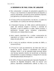
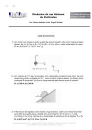
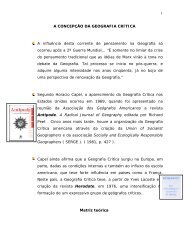
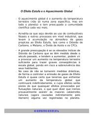
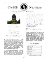
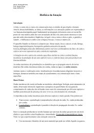
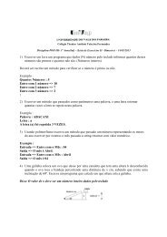
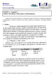
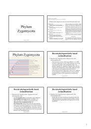
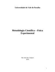
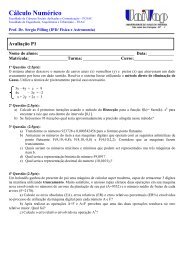
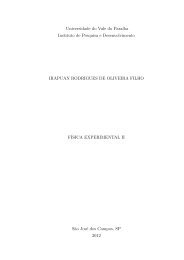
With regard to Ca <strong>and</strong> P content, the mapping <strong>of</strong> treated<br />
dentin showed an elemental uniformity in the control group<br />
CG specimens, when comparing groups A <strong>and</strong> B Figs. 1a<br />
<strong>and</strong> 2a Ca <strong>and</strong> 3a <strong>and</strong> 4a P. Isolated regions with<br />
blue spots in the GI <strong>and</strong> GII areas were observed for both<br />
decontamination treatments groups A <strong>and</strong> B. This is shown<br />
in Figs. 1b, 2b, 1c, <strong>and</strong> 2c Ca <strong>and</strong> 3b, 4b, 3c, <strong>and</strong><br />
4c, P <strong>and</strong> indicates lower calcium <strong>and</strong> phosphorus concentration<br />
than that found in the CG area. A significant demineralization<br />
dark blue areas, as shown in Figs. 1d Ca <strong>and</strong><br />
3d P, was found in the GIIIA area when compared to the<br />
GIIIB area, as shown in Figs. 2d Ca <strong>and</strong> 4d P.<br />
Elemental mapping by EDXRF showed that the autoclaving<br />
treatment caused greater demineralization after acid etching<br />
<strong>and</strong> <strong>laser</strong> <strong>irradiation</strong> than treatment with thymol solution.<br />
Acid etching produced demineralization that was chemically<br />
homogeneous compared to that induced by <strong>laser</strong> treatment, as<br />
shown by the yellow areas in the mapping. Surface distributions<br />
<strong>of</strong> calcium <strong>and</strong> phosphorus were more homogeneous in<br />
the acid-treated specimens <strong>of</strong> the group decontaminated with<br />
thymol than in the autoclaved samples. Laser treatment produced<br />
irregular patterns <strong>of</strong> Ca <strong>and</strong> P distribution throughout<br />
the dentin surface; 180 mJ <strong>of</strong> <strong>laser</strong> energy produced higher<br />
demineralization in autoclaved specimens compared to lower<br />
<strong>laser</strong> energies <strong>and</strong> compared to the specimens treated with<br />
thymol. Previous reports <strong>of</strong> <strong>Er</strong>:<strong>YAG</strong> <strong>laser</strong> etching on dentin,<br />
subjected to SEM analysis, showed a scaly, flaky surface <strong>of</strong><br />
Table 3 Results <strong>of</strong> Mann–Whitney unpaired-sample test.<br />
Content Group comparison P-value<br />
Calcium ACG normal versus ACG treated 0.0145<br />
Phosphorus ACG normal versus ACG treated 0.0012<br />
ACG treated versus BCG treated 0.0145<br />
Ca/P ratio ACG normal versus ACG treated 0.0005<br />
ACG treated versus BCG treated 0.0012<br />
Journal <strong>of</strong> Biomedical Optics 024002-4<br />
March/April 2009 Vol. 142
Silva Soares et al.: <strong>Effects</strong> <strong>of</strong> <strong>Er</strong>:<strong>YAG</strong> <strong>laser</strong> <strong>irradiation</strong> <strong>and</strong> <strong>manipulation</strong> treatment on dentin components, part 2…<br />
Fig. 1 Calcium mapping results obtained by X-ray fluorescence <strong>of</strong> specimens in group A with each subgroup <strong>of</strong> treatment: a CG, b GI, c GII,<br />
<strong>and</strong> d GIII.<br />
Fig. 2 Calcium mapping results obtained by X-ray fluorescence <strong>of</strong> specimens in group B with each subgroup <strong>of</strong> treatment: a CG, b GI, c GII,<br />
<strong>and</strong> d GIII.<br />
Journal <strong>of</strong> Biomedical Optics 024002-5<br />
March/April 2009 Vol. 142
Silva Soares et al.: <strong>Effects</strong> <strong>of</strong> <strong>Er</strong>:<strong>YAG</strong> <strong>laser</strong> <strong>irradiation</strong> <strong>and</strong> <strong>manipulation</strong> treatment on dentin components, part 2…<br />
Fig. 3 Phosphorus mapping results obtained by X-ray fluorescence <strong>of</strong> specimens in group A with each subgroup <strong>of</strong> treatment: a CG, b GI,<br />
c GII, <strong>and</strong> d GIII.<br />
Fig. 4 Phosphorus mapping results obtained by X-ray fluorescence <strong>of</strong> specimens in group B with each subgroup <strong>of</strong> treatment: a CG, b GI,<br />
c GII, <strong>and</strong> d GIII.<br />
Journal <strong>of</strong> Biomedical Optics 024002-6<br />
March/April 2009 Vol. 142
Silva Soares et al.: <strong>Effects</strong> <strong>of</strong> <strong>Er</strong>:<strong>YAG</strong> <strong>laser</strong> <strong>irradiation</strong> <strong>and</strong> <strong>manipulation</strong> treatment on dentin components, part 2…<br />
highly irregular shape with partially opened dentin tubules. In<br />
contrast, acid-etched dentin retained a smoother surface with<br />
opened dentin tubules. 3,5,8,9 Our results are in agreement with<br />
those previous reports; we found irregular patterns <strong>of</strong> elemental<br />
chemical distribution throughout the dentin surface after<br />
<strong>Er</strong>:<strong>YAG</strong> <strong>laser</strong> <strong>irradiation</strong>.<br />
The surface produced by <strong>laser</strong> etching is also acidresistant.<br />
According to Secilmis et al., 25 <strong>laser</strong> <strong>irradiation</strong> <strong>of</strong><br />
dental hard tissues modifies the Ca/P ratio <strong>and</strong> leads to the<br />
formation <strong>of</strong> a more stable <strong>and</strong> less acid-soluble structure. The<br />
alterations in chemical composition may affect the permeability<br />
<strong>and</strong> solubility characteristics <strong>of</strong> dentin, as well as the adhesion<br />
<strong>of</strong> dental materials to dentin.<br />
In summary, EDXRF analysis proved an adequate research<br />
tool to study the changes that occurred in dentin following<br />
decontamination or storage <strong>and</strong> <strong>laser</strong> or chemical etching.<br />
Sample preparation is fairly simple, since it does not require a<br />
specific tooth size or dehydration procedure. The measurements<br />
can be performed under normal atmospheric conditions,<br />
without the need for high vacuum. Finally, because this<br />
technique is nondestructive, samples can be used in multiple<br />
analyses.<br />
4 Conclusions<br />
The results <strong>of</strong> EDXRF measurements showed that the<br />
amounts <strong>of</strong> Ca Ca weight percent <strong>and</strong> P P weight percent<br />
decreased significantly in the specimens that were autoclaved<br />
<strong>and</strong> treated with phosphoric acid ACG. The results suggest<br />
that thymol storage treatment is advised for in vitro studies.<br />
<strong>Er</strong>:<strong>YAG</strong> <strong>laser</strong> etching at lower <strong>laser</strong> energies did not produce<br />
significant changes in dentin components. The EDXRF mapping<br />
data support the hypothesis that the acid etching on dentin<br />
produced a more chemically homogeneous surface, yielding<br />
a more favorable surface for the diffusion <strong>of</strong> adhesive<br />
monomers. These findings elucidate the chemical structure <strong>of</strong><br />
dentin after <strong>laser</strong> etching, facilitating the development <strong>of</strong><br />
effective guidelines for <strong>laser</strong> use.<br />
Acknowledgments<br />
This work was supported by FAPESP 05/50811-9 <strong>and</strong> CNPq<br />
Grant No. 302393/2003-0.<br />
References<br />
1. J.-T. Cheng, K. Itoh, M. Kusunoki, T. Hasegawa, S. Wakumoto, <strong>and</strong><br />
H. Hisamitsu, “Effect <strong>of</strong> dentine conditioners on the bonding efficacy<br />
<strong>of</strong> one-bottle adhesives,” J. Oral Rehabil. 32, 28–33 2005.<br />
2. F. Watari, “In situ quantitative analysis <strong>of</strong> etching process <strong>of</strong> human<br />
teeth by atomic force microscopy,” J. Electron Microsc. 54, 299–308<br />
2005.<br />
3. L. E. S. Soares, E. B. P. S. Resende, A. Brugnera Junior, F. A. A.<br />
Zanin, <strong>and</strong> A. A. Martin, “Combined FT-Raman <strong>and</strong> SEM studies <strong>of</strong><br />
the effects <strong>of</strong> <strong>Er</strong>:<strong>YAG</strong> <strong>laser</strong> <strong>irradiation</strong> on dentin,” Photomed. Laser<br />
Surg. 25, 239–244 2007.<br />
4. R. Rohanizadeh, R. Z. LeGeros, D. Fan, A. Jean, <strong>and</strong> G. Daculsi,<br />
“Ultrastructural properties <strong>of</strong> <strong>laser</strong>-irradiated <strong>and</strong> heat-treated dentin,”<br />
J. Dent. Res. 78, 1829–1835 1999.<br />
5. A. Martinez-Insua, L. D. Dominguez, F. G. Rivera, <strong>and</strong> U. A.<br />
Santana-Penin, “Differences in bonding to acid-etched or <strong>Er</strong>:<strong>YAG</strong><strong>laser</strong>-treated<br />
enamel <strong>and</strong> dentin surfaces,” J. Prosthet. Dent. 84, 280–<br />
288 2000.<br />
6. L. Ceballos, M. Toledano, R. Osorio, F. R. Tay, <strong>and</strong> G. W. Marshall,<br />
“Bonding to <strong>Er</strong>-<strong>YAG</strong>-<strong>laser</strong>-treated dentin,” J. Dent. Res. 81, 119–122<br />
2002.<br />
7. A. Kameyama, E. Kawada, T. Amagai, M. Takizawa, Y. Oda, <strong>and</strong> Y.<br />
Hirai, “Effect <strong>of</strong> HEMA on bonding <strong>of</strong> <strong>Er</strong>:<strong>YAG</strong> <strong>laser</strong>-irradiated bovine<br />
dentine <strong>and</strong> 4-META/MMA-TBB resin,” J. Oral Rehabil. 29,<br />
749–755 2002.<br />
8. A. E. Souza, S. A. M. Corona, R. G. P. Dibb, M. C. Borsatto, <strong>and</strong> J.<br />
D. Pécora, “Influence <strong>of</strong> <strong>Er</strong>:<strong>YAG</strong> <strong>laser</strong> on tensile bond strength <strong>of</strong> a<br />
self-etching system <strong>and</strong> a flowable resin in different dentin depths,” J.<br />
Dent. 32, 269–275 2004.<br />
9. W. J. Dunn, J. T. Davis, <strong>and</strong> D. A. Bush, “Shear bond strength <strong>and</strong><br />
SEM evaluation <strong>of</strong> composite bonded to <strong>Er</strong>:<strong>YAG</strong> <strong>laser</strong>-prepared dentin<br />
<strong>and</strong> enamel,” Dent. Mater. 21, 616–624 2005.<br />
10. Y. Sakakibara, K. Ishimaru, <strong>and</strong> M. Takamizu, “A study on bond<br />
strength to dentin irradiated by erbium:<strong>YAG</strong> <strong>laser</strong>,” Jpn. J. Conserv.<br />
Dent. 41, 207–219 1998.<br />
11. A. Kameyama, E. Kawada, M. Takizawa, Y. Oda, <strong>and</strong> Y. Hirai, “Influence<br />
<strong>of</strong> different acid conditioners on the tensile bond strength <strong>of</strong><br />
4-META/MMA-TBB resin to <strong>Er</strong>:<strong>YAG</strong> <strong>laser</strong>-irradiated bovine dentin,”<br />
J. Adhes. Dent. 2, 297–204 2000.<br />
12. T. Harima, “Adhesive properties <strong>of</strong> resinous materials to <strong>laser</strong>irradiated<br />
dentin,” Ph.D. Thesis, Hiroshima University School <strong>of</strong><br />
Dentistry, Hiroshima, Japan 2001.<br />
13. J. P. DeWald, “The use <strong>of</strong> extracted teeth for in vitro bonding studies:<br />
a review <strong>of</strong> infection control considerations,” Dent. Mater. 13, 74–81<br />
1997.<br />
14. L. E. S. Soares, A. Brugnera Junior, F. A. A. Zanin, M. T. T. Pacheco,<br />
<strong>and</strong> A. A. Martin, “<strong>Effects</strong> <strong>of</strong> treatment for <strong>manipulation</strong> <strong>of</strong> teeth <strong>and</strong><br />
<strong>Er</strong>:<strong>YAG</strong> <strong>laser</strong> <strong>irradiation</strong> on dentin: A Raman spectroscopy analysis,”<br />
Photomed. Laser Surg. 25, 50–57 2007.<br />
15. M. J. Toro, L. L. Lukantsova, M. Williamson, R. Hinesley, G. J.<br />
Eckert, <strong>and</strong> A. J. Dunipace, “In vitro fluoride dose–response study <strong>of</strong><br />
sterilized enamel lesions,” Caries Res. 34, 246–253 2000.<br />
16. M. S. Cenci, S. E. Piva, F. Potrich, E. Formolo, F. F. Demarco, <strong>and</strong> J.<br />
M. Powers, “Microleakage in bonded amalgam restorations using different<br />
adhesive materials,” Braz Dent. J. 15, 13–18 2004.<br />
17. J. M. White, H. E. Goodis, S. J. Marshall, <strong>and</strong> G. W. Marshall,<br />
“Sterilization <strong>of</strong> teeth by gamma radiation,” J. Dent. Res. 73, 1560–<br />
1567 1994.<br />
18. M. Sper<strong>and</strong>io, J. B. Souza, <strong>and</strong> D. T. Oliveira, “Effect <strong>of</strong> gamma<br />
radiation on dentin bond strength <strong>and</strong> morphology,” Braz Dent. J. 12,<br />
205–208 2001.<br />
19. C. P. Lin, B. S. Lee, F. H. Lin, S. H. Kok, <strong>and</strong> W. H. Lan, “Phase,<br />
compositional, <strong>and</strong> morphological changes <strong>of</strong> human dentin after<br />
Nd:<strong>YAG</strong> <strong>laser</strong> treatment,” J. Endod. 27, 389–393 2001.<br />
20. M. Hossain, Y. Nakamura, Y. Tamaki, Y. Yamada, Y. Murakami, <strong>and</strong><br />
K. Matsumoto, “Atomic analysis <strong>and</strong> knoop hardness measurement<br />
<strong>of</strong> the cavity floor prepared by <strong>Er</strong>,Cr:YSGG <strong>laser</strong> <strong>irradiation</strong> in vitro,”<br />
J. Oral Rehabil. 30, 515–521 2003.<br />
21. M. E. Barbour <strong>and</strong> J. S. Rees, “The laboratory assessment <strong>of</strong> enamel<br />
erosion: A review,” J. Dent. 32, 591–602 2004.<br />
22. D. G. Yu, Y. Kimura, J. Kinoshita, <strong>and</strong> K. Matsumoto, “Morphological<br />
<strong>and</strong> atomic analytical studies on enamel <strong>and</strong> dentin irradiated by<br />
an erbium, chromium:YSGG <strong>laser</strong>,” J. Clin. Laser Med. Surg. 18,<br />
139–143 2000.<br />
23. F. O. Falla-Sotelo, M. A. Rizzutto, M. H. Tabacniks, N. Added, <strong>and</strong><br />
M. D. L. Barbosa, “Analysis <strong>and</strong> discussion <strong>of</strong> trace elements in teeth<br />
<strong>of</strong> different animal species,” Braz. J. Phys. 35, 761–762, 2005.<br />
24. L. Bachmann, R. Diebolder, R. Hibst, <strong>and</strong> D. M. Zezell, “Changes in<br />
chemical composition <strong>and</strong> collagen structure <strong>of</strong> dentine tissue after<br />
erbium <strong>laser</strong> <strong>irradiation</strong>,” Spectrochim. Acta, Part A 61, 2634–2639<br />
2005.<br />
25. A. Secilmis, S. Altintas, A. Usumez, <strong>and</strong> G. Berk, “Evaluation <strong>of</strong><br />
mineral content <strong>of</strong> dentin prepared by erbium, chromium:yttrium<br />
sc<strong>and</strong>ium gallium garnet <strong>laser</strong>,” Lasers Med. Sci. 23, 421–425<br />
2008.<br />
Journal <strong>of</strong> Biomedical Optics 024002-7<br />
March/April 2009 Vol. 142