

The Helminthological Society of Washington - Peru State College
The Helminthological Society of Washington - Peru State College
The Helminthological Society of Washington - Peru State College

Create successful ePaper yourself
Turn your PDF publications into a flip-book with our unique Google optimized e-Paper software.
RESEARCH NOTES 203<br />
Tenebrio molitar Linnaeus, 1758. Radiographic<br />
studies were conducted at 21 days postinfection.<br />
Baseline methodologies were established using<br />
an uninfected control rat. Each rat was lightly<br />
anesthetized with the inhalation anesthesia Halothane®<br />
(Halocarbon Laboratories, River Edge,<br />
New Jersey), and a 1.5 cc bolus <strong>of</strong> a water-soluble<br />
iodinated radiographic contrast medium,<br />
diatrizoate meglumine sodium solution (Gastrografin®;<br />
Squibb Diagnostics, Princeton, New<br />
Jersey), was administered through a 6 French<br />
teflon catheter inserted into the rat's stomach. X-<br />
rays were taken at various exposure factors and<br />
with various films to determine the optimal radiographic<br />
technique. Optimal imaging was<br />
achieved with an exposure factor <strong>of</strong> 3.75 mAs<br />
at 54 kVp with Kodak Min-R® single-emulsion<br />
mammography film. <strong>The</strong> rat was placed in a<br />
posterior-anterior or dorsal-ventral position and<br />
x-rays were taken at 5-min intervals to establish<br />
the length <strong>of</strong> time required for the contrast medium<br />
to reach the ileocecal junction. By 30 min,<br />
Gastrografin had filled the entire small intestine.<br />
Food material and fecal shadows were evident<br />
in the control rat. Next, Gastrografin was administered<br />
to Rat I, which had been fed 10 cysticercoids.<br />
At 30 min, posterior-anterior and lateral<br />
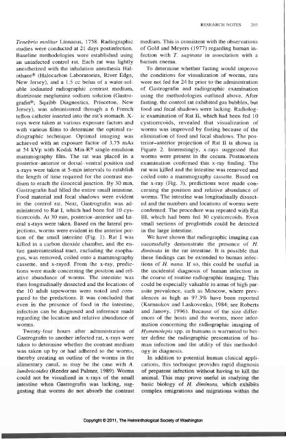
x-rays were taken. Based on the lateral projections,<br />
worms were evident in the anterior portion<br />
<strong>of</strong> the small intestine (Fig. 1). Rat I was<br />
killed in a carbon dioxide chamber, and the entire<br />
gastrointestinal tract, excluding the esophagus,<br />
was removed, coiled onto a mammography<br />
cassette, and x-rayed. From the x-ray, predictions<br />
were made concerning the position and relative<br />
abundance <strong>of</strong> worms. <strong>The</strong> intestine was<br />
then longitudinally dissected and the locations <strong>of</strong><br />
the 10 adult tapeworms were noted and compared<br />
to the predictions. It was concluded that<br />
even in the presence <strong>of</strong> food in the intestine,<br />
infection can be diagnosed and inference made<br />
regarding the location and relative abundance <strong>of</strong><br />
worms.<br />
Twenty-four hours after administration <strong>of</strong><br />
Gastrografin to another infected rat, x-rays were<br />
taken to determine whether the contrast medium<br />
was taken up by or had adhered to the worms,<br />
thereby creating an outline <strong>of</strong> the worms in the<br />
alimentary canal, as may be the case with A.<br />
lumbricoides (Reeder and Palmer, 1989). Worms<br />
could not be visualized in x-rays <strong>of</strong> the small<br />
intestine when Gastrografin was lacking, suggesting<br />
that worms do not absorb the contrast<br />
medium. This is consistent with the observations<br />
<strong>of</strong> Gold and Meyers (1977) regarding human infection<br />
with T. saginata in association with a<br />
barium enema.<br />
To determine whether fasting would improve<br />
the conditions for visualization <strong>of</strong> worms, rats<br />
were not fed for 24 hr prior to the administration<br />
<strong>of</strong> Gastrografin and radiographic examination<br />
using the methodologies outlined above. After<br />
fasting, the control rat exhibited gas bubbles, but<br />
food and fecal shadows were lacking. Radiologic<br />
examination <strong>of</strong> Rat II, which had been fed 10<br />
cysticercoids, revealed that visualization <strong>of</strong><br />
worms was improved by fasting because <strong>of</strong> the<br />
elimination <strong>of</strong> food and fecal shadows. <strong>The</strong> posterior-anterior<br />
projection <strong>of</strong> Rat II is shown in<br />
Figure 2. Interestingly, x-rays suggested that<br />
worms were present in the cecum. Postmortem<br />
examination confirmed this x-ray finding. <strong>The</strong><br />
rat was killed and the intestine was removed and<br />
coiled onto a mammography cassette. Based on<br />
the x-ray (Fig. 3), predictions were made concerning<br />
the position and relative abundance <strong>of</strong><br />
worms. <strong>The</strong> intestine was longitudinally dissected<br />
and the numbers and locations <strong>of</strong> worms were<br />
confirmed. <strong>The</strong> procedure was repeated with Rat<br />
III, which had been fed 30 cysticercoids. Even<br />
small sections <strong>of</strong> proglottids could be detected<br />
in the large intestine.<br />
We have shown that radiographic imaging can<br />
successfully demonstrate the presence <strong>of</strong> H.<br />
diminuta in the rat intestine. It is possible that<br />
these findings can be extended to human infections<br />
<strong>of</strong> H. nana. If so, this could be useful in<br />
the incidental diagnosis <strong>of</strong> human infection in<br />
the course <strong>of</strong> routine radiographic imaging. This<br />
could be especially valuable in areas <strong>of</strong> high parasite<br />
prevalence, such as Moscow, where prevalences<br />
as high as 97.3% have been reported<br />
(Karnaukov and Laskovenko, 1984; see Roberts<br />
and Janovy, 1996). Because <strong>of</strong> the size differences<br />
<strong>of</strong> the hosts and the worms, more information<br />
concerning the radiographic imaging <strong>of</strong><br />
Hymenolepis spp. in humans is warranted to better<br />
define the radiographic presentation <strong>of</strong> human<br />
infection and the utility <strong>of</strong> this methodology<br />
in diagnosis.<br />
In addition to potential human clinical applications,<br />
this technique provides rapid diagnosis<br />
<strong>of</strong> prepatent infection without having to kill the<br />
animal. This may prove useful in studying the<br />
basic biology <strong>of</strong> H. diminuta, which exhibits<br />
complex emigrations and migrations within the<br />
Copyright © 2011, <strong>The</strong> <strong>Helminthological</strong> <strong>Society</strong> <strong>of</strong> <strong>Washington</strong>


