

The Helminthological Society of Washington - Peru State College
The Helminthological Society of Washington - Peru State College
The Helminthological Society of Washington - Peru State College

You also want an ePaper? Increase the reach of your titles
YUMPU automatically turns print PDFs into web optimized ePapers that Google loves.
162 JOURNAL OF THE HELMINTHOLOGICAL SOCIETY OF WASHINGTON, 66(2). JULY 1999<br />
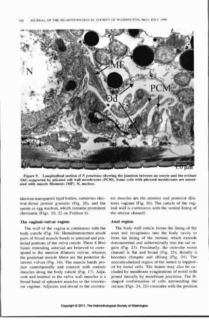
Figure 9. Longitudinal section <strong>of</strong> P. penetrans showing the junction between an oocyte and the oviduct<br />
(Od) supported by plicated cell wall membranes (PCM). Some cells with plicated membranes are associated<br />
with muscle filaments (MF). N, nucleus.<br />
electron-transparent lipid bodies, numerous electron-dense<br />
protein granules (Fig. 20), and the<br />
sperm or egg nucleus, which contains prominent<br />
chromatin (Figs. 20, 22 on Foldout 6).<br />
<strong>The</strong> vaginal-vulvar region<br />
<strong>The</strong> wall <strong>of</strong> the vagina is continuous with the<br />
body cuticle (Fig. 16). Hemidesmosomes attach<br />
pairs <strong>of</strong> broad muscle bands to anteriad and posteriad<br />
portions <strong>of</strong> the vulva cuticle. <strong>The</strong>se 4 fiber<br />
bands extending anteriad are believed to correspond<br />
to the anterior dilatores vulvae, whereas<br />
the posteriad muscle fibers are the posterior dilatores<br />
vulvae (Fig. 16). <strong>The</strong> muscle bands project<br />
ventrolaterally and connect with somatic<br />
muscles along the body cuticle (Fig. 17). Adjacent<br />
and internal to the vulva wall muscles is a<br />
broad band <strong>of</strong> sphincter muscles or the constrictor<br />
vaginae. Adjacent and dorsal to the constrictor<br />
muscles are the anterior and posterior dilatores<br />
vaginae (Fig. 16). <strong>The</strong> cuticle <strong>of</strong> the vaginal<br />
wall is continuous with the ventral lining <strong>of</strong><br />
the uterine channel.<br />
Anal region<br />
<strong>The</strong> body wall cuticle forms the lining <strong>of</strong> the<br />
anus and invaginates into the body cavity to<br />
form the lining <strong>of</strong> the rectum, which extends<br />
dorsoanteriad and subterminally into the tail region<br />
(Fig. 23). Proximally, the cuticular rectal<br />
channel is flat and broad (Fig. 25); distally it<br />
becomes elongate and oblong (Fig. 24). <strong>The</strong><br />
noncuticularized region <strong>of</strong> the lumen is supported<br />
by rectal cells. <strong>The</strong> lumen may also be occluded<br />
by membrane evaginations <strong>of</strong> rectal cells<br />
joined laterally by membrane junctions. <strong>The</strong> H-<br />
shaped conformation <strong>of</strong> cells surrounding the<br />
rectum (Figs. 24, 25) coincides with the position<br />
Copyright © 2011, <strong>The</strong> <strong>Helminthological</strong> <strong>Society</strong> <strong>of</strong> <strong>Washington</strong>


