

The Helminthological Society of Washington - Peru State College
The Helminthological Society of Washington - Peru State College
The Helminthological Society of Washington - Peru State College

Create successful ePaper yourself
Turn your PDF publications into a flip-book with our unique Google optimized e-Paper software.
160 JOURNAL OF THE HELMINTHOLOGICAL SOCIETY OF WASHINGTON, 66(2), JULY 1999<br />
. '~SM$mm^?i••<br />
/.r» •' ' '-'-'-^^^<br />
yfa *• ^iv ' ;^;;!^:^"!tS^^?^^^S^^ ^^'^^^P^ff<br />
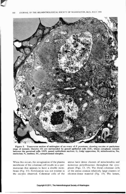
Figure 5. Transverse section <strong>of</strong> midregion <strong>of</strong> an ovary <strong>of</strong> P. penetrans, showing oocytes at pachytene<br />
stage <strong>of</strong> meiosis. Oocytes (O) are surrounded by gonad epithelial cells (GE), whose cytoplasm extends<br />
between the germinal cells. GEN, gonad epithelium nucleus; G, Golgi apparatus; M, mitochondria; Nu,<br />
nucleolus; N, nucleus; SC, synaptonemal complex.<br />
uterus have dense clusters <strong>of</strong> mitochondria and<br />
numerous polyribosomes throughout the cyto-<br />
plasm (Figs. 13, 15). <strong>The</strong> distal columnar cells<br />
<strong>of</strong> the uterus contain relatively large clusters <strong>of</strong><br />
electron-dense material (Fig. 15). <strong>The</strong> lumen,<br />
When this occurs, the invagination <strong>of</strong> the plasma<br />
membrane <strong>of</strong> the columnar cell results in a spermatozoan<br />
that appears to have a double membrane<br />
(Fig. 13). Fertilization was not evident in<br />
the oocytes observed. Columnar cells <strong>of</strong> the<br />
Copyright © 2011, <strong>The</strong> <strong>Helminthological</strong> <strong>Society</strong> <strong>of</strong> <strong>Washington</strong>


