

The Helminthological Society of Washington - Peru State College
The Helminthological Society of Washington - Peru State College
The Helminthological Society of Washington - Peru State College

Create successful ePaper yourself
Turn your PDF publications into a flip-book with our unique Google optimized e-Paper software.
EN DO ET AL.—ULTRASTRUCTURE OF THE LESION NEMATODE 167<br />
17<br />
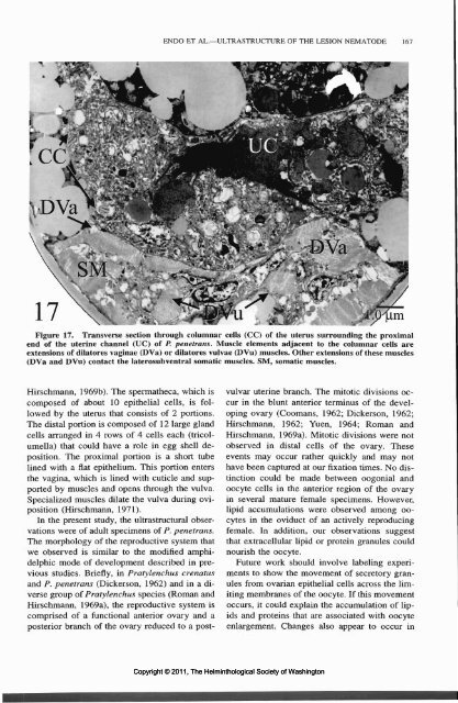
SM . -.-.<br />
tfPlBFti<br />
. " , >'> - ',<br />
Figure 17. Transverse section through columnar cells (CC) <strong>of</strong> the uterus surrounding the proximal<br />
end <strong>of</strong> the uterine channel (UC) <strong>of</strong> P. penetrans. Muscle elements adjacent to the columnar cells are<br />
extensions <strong>of</strong> dilatores vaginae (DVa) or dilatores vulvae (DVu) muscles. Other extensions <strong>of</strong> these muscles<br />
(DVa and DVu) contact the laterosubventral somatic muscles. SM, somatic muscles.<br />
Hirschmann, 1969b). <strong>The</strong> spermatheca, which is<br />
composed <strong>of</strong> about 10 epithelial cells, is followed<br />
by the uterus that consists <strong>of</strong> 2 portions.<br />
<strong>The</strong> distal portion is composed <strong>of</strong> 12 large gland<br />
cells arranged in 4 rows <strong>of</strong> 4 cells each (tricolumella)<br />
that could have a role in egg shell deposition.<br />
<strong>The</strong> proximal portion is a short tube<br />
lined with a flat epithelium. This portion enters<br />
the vagina, which is lined with cuticle and supported<br />
by muscles and opens through the vulva.<br />
Specialized muscles dilate the vulva during oviposition<br />
(Hirschmann, 1971).<br />
In the present study, the ultrastructural observations<br />
were <strong>of</strong> adult specimens <strong>of</strong> P. penetrans.<br />
<strong>The</strong> morphology <strong>of</strong> the reproductive system that<br />
we observed is similar to the modified amphidelphic<br />
mode <strong>of</strong> development described in previous<br />
studies. Briefly, in Pratylenchus crenatus<br />
and P. penetrans (Dickerson, 1962) and in a diverse<br />
group <strong>of</strong> Pratylenchus species (Roman and<br />
Hirschmann, 1969a), the reproductive system is<br />
comprised <strong>of</strong> a functional anterior ovary and a<br />
posterior branch <strong>of</strong> the ovary reduced to a postvulvar<br />
uterine branch. <strong>The</strong> mitotic divisions occur<br />
in the blunt anterior terminus <strong>of</strong> the developing<br />
ovary (Coomans, 1962; Dickerson, 1962;<br />
Hirschmann, 1962; Yuen, 1964; Roman and<br />
Hirschmann, 1969a). Mitotic divisions were not<br />
observed in distal cells <strong>of</strong> the ovary. <strong>The</strong>se<br />
events may occur rather quickly and may not<br />
have been captured at our fixation times. No distinction<br />
could be made between oogonial and<br />
oocyte cells in the anterior region <strong>of</strong> the ovary<br />
in several mature female specimens. However,<br />
lipid accumulations were observed among oocytes<br />
in the oviduct <strong>of</strong> an actively reproducing<br />
female. In addition, our observations suggest<br />
that extracellular lipid or protein granules could<br />
nourish the oocyte.<br />
Future work should involve labeling experiments<br />
to show the movement <strong>of</strong> secretory granules<br />
from ovarian epithelial cells across the limiting<br />
membranes <strong>of</strong> the oocyte. If this movement<br />
occurs, it could explain the accumulation <strong>of</strong> lipids<br />
and proteins that are associated with oocyte<br />
enlargement. Changes also appear to occur in<br />
Copyright © 2011, <strong>The</strong> <strong>Helminthological</strong> <strong>Society</strong> <strong>of</strong> <strong>Washington</strong>


