Case studies - PULSION Medical Systems SE
Case studies - PULSION Medical Systems SE
Case studies - PULSION Medical Systems SE

You also want an ePaper? Increase the reach of your titles
YUMPU automatically turns print PDFs into web optimized ePapers that Google loves.
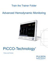
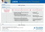
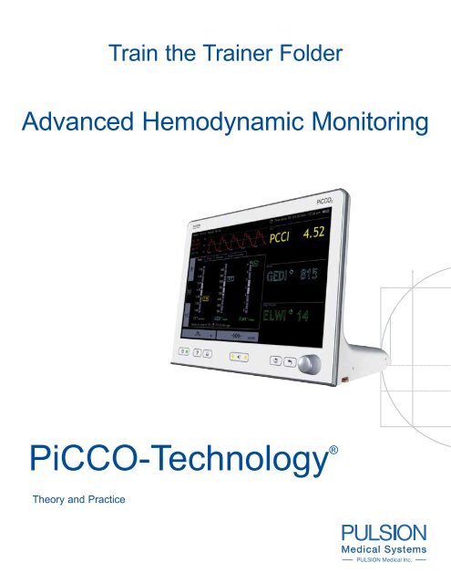
Train the Trainer Folder<br />
Advanced Hemodynamic Monitoring<br />
PiCCO-Technology ®<br />
Theory and Practice
<strong>Case</strong> <strong>studies</strong><br />
Cardiac<br />
Preload<br />
Cardiac Output<br />
Volume<br />
or<br />
catecholamines?<br />
<strong>Case</strong> <strong>studies</strong><br />
Extravascular<br />
Lung Water
<strong>Case</strong> <strong>studies</strong><br />
Cardiac Output<br />
Volume<br />
or<br />
catecholamines?<br />
Hemodynamic monitoring:<br />
More or Less?<br />
Azriel Perel, MD,<br />
Professor and Chairman<br />
Department of Anesthesiology and Intensive Care<br />
Sheba <strong>Medical</strong> Center, Tel Aviv University, Tel Hashomer, Israel<br />
Optimal management of critically ill patients demands<br />
accurate and continuous monitoring of their hemodynamic<br />
status. Such monitoring can be done by clinical<br />
assessment and by using a variety of available advanced<br />
monitoring techniques. It seems, however, that in recent<br />
years many clinicians have become wary of these techniques,<br />
and have either minimized or altogether stopped<br />
using any advanced methods of hemodynamic monitoring<br />
when managing critically ill patients.<br />
An illustrative case of what I perceive to be insufficient<br />
monitoring has been presented in one of the Internet critical<br />
care discussion groups. The presentation described<br />
a 72 year old man with a significant cardiac history, who<br />
became septic (worsening acidosis, oliguria and hypotension)<br />
following major surgery that included the removal<br />
of a large renal tumor and a necrotic gallbladder.<br />
The patient had a positive fluid balance of 20 l over 24h,<br />
received „a bit of noradrenaline“ and eventually had a<br />
sudden cardiac arrest. In answer to a comment that the<br />
patient may have been under-monitored, the response<br />
was: “He was actually on noradrenaline to achieve a target<br />
blood pressure of 70 mmHg…we actually monitored<br />
metabolic function of the liver (lactate), skin perfusion<br />
(clinical assessment), urine output - all good measures<br />
of organ function and perfusion rather than simply arbitrary<br />
pressures, volumes or flows….So what cardiac output<br />
is the right one for this patient? What level of preload<br />
is right? What level of lung water is right?...I would be<br />
happy to use more monitors if somebody could show me<br />
they made a difference… The biggest problem with ALL<br />
the fancy numbers…is that…in the individual patient…<br />
you have NO idea what the „best“ number is supposed<br />
to be“.<br />
One of the main reasons for this ‚back to basics‘ movement<br />
is the repeatedly reported failure of the PAC to<br />
improve patient outcome. These reports have led not<br />
only to repeated pleas to discontinue the use of the<br />
PAC and to a significant decrease in its clinical use,<br />
but also to a general feeling of mistrust towards any of<br />
the new alternative monitoring techniques which have<br />
emerged in the meantime. We have to remind ourselves,<br />
however, that earlier <strong>studies</strong> have repeatedly shown<br />
that the PAC is superior to clinical evaluation in the<br />
hemodynamic assessment of critically ill patients, and<br />
that as many as half of the significant hemodynamic<br />
abnormalities cannot be adequately assessed based<br />
on clinical experience and physical examination alone.<br />
These <strong>studies</strong> have also shown that physicians were<br />
generally confident of their clinical estimates of hemodynamic<br />
variables, but there was no relation between<br />
confidence and accuracy. Moreover, experienced physicians<br />
were no more accurate than less experienced<br />
ones, although they were significantly more confident.<br />
Hence the critical care community seemed to have<br />
learned at that time that clinical examination and vital<br />
signs alone are unreliable in the evaluation of the hemodynamic<br />
status and that more advanced monitoring<br />
tools may give us new important information that is relevant<br />
to patient care.<br />
The major value of the PAC lies in its ability to<br />
measure the cardiac output (CO). Although the<br />
conflicting results of the various <strong>studies</strong> that were<br />
aimed at optimizing oxygen delivery made it unclear<br />
what target CO values we should aim for, it is still imperative<br />
to identify those instances in which a very low<br />
or a very high CO is undetectable by clinical examination<br />
alone. However, although identifying a low CO is<br />
of great importance, it still does not necessarily point<br />
to the right therapeutic decision, e.g., fluids, inotropes,<br />
vasopressors. The next step in the hemodynamic assessment<br />
is the evaluation of the volume status, which,<br />
when the PAC is being used, is based on the measurement<br />
of the filling pressures (CVP, PAOP). This indeed<br />
is the major flaw of the PAC since filling pressures have<br />
been repeatedly shown to be unreliable in assessing<br />
the volume of the heart chambers and in predicting the<br />
response of the patient to fluid loading.<br />
It is therefore unclear why so many clinicians still rely<br />
on filling pressures to guide fluid therapy. What is even<br />
more disturbing is the fact that pre-determined levels of<br />
filling pressures are being used as end-points of resuscitation,<br />
as is the case in both the 2004 Update of the<br />
5-1
<strong>Case</strong> <strong>studies</strong><br />
Practice Parameters for Hemodynamic Support of Sepsis<br />
in Adult Patients and in the Surviving Sepsis Campaign<br />
Guidelines for Management of Severe Sepsis and<br />
Septic Shock. The current literature clearly shows that<br />
volumetric parameters of preload (e.g., the LV end-diastolic<br />
area measured by echo or the global end-diastolic<br />
volume – GEDV – measured by the PiCCO, reflect better<br />
the status of preload than the filling pressures. In addition,<br />
in patients who are on fully controlled mechanical<br />
ventilation, functional hemodynamic parameters (i.e.,<br />
SPV, SVV, PPV, RSVT) are far superior to both filling<br />
pressures and volumetric parameters in the prediction<br />
of fluid responsiveness. Hence the information provided<br />
by the PAC is not reliable enough to identify latent hypovolemia,<br />
neither can it reliably prevent fluid overload<br />
or alert the care-giver to the development of pulmonary<br />
edema. This is especially true in the presence of increased<br />
pulmonary microvascular permeability, where<br />
aggressive optimization of the cardiovascular status<br />
may have grave pulmonary consequences. It is in these<br />
situations that a direct measurement of extravascular<br />
lung water is of utmost importance.<br />
These shortcomings of the PAC may explain the<br />
claim that the use of the PAC is frequently associated<br />
with an aggressive style of treatment which, in<br />
turn, leads to adverse outcomes. In particular the<br />
PAC was shown in a number of <strong>studies</strong> to be associated<br />
with a high positive fluid balance. Obviously the<br />
non-uniformity of patient management in response to<br />
hemodynamic data obtained from PAC both within and<br />
between institutions may have caused great impact on<br />
outcome. This confusion has led to the performance of<br />
a few large randomized trials which were aimed at elucidating<br />
the effect of the PAC on patient outcome. To date<br />
none of these <strong>studies</strong> have shown that the PAC has any<br />
beneficial effect on outcome.<br />
The conclusion of all this is that even with improved<br />
training in the insertion, interpretation, and implementation<br />
of the PAC and the data it generates, the PAC<br />
has inherent limitations as an advanced hemodynamic<br />
monitoring tool. This is why, as an alternative to expensive<br />
clinical trials on the PAC, it was proposed that our<br />
limited financial resources for clinical investigation be invested<br />
in the development of innovative techniques that<br />
may replace the PAC. These techniques are already out<br />
there, each trying to prove its superiority over the others.<br />
This of course is a very natural and very necessary<br />
process. Nevertheless, what is needed more is the realization<br />
that advanced hemodynamic monitoring should<br />
be further explored rather than completely abandoned.<br />
Admittedly, it is difficult to use published data as a basis<br />
from which to draw meaningful conclusions about the effects<br />
of any monitoring technique on outcome. However,<br />
these techniques provide us with a road map which, although<br />
incomplete by nature, may be more helpful than<br />
having no map at all, provided that one is aware of its<br />
pitfalls and limitations. The same logic is being employed<br />
whenever any physiological parameter is being<br />
measured. The ultimate study that will tell us, once and<br />
for all, how to best monitor hemodynamic status at any<br />
circumstance, at any time, is not out yet, and may never<br />
be. In the meantime we can either do nothing, or further<br />
explore the available technologies as well as our understanding<br />
of the pathophysiological processes that occur<br />
in the critically ill.<br />
Cardiac Output<br />
Cardiac Preload<br />
Volume<br />
or<br />
catecholamines?<br />
Extravascular<br />
Lung Water<br />
5-2<br />
Volume or catecholamines? The „Hemodynamic triangle“ gives the answer
Cardiac Output<br />
Volume<br />
or<br />
catecholamines?<br />
Acute lung injury post trauma<br />
Greg Martin, MD, Assistant Professor<br />
Director of <strong>Medical</strong> and Coronary Intensive Care Units,<br />
Emory University School of Medicine, Grady Memorial Hospital,<br />
Atlanta, Georgia, USA<br />
Situation:<br />
• 34 year old male passenger in motor vehicle accident<br />
with pelvic and femur fractures<br />
• Hemorrhagic shock upon arrival in the emergency<br />
department, requiring fluid and blood product resuscitation<br />
• Taken to theatre for pelvic stabilization<br />
• Post-operative day 2 developed worsening hypoxemia<br />
consistent with Acute Lung Injury (ALI): chest<br />
X-ray showed bilateral pulmonary infiltrates and<br />
poor oxygenation ratio of 211 (PaO 2 /FiO 2 ratio)<br />
•<br />
A PiCCO was inserted showing the following parameters:<br />
• Mean Arterial Pressure (MAP) 87 mmHg<br />
• Global End-diastolic Volume Index GEDI 800 ml/m 2<br />
(normal: 680 – 800 ml/m 2 )<br />
• Cardiac Index CI 4.8 l/min/m 2<br />
• Stroke Volume Variation SVV 21 % (normal: ≤ 10 %)<br />
• Extravascular Lung Water Index ELWI 19 ml/kg<br />
(normal: 3 – 7 ml/kg)<br />
• Central Venous Pressure CVP 13 mm Hg<br />
Therapeutic Problems:<br />
Traumatically injured patient with pulmonary edema, high<br />
ELWI and GEDI after resuscitation. Acute Lung Injury<br />
(ALI) requiring conservative fluid management and active<br />
diuresis, as tolerated.<br />
Further Therapeutic Interventions:<br />
• Patient managed with pressure-limited low tidal volume<br />
ventilation to protect lungs against further injury<br />
• Monitoring of GEDI and CI against the ELWI and PaO 2 /<br />
FiO 2 permitting successful fluid management with active<br />
diuresis<br />
• After 96 hours of ventilator support, nutrition and diuretic-based<br />
therapy, PaO 2 /FiO 2 ratio increased to 345 on<br />
FiO 2 0.40 indicating resolution of ALI<br />
Further Progress:<br />
• Patient underwent operative fixation of femoral neck fracture<br />
• He began spontaneous breathing trials on post-operative<br />
day 1<br />
Comments:<br />
Clinical management after acute fluid resuscitation and<br />
development of ALI can be successfully guided by the PiC-<br />
CO parameters.<br />
Simultaneous monitoring of GEDI and ELWI allows fluid<br />
restriction (reduction of pulmonary edema) whilst avoiding<br />
organ hypoperfusion (maintenance of preload).<br />
5-3
<strong>Case</strong> <strong>studies</strong><br />
Clinical Measurement of Extravascular Lung Water:<br />
Its time has come<br />
Charles Philips, MD, Assistant Professor<br />
Director of Center for Intensive Care Research OHSU,<br />
Oregon Health and Science University<br />
Portland, Oregon, USA<br />
Introduction<br />
Increased extravascular lung water is an important cause<br />
of acute respiratory failure in the critically ill. The amount<br />
of extravascular lung water (ELWI) in the lung results as a<br />
balance of factors that cause pulmonary edema formation<br />
and those that cause edema clearance from the distal airways<br />
and interstitum. Inflammatory and coagulation processes<br />
in acute lung injury (ALI) increase the microvascular<br />
permeability while causing a decrease in alveolar fluid<br />
clearance, resulting in a net accumulation of protein rich<br />
pulmonary edema fluid in the interstitium and distal airways<br />
[1]. In patients with acute exacerbations of chronic left heart<br />
failure or with acute myocardial infarction, hydrostatic pulmonary<br />
edema frequently develops leading to hypoxemia<br />
and decreases in lung compliance and ultimately to respiratory<br />
failure. Resolution of edema from the alveolar space<br />
predicts outcome and is essential for survival in ALI [2] In<br />
fact there is a growing body of evidence to suggest that a<br />
strategy to limit or reduce the amount of extra-vascular<br />
lung water (ELWI) from all causes improves<br />
outcome [3-8].<br />
5-4<br />
Measuring ELWI – The How and the Why<br />
Pulmonary edema can be detected on physical examine<br />
by the presence of rales and confirmed roughly through<br />
chest radiography. But clinical examination, chest radiography<br />
and blood gases, either alone or together, have<br />
proven to be relatively poor indicators of the amount of<br />
lung edema or in changes in edema with treatment, of various<br />
etiologies [9]. The direct measurement of ELWI has<br />
been shown to be more sensitive than any non-invasive<br />
indirect method [10].<br />
The ability to directly measure ELWI at the bedside has<br />
been available for over 25 years now. Lewis and colleagues<br />
developed the first bedside method in 1982<br />
using the thermal-green dye method [11]. Subsequent<br />
advances by <strong>PULSION</strong> <strong>Medical</strong> <strong>Systems</strong> perfected the<br />
single thermal indicator technique employed today –<br />
representing a significant advance in reliability and ease<br />
of use.<br />
The PiCCO ® catheter system uses a single thermal indicator<br />
technique to determine ELWI, CO, and volumetric<br />
parameters. CO is calculated using the modified Stewart–Hamilton<br />
equation for thermodilution, with accuracy<br />
comparable with that of pulmonary artery thermodilution.<br />
Cold water injectate is introduced into a central vein and<br />
detected in the distal aorta. The volume of distribution of<br />
the thermal indicator represents the intrathoracic thermal<br />
volume (ITTV), where ITTV (ml) = CO × mean transit time<br />
(MTt) of the thermal indicator. The pulmonary thermal volume<br />
(PTV) is given by PTV (ml) = CO × down slope time<br />
(DSt), where DSt is the exponential decay time of the thermodilution<br />
curve. Global end-diastolic volume (GEDV) is<br />
given by ITTV – PTV (ml). This permits calculation of intrathoracic<br />
blood volume (ITBV) from the linear relationship<br />
with GEDV: ITBV = 1.25 × GEDV – 28.4 (ml). ELWI is the<br />
difference between the thermal indicator distribution in the<br />
chest (ITTV) and the blood volume of the chest (ITBV):<br />
ELWI = ITTV – ITBV (ml).<br />
Concern has been expressed that the thermal dilution<br />
method would lose accuracy in patients with high deadspace<br />
fractions and areas of lung with poor perfusion.<br />
This is an especially important consideration in patients<br />
with ALI where deadspace fractions can be as high as<br />
60-70%. But the accuracy of this method has been validated<br />
by comparison with the ”gold standard” postmortem<br />
gravimetric technique, and with the double dilution<br />
(thermo-dye) technique in animals and humans in a<br />
variety of disease states [12-14]. Moreover it has been<br />
shown to have a sensitivity that allows detection of clinically<br />
relevant changes in ELWI in animal models of severe<br />
ALI and congestive heart failure [15]. Furthermore ELWI<br />
measured in this way has been shown in several <strong>studies</strong><br />
to reliably predict disease severity and outcome in sepsis<br />
and sepsis associated with ALI. In fact it has been shown<br />
to be the best pulmonary specific predictor for mortality<br />
we have available – even in patients with high dead space<br />
fractions.<br />
Eisenberg et al and Mitchell et al, in two separate<br />
<strong>studies</strong>, first demonstrated that when fluid and hemodynamic<br />
management is guided by measured ELWI<br />
as opposed to central pressures and usual care, out-
Cardiac Output<br />
Volume<br />
or<br />
catecholamines?<br />
come is significantly improved. ELWI fell to a greater<br />
extent in patients with ALI and in patients with heart<br />
failure and the duration of time spent on the ventilator<br />
and in the ICU was less in both <strong>studies</strong> [3,8]. Eisenberg<br />
showed that mortality was improved in patients with<br />
higher initial ELWI and ALI (p
<strong>Case</strong> <strong>studies</strong><br />
Measurement of extravascular lung water at the bedside:<br />
Why and how?<br />
Mikhail Kirov MD, PhD, Professor<br />
Department of Anesthesiology and Intensive Care Medicine,<br />
Northern State <strong>Medical</strong> University<br />
Arkhangelsk, Russian Federation<br />
Why do we measure Extravascular Lung Water?<br />
Many critical conditions lead to pulmonary edema.<br />
During systemic inflammation and sepsis, acute lung<br />
injury (ALI), burns, pancreatitis, multiple trauma with<br />
severe blood loss, ischemia-reperfusion injury and other<br />
states, the release of inflammatory mediators may<br />
enhance pulmonary microvascular pressure and permeability,<br />
thus promoting the accumulation of fluid in<br />
the lungs. In contrast to hyperpermeability states, during<br />
cardiac failure the main mechanism for edema includes<br />
increased hydrostatic pressure in the pulmonary<br />
circulation. However, both cardiogenic and non-cardiogenic<br />
origins of pulmonary edema have one common sign –<br />
increased extravascular lung water (ELWI). Moreover,<br />
both types of lung edema are accompanied by a high<br />
mortality rate that necessitates a search for strategies<br />
that will improve our therapy. Consequently, reliable<br />
tools for monitoring lung fluid balance are increasingly<br />
needed in modern intensive care.<br />
The amount of edema is, however, difficult to<br />
estimate at the bedside. Clinical examination, chest<br />
radiography, and blood gases have proven to be of limited<br />
significance in quantifying pulmonary edema.<br />
Therefore, several techniques have been developed<br />
to assess ELWI. Among them, single transpulmonary<br />
thermodilution today is used most frequently.<br />
How can we determine Extravascular Lung Water?<br />
Originally, the thermo-dye dilution (TDD) was used for<br />
measuring lung water. This technique is based on the<br />
simultaneous detection of two indicators with different<br />
properties: a freely diffusible indicator (“cold”) and a dye<br />
(indocyanine green), which binds to the plasma albumin.<br />
Based on the Stewart-Hamilton principle, “cold”<br />
and dye allow the calculation of the intrathoracic thermal<br />
volume (ITTV) and the intrathoracic blood volume<br />
(ITBV), respectively. The difference between the two<br />
distribution volumes is used for estimation of ELWI<br />
(ELWI = ITTV – ITBV). The TDD method has been validated<br />
in animal models of lung edema and in the clinical<br />
setting. However, TDD is relatively time consuming,<br />
5-6<br />
cumbersome and expensive, thus motivating the search<br />
for a reasonable bedside alternative.<br />
Employing the PiCCO technique based on the injection<br />
of a single thermo-indicator that can be detected<br />
with an indwelling arterial thermodilution catheter, is an<br />
appealing idea. ELWI determined by single thermodilution<br />
(STD) can be calculated using the specific<br />
analysis of the thermodilution curve.<br />
In addition to ELWI, STD combined with pulse contour<br />
analysis of cardiac output also gives the possibility of<br />
displaying a variety of cardiopulmonary variables, thus<br />
expanding the options for hemodynamic monitoring.<br />
Recent experimental and clinical <strong>studies</strong> have shown<br />
that ELWI assessed by STD demonstrates good<br />
reproducibility and close agreement with the double<br />
indicator technique and postmortem gravimetry.<br />
Compared with both TDD and right heart<br />
catheterization, STD is simpler to apply, less invasive<br />
and more cost-effective, all factors that make it more<br />
suitable for use at the bedside. However, the detection<br />
of ELWI by the thermodilution method can be impaired<br />
by several factors, for example, severe changes in<br />
cardiac output and pulmonary blood volume and positive<br />
end-expiratory pressure (PEEP). Therefore, determination<br />
of ELWI by TDD and STD requires repeated<br />
measurements.<br />
What do we measure Extravascular Lung Water for?<br />
Several categories of both pediatric and adults intensive<br />
care patients have been shown to benefit from monitoring<br />
ELWI, including any patient who has cardiogenic and<br />
non-cardiogenic pulmonary edema, massive fluid shifts<br />
and severe changes in microvascular permeability. Thus,<br />
I consider any critical illness that results in shock and<br />
tissue hypoperfusion refractory to fluid resuscitation is<br />
a valid subject for ELWI monitoring. In addition, ELWI<br />
monitoring may also be of value in patients undergoing<br />
major surgical procedures, particularly cardiothoracic<br />
surgery and organ transplantation.<br />
In septic shock, invasive cardiovascular monitoring<br />
with arterial catheterization and “beat-to-beat”
Cardiac Output<br />
Volume<br />
or<br />
catecholamines?<br />
analysis facilitates the administration of large<br />
quantities of fluids, vasopressor/inotropic support,<br />
and ventilatory settings. Hence, such monitoring<br />
has recently been recommended as one of the<br />
guiding parameters for hemodynamic support in sepsis.<br />
During sepsis-induced pulmonary edema, accumulation<br />
of ELWI occurs before changes in blood gases,<br />
chest radiogram and pressure variables such as right<br />
atrial pressure (RAP) and pulmonary artery occlusion<br />
pressure (PAOP). It is important to emphasize that the<br />
latter variables are in fact unspecific diagnostic tools<br />
and influenced by a variety of factors. In contrast to<br />
RAP and PAOP, ELWI in severe sepsis correlates with<br />
markers of lung injury such as the oxygenation ratio, lung<br />
compliance, and the number of affected roentgenogram<br />
quadrants, as well as with the total lung injury score.<br />
During the onset of septic shock, ELWI is increased in<br />
three out of four patients. Therefore, in sepsis, ELWI<br />
serves as a marker of ALI, provides a valid estimate<br />
of the interstitial water content in the lungs and might<br />
become an alternative to RAP and PAOP in the<br />
management of fluid resuscitation.<br />
In critically ill patients, ELWI has important prognostic<br />
value and increase in non-survivors. When evaluated<br />
in combination with other cardiopulmonary parameters,<br />
ELWI may reduce the duration of mechanical ventilation<br />
and shorten the periods of stay in ICU and hospital.<br />
Moreover, measurement of ELWI can support the<br />
diagnosis and therefore improve the clinical outcome<br />
of pulmonary edema, if used cautiously in combination<br />
with treatment protocols known to hasten its resolution.<br />
In patients with increased ELWI, such protocols include<br />
fluid restriction, administration of diuretics and inotropes,<br />
ultrafiltration, PEEP, and so on.<br />
Summary:<br />
Many critical states can be accompanied by the<br />
accumulation of Lung Water and development<br />
of pulmonary edema. Among the various methods<br />
for measurement of ELWI at the bedside,<br />
single transpulmonary thermodilution is most useful.<br />
Recent clinical <strong>studies</strong> have shown that ELWI<br />
correlates with the severity of lung injury and has a prognostic<br />
value, especially in sepsis and ALI.<br />
Moreover, monitoring ELWI is an important tool<br />
for prevention and the goal-directed treatment of pulmonary<br />
edema of both cardiogenic and non-cardiogenic<br />
origins. Thus, the success of our therapy often<br />
depends on the correct answers to the following<br />
questions: (1) How much water is in the lungs, (2) Why<br />
is it there, and (3) What can we do to return lung water to<br />
the normal limits. I suspect that if we can answer these<br />
questions correctly, the measurement of ELWI and the<br />
individually therapeutic implications can contribute to improvement<br />
of outcome in many critically ill patients.<br />
5-7
<strong>Case</strong> <strong>studies</strong><br />
„Failure to thrive“ post abdominal laparotomy for gall stone ileus<br />
Professor David Bihari, MD<br />
Department Intensive Care Unit<br />
Lismore Base Hospital, Lismore<br />
New South Wales 2480, Australia<br />
Patient Diagnosis:<br />
Pulmonary aspiration and acute respiratory distress syndrome<br />
following an abdominal laparotomy for gallstone ileus.<br />
<strong>Medical</strong> History:<br />
An 85 year woman was admitted to the ICU following an<br />
abdominal laparotomy for the treatment of a gall<br />
stone ileus. A limited small bowel resection was<br />
performed and the patient was extubated after 48<br />
hours. Over a period of 72 hours, she “failed to thrive”<br />
complaining of abdominal pain and distension. Abdominal<br />
examination revealed some rebound tenderness<br />
and the absence of bowel sounds. TPN was initiated on<br />
the 3rd post operative day and she required CPAP for<br />
respiratory support. On the 4th day, the intra-abdominal<br />
pressure was measured at 18 mmHg (bladder catheter<br />
technique using 100 mL 0.9% saline) and the patient had<br />
an abdominal CT scan with contrast that suggested the<br />
presence of a post-operative ileus. On her return from the<br />
CT scanner, the patient vomited and became very short<br />
of breath suggesting pulmonary aspiration. She required<br />
emergency intubation and gastric contents were aspirated<br />
from the airway. She was immediately bronchoscoped and<br />
her airway washed out with 250 ml of 0.9% saline. Subsequently,<br />
she was difficult to ventilate, requiring pressure controlled<br />
ventilation (inspiratory pressure of 20 cmH 2 O, PEEP<br />
of 15 cmH 2 O, inspiratory time 2 seconds, respiratory rate 15<br />
breaths/minute, FiO 2 of 0.8) to achieve a PaO 2 of 58 mmHg.<br />
She was also hypotensive despite 7.5 mcg/kg/min of dobutamine<br />
(80/45, MAP 55 mmHg) and her CVP was 15 mmHg<br />
(central venous saturation 66%). An ECHO cardiogram<br />
(poor views) suggested a hyperdynamic left ventricle that was<br />
reasonably well filled. A chest X-ray demonstrated bilateral<br />
fluffy infiltrates consistent with an acute lung injury secondary<br />
to aspiration. Her urine output had fallen to less than<br />
0.5 ml/kg/hr and she had a metabolic acidosis with a blood<br />
lactate level of 2.9 mmol/l.<br />
Clinical Course:<br />
At this stage, the attending physician wished to measure this<br />
patient’s cardiac output, preload (GEDV) and extravascular<br />
lung water so as to optimize cardiovascular performance. He<br />
especially wanted to know the lung water in order to understand<br />
how aggressive he should be with fluid therapy. Before obtaining<br />
any hemodynamic data other than the CVP and central<br />
venous saturation (ScvO 2 ), he was inclined to give this lady at<br />
least 20 ml/kg of colloid (4% albumin) in an attempt to improve<br />
her hemodynamics. He was also concerned to administer a<br />
vasopressor (dopamine or nor-adrenaline) to such an elderly<br />
patient without some measure of cardiac output. The resident<br />
medical officer inserted a PiCCO arterial catheter into the right<br />
femoral artery without difficulty. The first set of measurements<br />
(mean of three) was as follows: cardiac index 1.8l/min/m 2 ,<br />
GEDI 880 ml/m 2 , SVV 7%, ELWI 18 ml/kg (see Table 1). The<br />
intensive care specialist interpreted these data as suggesting<br />
that although preload was probably adequate, flow was<br />
too low. Given the elevated ELWI (normal < 7 ml/kg), he did<br />
not think further fluid therapy was appropriate since she was<br />
unlikely to be “fluid responsive” (GEDI at the upper limit of<br />
normal, SVV < 10% during controlled ventilation [tidal volume<br />
7ml/kg]). Instead, the dose of dobutamine was increased<br />
to 15 mcg/kg/min and a low dose noradrenaline infusion<br />
(0.15 mcg/kg/min) was added to increase the MAP to greater<br />
than 70 mmHg. Three units of blood were administered to<br />
maintain a hematocrit of 32% and hydrocortine 50 mg qid<br />
was also prescribed.<br />
Subsequently:<br />
The patient was treated by fluid restriction, a 20% albumin<br />
infusion (12 ml/hour to maintain serum albumin greater than<br />
30 G/l) and a fursemide infusion (2-15 mg/hour to maintain<br />
urine output greater than 150 ml/hour). After 48 hours<br />
of therapy, the patient was in negative fluid balance and<br />
inotropic and vasopressor support could be reduced over<br />
the next five days as oxygenation improved. The patient was<br />
successfully extubated on day 8.<br />
Summary:<br />
Advanced hemodynamic monitoring with the PiCCO allowed<br />
the attending physicians to manipulate fluid therapy, inotropes/vasopressors<br />
and diuresis in such a way as to improve<br />
hemodynamics and pulmonary gas exchange. Excessive<br />
fluid therapy was avoided and active measures were taken<br />
to maintain colloid osmotic pressure and to obtain negative<br />
fluid balance.<br />
Table 1: PiCCO Measurements<br />
Measurement Day 0 Day 1 Day 2 Day 4 Day 5 Day 6<br />
CVP 15 14 12 11 10 8<br />
GEDI 880 680 810 825 830 790<br />
SVV 8 12 N/A N/A N/A N/A<br />
CI 1.8 2.7 3 3.5 3.3 3.1<br />
ELWI 18 18 14 12 10 9<br />
PaO 2 :FiO 2 95 125 210 270 295 330<br />
PEEP 15 15 15 12 12 10<br />
CVP Central Venous Pressure; GEDI Global End Diastolic Volume Index;<br />
SVV Stroke Volume Variation; CI Cardiac Index; ELWI Extra Vascular Lung<br />
Water Index; PaO 2 :FiO 2 Partial Pressure Oxygen divided by Fraction of<br />
Inspired Oxygen; PEEP Positive End Expiratory Pressure<br />
5-8
Cardiac Output<br />
Volume<br />
or<br />
catecholamines?<br />
Incipient pulmonary edema in systemic inflammatory<br />
response after multiple trauma<br />
Professor Enrique Fernandez Mondéjar, MD, Associate Professor<br />
Intensive Care Unit,<br />
Hospital Universitario Virgen de las Nieves<br />
Granada, Spain<br />
<strong>Medical</strong> History<br />
A 28 year old man suffering multiple trauma after an accidental<br />
fall from approximately 10 meters.<br />
Injuries included:<br />
- Complex facial fractures<br />
- Open fractures of left humerus and left tibia<br />
- Closed fracture of right femur<br />
- Transverse fracture of sacrum<br />
- Absence of pulse in left arm<br />
On arriving to our Trauma Centre Emergency Room<br />
(35 min after the accident) the patient was alert, conscious<br />
and breathing spontaneously O 2 . Arterial oxygen<br />
saturation was 96% with an oxygen mask. The hemodynamic<br />
status was volume dependent and the patient was intubated<br />
and connected to a mechanical ventilator. Arterial blood<br />
gas analysis on mechanical ventilation with FiO 2 0.4 and<br />
zero-PEEP: PaO 175, PaCO 2 2 33, pH 7.29, Base deficit<br />
-6.5, lactate 2.3 mmol/l and hemoglobin 9.8 g/dl. After<br />
radiological and sonographic explorations to rule out internal<br />
injuries the patient was transferred to OR (approximately<br />
30 minutes after admittance) for external fixation of the long<br />
bone fractures and vascular repair of left axilla artery.<br />
First 24 hours<br />
The principal problems early after OR were:<br />
1. Extreme hemodynamic instability requiring massive‚<br />
volume replacement (positive balance of 12 litres in the<br />
first 12 hrs) and vasoactive support with noradrenalin in<br />
increasing doses (from 0.35 to 2.7 µg/kg/min).<br />
2. Coagulopathy requiring replacement of hemo-derivates:<br />
Plasma 1500 ml, Red Blood Cells 2000 ml, platelets 12 units.<br />
After 12 hrs the hemodynamic picture remained unstable<br />
with noradrenalin at 1.5 µg/kg/min (BP: 120/70 mmHg,<br />
CVP 5-7 mmHg, urine output 1.5 ml/kg/h, heart rate 120 beat/<br />
min and a positive fluid balance of 2 liters in the last 12 hrs.<br />
The patient became febrile (38.5 ºC) and developed a<br />
generalized skin erythema. Blood cultures were taken. Periph-<br />
14<br />
12<br />
10<br />
8<br />
6<br />
4<br />
ELWI<br />
GEDI x 100<br />
CI<br />
2 1 2 3 4 5 6 7 8 9 10 11 12<br />
Simultaneous monitoring of lung water and preload allows reduction of<br />
pulmonary edema while maintaining preload and cardiac output<br />
eral edema was apparent. Severe Systemic Inflammatory<br />
Response was considered as responsible for the hemodynamic<br />
situation. Chest radiograph was normal and the patient<br />
showed excellent oxygenation on mechanical ventilation<br />
with FiO 2 0.4 and 6 cmH 2 O of PEEP (PaO 2 166, PaCO 2 27,<br />
pH 7.37, Base deficits -4, Lactate 1.6 mmol/L).<br />
Fluid therapy for the next days<br />
Based on a relatively low CVP in a patient with high vasopressor<br />
support and normal lung function, fluid<br />
therapy for the next 24 hours was planned to maintain<br />
the fluid infusion as necessary and to maintain the noradrenalin<br />
infusion or, if possible to reduce it. A PiCCO<br />
catheter was inserted which provided the following data:<br />
Cardiac Index (CI): 4.7 l/min/m 2 , Global End-Diastolic Volume<br />
Index (GEDVI): 630 ml/m 2 , Extravascular Lung Water Index<br />
(ELWI): 13 ml/kg and Stroke Volume Variation (SVV): 17%.<br />
As SVV is only applicable in patients receiving fully controlled<br />
mechanical ventilation, so we did not use this parameter.<br />
As the GEDV was in the low range of normal this indicated<br />
that the patient could tolerate more fluids. However, by<br />
contrast, the high ELWI of 13 ml/kg (normal
<strong>Case</strong> <strong>studies</strong><br />
Postoperative volume management after total hip replacement<br />
Azriel Perel, MD,<br />
Professor and Chairman<br />
Department of Anesthesiology and Intensive Care<br />
Sheba <strong>Medical</strong> Center, Tel Aviv University<br />
Tel Hashomer, Israel<br />
Situation:<br />
• 63 year old patient undergoing total hip replacement<br />
• uneventful course of anaesthesia<br />
• stable hemodynamics intraoperatively; after 4h surgery<br />
clinical signs of pulmonary edema with oxygen saturation<br />
(SaO 2 ) dropping below 80%<br />
• recovery room: blood pressure 63/40mmHg, heart rate<br />
137 bpm<br />
• stabilization of blood pressure with dobutamine and<br />
epinephrine<br />
• blood gas analysis (ventilation with 100% oxygen):<br />
pH 7.23 (norm 7.35-7.45), pCO 2 42mmHg (norm 35-46),<br />
pO 2 75mmHg (norm 70-104), Hct. 37% (norm 40-52%)<br />
Possible diagnoses:<br />
• acute myocardial infarction with cardiac decompensation<br />
(cardiogenic shock)<br />
• hypovolemia (hypovolemic shock)<br />
• reaction to transfusion (anaphylactic shock)<br />
• reaction to bone cement (anaphylactic shock)<br />
• pulmonary embolism<br />
Hemodynamic stabilization:<br />
• installation of PiCCO-system: CI 1.91, GEDI 623,<br />
ELWI 23, SVV 22%<br />
• despite the high lung water, volume loading started<br />
because of low GEDI and high SVV<br />
• reduction of catecholamine dosage with volume<br />
therapy<br />
• TEE showed a hyperdynamic left ventricle with small<br />
enddiastolic volume<br />
Further course:<br />
• further decrease and eventually cessation of catecholamines<br />
• fluid withdrawal after hemodynamic stabilization<br />
• extubation on 2nd postoperative day with normal ELWI<br />
• at the time of extubation there were still radiologic signs<br />
of significant pulmonary edema<br />
• extubation was nevertheless successful<br />
Conclusion:<br />
• detection of hypovolemia by means of PiCCO (low<br />
preload volume, high volume responsiveness) as the<br />
cause of hemodynamic instability.<br />
• recognition and subsequent monitoring of pulmonary<br />
fluid accumulation.<br />
• goal-oriented fluid- and catecholamine therapy with PiC-<br />
CO: initial volume loading, and then volume withdrawal<br />
after circulatory stabilization.<br />
• despite persisting radiologic signs of pulmonary edema<br />
the low ELWI showed an acceptable pulmonary water<br />
content allowing a successful extubation (Lung Water as<br />
a weaning parameter).<br />
ELWI in ml/kg BW<br />
GEDI (x1000)<br />
25 –<br />
23<br />
Course of PiCCO-Parameters for Volume Management<br />
3,8<br />
volume<br />
withdrawal<br />
3,5<br />
CI<br />
CI in l/min<br />
– 4<br />
– 3,5<br />
20 –<br />
2,9<br />
– 3<br />
15 –<br />
10 –<br />
1,9<br />
623<br />
15<br />
778<br />
875<br />
GEDI<br />
– 2,5<br />
– 2<br />
– 1,5<br />
5 –<br />
5<br />
4<br />
ELWI<br />
– 1<br />
5-10<br />
0 –<br />
volume loading<br />
post surgery after volume 1. postop day 2. postop day<br />
– 0,5<br />
– 0
Cardiac Output<br />
Volume<br />
or<br />
catecholamines?<br />
Admission to ICU for loss of consciousness<br />
Jean-Louis Teboul, MD, Professor<br />
Service de Réanimation médicale,<br />
Centre Hospitalier Universitaire de Bicètre<br />
Paris, France<br />
Situation:<br />
• 39 year old woman presented to the emergency department<br />
following coma related to benzodiazepine and antidepressant<br />
self poisoning<br />
• Previous psychological disorder for which she was treated<br />
with antidepressants and benzodiazepines<br />
• Intubated and mechanically ventilated then transferred to<br />
ICU<br />
• On admission laboratory results normal with exception of<br />
Lactate 1.6 mmol/L, White Blood Cells (WBC) 24.8 10 9 /L,<br />
Creatinine 120 µmol/L, CK 13400 IU, CRP 382 mg/dL<br />
Therapeutic problems:<br />
Aspiration pneumonia and moderate renal failure, with staph<br />
aureus found in tracheal secretions. Treated with fluids<br />
(saline), antibiotics and continued on mechanical ventilation<br />
Ongoing problems:<br />
Day 3 patient developed ARDS (Acute Respiratory Distress<br />
Syndrome) P/F ratio decreased from 280 to 120.<br />
Day 2<br />
Day 3<br />
Patient remained hemodynamically stable from day 3 – 7 with<br />
no other organ dysfunction and resolution of renal failure following<br />
fluid management.<br />
Further problems:<br />
• Day 7 sudden development of severe acute circulatory<br />
shock - blood pressure 42/20 mmHg, tachycardia 130 bpm,<br />
oliguria and pyrexia 40 °C<br />
• Grossly abnormal arterial blood gases (pH 6.89, lactate<br />
24 mmol/L), WBC 79.3 10 9 /L, Creatinine 169 µmol/L, potassium<br />
6.3 mmol/L, CK 1157 IU, CRP 394 mg/L<br />
• X-ray, abdominal echo, transthoracic echo – all normal<br />
• Patient given 2 liters normal saline and commenced on<br />
noradrenaline.<br />
• PiCCO was inserted showing the following;<br />
--<br />
Cardiac Index: 3.85 l/min/m 2<br />
--<br />
Stroke Volume Variation (SVV) 21 % (normal: ≤ 10%)<br />
--<br />
Stroke Volume Index: 31 ml/m 2 (normal: 40-60 ml/m 2 )<br />
--<br />
Global End-Diastolic Volume Index (GEDI): 630 ml/m 2<br />
(normal: 680-800 ml/m 2 )<br />
--<br />
Extra-Vascular Lung Water Index (ELWI): 29 ml/kg<br />
(normal: 3 – 7 ml/kg)<br />
--<br />
Heart Rate: 125 beats/min<br />
--<br />
Arterial pressure: 80 / 35 mmHg<br />
In the light of the very high Lung Water (ELWI) and despite<br />
the low preload (GEDI) and high stroke volume variation, the<br />
noradrenaline was increased to 8 µg/kg/min by hour 10 to achieve<br />
a MAP > 65 mmHg. Continuous veno-venous hemofiltration<br />
was started on hour 6. Activated Protein C was started at hour 10.<br />
Finally the antibiotics were changed.<br />
Following noradrenaline increase PiCCO parameters were:<br />
• Cardiac Index: 4.42 l/min/m 2 , SVV: 13 %,<br />
Stroke Volume Index: 37 ml/m 2, , GEDI 700 ml/m 2 ,<br />
ELWI 24 ml/kg, HR: 120 beats/min,<br />
Arterial pressure: 100/50 mmHg<br />
Further Therapeutic Interventions:<br />
From hour 18 the hemodynamic condition improved dramatically<br />
resulting in progressive reduction in the dose of noradrenaline,<br />
which was stopped at hour 36. At that time, blood lactate<br />
level returned to a normal value.<br />
At hour 24, continuous hemofiltration was replaced by intermittent<br />
hemodialysis for the following two weeks until recovery<br />
of renal function. The patient also experienced an episode of<br />
nosocomial pneumonia on day 12.<br />
Further Progress:<br />
Patient was extubated on day 19 and was discharged to the<br />
ward on day 26 with normal renal function and normal chest<br />
X-ray.<br />
Comments:<br />
Despite the obvious need for fluid therapy (low preload –<br />
GEDI 630 and high value of SVV) during the development of<br />
acute severe circulatory shock, the patient was treated with<br />
noradrenaline to support her blood pressure because the priority<br />
was to avoid worsening the pulmonary edema (ELWI 29).<br />
In this case the knowledge of the very high ELWI allowed a more<br />
complete cardiopulmonary picture which provided the treating<br />
physicians with beneficial tools to make the right therapeutic<br />
decision.<br />
5-11
<strong>Case</strong> <strong>studies</strong><br />
Why filling pressures alone are not enough - Patient with acute<br />
respiratory failure and abdominal compartment syndrome<br />
Manu Malbrain, MD<br />
Director <strong>Medical</strong> ICU<br />
ACZA Campus, Stuivenberg<br />
Antwerp, Belgium<br />
Patient History:<br />
A 55 year old man with a previous history of acute<br />
myeloid leukemia was admitted to the ICU because of acute<br />
respiratory failure. He had gained 7kg in weight the<br />
previous week on the ward where he was diagnosed as<br />
having a gastro-enteritis related to the chemotherapy<br />
(cytosar). His central venous pressure measured on the<br />
ward was 32cm H 2 O. The tentative diagnosis hence was<br />
acute lung edema and a bolus of 40 mg fursemide was<br />
administered.<br />
Clinical Course:<br />
On admission to ICU he was in distress with a respiratory<br />
rate of 34 breaths per minute. Further examination of his<br />
vital signs showed a core temperature of 34.4°C, a MAP<br />
of 59 mmHg and a sinus tachycardia of 140 beats per<br />
minute. Because of clinical exhaustion, he was intubated<br />
and mechanically ventilated (machine rate 24 x 500ml,<br />
inspiration: expiration ratio 1:1 and a PEEP of 15) however<br />
oxygenation was poor with a pO 2 /FiO 2 ratio of 115.<br />
Breaths sounds were diminished and fine crackles were<br />
heard over both lungs. The abdomen was tender, firm and<br />
distended with an intra-abdominal pressure of 26 mmHg.<br />
Neurological and extremities examination were unremarkable,<br />
however, the patient was oliguric. A PiCCO<br />
catheter was inserted and confirmed the diagnosis of<br />
septic shock with a cardiac index of 5.1 l/min/m 2 (normal<br />
range 3.0-5.0) and low SVRI. The CVP was<br />
24 mmHg with a SVV of 15% and a GEDI of 650 ml/m 2 ,<br />
confirming intravascular under-filling and fluid responsiveness,<br />
despite the high CVP. Blood cultures grew<br />
enterococcus faecalis and clostridium difficile toxins were<br />
positive on a recent stool sample. The patient’s MAP<br />
was initially responsive to fluids together with doses of<br />
noradrenaline up to 1 mcg/kg/min and dobutamine up<br />
to 15 mcg/kg/min, however he soon became anuric and<br />
the cumulative fluid balance was positive for another 12 l.<br />
Due to ongoing fluid resuscitation and profound<br />
capillary leak his pO 2 /FiO 2 ratio further deteriorated to 75.<br />
At that time CVP was 29 mmHg, MAP 65 mmHg, SVV<br />
13%, GEDI 780 ml/m 2 , IAP 28 mmHg, but ELWI increased<br />
from 12 initially to 17 ml/kg.<br />
Subsequently:<br />
The patient was diagnosed having an abdominal compartment<br />
syndrome with abdominal sepsis related to the<br />
toxic megacolon following diffuse clostridium difficile<br />
5-12<br />
pseudomembraneus colitis. On abdominal CT the<br />
caecum diameter was 18cm with wall thickening up to 3.5cm,<br />
the whole colon was infiltrated and dilated. Therefore the<br />
option was taken to perform a total colectomy and<br />
decompressive laparotomy with temporal abdominal vacuum<br />
assisted fascial closure. After decompression despite the<br />
good CI and SVV parameters the patient was put on CVVH<br />
with aggressive ultra-filtration combined with albumin<br />
substitution because of the high ELWI and low pO 2 /FiO 2<br />
ratio. Over the following days his condition improved with<br />
a decrease in IAP to 16 mmHg and ELWI to 13 ml/kg and<br />
a concomitant rise in pO 2 /FiO 2 ratio to 175. The CVP<br />
remained stable at 18 to 22 mmHg while SVV normalized<br />
at 10-13%.<br />
Summary:<br />
• Traditional filling pressures are erroneously increased<br />
in incidences of high intra-thoracic pressures<br />
(related to IAP or PEEP). In this situation volumes are<br />
better preload indicators.<br />
• SVV is NOT an indicator of preload but a marker of fluid<br />
responsiveness (in fully ventilated patients).<br />
• Measurement of flow (CI) does not allow you to<br />
discriminate between over-or under filling.<br />
• After the initial resuscitation phase an even more<br />
important question that needs to be answered is:<br />
“when to stop filling?”<br />
• ELWI can guide you to get rid of the excess fluids.<br />
SV<br />
∆ V<br />
∆ SV<br />
∆ V<br />
∆ SV<br />
∆ V<br />
∆ SV<br />
Volume Responsiveness Target Volume Overload<br />
Relation of preload and stroke volume in different fluid<br />
loading conditions<br />
Preload<br />
5-19
Cardiac Output<br />
Volume<br />
or<br />
catecholamines?<br />
Hemodynamic management of a patient with pneumonia<br />
Greg Martin, MD, Assistant Professor<br />
Director of <strong>Medical</strong> and Coronary Intensive Care Units,<br />
Emory University School of Medicine, Grady Memorial Hospital,<br />
Atlanta, Georgia, USA<br />
Situation<br />
• 66 year old male patient with pneumonia<br />
• Difficult to oxygenate with PaO 2 /FiO 2 ratio 128 on<br />
Positive End Expiratory Pressure (PEEP) of 18 cmH 2 O<br />
• A PiCCO was inserted showing the following<br />
parameters:<br />
--<br />
Mean Arterial Pressure (MAP) 58 mm Hg<br />
--<br />
Global End-diastolic Volume Index GEDI 560 ml/m 2<br />
(normal: 680 – 800 ml/m 2 )<br />
--<br />
Cardiac Index CI 3.8 l/min/m 2<br />
--<br />
Stroke Volume Variation SVV 21 % (normal: ≤ 10 %)<br />
--<br />
Extravascular Lung Water Index ELWI 16 ml/kg<br />
(normal: 3 – 7 ml/kg)<br />
--<br />
Central Venous Pressure CVP 12 mm Hg<br />
Therapeutic Problems:<br />
Patient with severe sepsis due to pneumonia, low MAP<br />
with elevated SVV, inappropriate CI (sepsis!), Acute Lung<br />
Injury (ALI) with increased ELWI.<br />
Further Therapeutic Interventions:<br />
• Patient managed with pressure-limited low tidal volume<br />
ventilation to protect lungs against further injury<br />
• Fluid resuscitated to target GEDI, increased CI,<br />
lower SVV<br />
• Patient remained hypotensive (MAP< 60, SVV 14%,<br />
ELWI 19), so vasopressors (norepinephrine) started for<br />
blood pressure support<br />
• ScvO 2 measured at 58% → early goal-directed therapy<br />
used to normalize ScvO 2 at > 70% (normal: 70% - 80%)<br />
• PaO 2 /FiO 2 decreased further to 110<br />
Further Progress:<br />
• Septic shock eventually resolved with antibiotics<br />
• Invasiveness of mechanical ventilation decreased,<br />
PaO 2 /FiO 2 increased and ELWI decreased to 7 ml/kg<br />
Summary<br />
• Patient required mechanical ventilation for 16 days<br />
• After extubation, patient transferred to rehabilitation<br />
facility before going home<br />
Comments<br />
• PiCCO parameters GEDI and SVV – in contrast to the<br />
CVP of 12 mmHg - indicated and guided fluid resuscitation<br />
to improve hemodynamics.<br />
• ScvO 2 tracked the effectiveness of the therapy by showing<br />
an increase to the normal range.<br />
5-13
<strong>Case</strong> <strong>studies</strong><br />
Hemodynamic stabilization of a burn patient<br />
Daniel Wyder, MD<br />
Surgical Intensive Care Medicine<br />
University hospital Zurich<br />
Zurich, Switzerland<br />
Situation:<br />
• 36 year old patient with 20% burns of face and upper body<br />
(3% 3rd degree, 17% 2nd degree)<br />
• Patient was admitted intubated, hypothermic but hemodynamically<br />
stable to Burns ICU<br />
• Gas exchange adequate with FiO 2 65, PEEP 10 cmH 2 O<br />
• Bronchoscopy showed inhalation trauma<br />
• Laboratory parameters consistent with rhabdomyolysis<br />
• Need for comprehensive monitoring due to complexity<br />
of case<br />
Hemodynamic Stabilization:<br />
• PiCCO monitoring installed; low CI 3.0, very low GEDI<br />
520, and high-normal ELWI 7 and SVRI 2500 indicating<br />
patient was hypovolemic with a capillary leak syndrome,<br />
typical in burns patients<br />
• Guided by further thermodilution measurements, patient<br />
was given in total 18 liters of crystalloids over first 24<br />
hours, allowing sufficient diuresis with decrease in rhabodomyolysis<br />
parameters<br />
• Despite the high need for fluid, pulmonary function improved<br />
and the patient was able to breathe spontaneously<br />
(FiO 2 .4. CI 3.8, GEDI 600, ELWI 8, SVRI 1500)<br />
• Capillary leak syndrome continued causing ongoing<br />
need for volume (CI 5.0, GEDI 480, ELWI 9, SVRI 750)<br />
• Under continued fluid therapy there was a slight decrease<br />
in gas exchange and increase in lung water, but<br />
normalization of the GEDI was possible with careful fluid<br />
loading<br />
• Once preload was normalized, a reduction in volume<br />
administration was possible<br />
• On day 5 patient became hemodynamically unstable<br />
(CI 7.9, GEDI 760, ELWI 13, SVRI 560, with pulmonary<br />
deterioration requiring FiO 2 1.0 with IRV (inverse ratio<br />
ventilation)<br />
• General picture of a fulminant SIRS / sepsis reaction,<br />
noradrenaline commenced. Diagnosed with sepsis<br />
(streptococcal cultures found in tracheal secretions and<br />
enterococcus in wound swabs)<br />
• Further requirement for fluid boluses, catecholamines<br />
and antibiotics resulted in a slow recovery<br />
• After 9 days a negative fluid balance was possible, with<br />
cessation of catecholamines from day 12. Throughout<br />
this PiCCO parameters (CI, GEDI, ELWI and SVRI) remained<br />
within the normal range<br />
• Following completion of a course of operative wound<br />
management, patient discharged to rehabilitation<br />
Summary<br />
• Volume status is difficult to predict in burn patients due<br />
to large fluid shifts<br />
• In this case the hypovolemic episodes were reliably detected<br />
by the PiCCO<br />
• ELWI is a helpful parameter in the evaluation of<br />
pulmonary permeability in burns, showing good<br />
correlation with the pulmonary function<br />
• Because of the rapid initiation of appropriate volumemanagement,<br />
renal failure (rhabdomyolysis) was avoided<br />
30<br />
Course of PiCCO-parameters<br />
25<br />
20<br />
CI<br />
ELWI<br />
GEDI (x100)<br />
SVRI (x100)<br />
15<br />
10<br />
5<br />
5-14<br />
0<br />
Admission After volume Septic episode<br />
Time Course
Cardiac Output<br />
Volume<br />
or<br />
catecholamines?<br />
Resuscitation post Lung Embolism<br />
Silke van Hülst, MD<br />
Department for Anesthesiology and Surgical Intensive Care Medicine<br />
Westküstenklinikum Heide<br />
Heide, Germany<br />
Situation<br />
• 58 year old patient with history of vertebral disc operation<br />
• On 1 day postop patient had a cardiac arrest during mobilization<br />
• Successfully resuscitated, suspected to have fulminant<br />
pulmonary embolism, admitted to the Intensive Care Unit<br />
• Echocardiography confirmed right heart dysfunction<br />
• Started on thrombolysis therapy, hemodynamic stabilization<br />
with high dose catecholamines<br />
• Development of anuric renal failure<br />
Hemodynamic Stabilization<br />
Installation of the PiCCO-Technology in order to<br />
accurately assess the cardiac pump function and to<br />
differentiate between need for volume and need for<br />
catecholamine treatment:<br />
First results CI 5.87, GEDI 698, ELWI 5.<br />
• Also had CeVOX-Technology* installed for the<br />
continuous measurement of the central venous oxygen<br />
saturation (ScvO 2 ) 63 % (normal range 70% - 80%)<br />
• Rapidly began to display early characteristics of a<br />
developing SIRS with significant capillary leak (CI 4.78,<br />
GEDI 578, ELWI 5).<br />
• Stabilization of Mean Arterial Pressure >80 mmHg possible<br />
with adrenaline and noradrenaline, and ScvO 2<br />
reached >70%<br />
• Reduction of adrenaline possible on day 3.<br />
• With PiCCO monitoring stabilization of GEDI and ELWI to<br />
within normal range (CI 7.2, GEDI 696, ELWI 5).<br />
• On day 3 development of a significant pulmonary<br />
dysfunction with clear requirement for high PEEP (18<br />
mbar) and FiO 2 (0.8)<br />
• Because ELWI remained at 5, a CT-scan of the thorax<br />
was done showing marked ventilation disturbances in the<br />
dorsal basal regions<br />
• Simultaneously, a cranial CT-scan showed multiple<br />
central bleeding spots in the brain tissue and<br />
generalized brain edema, probably resulting from the<br />
thrombolysis therapy<br />
• Patient died on day 4 from cardiac failure<br />
Summary:<br />
• Help in distinguishing between need for volume or<br />
catecholamine therapies possible by monitoring with the<br />
PiCCO and CeVOX-Technologies*<br />
• The ELWI parameter allowed the exclusion of<br />
pulmonary edema as the reason for the pulmonary<br />
deterioration (confirmed by diagnosis of atelectasis in the<br />
thoracic CT)<br />
5-15
<strong>Case</strong> <strong>studies</strong><br />
Acute pancreatitis and myocardial insufficiency<br />
Bernhard Fischer, MD<br />
<strong>Medical</strong> Clinic 2<br />
Clinic Fürth<br />
Fürth, Germany<br />
Situation:<br />
• 82 year old patient with one day history of acute<br />
pancreatitis, CT showed extensive exudate around the<br />
head of the pancreas<br />
• Biliary source suspected, so patient admitted for ERCP<br />
• Previous medical history: compensated cardiac<br />
insufficiency & renal insufficiency due to right<br />
nephrectomy, diabetes mellitus II, arterial hypertension,<br />
general atherosclerosis<br />
• On day 1 of admission clinically under resuscitated as<br />
shown by breath dependent variations on the arterial<br />
pressure curve, CVP 12 mmHg however<br />
Therapeutic Problems:<br />
• Apparent need for further volume management in patient<br />
with pancreatitis and atherosclerosis<br />
• Previous history of cardiac and renal insufficiency<br />
requiring extreme caution with any volume loading<br />
Hemodynamic Stabilization:<br />
• Installation of the PiCCO-System, CI 4.67, GEDI 833,<br />
ELWI 5 , SVRI 870, CVP 12, SVV 28.7%<br />
• Patient given 5 liters fluid<br />
• PiCCO thermodilution measurement 7 hours later:<br />
CI 4.67, GEDI 834, ELWI 5, SVRI 598, CVP 12,<br />
SVV 33.8%<br />
• Noradrenalin commenced and digitoxine was given for<br />
frequent tachy-arrhythmias<br />
• PiCCO thermodilution measurement 16 hours later:<br />
CI 4.53, GEDI 778, ELWI 6, SVRI 1051, CVP 14,<br />
SVV 16.6%<br />
• Following this, and because of cardiac decompensation<br />
with respiratory failure patient started on high dose<br />
diuretics and NIV-BIPAP (non-invasive ventilation)<br />
Further Course:<br />
• With the negative fluid balance and NIV the patient<br />
condition began to stabilize<br />
• Resolution of the acute pancreatitis (aetiology presumed<br />
ischemic because of absence of gallstones in ERCP)<br />
• Discharge to original hospital on day 9<br />
Summary:<br />
• Despite the volume therapy there was no increase in<br />
GEDI or ELWI, thereby pancreatitis responsible for<br />
large losses into the third space<br />
• Noradrenalin was probably responsible for the decompensation<br />
in the cardio-respiratory function because of<br />
the increase in afterload<br />
• Unfortunately no measurements are available once the<br />
patient began to recover<br />
• SVV was not valid in this example as the patient was<br />
not receiving controlled ventilation and was having intermittent<br />
arrhythmias.<br />
Course of PiCCO-parameters<br />
5-16
Cardiac Output<br />
Volume<br />
or<br />
catecholamines?<br />
Multiple trauma<br />
Andreas Weyland, MD, Professor<br />
Department of Anesthesiology and Intensive Care Medicine<br />
Städtische Kliniken Oldenburg<br />
Oldenburg, Germany<br />
Situation:<br />
• 20 year old patient with multiple trauma including<br />
ruptured spleen, open fracture of lower leg, open fracture<br />
of lower jaw, craniocerebral injury (grade 2) as well as<br />
severe bilateral lung contusions and pneumothoraces<br />
and pneumomediastinum<br />
• Patient had already undergone emergency laparotomy<br />
with development of severe oxygenation problems<br />
requiring invasive ventilation with FiO 2 of 1.0<br />
• Massive blood transfusion required for hemodynamic<br />
stabilization<br />
• On admission to the ICU, very gradual improvement seen<br />
in the pulmonary situation (FiO 2 7) with airway pressure<br />
release ventilation (APRV) and patient positioning<br />
Hemodynamic Stabilization<br />
• After installation of the PiCCO system, CI was high<br />
normal, GEDI normal with 720 after massive transfusion<br />
and ELWI elevated at 15<br />
• As a result of the high ELWI with poor pulmonary<br />
function, aggressive diuresis started with fursemide,<br />
guided by the GEDI. After only a few hours ELWI<br />
decreased to 11, with a corresponding decrease in<br />
FiO 2 to 4<br />
• Further negative fluid balance possible with close<br />
attention paid to GEDI<br />
• On day 4 post trauma ELWI had decreased to 8, with<br />
a corresponding improvement in the pulmonary function<br />
(FiO 2 3), and increasing rate of spontaneous breathing<br />
• Stable hemodynamically (CI 4.5) with low dose<br />
dobutamine therapy<br />
• Patient condition continued to slowly improve with<br />
intensive care treatment, and still in ICU at time of<br />
writing<br />
Summary:<br />
• There was a direct correlation between pulmonary<br />
function, ELWI and need for mechanical ventilation<br />
• By simultaneous monitoring of ELWI and GEDI,<br />
an effective reduction of pulmonary edema was possible<br />
while maintaining adequate preload and hemodynamic<br />
stability.<br />
Course of PiCCO-parameters<br />
30<br />
25<br />
20<br />
FiO 2 (timed by 10)<br />
ELWI<br />
15<br />
10<br />
5<br />
0<br />
During OR<br />
After admission<br />
to ICU<br />
After aggressive<br />
diuresis<br />
Day 4 post trauma<br />
5-17
<strong>Case</strong> <strong>studies</strong><br />
Respiratory failure post aspiration<br />
Tilmann von Spiegel<br />
Clinic for Anesthesia and Intensive Care Medicine<br />
University Clinic Bonn<br />
Bonn, Germany<br />
Situation<br />
• 59 year old patient with previous history of liver<br />
cirrhosis Child A from Hepatitis C<br />
• Portal hypertension with long standing esophageal<br />
varices<br />
• Need for emergency laparotomy because of small<br />
bowel ileus (cause: perforated appendicitis)<br />
• During anesthetic induction massive vomiting with<br />
aspiration<br />
• Consequently difficult to ventilate intraoperatively<br />
• On first day post op patient transferred to larger<br />
hospital for further management<br />
• On admission to ICU marked respiratory<br />
insufficiency with PaO 2 85 mmHg on FiO 2 8,<br />
hypotension, with SBP 100 mmHg with dopamine<br />
and dobutamine infusions, CVP 12 mmHg,<br />
diuresis 0.5 ml/kg/h<br />
• Radiological findings: basal delineation increase<br />
and central congestion<br />
• Ventilation adjusted to inverse ratio BiPAP<br />
24/14 mmHg<br />
• Laboratory parameters showed a worsening liver<br />
function along with a ascitic picture, and high CVP<br />
of 20 cm H 2O<br />
Therapeutic Problems:<br />
• Hemodynamic assessment indicating volume<br />
depletion<br />
• X-ray showing pulmonary congestion, hence maybe<br />
volume restriction would be more appropriate?<br />
• Deterioration of the liver function as the result of<br />
cardiac congestion or of volume depletion?<br />
Further Therapeutic Interventions:<br />
• Installation of the PiCCO system, CI 5.2, low SVR,<br />
therefore suggestive of a SIRS / sepsis reaction,<br />
GEDI 520, ELWI 8<br />
• Given 5 liters fluid over 36 hours, later GEDI 680<br />
• Low dose noradrenaline started<br />
• With gradual volume loading no significant increase<br />
in ELWI<br />
• Patient treated in different positions including<br />
prone, bronchoscopy for suction of secretions, antibiotic<br />
therapy<br />
Further Progress:<br />
• Improvement in pulmonary status over the<br />
following days<br />
• Stabilization of the hemodynamic status with<br />
further volume administration, gradual cessation of<br />
noradrenaline infusion<br />
• Normalization of the liver and renal function<br />
• Extubation on day 10 post op<br />
Summary<br />
• Through the parameters provided by the PiCCO<br />
rapid detection of the volume depletion despite high<br />
CVP and radiology showing congestion<br />
• Control of the volume loading by close monitoring<br />
of GEDI and simultaneously avoidance of any<br />
relevant increase in ELWI<br />
• Through rapid hemodynamic stabilization<br />
prevention of any further deterioration in the liver<br />
and renal function.<br />
5-18
See instructions for use and package insert for full prescribing information. Technical specifications are subject<br />
to change without further notice. © 2009 <strong>PULSION</strong> <strong>Medical</strong> <strong>Systems</strong> all rights reserved.<br />
<strong>PULSION</strong> <strong>Medical</strong> Inc. • 2445 Gateway Drive • Suite 110 • TX 75063 Irving, USA<br />
Tel. +1 (214) 446 8500 • Fax +1 (214) 446 6702 • Toll free +1 877 655 8844<br />
info@pulsion.com • www.<strong>PULSION</strong>.com<br />
MPI851405US_R00 Train the Trainer Folder US<br />
© <strong>PULSION</strong> 08/2009