FINAL PROGRAM - Imo
FINAL PROGRAM - Imo
FINAL PROGRAM - Imo
Create successful ePaper yourself
Turn your PDF publications into a flip-book with our unique Google optimized e-Paper software.
Scientific Posters<br />
Scientific Posters<br />
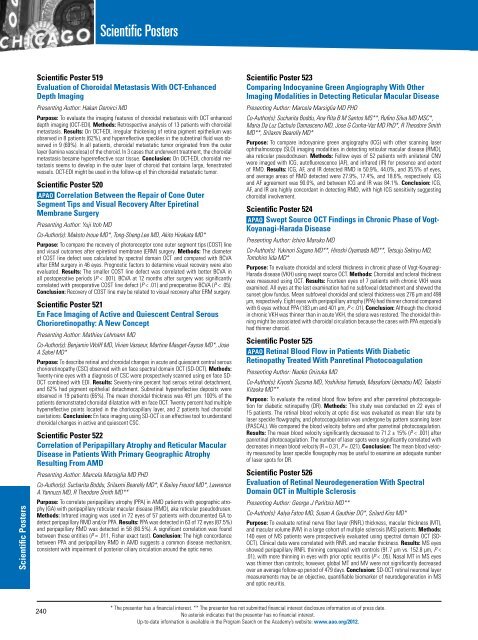
Scientific Poster 519<br />
Evaluation of Choroidal Metastasis With OCT-Enhanced<br />
Depth Imaging<br />
Presenting Author: Hakan Demirci MD<br />
Purpose: To evaluate the imaging features of choroidal metastasis with OCT enhanced<br />
depth imaging (OCT-EDI). Methods: Retrospective analysis of 13 patients with choroidal<br />
metastasis. Results: On OCT-EDI, irregular thickening of retina pigment epithelium was<br />
observed in 8 patients (62%), and hyperreflective speckles in the subretinal fluid was observed<br />
in 9 (69%). In all patients, choroidal metastatic tumor originated from the outer<br />
layer (lamina vasculosa) of the choroid. In 3 cases that underwent treatment, the choroidal<br />
metastasis became hyperreflective scar tissue. Conclusion: On OCT-EDI, choroidal metastasis<br />
seems to develop in the outer layer of choroid that contains large, fenestrated<br />
vessels. OCT-EDI might be used in the follow-up of thin choroidal metastatic tumor.<br />
Scientific Poster 520<br />
APAO Correlation Between the Repair of Cone Outer<br />
Segment Tips and Visual Recovery After Epiretinal<br />
Membrane Surgery<br />
Presenting Author: Yuji Itoh MD<br />
Co-Author(s): Makoto Inoue MD*, Tong-Sheng Lee MD, Akito Hirakata MD*<br />
Purpose: To compare the recovery of photoreceptor cone outer segment tips (COST) line<br />
and visual outcomes after epiretinal membrane (ERM) surgery. Methods: The diameter<br />
of COST line defect was calculated by spectral domain OCT and compared with BCVA<br />
after ERM surgery in 46 eyes. Prognostic factors to determine visual recovery were also<br />
evaluated. Results: The smaller COST line defect was correlated with better BCVA in<br />
all postoperative periods (P < .001). BCVA at 12 months after surgery was significantly<br />
correlated with preoperative COST line defect (P < .01) and preoperative BCVA (P < .05).<br />
Conclusion: Recovery of COST line may be related to visual recovery after ERM surgery.<br />
Scientific Poster 521<br />
En Face Imaging of Active and Quiescent Central Serous<br />
Chorioretinopathy: A New Concept<br />
Presenting Author: Mathieu Lehmann MD<br />
Co-Author(s): Benjamin Wolff MD, Vivien Vasseur, Martine Mauget-Faysse MD*, Jose<br />
A Sahel MD*<br />
Purpose: To describe retinal and choroidal changes in acute and quiescent central serous<br />
chorioretinopathy (CSC) observed with en face spectral domain OCT (SD-OCT). Methods:<br />
Twenty-nine eyes with a diagnosis of CSC were prospectively scanned using en face SD-<br />
OCT combined with EDI. Results: Seventy-nine percent had serous retinal detachment,<br />
and 62% had pigment epithelial detachment. Subretinal hyperreflective deposits were<br />
observed in 19 patients (65%). The mean choroidal thickness was 491 µm. 100% of the<br />
patients demonstrated choroidal dilatation with en face OCT. Twenty percent had multiple<br />
hyperreflective points located in the choriocapillary layer, and 2 patients had choroidal<br />
cavitations. Conclusion: En face imaging using SD-OCT is an effective tool to understand<br />
choroidal changes in active and quiescent CSC.<br />
Scientific Poster 522<br />
Correlation of Peripapillary Atrophy and Reticular Macular<br />
Disease in Patients With Primary Geographic Atrophy<br />
Resulting From AMD<br />
Presenting Author: Marcela Marsiglia MD PHD<br />
Co-Author(s): Sucharita Boddu, Srilaxmi Bearelly MD*, K Bailey Freund MD*, Lawrence<br />
A Yannuzzi MD, R Theodore Smith MD**<br />
Purpose: To correlate peripapillary atrophy (PPA) in AMD patients with geographic atrophy<br />
(GA) with peripapillary reticular macular disease (RMD), aka reticular pseudodrusen.<br />
Methods: Infrared imaging was used in 72 eyes of 57 patients with documented GA to<br />
detect peripapillary RMD and/or PPA. Results: PPA was detected in 63 of 72 eyes (87.5%)<br />
and peripapillary RMD was detected in 58 (80.5%). A significant correlation was found<br />
between these entities (P = .011, Fisher exact test). Conclusion: The high concordance<br />
between PPA and peripapillary RMD in AMD suggests a common disease mechanism,<br />
consistent with impairment of posterior ciliary circulation around the optic nerve.<br />
Scientific Poster 523<br />
Comparing Indocyanine Green Angiography With Other<br />
Imaging Modalities in Detecting Reticular Macular Disease<br />
Presenting Author: Marcela Marsiglia MD PHD<br />
Co-Author(s): Sucharita Boddu, Ana Rita B M Santos MS**, Rufino Silva MD MSC*,<br />
Maria Da Luz Cachulo Damasceno MD, Jose G Cunha-Vaz MD PhD*, R Theodore Smith<br />
MD**, Srilaxmi Bearelly MD*<br />
Purpose: To compare indocyanine green angiography (ICG) with other scanning laser<br />
ophthalmoscopy (SLO) imaging modalities in detecting reticular macular disease (RMD),<br />
aka reticular pseudodrusen. Methods: Fellow eyes of 52 patients with unilateral CNV<br />
were imaged with ICG, autofluorescence (AF), and infrared (IR) for presence and extent<br />
of RMD. Results: ICG, AF, and IR detected RMD in 50.9%, 44.0%, and 35.5% of eyes,<br />
and average areas of RMD detected were 27.9%, 17.4%, and 18.6%, respectively. ICG<br />
and AF agreement was 90.0%, and between ICG and IR was 84.1%. Conclusion: ICG,<br />
AF, and IR are highly concordant in detecting RMD, with high ICG sensitivity suggesting<br />
choroidal involvement.<br />
Scientific Poster 524<br />
APAO Swept Source OCT Findings in Chronic Phase of Vogt-<br />
Koyanagi-Harada Disease<br />
Presenting Author: Ichiro Maruko MD<br />
Co-Author(s): Yukinori Sugano MD**, Hiroshi Oyamada MD**, Tetsuju Sekiryu MD,<br />
Tomohiro Iida MD*<br />
Purpose: To evaluate choroidal and scleral thickness in chronic phase of Vogt-Koyanagi-<br />
Harada disease (VKH) using swept source OCT. Methods: Choroidal and scleral thickness<br />
was measured using OCT. Results: Fourteen eyes of 7 patients with chronic VKH were<br />
examined. All eyes at the last examination had no subfoveal detachment and showed the<br />
sunset glow fundus. Mean subfoveal choroidal and scleral thickness was 276 µm and 498<br />
µm, respectively. Eight eyes with peripapillary atrophy (PPA) had thinner choroid compared<br />
with 6 eyes without PPA (183 µm and 401 µm, P < .01). Conclusion: Although the choroid<br />
in chronic VKH was thinner than in acute VKH, the sclera was restored. The choroidal thinning<br />
might be associated with choroidal circulation because the cases with PPA especially<br />
had thinner choroid.<br />
Scientific Poster 525<br />
APAO Retinal Blood Flow in Patients With Diabetic<br />
Retinopathy Treated With Panretinal Photocoagulation<br />
Presenting Author: Naoko Onizuka MD<br />
Co-Author(s): Kiyoshi Suzuma MD, Yoshihisa Yamada, Masafumi Uematsu MD, Takashi<br />
Kitaoka MD**<br />
Purpose: To evaluate the retinal blood flow before and after panretinal photocoagulation<br />
for diabetic retinopathy (DR). Methods: This study was conducted on 22 eyes of<br />
15 patients. The retinal blood velocity at optic disc was evaluated as mean blur rate by<br />
laser speckle flowgraphy, and photocoagulation was undergone by pattern scanning laser<br />
(PASCAL). We compared the blood velocity before and after panretinal photocoagulation.<br />
Results: The mean blood velocity significantly decreased to 71.2 ± 15% (P < .001) after<br />
panretinal photocoagulation. The number of laser spots were significantly correlated with<br />
decreases in mean blood velocity (R = 0.31, P = .021). Conclusion: The mean blood velocity<br />
measured by laser speckle flowgraphy may be useful to examine an adequate number<br />
of laser spots for DR.<br />
Scientific Poster 526<br />
Evaluation of Retinal Neurodegeneration With Spectral<br />
Domain OCT in Multiple Sclerosis<br />
Presenting Author: George J Parlitsis MD**<br />
Co-Author(s): Aalya Fatoo MD, Susan A Gauthier DO*, Szilard Kiss MD*<br />
Purpose: To evaluate retinal nerve fiber layer (RNFL) thickness, macular thickness (MT),<br />
and macular volume (MV) in a large cohort of multiple sclerosis (MS) patients. Methods:<br />
140 eyes of MS patients were prospectively evaluated using spectral domain OCT (SD-<br />
OCT). Clinical data were correlated with RNFL and macular thickness. Results: MS eyes<br />
showed peripapillary RNFL thinning compared with controls (91.7 µm vs. 152.8 µm, P <<br />
.01), with more thinning in eyes with prior optic neuritis (P < .05). Nasal MT in MS eyes<br />
was thinner than controls; however, global MT and MV were not significantly decreased<br />
over an average follow-up period of 479 days. Conclusion: SD-OCT retinal neuronal layer<br />
measurements may be an objective, quantifiable biomarker of neurodegeneration in MS<br />
and optic neuritis.<br />
240<br />
* The presenter has a financial interest. ** The presenter has not submitted financial interest disclosure information as of press date.<br />
No asterisk indicates that the presenter has no financial interest.<br />
Up-to-date information is available in the Program Search on the Academy’s website: www.aao.org/2012.


