Anthropometrics/Body Segment Parameters
Anthropometrics/Body Segment Parameters
Anthropometrics/Body Segment Parameters

Create successful ePaper yourself
Turn your PDF publications into a flip-book with our unique Google optimized e-Paper software.
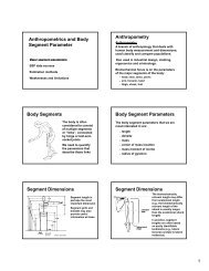
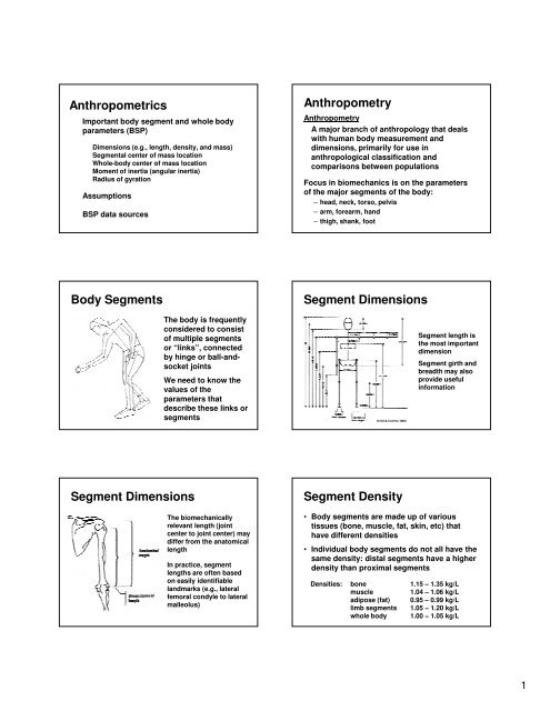
<strong>Anthropometrics</strong><br />
Important body segment and whole body<br />
parameters (BSP)<br />
Dimensions (e.g., length, density, and mass)<br />
<strong>Segment</strong>al center of mass location<br />
Whole-body center of mass location<br />
Moment of inertia (angular inertia)<br />
Radius of gyration<br />
Assumptions<br />
BSP data sources<br />
Anthropometry<br />
Anthropometry<br />
A major branch of anthropology that deals<br />
with human body measurement and<br />
dimensions, primarily for use in<br />
anthropological classification and<br />
comparisons between populations<br />
Focus in biomechanics is on the parameters<br />
of the major segments of the body:<br />
– head, neck, torso, pelvis<br />
– arm, forearm, hand<br />
– thigh, shank, foot<br />
<strong>Body</strong> <strong>Segment</strong>s<br />
<strong>Segment</strong> Dimensions<br />
The body is frequently<br />
considered to consist<br />
of multiple segments<br />
or “links”, connected<br />
by hinge or ball-andsocket<br />
joints<br />
We need to know the<br />
values of the<br />
parameters that<br />
describe these links or<br />
segments<br />
Drillis & Contini (1966)<br />
<strong>Segment</strong> length is<br />
the most important<br />
dimension<br />
<strong>Segment</strong> girth and<br />
breadth may also<br />
provide useful<br />
information<br />
<strong>Segment</strong> Dimensions<br />
<strong>Segment</strong> Density<br />
The biomechanically<br />
relevant length (joint<br />
center to joint center) may<br />
differ from the anatomical<br />
length<br />
In practice, segment<br />
lengths are often based<br />
on easily identifiable<br />
landmarks (e.g., lateral<br />
femoral condyle to lateral<br />
malleolus)<br />
• <strong>Body</strong> segments are made up of various<br />
tissues (bone, muscle, fat, skin, etc) that<br />
have different densities<br />
• Individual body segments do not all have the<br />
same density: distal segments have a higher<br />
density than proximal segments<br />
Densities: bone 1.15 – 1.35 kg/L<br />
muscle 1.04 – 1.06 kg/L<br />
adipose (fat) 0.95 – 0.99 kg/L<br />
limb segments 1.05 − 1.20 kg/L<br />
whole body 1.00 − 1.05 kg/L<br />
1
<strong>Segment</strong> Mass<br />
• <strong>Segment</strong> mass is a<br />
quantitative indicator of a<br />
segment’s inertia<br />
• Determine by segment<br />
density and volume<br />
• For many segments<br />
density can be estimated<br />
from literature data, and<br />
volume can be measured<br />
<strong>Segment</strong> Mass<br />
• Tables also exist that allow one to predict<br />
segment masses, usually as a percentage of<br />
whole body mass<br />
• Finally, the volume represented by different<br />
tissues (fat, muscle, bone) can also be<br />
determined using modern imaging/scanning<br />
techniques (MRI, CT, DEXA)<br />
• These volumes can be combined with more<br />
accurate individual tissue densities to yield<br />
good estimates of segment mass<br />
Center of Mass (COM)<br />
• <strong>Body</strong> segments are made up of an immense<br />
number of distributed mass particles<br />
• Mechanical analysis is facilitated by<br />
identifying the COM: a single point about<br />
which the mass is equally distributed (i.e.,<br />
the balance point)<br />
• Typically specified as a distance (% of<br />
segment length) relative to some unique<br />
point (e.g. proximal joint center)<br />
<strong>Segment</strong>al COM<br />
The location of the COM can be defined as<br />
follows:<br />
x 1<br />
m 1 m 2 m 3<br />
x 2<br />
x 3<br />
X<br />
M<br />
M X = Σ m i x i so X = Σ m i x i<br />
M<br />
<strong>Segment</strong>al COM<br />
The COM of an object can be determined using<br />
a “reaction board”<br />
COM is found by knowing that all moments<br />
about the knife edge must sum to zero<br />
<strong>Segment</strong>al COM<br />
cog = X<br />
X<br />
cog = Y<br />
Y<br />
prox<br />
prox<br />
+ L%(X<br />
+ L%(Y<br />
dist<br />
dist<br />
- X<br />
- Y<br />
prox<br />
prox<br />
)<br />
)<br />
The x and y coordinates for the<br />
right shoulder of a female tennis<br />
player are . The x and y<br />
coordinates for the elbow are<br />
. What is the location of<br />
the center of gravity of the upper<br />
arm?<br />
should be at<br />
knife edge<br />
2
Whole-body COM<br />
Whole-body COM<br />
Σ(w r) = (Σw) r com<br />
Σ(w r) = (ΣW) r com<br />
A bar (length = 2.2 m; mass = 20<br />
kg) is loaded with two 20-kg<br />
plates on its right side (locked in<br />
place, 35 and 40 cm from the<br />
right end) and two 20-kg plates<br />
on its left side (locked in place,<br />
30 and 20 cm from the left end).<br />
Where is the COM of this bar and<br />
the four plates, relative to the left<br />
end of the bar?<br />
chapter05_files/wholebodycom.xls<br />
Moment of Inertia (I)<br />
I is the sum of the products of the (1) mass<br />
element of an object and (2) square of the<br />
distance between the mass element and axis of<br />
rotation<br />
axis<br />
I AXIS = Σm i r i<br />
2<br />
r 1<br />
m 1 m 2<br />
r 2<br />
Radius of Gyration<br />
A more practical approach:<br />
I AXIS = (m BODY )(k 2 )<br />
k indicates radius of gyration,<br />
which is an experimentally<br />
determined length that applies to<br />
the whole object at once<br />
k depends on the<br />
location of the axis<br />
and location of the<br />
mass<br />
I AXIS = m 1 r 12 + m 2 r 22 + m 3 r 32 + .... + m n r n<br />
2<br />
Radius of Gyration<br />
Mass Moment of Inertia<br />
• Radius of gyration is the distance a particle<br />
with the same mass as the segment would<br />
need to be from the axis of rotation to have<br />
the same moment of inertia (angular inertia)<br />
as the segment<br />
• Radius of gyration is really just a convenient<br />
way of “packaging” mass moment of inertia<br />
information<br />
• In anthropometric data sets, k is often<br />
expressed relative to segment length<br />
For a three dimensional<br />
object there will be 3<br />
moments of inertia,<br />
typically expressed<br />
about the 3 principle<br />
axes of the segment<br />
For human limb<br />
segments, I X and I Z will<br />
be similar, I Y will be<br />
much smaller<br />
Z SEG<br />
X SEG<br />
Y SEG<br />
3
Parallel Axis Theorem<br />
• Moment of inertia values are commonly<br />
specified relative to an axis through the<br />
segment center of mass<br />
• The moment of inertia about a different axis<br />
(e.g., through the proximal joint) can be<br />
determined using the parallel axis theorem<br />
I A = I CM + md 2<br />
Where I A is moment of inertia about the new axis, I CM is<br />
moment of inertia about an axis through the CM, m is<br />
segment mass, and d is distance between the two axes<br />
Parallel Axis Theorem<br />
A prosthetic lower leg has a mass of 3 kg and<br />
a center of mass 20 cm from the knee joint.<br />
The radius of gyration is 14.1 cm. What is the<br />
moment of inertia about the knee joint (I KNEE )?<br />
I CM = mk 2<br />
I CM = (3 kg)(0.141 m) 2 = 0.06 kg⋅m 2<br />
I KNEE = I CM + md 2<br />
I KNEE = 0.06 kg⋅m 2 + (3 kg)(0.2 m) 2 = 0.18<br />
kg⋅m 2<br />
Parallel Axis Theorem<br />
The combined moment of inertia<br />
of several segments about a<br />
remote axis can be calculated by<br />
using the parallel axis theorem,<br />
and summing across segments<br />
I LEG(HIP) = I T(HIP) + I S(HIP) + I F(HIP)<br />
where:<br />
I T(HIP) = I T(CM) + m T d T<br />
2<br />
I S(HIP) = I S(CM) + m S d S<br />
2<br />
I F(HIP) = I F(CM) + m F d F<br />
2<br />
Hip<br />
d F<br />
d S<br />
d T<br />
m F<br />
m T<br />
m S<br />
<strong>Body</strong> segment parameter<br />
assumptions<br />
During the period of data collection:<br />
• The segments are perfectly rigid<br />
• The segments are connect by frictionless<br />
hinge or ball-and-socket joints<br />
• The length of each segment remains constant<br />
• The CM location of each segment remains<br />
constant<br />
• The mass moment of inertia of each segment<br />
remains constant<br />
BSP Studies/Data<br />
Harless (1860)<br />
– One of the earliest quantitative study of BSPs<br />
– Dissected 2 cadavers (former prisoners who had<br />
recently been decapitated)<br />
– Determined segment mass & CM locations<br />
Braun & Fischer (1889)<br />
– Determined BSPs in 3 cadaver specimens<br />
– Divided body into 14 segments<br />
– Used data from individual segments to determine<br />
center of gravity of whole body during locomotion<br />
BSP Studies/Data<br />
Dempster (1955)<br />
– Did most detailed and extensive dissection to<br />
date in 8 cadavers specimens<br />
– Determined mass, CM location, & moment of<br />
inertia values for all major body segments<br />
– Expressed data as proportion of total body mass,<br />
and relative to segment lengths<br />
– Results have been used extensively in<br />
biomechanics research<br />
– However, data were from only 8 cadavers, who<br />
were older (52-83 yrs), Caucasian males, most of<br />
whom were reported to be emaciated<br />
4
BSP Studies/Data<br />
Hanavan (1964)<br />
– Used simple geometric shapes<br />
to model 15 major segments<br />
– Assumed uniform density within<br />
each segment<br />
– Used anthropometric data from<br />
the subject to personalize BSP<br />
estimates (still up to 10% error)<br />
Hatze (1980)<br />
– More advanced model; included<br />
17, irregularly shaped segments<br />
– Requires 242 anthropometric<br />
measurements from the subject<br />
BSP Studies/Data<br />
Clauser et al. (1969)<br />
– Determined mass and CM location in 13 cadaver<br />
specimens<br />
– Weakness: did not determine moments of inertia<br />
– <strong>Segment</strong> endpoints adjusted by Hinrichs (1990)<br />
Chandler et al. (1974)<br />
– Determined 3-D moments of inertia in 6 cadaver<br />
specimens<br />
– Did not generate prediction equations<br />
– Equations later developed by Hinrichs (1985)<br />
BSP Studies/Data<br />
Zatsiorsky & Seluyanov (1980-1990)<br />
– Use a gamma-ray scanner to estimate segment<br />
mass, CM, and moment of inertia<br />
– Used 100 male and 15 female young Caucasian<br />
adults<br />
– Developed regression equation to predict BSPs<br />
de Leva (1996)<br />
– Made Zatsiorsky’s data more user-friendly<br />
– Adjusted prediction equations to use more<br />
relevant segment endpoints<br />
– Used as the current standard by many (appropriate?)<br />
BSPs Summary<br />
Issues and Shortcomings:<br />
• Prior to 1970’s fewer than 50 cadavers had<br />
been studied<br />
• Most were adult, Caucasian males<br />
• Data on other populations (women, children,<br />
different ethnic groups) are scant<br />
• Can cadaver data safely be applied to living<br />
humans?<br />
• Techniques on living subjects often suffer<br />
from difficulty in validating results<br />
BSPs Summary<br />
• BSPs arguably represent the greatest source<br />
of error in biomechanical analyses (errors<br />
can easily be ±10%)<br />
• The influence of these errors depends<br />
greatly on the nature of the movement being<br />
studied (magnitude of the accelerations)<br />
• Several investigators have used techniques<br />
like CT, MRI, DEXA to get more accurate,<br />
personalized BSP estimates<br />
– This is still far from being routine (cost & time)<br />
5