

Myringotomy and Ventilation tube insertion - Vula - University of ...
Myringotomy and Ventilation tube insertion - Vula - University of ...
Myringotomy and Ventilation tube insertion - Vula - University of ...

You also want an ePaper? Increase the reach of your titles
YUMPU automatically turns print PDFs into web optimized ePapers that Google loves.
OPEN ACCESS ATLAS OF OTOLARYNGOLOGY, HEAD &<br />
NECK OPERATIVE SURGERY<br />
MYRINGOTOMY WITH VENTILATION<br />
TUBE INSERTION<br />
<strong>Ventilation</strong> <strong>tube</strong>s (grommets) are generally<br />
inserted for refractory middle ear effusions<br />
with persistent conductive hearing loss,<br />
present for a minimum duration <strong>of</strong><br />
3months <strong>and</strong> with hearing loss exceeding<br />
25dB. They may also be inserted as an<br />
adjunct procedure in acute mastoiditis<br />
secondary to acute suppurative otitis<br />
media.<br />
Tashneem Harris & Thomas Linder<br />
membrane 30 minutes prior to the<br />
procedure, or the deep ear canal may<br />
be injected with local anaesthesia wiht<br />
a dental needle<br />
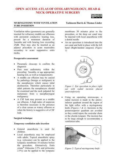
An ear speculum is introduced into the<br />
ear canal <strong>and</strong> held in place with the left<br />
h<strong>and</strong> (Right-h<strong>and</strong>ed surgeon) (Figure<br />
1)<br />
Preoperative assessment<br />
Pneumatic otoscopy to confirm the<br />
diagnosis<br />
Pure tone audiometry within the<br />
preceding 3months, or age appropriate<br />
hearing test, as well as tympanometry<br />
A middle ear effusion may be caused<br />
by pathology (benign or malignant) in<br />
the nasopharynx which causes tubal<br />
dysfunction. Therefore particularly in<br />
adult patients the nasopharynx should<br />
be examined <strong>and</strong> the neck palpated for<br />
metastases from a nasopharyngeal<br />
malignancy<br />
A CSF leak may present as a middle<br />
ear effusion. A high index <strong>of</strong> suspicion<br />
is therefore necessary in the presence<br />
<strong>of</strong> a clear serous or watery effusion or<br />
when the history is suggestive <strong>of</strong> a CSF<br />
leak.<br />
Surgical technique<br />
Temporary ventilation <strong>tube</strong> <strong>insertion</strong><br />
General anaesthesia is used for<br />
children<br />
Local anaesthesia may be employed<br />
with adults. Topical anaesthetic spray<br />
(e.g. xylocaine) can be applied to the<br />
tympanic membrane 10 minutes before<br />
the procedure. Alternatively Emla<br />
cream ® (lidocaine 2.5% <strong>and</strong> prilocaine<br />
2.5%) can be applied to the tympanic<br />
Figure 1: Ear speculum in place right<br />
ear with radial incision placed<br />
anteroinferiorly<br />
Using an operating microscope, a<br />
radial incision is made in the anteroinferior<br />
quadrant around the region <strong>of</strong><br />
the light reflex with a myringotomy<br />
knife (Figures 1 & 2). Incisions in the<br />
posterosuperior quadrant are avoided<br />
as they could injure the ossicular chain<br />
or the chorda tympani. The incision has<br />
to be large enough to accommodate a<br />
ventilation <strong>tube</strong>.<br />
Figure 2: Typical myringotomy knife
The middle ear effusion may be<br />
aspirated with a microsuction <strong>tube</strong><br />
before inserting the grommet<br />
A ventilation <strong>tube</strong> is picked up with<br />
crocodile forceps <strong>and</strong> introduced into<br />
the ear canal using the right h<strong>and</strong><br />
(Figure 3)<br />
extrusion or removal it results in a chronic<br />
perforation <strong>of</strong> the tympanic membrane in<br />
about 16-19% <strong>of</strong> cases. 2,3<br />
Figure 5: Example <strong>of</strong> a T-<strong>tube</strong><br />
Figure 3: Examples <strong>of</strong> short stay <strong>tube</strong>s<br />
The <strong>tube</strong> is placed on the tympanic<br />
membrane adjacent to the<br />
myringotomy opening (Figure 4)<br />
The flanges <strong>of</strong> the T-<strong>tube</strong> are grasped<br />
with crocodile forceps<br />
The flanges are then trimmed so that<br />
the ends are pointed; this facilitates<br />
<strong>insertion</strong> <strong>of</strong> the <strong>tube</strong> through the<br />
myringotomy opening (Figure 6)<br />
Figure 6: The flanges are both trimmed<br />
Figure 4: Placement <strong>of</strong> <strong>tube</strong> on right<br />
tympanic membrane, followed by<br />
advancement <strong>of</strong> <strong>tube</strong> with a hook<br />
Using a 1,5mm, 45° hook the inner<br />
flange is rotated through the<br />
myringotomy incision so that the <strong>tube</strong><br />
straddles the tympanic membrane<br />
(Figure 4)<br />
Long-term ventilation <strong>tube</strong> <strong>insertion</strong><br />
For long-term middle ear ventilation a<br />
ventilating T-<strong>tube</strong> is used (Figure 5). It<br />
remains in place for up to 3 years. After<br />
A myringotomy is made in the<br />
anteroinferior quadrant <strong>of</strong> the tympanic<br />
membrane (Figure 1)<br />
The T-Tube is grasped with a fine<br />
crocodile forceps <strong>and</strong> the pointed end<br />
<strong>of</strong> the flange is inserted through the<br />
myringotomy incision<br />
Special problem: <strong>Ventilation</strong> <strong>tube</strong> falls<br />
into middle ear<br />
Although <strong>tube</strong>s are inert <strong>and</strong> are<br />
unlikely to cause damage when left in<br />
the middle ear, removal should be<br />
attempted because <strong>of</strong> the potential for<br />
foreign body reaction. 4<br />
2
If the grommet lies close to <strong>and</strong> can be<br />
seen through the myringotomy incision,<br />
then it may be possible to retrieve it<br />
using small crocodile forceps, <strong>and</strong> then<br />
reinserted correctly<br />
If the <strong>tube</strong> however lies beyond the<br />
confines <strong>of</strong> the mesotympanum, cannot<br />
be seen <strong>and</strong> removal would be difficult,<br />
then one option is to leave it in situ <strong>and</strong><br />
for the patient to return regularly for<br />
surveillance <strong>and</strong> otomicroscopy 4<br />
Surgical removal when one has a<br />
healed, intact tympanic membrane<br />
entails a wide myringotomy <strong>and</strong><br />
removal <strong>of</strong> ventilation <strong>tube</strong><br />
Very rarely an exploratory tympanotomy<br />
may be required<br />
References<br />
1. Fisch U, May J. Tympanoplasty,<br />
Mastoidectomy <strong>and</strong> Stapes Surgery.<br />
New York: Thieme; 1994<br />
2. Van Heerbeek N, De Saar GM,<br />
Mulder JJ. Long term ventilation<br />
<strong>tube</strong>s: results <strong>of</strong> 726 <strong>insertion</strong>s. Clin<br />
Otolaryngol Allied Sci.<br />
2002;27(5):378-83<br />
3. Kay DJ, Nelson M, Rosenfeld RM.<br />
Meta-analysis <strong>of</strong> tympanostomy <strong>tube</strong><br />
sequelae. Otolaryngol Head Neck<br />
Surg. 2001;124(4):374-80<br />
4. Rosenfeld RM, Bluestone CD.<br />
Evidence Based Otitis Media. 2nd Ed.<br />
Hamilton: BC Decker Inc; 2003<br />
Author<br />
Tashneem Harris MBChB, FCORL,<br />
MMED (Otol), Fisch Instrument<br />
Microsurgical Fellow<br />
ENT Specialist<br />
Division <strong>of</strong> Otolaryngology<br />
<strong>University</strong> <strong>of</strong> Cape Town<br />
Cape Town, South Africa<br />
harristasneem@yahoo.com<br />
Senior Author<br />
Thomas Linder, M.D.<br />
Pr<strong>of</strong>essor, Chairman <strong>and</strong> Head <strong>of</strong><br />
Department <strong>of</strong> Otorhinolaryngology,<br />
Head, Neck <strong>and</strong> Facial Plastic Surgery<br />
Lucerne Canton Hospital, Switzerl<strong>and</strong><br />
thomas.linder@ksl.ch<br />
Editor<br />
Johan Fagan MBChB, FCORL, MMed<br />
Pr<strong>of</strong>essor <strong>and</strong> Chairman<br />
Division <strong>of</strong> Otolaryngology<br />
<strong>University</strong> <strong>of</strong> Cape Town<br />
Cape Town<br />
South Africa<br />
johannes.fagan@uct.ac.za<br />
THE OPEN ACCESS ATLAS OF<br />
OTOLARYNGOLOGY, HEAD &<br />
NECK OPERATIVE SURGERY<br />
www.entdev.uct.ac.za<br />
The Open Access Atlas <strong>of</strong> Otolaryngology, Head &<br />
Neck Operative Surgery by Johan Fagan (Editor)<br />
johannes.fagan@uct.ac.za is licensed under a Creative<br />
Commons Attribution - Non-Commercial 3.0 Unported<br />
License<br />
3


