Unusual cause for V pattern XT - The Private Eye Clinic
Unusual cause for V pattern XT - The Private Eye Clinic
Unusual cause for V pattern XT - The Private Eye Clinic

Create successful ePaper yourself
Turn your PDF publications into a flip-book with our unique Google optimized e-Paper software.
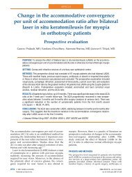
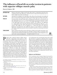
Case 1: RH<br />
An unusual <strong>cause</strong> <strong>for</strong> “V” <strong>pattern</strong> exotropia
RH<br />
12yo<br />
Bilateral asymmetric astigmatism<br />
POHx: <strong>XT</strong> first presented in 2005.<br />
OU LR Rc 5.00mm and IO Rc in 2006<br />
C/o progressive residual / recurrent <strong>XT</strong> with persistent<br />
“V” <strong>pattern</strong> despite IO weakening.<br />
No diplopia
Preop II (June 2011)<br />
Gls: R -3.00x180 L+0.50-1.50x165<br />
VA R+L 6/9cgl N3<br />
Stereo: Titmus 400‟‟<br />
Fundus torsion was not detected.
O/E in 2005, be<strong>for</strong>e Sx I: Bil<br />
LRc+IOrec<br />
• C-T (cc+sc )<br />
N: X‟T 20pd<br />
D: <strong>XT</strong> 25<br />
25<br />
10<br />
Dg: “V” <strong>pattern</strong> <strong>XT</strong> with Bil IO OA,<br />
Asymmetric astigmatism
O/E: Be<strong>for</strong>e the 2nd Sx in 2011<br />
• C-T (cc)<br />
C-T: N: X‟ 35pd, minimal fusion reserve<br />
D: X(T) 30<br />
25<br />
20<br />
“V” Pattern intermittent exotropia<br />
Full correction with prism did not <strong>cause</strong> diplopia<br />
Overcorrection with prism did <strong>cause</strong> diplopia [patient had<br />
never been ET there<strong>for</strong>e had no sensory wiring to cope with<br />
being ET unlike a pt with consec <strong>XT</strong>]
<strong>The</strong> challenge is:<br />
How to manage the V <strong>pattern</strong> in pt with previous IO<br />
weakening and no fundus torsion
MRI Brain and Orbits<br />
3-6-2011
Coronal<br />
MRI T1: inf<br />
positioning<br />
of LR<br />
(L>R), and<br />
nasal IR
Axial T1:<br />
LR appears<br />
inferiorly to<br />
the MR
Re<br />
Le<br />
Normal<br />
Inf displ of LR<br />
Nasal disp of<br />
IR
Heterotopy of extraocular muscle<br />
pulleys <strong>cause</strong>s incomitant strabismus<br />
Muscle pulley: connective tissue sleeves in the post tenon‟s fascia,<br />
that act as functional origins of the muscles. MRI analysis shows<br />
that the location of the pulleys are highly uni<strong>for</strong>m (
“V” <strong>pattern</strong> ET<br />
Inf displacement of LR<br />
centroid (R>L)<br />
“A”<strong>pattern</strong> ET<br />
LR displaced sup to MR and<br />
SR displaced nasal to IR<br />
Abnormal location of the pulleys could explain many of the cases of<br />
incomitant strabismus, that conventionally attributed to „oblique<br />
muscle dysfunction‟ .<br />
(Joseph Demer, Robert Clark, Joel Miller,- Advances in strabismology)
Sx 11-7-11 :Ou Upper ¼ width LR sutured<br />
and hitched superiorly and MR resected
Post op<br />
measurements in D1,<br />
D7<br />
Day Alternate CT Sensorydiplopia<br />
Angle of<br />
anomaly<br />
D1 D ET 20 ET>40 >20<br />
N EX‟ 0 ET‟>40 >40<br />
W2 D <strong>XT</strong> 2 ET 30 32<br />
N <strong>XT</strong>‟ 8 ET‟ 16 24
Now , what is going on ?
In this case, the pt had <strong>XT</strong> with no diplopia be<strong>for</strong>e Sx, then prior<br />
to Sx she had one or both [usually both] of:<br />
Suppression<br />
Anomalous Retinal Correspondence (ARC)<br />
<strong>The</strong> cortex has the ability to shift the visual direction associated<br />
with the fovea of the deviating eye (ARC), as well as the ability to<br />
suppress the image of the deviating eye
NRC ARC<br />
X- image point<br />
When strabismic eyes are straightened, ARC usually poorly resolves<br />
with constant esotropes, compared to Intermittent exotropes.<br />
Unresolved ARC will <strong>cause</strong> (paradoxical) diplopia despite straight<br />
eyes .
Prevalence of ARC<br />
Jampolsky : ARC in 90%<br />
of ET
A case series/ Dr Kowal<br />
Retrospective review of manifest /<br />
expected symptomatic paradoxical<br />
diplopia after surgery to straighten<br />
eyes<br />
Methods:<br />
15 pt‟s<br />
13 had Sx <strong>for</strong> ET 3, <strong>XT</strong> 12 (mostly<br />
consecutive)<br />
Age: 8 - 62 yrs
Results<br />
12/13 had paradoxical diplopia, 1/13 had suppression<br />
9/12 had resolution of diplopia within 12w (resolution <strong>for</strong> near be<strong>for</strong>e<br />
distance).<br />
3/12 patients had persistent diplopia after surgery (longest follow-up<br />
being 4 yrs)<br />
8 y o was the youngest pt to c/o symptomatic diplopia
Conclusion<br />
This case of paradoxical diplopia was NOT predicted with preoperative<br />
prism simulation of surgical correction. This is very<br />
rare.<br />
Most BUT NOT ALL patients with postop diplopia due to ARC resolve<br />
after Sx.<br />
Deferring surgery to adulthood is a risk factor <strong>for</strong> not resolved ARC.