Epidemiology of amblyopia - The Private Eye Clinic
Epidemiology of amblyopia - The Private Eye Clinic
Epidemiology of amblyopia - The Private Eye Clinic
You also want an ePaper? Increase the reach of your titles
YUMPU automatically turns print PDFs into web optimized ePapers that Google loves.
Seminar<br />
Amblyopia<br />
Jonathan M Holmes, Michael P Clarke<br />
Results from recent randomised clinical trials in <strong>amblyopia</strong> should change our approach to screening for and<br />
treatment <strong>of</strong> <strong>amblyopia</strong>. Based on the current evidence, if one screening session is used, screening at school entry<br />
could be the most reasonable time. <strong>Clinic</strong>ians should preferably use age-appropriate LogMAR acuity tests, and<br />
treatment should only be considered for children who are clearly not in the typical range for their age. Any substantial<br />
refractive error should be corrected before further treatment is considered and the child should be followed in<br />
spectacles until no further improvement is recorded, which can take up to 6 months. Parents and carers should then<br />
be <strong>of</strong>fered an informed choice between patching and atropine drops. Successful patching regimens can last as little as<br />
1 h or 2 h a day, and successful atropine regimens as little as one drop twice a week. Intense and extended regimens<br />
might not be needed in initial therapy.<br />
This Seminar provides an update on recent develop ments<br />
in <strong>amblyopia</strong> research that affects screening, diagnosis,<br />
and treatment. Only in the past 5 years have multicentre,<br />
randomised controlled trials addressed <strong>amblyopia</strong><br />
treatment issues. <strong>The</strong>se studies not only help clinicians<br />
and parents in choosing appropriate therapies, but also<br />
inform screening policy, which is <strong>of</strong> general interest. We<br />
first summarise common definitions <strong>of</strong> <strong>amblyopia</strong>,<br />
discuss the diagnosis <strong>of</strong> <strong>amblyopia</strong>, and describe recent<br />
studies <strong>of</strong> screening for <strong>amblyopia</strong> and the implications<br />
<strong>of</strong> treatment trials on screening. We then present the<br />
existing evidence on whether the disorder should be<br />
treated and summarise recent treatment studies,<br />
providing specific recommendations.<br />
Definition <strong>of</strong> <strong>amblyopia</strong><br />
Amblyopia has traditionally been defined by what it is<br />
not, rather than by what it is. Definitions <strong>of</strong>ten include<br />
aphorisms such as a disorder “in which the patient sees<br />
nothing and the doctor sees nothing”. 1 Based on animal<br />
studies 2 and functional human neuroimaging, 3 <strong>amblyopia</strong><br />
can be defined as a disorder in which there is dysfunction<br />
<strong>of</strong> the processing <strong>of</strong> visual information. This dysfunction<br />
is usually detected and evident as reduced recognition<br />
visual acuity, although the abnormalities include many<br />
types <strong>of</strong> visual function. 4 Although clinical ocular<br />
examination is most <strong>of</strong>ten entirely normal in <strong>amblyopia</strong>,<br />
microscopic anatomical and structural abnormalities<br />
have been found in the retina, 5 lateral geniculate bodies, 6<br />
and visual cortex. 7<br />
Amblyopia results from degradation <strong>of</strong> the retinal<br />
image during a sensitive period <strong>of</strong> visual development,<br />
which historically has been thought to be the first 7 years<br />
<strong>of</strong> life. 8 <strong>The</strong> sensitive period for development <strong>of</strong> <strong>amblyopia</strong><br />
might not be the same as the sensitive period during<br />
which treatment is possible. <strong>The</strong> degradation <strong>of</strong> the<br />
image, and subsequent central suppression that leads to<br />
<strong>amblyopia</strong>, results from one <strong>of</strong> three causal processes<br />
(table 1).<br />
<strong>The</strong>refore, <strong>amblyopia</strong> never occurs in isolation. <strong>The</strong><br />
disorder is not the cause, but the effect <strong>of</strong> another<br />
pathological process. Amblyopia can also be thought <strong>of</strong><br />
as resulting from either disuse due to the absence <strong>of</strong> a<br />
clear image on the retina (anisometropia or deprivation),<br />
or misuse due to abnormal binocular interaction<br />
(strabismic). A widely accepted definition <strong>of</strong> <strong>amblyopia</strong><br />
based on visual acuity is 2 or more Snellen or logMAR<br />
lines difference between eyes in best-corrected visual<br />
acuity. A one-line difference is usually a normal result,<br />
based on test-retest variability. 9<br />
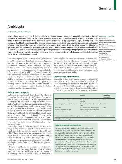
<strong>Epidemiology</strong> <strong>of</strong> <strong>amblyopia</strong><br />
Amblyopia is the most common cause <strong>of</strong> monocular<br />
vision loss in children with an estimated prevalence <strong>of</strong><br />
1–5%, depending on population and study. 1 Because <strong>of</strong><br />
the failure <strong>of</strong> detection or treatment, <strong>amblyopia</strong> continues<br />
to be an important cause <strong>of</strong> vision loss in adults, with an<br />
estimated prevalence <strong>of</strong> 2·9%. 10 A study by the National<br />
Strabismus (ocular misalignment)<br />
Anisometropia (difference in refractive error)<br />
Deprivation<br />
(including ametropia—ie, large<br />
symmetric refractive errors)*<br />
Search strategy and selection criteria<br />
Features<br />
Each eye does not have the same image on<br />
the fovea<br />
One foveal image is more blurred than the<br />
other<br />
Physical obstruction <strong>of</strong> one image (eg,<br />
cataract, ptosis, or bilateral blur from<br />
uncorrected refractive error)<br />
We searched MEDLINE (1966 to 2005) and the Cochrane<br />
Library (to 2005), and used the search term “<strong>amblyopia</strong>”. We<br />
mainly selected publications in the past 5 years, but did not<br />
exclude commonly referenced and highly regarded older<br />
publications. We also searched the reference lists <strong>of</strong> articles<br />
identified by this search strategy and selected those we<br />
judged relevant. Several review articles or book chapters were<br />
included because they provide comprehensive overviews that<br />
are beyond the scope <strong>of</strong> this Seminar. <strong>The</strong> reference list was<br />
subsequently modified during the peer-review process on the<br />
basis <strong>of</strong> comments from reviewers.<br />
Lancet 2006; 367: 1343–51<br />
Department <strong>of</strong><br />
Ophthalmology, Mayo <strong>Clinic</strong><br />
College <strong>of</strong> Medicine, Rochester,<br />
MN 55905, USA<br />
(Pr<strong>of</strong> J M Holmes BM BCh); and<br />
Department <strong>of</strong><br />
Ophthalmology, University <strong>of</strong><br />
Newcastle upon Tyne,<br />
Newcastle, UK<br />
(M P Clarke FRCOphth)<br />
Correspondence to:<br />
Pr<strong>of</strong> Jonathan M Holmes<br />
holmes.jonathan@mayo.edu<br />
Unilateral or<br />
bilateral effect<br />
Unilateral<br />
Unilateral<br />
Either<br />
*Amblyopia is the residual visual deficit after the physical obstruction is removed and appropriate optical correction is provided.<br />
Table 1: Causes <strong>of</strong> <strong>amblyopia</strong><br />
www.thelancet.com Vol 367 April 22, 2006 1343
Seminar<br />
<strong>Eye</strong> Institute in the USA, showed <strong>amblyopia</strong> to still be<br />
the leading cause <strong>of</strong> monocular visual loss in people aged<br />
between 20 and 70 years. 11<br />
Few data exist for the prevalence or incidence <strong>of</strong> the<br />
various types <strong>of</strong> <strong>amblyopia</strong>. Deprivation <strong>amblyopia</strong> seems<br />
to be rare, based on the incidence <strong>of</strong> the primary causative<br />
factors such as infantile cataract (2 to 4·5 <strong>of</strong> every 10 000<br />
births). 12,13 Many clinical studies have shown about a third<br />
<strong>of</strong> <strong>amblyopia</strong> to be caused by ani sometropia, a third by<br />
strabismus, and a third by a combination <strong>of</strong> both disorder<br />
types. 14,15 Nevertheless, these data are age-dependent, since<br />
strabismic <strong>amblyopia</strong> <strong>of</strong>ten presents earlier than<br />
anisometropic <strong>amblyopia</strong> because <strong>of</strong> parental observation<br />
<strong>of</strong> squint. <strong>The</strong> remainder <strong>of</strong> this Seminar will focus on<br />
unilateral <strong>amblyopia</strong> caused by anisometropia,<br />
strabismus, or both.<br />
Diagnosis <strong>of</strong> <strong>amblyopia</strong><br />
<strong>The</strong> diagnosis <strong>of</strong> unilateral <strong>amblyopia</strong> is made when<br />
reduced visual acuity is recorded in the presence <strong>of</strong> an<br />
amblyogenic factor, despite optimum refractive correction<br />
(ie, best-corrected visual acuity) and not explained by<br />
another ocular abnormality. Residual visual deficits after<br />
correction <strong>of</strong> any amblyogenic factor (eg, by spectacles<br />
prescription or cataract removal) are assumed to be due to<br />
<strong>amblyopia</strong>. <strong>The</strong>refore a critical component <strong>of</strong> <strong>amblyopia</strong><br />
diagnosis is the measurement <strong>of</strong> visual acuity.<br />
In children younger than 2·5 years, the diagnosis <strong>of</strong><br />
unilateral <strong>amblyopia</strong> relies on comparison <strong>of</strong> fixation<br />
preference on a light or small toy. If the child has an<br />
obvious squint, it is relatively easy to determine which<br />
eye the child prefers, but with a straight-eyed child the<br />
visual axes <strong>of</strong> the two eyes must be optically separated<br />
with the so-called induced tropia test to make this<br />
assessment. 16,17 More quantitative methods to assess<br />
visual acuity have been used in these younger children,<br />
such as preferential looking techniques (Teller acuity<br />
cards), 18 Kay pictures, 19 and Cardiff cards, 20 but assessment<br />
<strong>of</strong> grating acuity with Teller acuity cards has been shown<br />
to be relatively insensitive to <strong>amblyopia</strong>. 21<br />
Children aged 2·5 years or older can complete optotype<br />
visual acuity testing (identifying symbols or letters),<br />
allowing quantification <strong>of</strong> visual acuity on a Snellen or<br />
preferably a logMAR scale. Use <strong>of</strong> a non-logMAR scale,<br />
such as the classic Snellen chart, introduces errors and<br />
inefficiencies due to the non-equal increments between<br />
one level and the next. Very large increments between<br />
higher levels result in imprecise estimates <strong>of</strong> visual<br />
acuity, and smaller increments at lower levels result in<br />
increased testing time with little additional information.<br />
Picture charts have been used in children aged 2–3<br />
years, 22 but again they seem to be insensitive to <strong>amblyopia</strong>.<br />
Children younger than 5 years can undertake a matching<br />
task, which is the basis <strong>of</strong> the Amblyopia Treatment<br />
Study (ATS) visual acuity protocol using HOTV<br />
optotypes, 9 the Glasgow cards using XVOHUY<br />
optotypes, 23 the STYCAR test using HOTVLXAUC<br />
optotypes, 24 and the Lea symbol test. 25 Children as young<br />
as 5 years can be tested with conventional adult visualacuity<br />
charts, such as the standard Snellen charts and<br />
Early Treatment Diabetic Retinopathy Study (ETDRS)<br />
protocols. 26–28<br />
Most clinical visual acuity tests for <strong>amblyopia</strong> use an<br />
assessment in which isolated letters surrounded by<br />
crowding bars or letters are presented in a line <strong>of</strong> 4 or<br />
5 letters. Visual acuity tests with single uncrowded letters<br />
seem to be insensitive to <strong>amblyopia</strong>. 21 Crowding (a<br />
reduction <strong>of</strong> visual acuity when optotypes are presented<br />
in a line or surrounded by bars) seems to be a feature <strong>of</strong><br />
the developing visual system, which persists in <strong>amblyopia</strong><br />
and cerebral visual impairment. 29<br />
One important feature <strong>of</strong> visual acuity testing to<br />
diagnose <strong>amblyopia</strong> is that there is a distribution or range<br />
<strong>of</strong> typical visual acuity in any population. This range<br />
changes with age because <strong>of</strong> neural maturational<br />
processes. With age-appropriate logMAR tests in 4-yearold<br />
children, the mean visual acuity is about 0·1 (6/7·5,<br />
20/25) logMAR, with a typical range, as measured by<br />
2 SDs from the mean, extending from 0·0 (6/6, 20/20) to<br />
0·2 (6/9, 20/30) logMAR. 30 Thus, the visual system is not<br />
fully developed at this age, and therefore doctors should<br />
not use failure to reach 6/6 as a criterion to diagnose and<br />
treat <strong>amblyopia</strong>.<br />
Screening for <strong>amblyopia</strong>: how to screen<br />
Since measurement <strong>of</strong> best-corrected visual acuity is a<br />
critical part <strong>of</strong> <strong>amblyopia</strong> diagnosis, it might seem<br />
intuitive that screening for <strong>amblyopia</strong> would use a<br />
measurement <strong>of</strong> visual acuity. Indeed, many screening<br />
programmes use measurement <strong>of</strong> visual acuity as the<br />
only screening method or part <strong>of</strong> a screening battery.<br />
Other screening methods rely on detection <strong>of</strong> amblyogenic<br />
factors (or amblyopiogenic, as the proper term), such as<br />
refractive error (using automated autorefractors) or<br />
strabismus (using photoscreening techniques). Other<br />
methods test other types <strong>of</strong> visual function, such as<br />
stereoacuity that could be reduced or absent in<br />
<strong>amblyopia</strong>.<br />
In the Vision in Preschoolers study, 31 various screening<br />
methods were compared with each other and with gold<br />
standard eye examinations in an enriched population <strong>of</strong><br />
children aged 3–5 years (over-representing children who<br />
would probably have ocular problems). For detection <strong>of</strong><br />
<strong>amblyopia</strong>, the autorefractor methods had a higher<br />
sensitivity than visual-acuity screening methods using<br />
HOTV letters or Lea symbols, and photoscreener<br />
methods and stereoacuity screening did less well than<br />
visual acuity screening. 31<br />
In the UK, Williams and colleagues 32 did a randomised<br />
controlled trial to compare visual surveillance by health<br />
visitors and family practitioners with regular assessments<br />
by orthoptists (paramedical ophthalmic pr<strong>of</strong>essionals<br />
who treat childhood eye disease and adult strabismus),<br />
and tested for visual acuity, ocular alignment, stereopsis,<br />
1344 www.thelancet.com Vol 367 April 22, 2006
Seminar<br />
and non-cycloplegic photorefraction. <strong>The</strong> researchers<br />
concluded that photorefraction (to detect refractive<br />
errors) combined with a cover test (to detect strabismus)<br />
at age 37 months would have the highest sensitivity and<br />
specificity <strong>of</strong> any <strong>of</strong> the methods they included. 32<br />
Who should screen<br />
<strong>The</strong> Vision in Preschoolers study 31 had licensed eye<br />
pr<strong>of</strong>essionals (ophthalmologists and optometrists) as<br />
screeners to compare screening instruments. In a second<br />
phase, 33 nurses and trained lay people were used as<br />
screeners, comparing the best screening tests in phase I<br />
studies with gold standard examinations in a similar<br />
population. For detection <strong>of</strong> <strong>amblyopia</strong>, the two<br />
autorefractors continued to have a higher sensitivity than<br />
visual acuity testing, if visual acuity testing was done at the<br />
standard 10-feet testing distance. Sensitivity <strong>of</strong> visual acuity<br />
testing only became similar to that <strong>of</strong> autorefraction when<br />
the format was changed to single surrounded letters and<br />
the test distance was reduced to 5 feet.<br />
Based on this second study, 33 autorefraction could be<br />
used by nurses or trained members <strong>of</strong> the public to<br />
screen for <strong>amblyopia</strong>. If screening with visual acuity tests<br />
is used, its sensitivity seems to depends greatly on the<br />
screener, type <strong>of</strong> test, and testing distance.<br />
In the UK, orthoptists have been shown to be the most<br />
accurate screeners for <strong>amblyopia</strong> and orthoptic-led<br />
community screening programmes are currently in<br />
existence. In the study by Williams and co-workers, 32<br />
orthoptists did multiple examinations between ages<br />
8 and 31 months. Nevertheless, the availability and costs<br />
associated with the use <strong>of</strong> orthoptists for screening will<br />
be prohibitive in some countries. 34,35<br />
When to screen<br />
<strong>The</strong> controversy <strong>of</strong> when to screen is based on beliefs<br />
regarding the sensitive period for the development and<br />
treatment <strong>of</strong> <strong>amblyopia</strong>. Standard teaching has been that<br />
<strong>amblyopia</strong> caused by strabismus and anisometropia<br />
should be treated before age 7 years, 8 and the earlier the<br />
treatment, the better. This approach is supported by data<br />
from a randomised trial <strong>of</strong> screening strategies 36 and the<br />
philosophy to treat as early as possible has led to<br />
recommendations to screen for <strong>amblyopia</strong> as soon as a<br />
child can undertake a visual acuity measurement task,<br />
typically at 3 years old in many US states. 37<br />
Emerging data from recent randomised clinical<br />
trials 14,38–40 have led us to question whether earlier<br />
treatment does result in better outcomes, which has<br />
implications for screening. If visual acuity outcomes are<br />
similar in 3-year-old and 6-year-old children after<br />
treatment for <strong>amblyopia</strong>, then screening at school entry<br />
(age 5 years in the UK, 6 years in the USA) might be<br />
more reasonable, rather than at age 3 or 4 years as<br />
currently recommended by many authorities. 37,41,42<br />
Nevertheless, further studies are needed to establish<br />
whether earlier screening strategies, or multiple<br />
screening strategies, would be best in decreasing the<br />
ultimate burden <strong>of</strong> <strong>amblyopia</strong> in a population.<br />
Should <strong>amblyopia</strong> be treated<br />
Public-health authorities have questioned whether<br />
<strong>amblyopia</strong> should be treated at all, since individuals with<br />
<strong>amblyopia</strong> show little functional disability and treatment<br />
with patching is psychologically distressing. 43 Data for<br />
the natural history <strong>of</strong> untreated <strong>amblyopia</strong> are scarce, but<br />
they have indicated either no or minimum improvement<br />
with time. 39,44 Little work has been done so far on the<br />
degree <strong>of</strong> disability associated with unilateral <strong>amblyopia</strong><br />
and on the degree <strong>of</strong> disability associated with the<br />
resulting reduced stereoacuity (ie, loss <strong>of</strong> depth<br />
perception). 45 Few data indicate that unilateral <strong>amblyopia</strong><br />
greatly affects quality <strong>of</strong> life, as long as vision in the<br />
fellow eye remains good. Chua and Mitchell 46 found that<br />
<strong>amblyopia</strong> in people aged 49 years or older did not affect<br />
lifetime occupational class, but that fewer affected<br />
individuals completed university degrees than those<br />
unaffected. By contrast, Membreno and colleagues 47<br />
calculated the effect <strong>of</strong> unilateral <strong>amblyopia</strong> on the quality<br />
<strong>of</strong> life by estimating utility values for the effect <strong>of</strong> poor<br />
vision in one eye. With a time trade-<strong>of</strong>f estimation<br />
approach, treatment <strong>of</strong> <strong>amblyopia</strong> in childhood resulted<br />
in a substantial lifetime gain in quality-<strong>of</strong>-life years.<br />
If normal vision is assumed in the fellow eye, reduced<br />
binocular visual acuity could result from the temporary<br />
or permanent loss <strong>of</strong> acuity in this eye. Temporary loss <strong>of</strong><br />
acuity in the healthy eye could result from trauma, which<br />
might be why reduction in unilateral visual acuity<br />
precludes individuals from pr<strong>of</strong>essions such as the fire<br />
service and armed forces. 48<br />
Permanent loss <strong>of</strong> acuity in the healthy eye will result<br />
in reduced quality <strong>of</strong> life. Tommila and Tarkkanen 49 found<br />
that in 1958–78, 35 patients with <strong>amblyopia</strong> lost vision in<br />
the healthy eye. For more than 50% <strong>of</strong> these individuals,<br />
the cause was traumatic. <strong>The</strong> occurrence <strong>of</strong> loss <strong>of</strong> vision<br />
in healthy eyes was 1·75 per 1000 people. During the<br />
same period, the overall blindness rate was 0·11 per 1000<br />
in children and 0·66 per 1000 in adults. <strong>The</strong> researchers<br />
concluded that individuals with <strong>amblyopia</strong> are at<br />
increased risk <strong>of</strong> blindness. In a UK national survey <strong>of</strong><br />
the incidence <strong>of</strong> visual loss in the healthy eye, 50 an<br />
estimated 1·2% risk <strong>of</strong> loss <strong>of</strong> vision in the healthy eye to<br />
6/12 or less (lower than the UK driving standard) was<br />
recorded during the working lifetime <strong>of</strong> an individual<br />
with <strong>amblyopia</strong>. Even with possible partial improvement<br />
in visual acuity in the amblyopic eye in some individuals<br />
after vision loss in the healthy eye, 51,52 prevention <strong>of</strong> future<br />
disability is an important argument for the treatment <strong>of</strong><br />
<strong>amblyopia</strong> in childhood.<br />
Amblyopia treatment<br />
If <strong>amblyopia</strong> can be thought <strong>of</strong> as disuse or misuse,<br />
then all treatments for the disorder can be thought <strong>of</strong> as<br />
designed to increase the use <strong>of</strong> the amblyopic eye. In<br />
www.thelancet.com Vol 367 April 22, 2006 1345
Seminar<br />
Prescribing guidelines for<br />
children aged 2–3 years*<br />
Anisometropia (asymmetric<br />
refractive error)<br />
Hyperopic ≥1·50 ≥1·00<br />
Astigmatism ≥2·00 ≥1·50<br />
Myopic ≥–2·00 ≥–1·00<br />
Symmetric<br />
Hyperopia ≥4·50 >3·00<br />
Myopia ≥–3·00 >–3·00<br />
Spectacle requirements before entry into<br />
recent randomised trials†<br />
*Based on prescribing guidelines from the American Academy <strong>of</strong> Ophthalmology for refractive error recorded in a routine eye<br />
examination and the philosophy <strong>of</strong> preventing ambylopia.55 †Based on the minimum amount <strong>of</strong> refractive error that should be<br />
first treated with spectacles, with respect to reduced visual acuity in recent randomised trials by the Pediatric <strong>Eye</strong> Disease<br />
Investigator Group (PEDIG).14,38,54<br />
Table 2: Degrees <strong>of</strong> refractive error thought to induce <strong>amblyopia</strong><br />
Patching<br />
Atropine<br />
Effect on appearance <strong>of</strong> patient Obtrusive Unobtrusive<br />
Reversibility Immediate Effects last up to 2 weeks<br />
Local side-effects Irritation and allergy Light sensitivity and allergy<br />
Systemic side-effects None Rare but dangerous (possibly more<br />
common in trisomy 21): flushing, dry<br />
mouth, hyperactivity, tachycardia, and very<br />
rare possibility <strong>of</strong> seizures<br />
Compliance Easy for child to remove Compliance is assured once drop is instilled<br />
Binocularity Impaired during treatment Peripheral binocularity allowed<br />
State <strong>of</strong> child distress while treated Could be high Rarely more than very low<br />
Table 3: Comparison between atropine and patching treatments for <strong>amblyopia</strong><br />
general, treatment for <strong>amblyopia</strong> consists <strong>of</strong> depriving<br />
the healthy eye <strong>of</strong> visual input by patching or by optical<br />
or pharmaceutical penalisation.<br />
In deprivation <strong>amblyopia</strong>, the cause <strong>of</strong> the visual<br />
deprivation (eg, ptosis or cataract) needs to be addressed<br />
first and then the disorder should be treated similarly to<br />
other types <strong>of</strong> <strong>amblyopia</strong>. In anisometropic <strong>amblyopia</strong>,<br />
refractive errors need to be corrected with spectacles or<br />
contact lenses. In strabismic <strong>amblyopia</strong>, conventional<br />
wisdom states that the <strong>amblyopia</strong> should be treated<br />
first, and that correction <strong>of</strong> the strabismus will have<br />
little if any effect on the <strong>amblyopia</strong>, although the timing<br />
<strong>of</strong> surgery is controversial. 53<br />
Initial correction <strong>of</strong> refractive errors<br />
Table 2 14,38,54,55 summarises the degrees <strong>of</strong> refractive error<br />
thought to induce <strong>amblyopia</strong>. Correction <strong>of</strong> lower<br />
degrees <strong>of</strong> refractive error might be needed to yield the<br />
true best-corrected visual acuity, which is especially true<br />
<strong>of</strong> low amounts <strong>of</strong> myopia. With the optimum refractive<br />
correction in place, any residual visual deficit is, by<br />
definition, due to <strong>amblyopia</strong>. Convincing evidence<br />
indicates that continued spectacle wear is therapeutic in<br />
its own right, providing a clear image to the fovea <strong>of</strong> the<br />
amblyopic eye for perhaps the first time.<br />
Researchers 56,57 have shown a progressive improvement<br />
in acuity for up to 18 weeks in some patients after<br />
refractive correction alone, coining the term refractive<br />
adaptation. Clarke and colleagues 39 showed that refractive<br />
correction alone resulted in a significant improvement<br />
in acuity in a group <strong>of</strong> children failing preschool vision<br />
screening, compared with no treatment. Unexpectedly,<br />
improvement occurred not only in patients with pure<br />
anisometropic <strong>amblyopia</strong> but also in children with<br />
strabismic <strong>amblyopia</strong>. 57 Since most <strong>of</strong> these children<br />
with strabismus also had hyperopia, we speculate that<br />
correction <strong>of</strong> their refractive error treated a component<br />
<strong>of</strong> refractive deprivation <strong>amblyopia</strong>. Additionally, the<br />
US-based Pediatric <strong>Eye</strong> Disease Investigator Group<br />
(PEDIG) 58 will soon report the results <strong>of</strong> a similar study<br />
in which children with <strong>amblyopia</strong> were treated with<br />
refractive correction alone until they stopped improving.<br />
In all these studies, about a quarter <strong>of</strong> children with<br />
<strong>amblyopia</strong> reached equal visual acuity with refractive<br />
correction alone, and therefore did not need other<br />
treatments.<br />
Patching versus atropine for ambloypia<br />
treatment<br />
Patching has been used to treat <strong>amblyopia</strong> for centuries 59<br />
whereas the use <strong>of</strong> atropine was first described for use<br />
more recently. 60,61 Atropine is used as a 1% drop to the<br />
healthy eye, blocking parasympathetic innervation <strong>of</strong> the<br />
pupil and ciliary muscle and causing pupillary dilatation<br />
and loss <strong>of</strong> accommodation. <strong>The</strong> blurring that occurs is<br />
much greater in eyes with hypermetropic refractive errors<br />
since accommodation can no longer correct blur.<br />
Historically, patching has been more popular than<br />
atropine, based partly on a belief that patching is more<br />
effective. Atropine has <strong>of</strong>ten been reserved for instances<br />
when the child is intolerant <strong>of</strong> patching, which thus<br />
selects cases more likely to have unsuccessful outcomes,<br />
reinforcing a potentially erroneous belief. Table 3 lists<br />
theoretical and practical advantages <strong>of</strong> each treatment.<br />
In a PEDIG randomised trial, 14,61 patching for at least 6 h<br />
per day was compared with a 1% atropine drop every<br />
morning in 419 children aged 3–7 years with acuities <strong>of</strong><br />
6/12 to 6/30. At the 6-month primary outcome, mean<br />
improvement was 3·16 lines in the patching group and<br />
2·84 lines in the atropine group. <strong>The</strong> researchers<br />
concluded that atropine was as effective as patching, but<br />
that patching was initially faster and atropine had a<br />
somewhat higher acceptability based on a parental<br />
questionnaire. 62,63 <strong>The</strong> 6-month trial was followed by 18<br />
months <strong>of</strong> the best possible clinical care. At 2 years <strong>of</strong><br />
follow-up, mean improvements were 3·7 lines in the<br />
patching group and 3·6 lines in the atropine group. 61 <strong>The</strong><br />
suggestion that atropine would result in better stereoacuity<br />
outcomes than patching was not supported by the data. 61<br />
Other atropine issues<br />
With atropine therapy, the hypermetropic spectacle<br />
correction over the treated eye can be reduced to enhance<br />
the effect <strong>of</strong> atropine on visual acuity in the healthy eye. In<br />
1346 www.thelancet.com Vol 367 April 22, 2006
Seminar<br />
Visual acuity Age (years) Prescribed regimens<br />
(h/day)<br />
PEDIG (n=189) 38 6/12–6/24 3 to 6–12 h: 3·0<br />
n/a=not available. NS=non-significant differences between groups. *Measured with occlusion dose monitor. †None <strong>of</strong> the outcomes differed significantly between groups within each trial,<br />
apart from the ROTAS study, which was analysed by actual patching hours. ‡With 1 h <strong>of</strong> near visual activities.<br />
Table 4: Patching dose studies<br />
the PEDIG comparison <strong>of</strong> atropine with patching, 14,61<br />
reduction <strong>of</strong> the hypermetropic spectacle correction was<br />
undertaken at an interim visit if the child had not<br />
responded. Another randomised trial is currently being<br />
done by PEDIG, comparing atropine with and without a<br />
plano spectacle lens (results expected in 2006 or 2007).<br />
Since one dose <strong>of</strong> 1% atropine lasts up to 2 weeks, a less<br />
than daily dosing schedule might also be reasonable.<br />
PEDIG compared daily atropine with twice weekly<br />
atropine (given on Saturday and Sunday) in moderate<br />
<strong>amblyopia</strong> (6/12 to 6/24). 64 <strong>The</strong> improve ment in visual<br />
acuity was 2·3 lines in each group, and the researchers<br />
concluded that weekend atropine provides an improvement<br />
in visual acuity <strong>of</strong> similar magnitude as daily atropine.<br />
During atropine therapy, vision in the treated eye should<br />
be checked to ensure that no iatrogenic reverse <strong>amblyopia</strong><br />
has taken place. 65 This check <strong>of</strong> visual acuity poses a<br />
difficulty, since optical aberrations caused by pupillary<br />
dilatation <strong>of</strong>ten result in a slight reduction <strong>of</strong> visual acuity<br />
even if accommodative factors are corrected by full<br />
hypermetropic correction. Never theless, the two PEDIG<br />
studies 14,64 found only one <strong>of</strong> 372 patients treated with<br />
atropine was actively treated for reverse <strong>amblyopia</strong>, and<br />
only two patients had a drop <strong>of</strong> more than one line from<br />
baseline, at last follow-up.<br />
Another reason for previous unpopularity <strong>of</strong> atropine<br />
has been a perception that the treatment would not be<br />
effective unless fixation switched to the amblyopic eye.<br />
Consequently, atropine was thought not to be effective in<br />
severe <strong>amblyopia</strong>. PEDIG studies 64,66 have shown that<br />
fixation switch, or reduction <strong>of</strong> near visual acuity <strong>of</strong> the<br />
healthy eye beyond that <strong>of</strong> the amblyopic eye, is not<br />
needed for atropine to be effective. We speculate that<br />
atropine could be effective even without a fixation switch<br />
by blurring <strong>of</strong> higher spatial frequencies in the atropinised<br />
eye. Another PEDIG study is currently investigating<br />
atropine in severe <strong>amblyopia</strong>.<br />
How much patching<br />
Until recent trials, 67,68 the amount <strong>of</strong> patching prescribed<br />
has been entirely a matter <strong>of</strong> individual preference. Some<br />
researchers have argued for full-time occlusion,<br />
recommending at least three cycles 69 <strong>of</strong> a week <strong>of</strong> full-time<br />
occlusion per year <strong>of</strong> age. Others have preferred to patch<br />
less intensively (a few h per day), recognising that treatment<br />
could take longer than expected but could be just as<br />
effective with the advantage <strong>of</strong> being less disruptive.<br />
Patching has been investigated in several randomised<br />
trials (table 4). 38,54,70,71 Although the PEDIG studies 38,54 and<br />
the study by Awan and colleagues 70 were somewhat<br />
restricted by failure to wait for maximum improvement<br />
<strong>of</strong> visual acuity with spectacles alone, they show that<br />
many children improve with much less patching than<br />
has <strong>of</strong>ten been prescribed. Notably, substantial individual<br />
variability <strong>of</strong> response to patching has been recorded 72<br />
and recent data 73 suggests that 1 h or more <strong>of</strong> actual<br />
patching per day is effective in many children. <strong>The</strong> panel<br />
summarises interpretation <strong>of</strong> data from all these recent<br />
trials and observational studies with an evidence-based<br />
approach to treating <strong>amblyopia</strong>.<br />
Compliance issues and side-effects <strong>of</strong> patching<br />
Occlusion dose monitors have confirmed that some<br />
children and families comply well with patching whereas<br />
Panel: Current treatment recommendations for <strong>amblyopia</strong> secondary to<br />
anisometropia, strabismus, or both<br />
● For diagnosis and monitoring <strong>of</strong> <strong>amblyopia</strong>, measure best-corrected visual acuity with<br />
logMAR-based tests<br />
● Prescribe refractive correction based on cycloplegic retinoscopy<br />
● Wear spectacles full time and monitor visual acuity every 6–12 weeks until stable<br />
● If <strong>amblyopia</strong> remains, discuss options <strong>of</strong> patching versus atropine<br />
● If patching treatment is used, start with a low dose (eg, 1–2 h per day) and monitor<br />
visual acuity every 6–12 weeks<br />
● If atropine treatment is given, start with twice weekly dose, and monitor visual acuity<br />
every 6–12 weeks<br />
● If improvement stops and <strong>amblyopia</strong> remains, consider increasing treatment or<br />
switching treatment<br />
If no further improvement occurs or <strong>amblyopia</strong> resolves, consider weaning treatment or stopping treatment, but follow for at<br />
least a year after stopping treatment, because <strong>of</strong> risk <strong>of</strong> recurrence.<br />
www.thelancet.com Vol 367 April 22, 2006 1347
Seminar<br />
others do not. 72,74,75 Parents or carers having to deal with<br />
distressed, uncomfortable, and visually-impaired<br />
children wearing the patch should be given information,<br />
convinced <strong>of</strong> the need for treatment, 76,77 and appropriately<br />
motivated to treat. 78,79 Parents or carers giving older<br />
children a role in monitoring their own treatment—eg,<br />
with the use <strong>of</strong> patching diaries with stickers—could<br />
help. Active, unreasonable toddlers pose the biggest<br />
challenge. Behavioural modification programmes might<br />
also help children and families. 80<br />
Patches can also be stuck onto spectacles, but this<br />
method gives the child the opportunity to look around<br />
them. Felt patches, which slide over the spectacle lens,<br />
have a side-piece that helps prevent the child looking<br />
around the patch but are cosmetically obtrusive.<br />
Translucent material such as blenderm or Bangerter<br />
filters (Fresnel Prism and Lens Co LLC, Eden Praire,<br />
MN, USA) are more cosmetically acceptable but have<br />
not been rigorously studied.<br />
Some concerns have been raised regarding the<br />
emotional effect <strong>of</strong> <strong>amblyopia</strong> treatment, 43 but in a<br />
PEDIG study, 63 both atropine and patching treatments<br />
seemed to be well tolerated by assessment with a parental<br />
questionnaire. Additionally, several other studies 81,82 have<br />
shown minimum emotional effect from <strong>amblyopia</strong><br />
treatment.<br />
Effective ages at which to treat <strong>amblyopia</strong><br />
<strong>The</strong> duration <strong>of</strong> a sensitive period for <strong>amblyopia</strong><br />
treatment seems to vary depending on the cause <strong>of</strong> the<br />
disorder. Causes that severely degrade the retinal image<br />
early in infancy (usually the stimulus deprivation type <strong>of</strong><br />
<strong>amblyopia</strong>—eg, caused by congenital cataract) need<br />
early, vigorous treatment. Causes with a late onset could<br />
respond to treatment given well into late childhood and<br />
after.<br />
In a PEDIG trial, 14 no effect <strong>of</strong> age was found at the<br />
6-month primary outcome in children aged 3 to less than<br />
7 years, and only a very small effect was seen at the 2-<br />
year follow-up, 61 with children aged 6–7 years having a<br />
slightly worse outcome (3·2 lines improvement) than<br />
those aged less than 4 years (3·9), 4–5 years (3·7), and<br />
5–6 years (3·7). A similar absence <strong>of</strong> age effect was seen<br />
in a 2-h versus 6-h randomised trial; 38 however, a fulltime<br />
versus part-time trial 54 did show reduced<br />
improvement in the older children. Nevertheless, these<br />
two patching regimen trials were only designed to have<br />
4 months’ follow-up, and not to indicate maximum<br />
improvement.<br />
In a randomised trial 83 enrolling 7 to 17-year-old<br />
individuals with anisometropic and strabismic <strong>amblyopia</strong><br />
ranging from 6/12 to 6/120, 53% <strong>of</strong> 7 to 12-year-old<br />
children responded to patching, atropine, near activities,<br />
and optical correction, whereas 25% responded to optical<br />
correction alone (response was defined as at least ten<br />
letters on the ETDRS chart—ie, two lines). 83 In 13 to 17-<br />
year-old individuals, similar proportions responded to<br />
patching-optical correction and optical correction alone<br />
(25% and 23%, respectively), although those who had<br />
not been previously treated had a higher response rate<br />
than those who had been previously treated (47% vs<br />
20%). <strong>The</strong>se data support previous reports 84–86 that<br />
<strong>amblyopia</strong> can be treated beyond age 7 years. What is<br />
unclear, and will be forthcoming in long-term follow-up<br />
data, 83 is whether these improvements in visual acuity<br />
are sustained, similar to the younger age group, in which<br />
2-year follow-up data are available. 61<br />
Does <strong>amblyopia</strong> treatment work<br />
<strong>The</strong>re has been some skepticism 43 about the effectiveness<br />
<strong>of</strong> <strong>amblyopia</strong> treatment, because until recently, 70,87 few<br />
studies have included untreated controls in their design.<br />
Some researchers have suggested that many instances <strong>of</strong><br />
<strong>amblyopia</strong> are due to a congenital and permanent opticnerve<br />
abnormality, 88,89 which would be expected to be<br />
completely resistant to any intervention. <strong>The</strong> reluctance<br />
to design studies with untreated controls has resulted<br />
from the previous feeling <strong>of</strong> urgency to treat, due to the<br />
potential closing <strong>of</strong> a window <strong>of</strong> opportunity. <strong>The</strong> failure<br />
<strong>of</strong> several trials to find any relation between treatment<br />
effect and age in 3 to 7-year-old children, and the finding<br />
<strong>of</strong> response in 7 to 12-year-old children, increases the<br />
comfort level with studies that have an untreated control<br />
group.<br />
Of studies that have included untreated controls,<br />
Clarke and colleagues 87 showed that in a group <strong>of</strong><br />
children (mean age 4 years) who had failed preschool<br />
screening on account <strong>of</strong> poor vision in one eye, treatment<br />
resulted in a significant improvement in acuity. Subgroup<br />
analysis showed this benefit to be confined to children<br />
with visual acuity <strong>of</strong> 6/18 or worse in the eye with reduced<br />
acuity at presentation.<br />
Awan and co-workers 70 recorded no mean difference<br />
between 0, 3, and 6 h/day <strong>of</strong> prescribed patching in a<br />
short 12-week randomised trial, but the participants only<br />
had 6 weeks <strong>of</strong> spectacle wear before study entry, so<br />
some <strong>of</strong> the improvement in the 0-h group would have<br />
been expected to be due to continued optical treatment<br />
<strong>of</strong> <strong>amblyopia</strong> 57 and therefore potentially masked any<br />
dose-response treatment difference. In a secondary<br />
analysis, patients who actually wore the patch for 3–6 h/<br />
day had greater improvement than those who had no<br />
patching. A forthcoming PEDIG trial will report data<br />
comparing 2 h/day <strong>of</strong> patching with continued spectacle<br />
wear in children who had reached maximum visual<br />
acuity improvement with spectacle wear alone.<br />
Why is <strong>amblyopia</strong> treatment not always<br />
successful<br />
Evidence from retrospective case series 90 and more<br />
recent randomised trials 61 suggests that only about 50%<br />
<strong>of</strong> children achieve normal vision in the amblyopic eye.<br />
In the past, this effect has <strong>of</strong>ten been assumed to be<br />
because treatment has been started too late to be<br />
1348 www.thelancet.com Vol 367 April 22, 2006
Seminar<br />
effective, but recent data indicating an absence <strong>of</strong> age<br />
effect should question this assertion.<br />
Subtle ocular and cerebral pathology could underlie<br />
failure to respond to treatment. Optic nerve hypoplasia is<br />
easily missed on indirect ophthalmoscopy and should be<br />
specifically excluded. Inaccurate refractive correction,<br />
which inevitably occurs during periods <strong>of</strong> emmetropisation,<br />
should also be considered. Lack <strong>of</strong> compliance<br />
(concordance), as discussed earlier, is also a factor.<br />
<strong>The</strong> approach to an individual in whom vision initially<br />
improves and then seems to plateau is far from clear.<br />
Attempts to improve compliance with a specific regimen,<br />
followed by increasing the number <strong>of</strong> hours per day, are<br />
reasonable. Nevertheless, since only 50% <strong>of</strong> children ever<br />
reach normal visual acuity, to continue patching<br />
indefinitely until visual acuity reaches 6/6 would be<br />
unreasonable. Although some investigators feel all<br />
improvement is seen in the first 12 weeks, 72 other studies<br />
suggest extended courses. 61<br />
Other methods <strong>of</strong> treating <strong>amblyopia</strong><br />
Optical penalisation<br />
Blurring <strong>of</strong> the sound eye by use <strong>of</strong> optical means in a<br />
spectacle correction or contact lens has been reported to<br />
successfully treat <strong>amblyopia</strong> 91 but has not yet been<br />
subject to a randomised clinical trial.<br />
Near activities while patching<br />
Although many practitioners instruct children to do near<br />
activities or activities that need hand-eye coordination<br />
while patching, the issue has not been rigorously studied.<br />
A pilot study 92 was undertaken by PEDIG to determine<br />
whether children would stay in their assigned groups if<br />
randomised to near or distance activities, and data for<br />
visual acuity suggested a modest benefit <strong>of</strong> near activities.<br />
A full-scale randomised controlled trial is currently<br />
underway to address this issue. 92<br />
Levodopa and citocholine<br />
Oral levodopa has been reported in <strong>amblyopia</strong> treatment<br />
and has shown effects seen on both visual acuity and<br />
functional MRI. 93–96 Citocholine has been reported to<br />
have similar effects. 97,98 <strong>The</strong> neuro psychiatric side-effects<br />
<strong>of</strong> these drugs render their use unlikely in routine<br />
clinical practice for <strong>amblyopia</strong> treatment, but the<br />
studies do show the potential for such an approach to<br />
treat ment.<br />
Visual stimulation<br />
Since the use <strong>of</strong> the CAM (Cambridge) stimulator, 99 there<br />
has been interest in the use <strong>of</strong> positive visual stimulation<br />
compared with occlusion or penalisation, but this<br />
treatment has not shown to be beneficial in randomised<br />
trials. <strong>The</strong> role <strong>of</strong> near visual tasks as an adjunct to<br />
patching has been a feature <strong>of</strong> some trials, 38,54 and is<br />
currently being investigated, 92 as are other computerbased<br />
systems. 100,101<br />
Future developments and implications<br />
Continuing and planned research will provide further<br />
evidence on: the role <strong>of</strong> near-activities while patching,<br />
atropine in more severe <strong>amblyopia</strong>, combined optical and<br />
atropine penalisation, atropine versus patching in older<br />
children with <strong>amblyopia</strong>, and the effectiveness <strong>of</strong> blurring<br />
filters. <strong>The</strong> past few years have heralded a new era in<br />
evidence-based treatment for <strong>amblyopia</strong>, increasing the<br />
options and reducing the burden for the child and family.<br />
Conflict <strong>of</strong> interest statement<br />
We declare that we have no conflict <strong>of</strong> interest with respect to the writing<br />
<strong>of</strong> this Seminar.<br />
Acknowledgments<br />
J M Holmes is funded by grants from the National Institutes <strong>of</strong> Health,<br />
Bethesda, MD, USA (EY015799 and EY011751); Research to Prevent<br />
Blindness Inc, New York, NY, USA (Olga Keith Weiss Scholar); and an<br />
unrestricted grant to the Department <strong>of</strong> Ophthalmology, Mayo <strong>Clinic</strong><br />
College <strong>of</strong> Medicine.<br />
References<br />
1 von Noorden GK, Campos E. Binocular vision and ocular motility,<br />
6th edn. St Louis, MO: Mosby, 2002.<br />
2 Hubel DH, Wiesel TN. <strong>The</strong> period <strong>of</strong> susceptibility to the<br />
physiological effects <strong>of</strong> unilateral eye closure in kittens. J Physiol<br />
1970; 206: 419–36.<br />
3 Goodyear BG, Nicolle DA, Humphrey GK, Menon RS. Bold fMRI<br />
response <strong>of</strong> early visual areas to perceived contrast in human<br />
<strong>amblyopia</strong>. J Neurophysiol 2000; 84: 1907–13.<br />
4 McKee SP, Levi DL, Movshon JA. <strong>The</strong> pattern <strong>of</strong> visual deficits in<br />
<strong>amblyopia</strong>. J Vision 2003; 3: 380–405.<br />
5 Williams C, Papakostopoulos D. Electro-oculographic abnormalities<br />
in <strong>amblyopia</strong>. Br J Ophthalmol 1995; 79: 218–24.<br />
6 von Noorden GK, Crawford ML. <strong>The</strong> lateral geniculate nucleus<br />
inhuman strabismic <strong>amblyopia</strong>. Invest Ophthalmol Vis Sci 1992; 33:<br />
2729–32.<br />
7 Davis AR, Sloper JJ, Neveu MM, Hogg CR, Morgan MJ,<br />
Holder GE. Electrophysiological and psychophysical differences<br />
between early- and late-onset strabismic <strong>amblyopia</strong>.<br />
Invest Ophthalmol Vis Sci 2003; 44: 610–17.<br />
8 von Nooden GK, Crawford ML. <strong>The</strong> sensitive period.<br />
Trans Ophthalmol Soc UK 1979; 99: 442–46.<br />
9 Holmes JM, Beck RW, Repka MX, et al. <strong>The</strong> <strong>amblyopia</strong> treatment<br />
study visual acuity testing protocol. Arch Ophthalmol 2001; 119:<br />
1345–53.<br />
10 Attebo K, Mitchell P, Cumming R, et al. Prevalence and causes <strong>of</strong><br />
<strong>amblyopia</strong> in an adult population. Ophthalmology 1998; 105: 154–59.<br />
11 National <strong>Eye</strong> Institute Office <strong>of</strong> Biometry and <strong>Epidemiology</strong>. Report<br />
on the National <strong>Eye</strong> Institute’s Visual Acuity Impairment Survey<br />
Pilot Study. Washington, DC: US Department <strong>of</strong> Health and<br />
Human Services, 1984.<br />
12 Holmes JM, Leske DA, Burke JP, Hodge DO. Birth prevalence <strong>of</strong><br />
visually significant infantile cataract. Ophthalmic Epidemiol 2003; 10:<br />
67–74.<br />
13 Rahi JS, Dezateux C. National cross sectional study <strong>of</strong> detection <strong>of</strong><br />
congenital and infantile cataract in the United Kingdom: role <strong>of</strong><br />
childhood screening and surveillance. <strong>The</strong> British Congenital<br />
Cataract Interest Group. BMJ 1999; 318: 362–65.<br />
14 Pediatric <strong>Eye</strong> Disease Investigator Group. A randomized trial <strong>of</strong><br />
atropine vs patching for treatment <strong>of</strong> moderate <strong>amblyopia</strong> in<br />
children. Arch Ophthalmol 2002; 120: 268–78.<br />
15 Pediatric <strong>Eye</strong> Disease Investigator Group. <strong>The</strong> clinical pr<strong>of</strong>ile <strong>of</strong><br />
moderate <strong>amblyopia</strong> in children younger than 7 years.<br />
Arch Ophthalmol 2002; 120: 281–87.<br />
16 Wright KW, Edelman PM, Walonker F, Yiu S. Reliability <strong>of</strong> fixation<br />
preference testing in diagnosing <strong>amblyopia</strong>.<br />
Arch Ophthalmol 1986; 104: 549–53.<br />
17 Wallace D. Tests <strong>of</strong> fixation preference for <strong>amblyopia</strong>.<br />
Am Orthopt J 2005; 55: 76–81.<br />
18 Getz LM, Dobson V, Luna B, Mash C. Interobserver reliability <strong>of</strong> the<br />
Teller Acuity Card procedure in pediatric patients.<br />
Invest Ophthalmol Vis Sci 1996; 37: 180–87.<br />
www.thelancet.com Vol 367 April 22, 2006 1349
Seminar<br />
19 Kay H. New method <strong>of</strong> assessing visual acuity with pictures.<br />
Br J Ophthalmol 1983; 67: 131–33.<br />
20 Hazell CD. Evaluation <strong>of</strong> the Cardiff acuity test in uniocular<br />
<strong>amblyopia</strong>. Br Orthopt J 1995; 52: 8–15.<br />
21 Rydberg A, Ericson B, Lennerstrand G, Jacobson L, Lindstedt E.<br />
Assessment <strong>of</strong> visual acuity in children aged 1 1/2–6 years, with<br />
normal and subnormal vision. Strabismus 1999; 7: 1–24.<br />
22 Allen HF. A new picture series for preschool vision testing.<br />
Am J Ophthalmol 1957; 44: 38–41.<br />
23 McGraw PV, Winn B. Glasgow acuity cards: a new test for the<br />
measurement <strong>of</strong> letter acuity in children.<br />
Ophthalmic Physiol Opt 1993; 13: 400–04.<br />
24 Browder JA, Levy WJ. Vision testing <strong>of</strong> young and retarded<br />
children. Experience with the British STYCAR screening test.<br />
Clin Pediatr 1974; 13: 983–06.<br />
25 Hyvarinen L, Nasanen R, Laurinen P. New visual acuity test for preschool<br />
children. Acta Ophthalmologica 1980; 58: 507–11.<br />
26 Ferris III FL, Kass<strong>of</strong>f A, Bresnick GH, Bailey I. New visual acuity<br />
charts for clinical research. Am J Ophthalmol 1982; 94: 91–96.<br />
27 Beck RW, Moke PS, Turpin AH, et al. A computerized method <strong>of</strong><br />
visual acuity testing: adaptation <strong>of</strong> the early treatment <strong>of</strong> diabetic<br />
retinopathy study testing protocol. Am J Ophthalmol 2003; 135:<br />
194–205.<br />
28 Rice ML, Leske DA, Holmes JM. Comparison <strong>of</strong> the <strong>amblyopia</strong><br />
treatment study HOTV and electronic-early treatment <strong>of</strong> diabetic<br />
retinopathy study visual acuity protocols in children aged 5 to<br />
12 years. Am J Ophthalmol 2004; 137: 278–82.<br />
29 Atkinson J, Braddick O. Assessment <strong>of</strong> visual acuity in infancy and<br />
early childhood. Acta Ophthalmol Scand Suppl 1983: 18–26.<br />
30 Stewart C. Comparison <strong>of</strong> Snellen and log-based acuity scores for<br />
school-aged children. Br Orthopt J 2000; 57: 32–38.<br />
31 Schmidt P, Ciner E, Cyert L, et al. Comparison <strong>of</strong> preschool vision<br />
screening tests as administered by licensed eye care pr<strong>of</strong>essionals in<br />
the vision in preschoolers study. Ophthalmology 2004; 111: 637–50.<br />
32 Williams C, Harrad RA, Harvey I, Sparrow JM, ALSPAC study<br />
team. Screening for <strong>amblyopia</strong> in preschool children: results <strong>of</strong> a<br />
population-based randomized controlled trial.<br />
Ophthalmic Epidemiol 2001; 8: 279–95.<br />
33 Vision in Preschoolers Study Group. Preschool vision screening<br />
tests administered by nurse screeners compared with lay screeners<br />
in the vision in preschoolers study. Invest Ophthalmol Vis Sci 2005;<br />
46: 2639–48.<br />
34 Konig HH, Barry JC, Leidl R, Zrenner E. Cost-effectiveness <strong>of</strong><br />
orthoptic screening in kindergarten: a decision-anlytic model.<br />
Strabismus 2000; 8: 79–90.<br />
35 Konig HH, Barry JC. Cost-utility analysis <strong>of</strong> orthoptic screening in<br />
kindergarten: a Markov model based on data from Germany.<br />
Pediatrics 2004; 113: e95–108.<br />
36 Williams C, Northsone K, Harrad RA, Sparrow JM, Harvey I,<br />
ALSPAC study team. Ambylopia treatment outcomes after<br />
screening before and at age three years: follow up from randomised<br />
trial. BMJ 2002; 324: 1549–51.<br />
37 Hartmann EE, Dobson V, Hailine L, et al. Preschool vision<br />
screening: summary <strong>of</strong> a task force report. Pediatrics 2000; 106:<br />
1105–16.<br />
38 Pediatric <strong>Eye</strong> Disease Investigator Group. A randomized trial <strong>of</strong><br />
patching regimens for treatment <strong>of</strong> moderate <strong>amblyopia</strong> in<br />
children. Arch Ophthalmol 2003; 121: 603–11.<br />
39 Clarke MP, Wright CM, Hrisos S, Anderson JD, Henderson J,<br />
Richardson SR. Randomised controlled trial <strong>of</strong> treatment <strong>of</strong><br />
unilateral visual impairment detected at preschool vision screening.<br />
BMJ 2003; 327: 1251–56.<br />
40 Richardson SR, Wright CM, Hrisos S, Buck D, Clarke MP.<br />
Stereoacuity in unilateral visual impairment detected at preschool<br />
screening: outcomes from a randomized controlled trial.<br />
Invest Ophthalmol Vis Sci 2005; 46: 150–54.<br />
41 American Academy <strong>of</strong> Pediatric Committee on Practice and<br />
Ambulatory Medicine Section <strong>of</strong> Ophthalmology. <strong>Eye</strong> examination<br />
and vision screening guidelines in infants, children and young<br />
adults by pediatricians: policy statement. Pediatrics 2003; 111:<br />
902–07.<br />
42 US Preventative Services Task Force. Screening for visual<br />
impairment in children younger than age 5 years: recommendation<br />
statement. Ann Fam Med 2004; 2: 263–66.<br />
43 Snowdon SK, Stewart-Brown SL. Preschool vision screening.<br />
Health Technol Assess 1997; 1: 1–83.<br />
44 Simons K, Preslan M. Natural history <strong>of</strong> <strong>amblyopia</strong> untreated owing<br />
to lack <strong>of</strong> compliance. Br J Ophthalmol 1999; 83: 582–87.<br />
45 Simons K. Amblyopia characterization, treatment, and prophylaxis.<br />
Surv Ophthalmol 2005; 50: 123–66.<br />
46 Chua B, Mitchell P. Consequences <strong>of</strong> <strong>amblyopia</strong> on education,<br />
occupation and long term vision loss. Br J Ophthalmol 2004; 88:<br />
1119–21.<br />
47 Membreno JH, Brown MM, Brown GC, Sharma S, Beauchamp GR.<br />
A cost-utility analysis <strong>of</strong> therapy for <strong>amblyopia</strong>. Ophthalmology 2002;<br />
109: 2265–71.<br />
48 Adams GG, Karas MP. Effect <strong>of</strong> <strong>amblyopia</strong> on employment<br />
prospects. Br J Ophthalmol 1999; 83: 378.<br />
49 Tommila V, Tarkkanen A. Incidence <strong>of</strong> loss <strong>of</strong> vision in the healthy<br />
eye in <strong>amblyopia</strong>. Br J Ophthalmol 1981; 65: 575–77.<br />
50 Rahi JS, Logan S, Timms C, Russell-Eggitt I, Taylor D. Risk, causes,<br />
and outcomes <strong>of</strong> visual impairment after loss <strong>of</strong> vision in the nonamblyopic<br />
eye: a population-based study. Lancet 2002; 360: 597–602.<br />
51 Mallah MKE, Chakravarthy U, Hart PM. Amblyopia: is visual loss<br />
permanent Br J Ophthalmol 2000; 84: 952–56.<br />
52 Rahi JS, Logan S, Borja MC, Timms C, Russell-Eggitt I, Taylor D.<br />
Prediction <strong>of</strong> improved vision in the amblyopic eye after visual loss in<br />
the non-amblyopic eye. Lancet 2002; 360: 621–22.<br />
53 Lam GC, Repka MX, Guyton DL. Timing <strong>of</strong> <strong>amblyopia</strong> therapy<br />
relative to strabismus surgery. Ophthalmology 1993; 100: 1751–56.<br />
54 Pediatric <strong>Eye</strong> Disease Investigator Group. A randomized trial <strong>of</strong><br />
prescribed patching regimens for treatment <strong>of</strong> severe <strong>amblyopia</strong> in<br />
children. Ophthalmology 2003; 110: 2075–87.<br />
55 American Academy <strong>of</strong> Ophthalmology. Pediatric eye evaluations,<br />
preferred practice pattern. San Francisco, CA, USA: American<br />
Academy <strong>of</strong> Ophthalmology, 2002.<br />
56 Moseley MJ, Neufeld M, McCarry B, et al. Remediation <strong>of</strong> refractive<br />
<strong>amblyopia</strong> by optical correction alone. Ophthalmic Physiol Opt 2002;<br />
22: 296–99.<br />
57 Stewart CE, Moseley MJ, Fielder AR, Stephens DA, MOTAS<br />
Cooperative. Refractive adaptation in <strong>amblyopia</strong>: quantification <strong>of</strong><br />
effect and implications for practice. Br J Ophthalmol 2004; 88:<br />
1552–56.<br />
58 Beck RW. <strong>The</strong> pediatric eye disease investigator group. J AAPOS<br />
1998; 2: 255–56.<br />
59 Loudon SE, Simonsz HJ. <strong>The</strong> history <strong>of</strong> the treatment <strong>of</strong> <strong>amblyopia</strong>.<br />
Strabismus 2005; 13: 93–106.<br />
60 Baldone JA. Combined atropine-fluoropryl treatment for selected<br />
children with suppression <strong>amblyopia</strong>. J La State Med Soc 1963; 116:<br />
420–22.<br />
61 Pediatric <strong>Eye</strong> Disease Investigator Group. Two-year follow-up <strong>of</strong> a<br />
6-month randomized trial <strong>of</strong> atropine vs patching for treatment<br />
<strong>of</strong> moderate <strong>amblyopia</strong> in children. Arch Ophthalmol 2005; 123:<br />
149–57.<br />
62 Cole SR, Beck RW, Moke PS, et al. <strong>The</strong> <strong>amblyopia</strong> treatment index.<br />
J AAPOS 2001; 5: 250–54.<br />
63 Pediatric <strong>Eye</strong> Disease Investigator Group. Impact <strong>of</strong> patching and<br />
atropine treatment on the child and family in the <strong>amblyopia</strong><br />
treatment study. Arch Ophthalmol 2003; 121: 1625–32.<br />
64 Pediatric <strong>Eye</strong> Disease Investigator Group. A randomized trial <strong>of</strong><br />
atropine regimens for treatment <strong>of</strong> moderate <strong>amblyopia</strong> in children.<br />
Ophthalmology 2004; 111: 2076–85.<br />
65 Morrison DG, Palmer NJ, Sinatra RB, Donahue S. Severe <strong>amblyopia</strong><br />
<strong>of</strong> the sound eye resulting from atropine therapy combined with<br />
optical penalization. J Pediatr Ophthalmol Strabismus 2005; 42:<br />
52–53.<br />
66 Pediatric <strong>Eye</strong> Disease Investigator Group. <strong>The</strong> course <strong>of</strong> moderate<br />
<strong>amblyopia</strong> treated with atropine in children: experience <strong>of</strong> the<br />
<strong>amblyopia</strong> treatment study. Am J Ophthalmol 2003; 136: 630–39.<br />
67 Tan JHY, Thompson JR, Gottlob I. Differences in the management<br />
<strong>of</strong> <strong>amblyopia</strong> between European countries. Br J Ophthalmol 2003:<br />
291–96.<br />
68 Loudon SE, Polling J-R, Simonsz B, Simonsz HJ. Objective survey<br />
<strong>of</strong> the prescription <strong>of</strong> occlusion therapy for <strong>amblyopia</strong>.<br />
Graefes Arch Clin Exp Ophthalmol 2004; 242: 736–40.<br />
69 Keech RV, Ottar W, Zhang L. <strong>The</strong> minimum occulsion trial for the<br />
treatment <strong>of</strong> <strong>amblyopia</strong>. Ophthalmology 2002; 109: 2261–64.<br />
1350 www.thelancet.com Vol 367 April 22, 2006
Seminar<br />
70 Awan M, Proudlock FA, Gottlob I. A randomized controlled trial <strong>of</strong><br />
unilateral strabismic and mixed <strong>amblyopia</strong> using occlusion dose<br />
monitors to record compliance. Invest Ophthalmol Vis Sci 2005; 46:<br />
1435–39.<br />
71 Stewart CE, Moseley MJ, Stephens DA, Fielder AR, ROTAS<br />
Cooperative. Modelling <strong>of</strong> treatment dose-response in <strong>amblyopia</strong>.<br />
Invest Ophthalmol Vis Sci 2005; 46: 3595 (abstr). http://www.<br />
abstractsonline.com/viewer/mkey=%7B74423071%2D0FB2%2D42<br />
B6%2DB3C9%2D3787D20BDD73%7D (search “rotas”; accessed<br />
Feb 25, 2006).<br />
72 Stewart CE, Moseley MJ, Stephens DA, Fielder AR. Treatment doseresponse<br />
in <strong>amblyopia</strong> therapy: the Monitored Occlusion Treatment<br />
<strong>of</strong> Amblyopia Study (MOTAS). Invest Ophthalmol Vis Sci 2004;<br />
45: 3048–54.<br />
73 Stewart CE, Fielder AR, Stephens DA, Moseley MJ. Treatment <strong>of</strong><br />
unilateral <strong>amblyopia</strong>: factors influencing visual outcome.<br />
Invest Ophthalmol Vis Sci 2005; 46: 3152–60.<br />
74 Loudon SE, Polling JR, Simonsz HJ. Electronically measured<br />
compliance with occlusion therapy for <strong>amblyopia</strong> is related to visual<br />
acuity increase. Graefes Arch Clin Exp Ophthalmol 2003; 241: 176–80.<br />
75 Loudon SE, Polling JR, Simonsz HJ. A preliminary report about the<br />
relation between visual acuity increase and compliance in patching<br />
therapy for <strong>amblyopia</strong>. Strabismus 2002; 10: 79–82.<br />
76 Newsham D. Parental non-concordance with occlusion therapy.<br />
Br J Ophthalmol 2000; 84: 957–62.<br />
77 Newsham D. A randomised controlled trial <strong>of</strong> written information:<br />
the effect on parental non-concordance with occlusion therapy.<br />
Br J Ophthalmol 2002; 86: 787–91.<br />
78 Searle A, Norman P, Harrad R, Vedhara K. Psychosocial and clinical<br />
determinants <strong>of</strong> compliance with occlusion therapy for amblyopic<br />
children. <strong>Eye</strong> 2002; 16: 150–55.<br />
79 Gregson R. Why are we so bad at treating <strong>amblyopia</strong> <strong>Eye</strong> 2002; 16:<br />
461–62.<br />
80 Loudon SE, Verhoef BL, Joosse MV, et al. Electronic Recording <strong>of</strong><br />
Patching for Amblyopia Study (ERPAS): preliminary results.<br />
Invest Ophthalmol Vis Sci 2003; 44: 4246 (abstr). http://www.<br />
abstractsonline.com/viewer/mkey=%7BB7EE4F95%2D9F5C%<br />
2D4CE7%2DBE3E%2D4546DC966490%7D (search “loudon”;<br />
accessed Feb 25, 2006).<br />
81 Hrisos S, Clarke MP, Wright CM. <strong>The</strong> emotional impact <strong>of</strong> <strong>amblyopia</strong><br />
treatment in preschool children. Ophthamology 2004; 111: 1550–56.<br />
82 Choog YF, Lukman H, Martin S, Laws DE. Childhood <strong>amblyopia</strong><br />
treatment: psychosocial implications for patients and primary care<br />
givers. <strong>Eye</strong> 2004; 18: 369–75.<br />
83 Pediatric <strong>Eye</strong> Disease Investigator Group. Randomized trial <strong>of</strong><br />
treatment <strong>of</strong> <strong>amblyopia</strong> in children aged 7 to 17 years.<br />
Arch Ophthalmol 2005; 123: 437–47.<br />
84 Park KH, Hwang JM, Ahn JK. Efficacy <strong>of</strong> <strong>amblyopia</strong> therapy<br />
initiated after 9 years <strong>of</strong> age. <strong>Eye</strong> 2004; 18: 571–74.<br />
85 Oliver M, Neumann R, Chaimovitch Y, Gotesman N, Shimshoni M.<br />
Compliance and results <strong>of</strong> treatment for <strong>amblyopia</strong> in children<br />
more than 8 years old. Am J Ophthalmol 1986; 102: 340–45.<br />
86 Mintz-Hittner HA, Fernandez KM. Successful <strong>amblyopia</strong> therapy<br />
initiated after age 7 years: compliance cures. Arch Ophthalmol 2000;<br />
118: 1535–41.<br />
87 Clarke M, Richardson S, Hrisos S, et al. <strong>The</strong> UK <strong>amblyopia</strong><br />
treatment trial: visual acuity and stereoacuity values in treated and<br />
untreated unilateral straight eyed <strong>amblyopia</strong>. In: de Faber J-T, ed.<br />
9th meeting <strong>of</strong> the International Strabismological Association.<br />
Sydney, Australia: Swets & Zeitlinger, 2003: 167–68.<br />
88 Lempert P. Optic nerve hypoplasia and small eyes in presumed<br />
<strong>amblyopia</strong>. J AAPOS 2000; 4: 258–66.<br />
89 Lempert P. <strong>The</strong> Axial length/disc area ratio in anisometropic<br />
hyperopic <strong>amblyopia</strong>. Ophthalmology 2004; 111: 304–08.<br />
90 Woodruff G, Hiscox F, Thompson JR, Smith LK. Factors<br />
affecting the outcome <strong>of</strong> children treated for <strong>amblyopia</strong>. <strong>Eye</strong> 1994;<br />
8: 627–31.<br />
91 Simons K, Stein L, Sener EC, Vitale S, Guyton DL. Full-time<br />
atropine, intermittent atropine and optical penalization and<br />
binocular outcome in treatment <strong>of</strong> strabismic <strong>amblyopia</strong>.<br />
Ophthalmology 1997; 104: 2143–55.<br />
92 Pediatric <strong>Eye</strong> Disease Investigator Group. A randomized pilot study<br />
<strong>of</strong> near activities versus non-near activities during patching therapy<br />
for <strong>amblyopia</strong>. J AAPOS 2005; 9: 129–36.<br />
93 Gottlob I, Strangler-Zuschrott E. Effect <strong>of</strong> levodopa on contrast<br />
sensitivity and scotomas in human <strong>amblyopia</strong>.<br />
Invest Ophthalmol Vis Sci 1990; 31: 776–80.<br />
94 Gottlob I, Charlier J, Reinecke RD. Visual acuities and scotomas<br />
after one week <strong>of</strong> levodopa administration in human <strong>amblyopia</strong>.<br />
Invest Ophthalmol Vis Sci 1992; 33: 2722–28.<br />
95 Gottlob I, Wizov SS, Reinecke RD. Visual acuities and scotomas<br />
after 3 weeks’ levodopa adminstration in adult <strong>amblyopia</strong>.<br />
Graefes Arch Clin Exp Ophthalmol 1995; 233: 407–13.<br />
96 Leguire LE, Komaromy KL, Nairus TM, et al. Long-term follow-up<br />
<strong>of</strong> L-dopa treatment in children with <strong>amblyopia</strong>.<br />
J Pediatr Ophthalmol Strabismus 2002; 39: 326–30.<br />
97 Campos EC, Schiavi C, Benedetti P, Bolzani R, Porciatti V. Effect <strong>of</strong><br />
citicoline on visual acuity in <strong>amblyopia</strong>: preliminary results.<br />
Graefes Arch Clin Exp Ophthalmol 1995; 233: 307–12.<br />
98 Campos EC, Bolzani R, Schiavi C, Baldi A, Porciatti V. Cytidin-5´diphosphocholine<br />
enhances the effect <strong>of</strong> part-time occlusion in<br />
<strong>amblyopia</strong>. Doc Ophthalmol 1997; 93: 247–63.<br />
99 Campbell FW, Hess RF, Watson PG, Banks R. Preliminary results<br />
<strong>of</strong> a physiologically based treatment <strong>of</strong> <strong>amblyopia</strong>. Br J Ophthalmol<br />
1978; 62: 748–55.<br />
100 Eastgate, RM, Griffiths GD, Waddingham PE, et al. Modified virtual<br />
reality technology for treatment <strong>of</strong> <strong>amblyopia</strong>. <strong>Eye</strong> 2005; published<br />
online April 15. DOI:10.1038/sj.eye.6701882.<br />
101 Waddingham PE, Butler TKH, Cobb SV, et al. Preliminary results<br />
from the use <strong>of</strong> the novel Interactive Binocular Treatment (I-BiT)<br />
System in the treatment <strong>of</strong> <strong>amblyopia</strong>. <strong>Eye</strong> 2005; published online<br />
April 25. DOI:10.1038/sj.eye.6701883.<br />
www.thelancet.com Vol 367 April 22, 2006 1351


