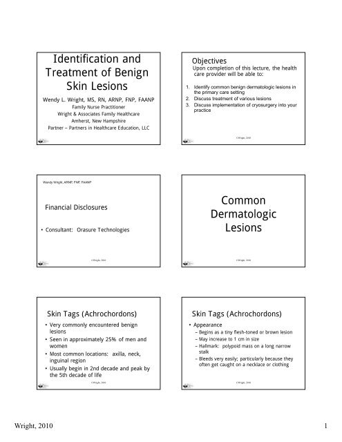
Identification and Treatment of Benign Skin Lesions Common ...
Identification and Treatment of Benign Skin Lesions Common ...
Identification and Treatment of Benign Skin Lesions Common ...

Create successful ePaper yourself
Turn your PDF publications into a flip-book with our unique Google optimized e-Paper software.
<strong>Identification</strong> <strong>and</strong><br />
<strong>Treatment</strong> <strong>of</strong> <strong>Benign</strong><br />
<strong>Skin</strong> <strong>Lesions</strong><br />
Wendy L. Wright, MS, RN, ARNP, FNP, FAANP<br />
Family Nurse Practitioner<br />
Wright & Associates Family Healthcare<br />
Amherst, New Hampshire<br />
Partner – Partners in Healthcare Education, LLC<br />
Objectives<br />
Upon completion <strong>of</strong> this lecture, the health<br />
care provider will be able to:<br />
1. Identify common benign dermatologic lesions in<br />
the primary care setting<br />
2. Discuss treatment <strong>of</strong> various lesions<br />
3. Discuss implementation <strong>of</strong> cryosurgery into your<br />
practice<br />
©Wright, 2010<br />
Wendy Wright, ARNP, FNP, FAANP<br />
Financial Disclosures<br />
• Consultant: Orasure Technologies<br />
<strong>Common</strong><br />
Dermatologic<br />
<strong>Lesions</strong><br />
©Wright, 2010<br />
©Wright, 2010<br />
<strong>Skin</strong> Tags (Achrochordons)<br />
• Very commonly encountered benign<br />
lesions<br />
• Seen in approximately 25% <strong>of</strong> men <strong>and</strong><br />
women<br />
• Most common locations: axilla, neck,<br />
inguinal region<br />
• Usually begin in 2nd decade <strong>and</strong> peak by<br />
the 5th decade <strong>of</strong> life<br />
©Wright, 2010<br />
<strong>Skin</strong> Tags (Achrochordons)<br />
• Appearance<br />
– Begins as a tiny flesh-toned or brown lesion<br />
– May increase to 1 cm in size<br />
– Hallmark: polypoid mass on a long narrow<br />
stalk<br />
– Bleeds very easily; particularly because they<br />
<strong>of</strong>ten get caught on a necklace or clothing<br />
©Wright, 2010<br />
Wright, 2010 1
Diagnosis<br />
Linked with____________<br />
<strong>Skin</strong> Tags<br />
©Wright, 2010<br />
©Wright, 2010<br />
<strong>Skin</strong> Tags (Achrochordons)<br />
•<strong>Treatment</strong><br />
–Shave excision<br />
– Cryosurgery<br />
– Electrocautery<br />
©Wright, 2010<br />
Dermat<strong>of</strong>ibroma<br />
• <strong>Common</strong>, benign asymptomatic lesions<br />
• May be slightly itchy; Retract beneath the<br />
skin when you try to elevate them<br />
• 1-1010 lesions occurring on the extremities;<br />
most common location is the anterior<br />
surface <strong>of</strong> the lower leg<br />
• Etiology: fibrous reaction to trauma, virus<br />
or an insect bite<br />
– Multiple lesions: Systemic lupus<br />
©Wright, 2010<br />
Dermat<strong>of</strong>ibroma<br />
Dermat<strong>of</strong>ibroma<br />
•<strong>Treatment</strong><br />
– Elliptical excision<br />
–Shave excision<br />
– Cryosurgery<br />
©Wright, 2010<br />
©Wright, 2010<br />
Wright, 2010 2
Verruca Vulgaris<br />
• <strong>Common</strong> warts<br />
• <strong>Benign</strong> lesions <strong>of</strong> the epidermis caused by a<br />
virus<br />
• Transmitted by touch <strong>and</strong> commonly appear<br />
at sites <strong>of</strong> trauma, on the h<strong>and</strong>s, around the<br />
periungual regions from nail biting <strong>and</strong> on the<br />
plantar surfaces <strong>of</strong> the feet<br />
Verruca Vulgaris<br />
• Appearance<br />
– Smooth, flesh colored papules which<br />
evolve into a dome-shaped growth with<br />
black dots on the surface<br />
– Black dots are thrombosed capillaries <strong>and</strong><br />
can be visualized with a 15 blade<br />
•<strong>Treatment</strong><br />
– OTC products: Compound W; Duoplant<br />
©Wright, 2010<br />
©Wright, 2010<br />
Verruca Vulgaris<br />
Verruca Vulgaris<br />
• <strong>Treatment</strong><br />
– Liquid nitrogen<br />
– Cryosurgery<br />
– Electrocautery<br />
–Duct Tape<br />
– Blunt dissection (plantar lesions)<br />
– Tagamet 600 mg bid x 2 - 4 weeks<br />
–Imiquimod<br />
©Wright, 2010<br />
©Wright, 2010<br />
Condyloma Acuminata<br />
•AKA: genital warts<br />
• Etiology: human papillomavirus<br />
– More than 30 strains <strong>of</strong> HPV infect the genital<br />
tract<br />
– Usually caused by HPV subtypes 6, 11, 40 –<br />
45, <strong>and</strong> 51<br />
• Transmitted through sexual contact<br />
• Incubation period <strong>of</strong> 1 – 6 months<br />
Condyloma Acuminata<br />
• Characteristics<br />
– Cauliflower appearing lesion<br />
– White or flesh toned<br />
– May be associated with abnormal pap<br />
smear<br />
©Wright, 2010<br />
©Wright, 2010<br />
Wright, 2010 3
Condylomata Acuminata<br />
Condyloma Acuminata<br />
•<strong>Treatment</strong><br />
– Cryosurgery<br />
–Imiquimod 5% cream<br />
– TCA<br />
– Electrodessication<br />
–Laser<br />
– Pod<strong>of</strong>ilox<br />
©Wright, 2010<br />
©Wright, 2010<br />
Seborrheic Keratoses<br />
• Most common benign skin lesion<br />
• Unknown origin<br />
•No potential for malignancy<br />
• Characteristics<br />
– Smooth surface with tiny round, embedded<br />
pearls<br />
– May be rough, dry <strong>and</strong> cracked<br />
– Appear stuck on the surface<br />
©Wright, 2010<br />
Seborrheic Keratoses<br />
©Wright, 2010<br />
Seborrheic Keratosis<br />
Can Mimic a Malignant Melanoma<br />
©Wright, 2010<br />
©Wright, 2010<br />
Wright, 2010 4
Seborrheic Keratoses<br />
•<strong>Treatment</strong><br />
– <strong>Lesions</strong> are only removed for cosmetic<br />
purposes or for a biopsy if pathology is<br />
unknown<br />
– If removed, shave excision<br />
– Cryosurgery<br />
©Wright, 2010<br />
Molluscum Contagiosum<br />
• Infection caused by the pox-virus<br />
• Most commonly seen on the face, trunk <strong>and</strong><br />
axillae<br />
• Self-limiting<br />
• Spread by auto-inoculation<br />
• Incubation period: 2-7 weeks after<br />
exposure<br />
• Contagious until gone<br />
©Wright, 2010<br />
Molluscum Contagiosum<br />
• Asymptomatic lumps<br />
• May have 1 - hundreds<br />
• Physical Examination<br />
– 2-5mm papule p with an umbilicated center<br />
– Flesh toned - white in color<br />
– Most <strong>of</strong>ten around the eye in children<br />
– Scaling <strong>and</strong> erythema around the periphery <strong>of</strong> the lesion<br />
is not unusual<br />
– If in the genital area <strong>of</strong> a child-should consider sexual<br />
abuse<br />
©Wright, 2010<br />
Molluscum Contagiosum<br />
©Wright, 2010<br />
Molluscum Contagiosum<br />
•Plan<br />
–Diagnostic:<br />
• None or KOH prep looking for inclusion bodies<br />
– Therapeutic:<br />
• Conservative treatment is the best for children<br />
–Curettage<br />
–Cryosurgery<br />
–Tretinoin<br />
Molluscum Contagiosum<br />
•Plan<br />
– Therapeutic:<br />
• Salicylic Acid (Occlusal)<br />
• Laser<br />
•TCA<br />
•Imiquimod<br />
©Wright, 2010<br />
©Wright, 2010<br />
Wright, 2010 5
Molluscum Contagiosum<br />
•Plan<br />
–Educational<br />
• May resolve on own in 6 - 9 months<br />
• Contagious until lesions are gone<br />
•<strong>Benign</strong><br />
• Recurrence very common<br />
©Wright, 2010<br />
Actinic Keratoses<br />
• <strong>Common</strong> sun-induced premalignant lesions<br />
• Incidence: Increases with age, light<br />
complexion<br />
• Clinical presentation: Slightly roughened<br />
area that <strong>of</strong>ten bleeds when excoriated<br />
– Progresses to an adherent yellow crust<br />
– Size 3-6 mm<br />
– <strong>Common</strong> location: scalp, temples, forehead,<br />
h<strong>and</strong>s ©Wright, 2010<br />
Actinic Keratoses<br />
•Prognosis<br />
– Can spontaneously regress if sun exposure is<br />
eliminated<br />
– Good prognosis if treated t adequately<br />
– Small percentage transform into a squamous<br />
cell carcinoma which can metastasize<br />
• 60% <strong>of</strong> all squamous cell carcinomas began<br />
as an actinic keratosis<br />
Actinic Keratosis<br />
©Wright, 2010<br />
©Wright, 2010<br />
Actinic Keratosis<br />
•Keratin may<br />
accumulate <strong>and</strong><br />
transform lesion<br />
into a cutaneous<br />
horn<br />
• Frequently seen on<br />
the pinna <strong>of</strong> the ear<br />
Actinic Keratoses<br />
• <strong>Treatment</strong><br />
– Cryosurgery<br />
• preferred method<br />
– Surgical Removal<br />
–Tretinoin<br />
–5-fluorouracil<br />
– Acid peels<br />
– Sunscreen<br />
–Imiquimod<br />
©Wright, 2010<br />
©Wright, 2010<br />
Wright, 2010 6
Cryosurgery<br />
• Definition: The process <strong>of</strong> applying extreme<br />
cold to a lesion for the purpose <strong>of</strong><br />
destruction<br />
• Indications<br />
– Seborrheic keratoses<br />
– Actinic keratoses<br />
–<strong>Skin</strong> tags<br />
– Verruca vulgaris<br />
Cryosurgery<br />
• Indications<br />
–Plantar warts<br />
– Condyloma acuminatum<br />
– Molluscum contagiosum<br />
• Advantages<br />
– Minimal discomfort<br />
– Minimal scarring<br />
– No sutures needed<br />
©Wright, 2010<br />
©Wright, 2010<br />
Cryosurgery<br />
• Disadvantages<br />
– May not be effective for all lesions, particularly<br />
warts<br />
– Some individuals report moderate pain during<br />
the procedure<br />
•Equipment<br />
–Betadine<br />
– 4X4 gauze<br />
– Freeze kit or nitrous oxide cryosurgery unit<br />
©Wright, 2010<br />
Cryosurgery<br />
•Equipment<br />
– Cotton applicators<br />
– Vaseline petroleum jelly<br />
– Dressing<br />
©Wright, 2010<br />
Cryosurgery<br />
• Procedure<br />
– Position the patient for provider comfort<br />
– Cleanse the lesion with betadine<br />
– Cover the lesion with a water soaked dressing for 5-10<br />
minutes<br />
– Using the cotton applicators, surround the lesion with<br />
vaseline petroleum jelly<br />
– Choose the appropriate w<strong>and</strong> for the lesion<br />
– Freeze the lesion for the appropriate amount <strong>of</strong> time<br />
Cryosurgery<br />
©Wright, 2010<br />
©Wright, 2010<br />
Wright, 2010 7
Cryosurgery<br />
Cryosurgery<br />
©Wright, 2010<br />
©Wright, 2010<br />
Cryosurgery<br />
• Time Frame<br />
– Utilize charts provided with your particular cryokit/device<br />
– I.e. 20 – 40 seconds<br />
– Liquid nitrogen – much, much less than this<br />
Cryosurgery<br />
•Time Frame<br />
– Another method is to apply the freeze until<br />
a frost ring appears approximately 1-2 mm<br />
around the lesion<br />
©Wright, 2010<br />
©Wright, 2010<br />
Cryosurgery<br />
• Procedure<br />
– Apply additional pressure for deeper tissue<br />
penetration<br />
– Cover with a dressing<br />
• Follow-up<br />
– Monitor for redness, discharge, fever, pain,<br />
streaking<br />
– Recheck lesion in 4 - 6 weeks<br />
©Wright, 2010<br />
Cryosurgery<br />
•Red Flags<br />
– Malignant lesions<br />
– Facial lesions<br />
– Infected lesion<br />
•CPT Code<br />
– 17000: Destruction, any method, all benign<br />
– 17110: Destruction, flat warts or molluscum<br />
contagiosum<br />
©Wright, 2010<br />
Wright, 2010 8
Case Study<br />
• 47 year-old female presents with the<br />
following:<br />
– 2 verruca vulgaris lesions on the soles <strong>of</strong> her feet<br />
– 1 actinic keratosis on her right h<strong>and</strong><br />
– Assessments:<br />
•1. Warts<br />
– CPT: 17110: 1-14 lesions (cryo)<br />
• 2. Actinic keratosis<br />
– CPT: 17000 (1 lesion) – destroy benign/premalig lesion<br />
– Mod 57 (see <strong>and</strong> perform destruction today)<br />
©Wright, 2010<br />
–<br />
Thank You<br />
I Would Be Happy To<br />
Entertain t Any<br />
Questions That You<br />
May Have!!<br />
©Wright, 2010<br />
Wendy L. Wright, MS, RN, ARNP, FNP, FAANP<br />
(W) 603-249-8883<br />
(H) 603-472-6776<br />
(F) 603-472-2597<br />
Email: WendyARNP@aol.com<br />
©Wright, 2010<br />
Wright, 2010 9