

Parathyroidectomy - Vula - University of Cape Town
Parathyroidectomy - Vula - University of Cape Town
Parathyroidectomy - Vula - University of Cape Town

Create successful ePaper yourself
Turn your PDF publications into a flip-book with our unique Google optimized e-Paper software.
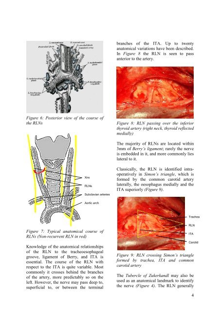
anches <strong>of</strong> the ITA. Up to twenty<br />
anatomical variations have been described.<br />
In Figure 8 the RLN is seen to pass<br />
anterior to the artery.<br />
Figure 6: Posterior view <strong>of</strong> the course <strong>of</strong><br />
the RLNs<br />
Figure 8: RLN passing over the inferior<br />
thyroid artery (right neck, thyroid reflected<br />
medially)<br />
The majority <strong>of</strong> RLNs are located within<br />
3mm <strong>of</strong> Berry’s ligament; rarely the nerve<br />
is embedded in it, and more commonly lies<br />
lateral to it.<br />
Xns<br />
RLNs<br />
Subclavian arteries<br />
Classically, the RLN is identified intraoperatively<br />
in Simon’s triangle, which is<br />
formed by the common carotid artery<br />
laterally, the oesophagus medially and the<br />
ITA superiorly (Figure 9).<br />
Aortic arch<br />
Trachea<br />
Figure 7: Typical anatomical course <strong>of</strong><br />
RLNs (Non-recurrent RLN in red)<br />
Knowledge <strong>of</strong> the anatomical relationships<br />
<strong>of</strong> the RLN to the tracheoesophageal<br />
groove, ligament <strong>of</strong> Berry, and ITA is<br />
essential. The course <strong>of</strong> the RLN with<br />
respect to the ITA is quite variable. Most<br />
commonly it crosses behind the branches<br />
<strong>of</strong> the artery, more predictably so on the<br />
left. However, the nerve may pass deep to,<br />
superficial to, or between the terminal<br />
RLN<br />
ITA<br />
Carotid<br />
Figure 9: RLN crossing Simon’s triangle<br />
formed by trachea, ITA and common<br />
carotid artery<br />
The Tubercle <strong>of</strong> Zukerkandl may also be<br />
used as an anatomical landmark to identify<br />
the nerve (Figure 4). The RLN generally<br />
4


