

Parathyroidectomy - Vula - University of Cape Town
Parathyroidectomy - Vula - University of Cape Town
Parathyroidectomy - Vula - University of Cape Town

Create successful ePaper yourself
Turn your PDF publications into a flip-book with our unique Google optimized e-Paper software.
OPEN ACCESS ATLAS OF OTOLARYNGOLOGY, HEAD &<br />
NECK OPERATIVE SURGERY<br />
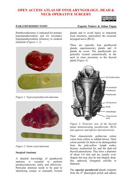
PARATHYROIDECTOMY<br />
<strong>Parathyroidectomy</strong> is indicated for primary<br />
hyperparathyroidism and for secondary<br />
hyperparathyroidism refractory to medical<br />
treatment (Figures 1, 2).<br />
Eugenio Panieri & Johan Fagan<br />
glands and to avoid injury to important<br />
local structures, particularly the recurrent<br />
laryngeal nerve (RLN).<br />
There are typically four parathyroid<br />
glands; supernumerary glands and
to the posterior surface <strong>of</strong> the caudally<br />
migrating thyroid. They have a much<br />
shorter distance to migrate than the inferior<br />
parathyroid glands; this would account for<br />
their more predictable location. They are<br />
embryologically and anatomically closely<br />
related to the Tubercle <strong>of</strong> Zuckerkandl, and<br />
are usually found posteriorly at the level <strong>of</strong><br />
the upper two-thirds <strong>of</strong> the thyroid about<br />
1cm above the crossing point <strong>of</strong> the RLN<br />
and inferior thyroid artery (ITA) (Figure<br />
4).<br />
Figure 4: Tubercle <strong>of</strong> Zuckerkandl (TZ)<br />
and its relationship to the superior<br />
parathyroid gland and RLN<br />
Ectopic superior parathyroids are<br />
uncommon (1%) and may be found in the<br />
posterior neck, retropharyngeal and retroesophageal<br />
spaces and intrathyroidally<br />
(Figure 5).<br />
TZ<br />
Sup parathyroid<br />
RLN<br />
The inferior parathyroid glands arise from<br />
the dorsal wings <strong>of</strong> the 3 rd pharyngeal<br />
pouches. They join the thymus as it<br />
migrates caudally and medially to its final<br />
position in the mediastinum. Ectopic<br />
inferior parathyroid glands can be found<br />
anywhere along this large area <strong>of</strong> descent<br />
from the proximal neck down to the<br />
superior border <strong>of</strong> the pericardium. Their<br />
commonest location is on the anterior or<br />
the posterolateral surfaces <strong>of</strong> the lower<br />
pole <strong>of</strong> the thyroid, between the lower pole<br />
<strong>of</strong> thyroid and thyroid isthmus (42%,<br />
Wang et al); or within the thyrothymic<br />
ligament in the lower neck in proximity to<br />
the thymus (39%). Other locations include<br />
lateral to the thyroid, within the carotid<br />
sheath (15%), or within mediastinal thymic<br />
tissue and pericardium (2%). (Figure 5)<br />
The inferior parathyroids are usually found<br />
in a plane more ventral to that <strong>of</strong> the<br />
superior glands. If the RLN’s course is<br />
viewed in a coronal plane, then the<br />
superior parathyroid glands are located<br />
deep (dorsal) and the inferior parathyroid<br />
superficial (ventral) to the plane <strong>of</strong> the<br />
nerve (Figures 6, 7).<br />
Thyroid cartilage<br />
Superior parathyroid<br />
Oesophagus<br />
Inferior parathyroid<br />
Thymus<br />
Figure 6: The superior parathyroid gland<br />
lies deep (dorsal) and the inferior<br />
parathyroid superficial (ventral) to a<br />
coronal plane along course <strong>of</strong> RLN<br />
Figure 5: Ectopic parathyroids in retropharyngeal<br />
space and mediastinum<br />
2
Figure 7: The superior and inferior<br />
parathyroids relative to a coronal plane<br />
along the course <strong>of</strong> the RLN<br />
The inferior thyroid artery (ITA) is a<br />
branch <strong>of</strong> the thyrocervical trunk, which in<br />
turn arises from the subclavian artery<br />
(Figures 3, 5). It is the predominant<br />
vascular supply to both the upper and<br />
lower parathyroids (Figures 3, 5). Consequently<br />
division <strong>of</strong> the main trunk <strong>of</strong> the<br />
ITA during thyroidectomy is discouraged<br />
as it places both parathyroids at risk <strong>of</strong><br />
ischaemic injury.<br />
STA<br />
ITA<br />
Inferior PT<br />
Superior PT<br />
RLN<br />
Thyrocervical<br />
Subclavian<br />
Figure 5: Superior thyroid artery (STA),<br />
subclavian artery, thyrocervical trunk and<br />
inferior thyroid artery (ITA)<br />
The ITA courses superiorly along the<br />
surface <strong>of</strong> the anterior scalene muscle<br />
before turning medially behind the carotid<br />
sheath from where it reaches the inferior<br />
pole <strong>of</strong> the thyroid gland. It provides blood<br />
supply to the parathyroids, thyroid, upper<br />
oesophagus and trachea. Its branches<br />
communicate with the superior thyroid<br />
artery (STA) and with the blood supply <strong>of</strong><br />
the contralateral thyroid lobe via the<br />
thyroid isthmus.<br />
The Recurrent Laryngeal Nerve (RLN) is<br />
a key structure in any exploration <strong>of</strong> the<br />
central neck. Identification and preservation<br />
<strong>of</strong> the RLN during thyroid and parathyroid<br />
surgery is essential to minimise<br />
morbidity. The RLN innervates all the<br />
intrinsic muscles <strong>of</strong> the larynx except the<br />
cricothyroid muscle (SLN) and provides<br />
sensory innervation to the larynx. Even<br />
minor neuropraxia may cause dysphonia;<br />
irreversible injury confers permanent<br />
hoarseness. The incidence <strong>of</strong> RLN injury<br />
during thyroidectomy is 0-28% and is the<br />
most common reason for medicolegal<br />
claims following thyroidectomy; the<br />
incidence <strong>of</strong> injury during parathyroidectomy<br />
is much lower.<br />
The RLNs originate from the Xn. After<br />
circling around the subclavian artery<br />
(right) and aortic arch (left) the RLNs<br />
course superiorly and medially toward the<br />
tracheoesophageal groove (Figures 6, 7).<br />
The right RLN enters the root <strong>of</strong> the neck<br />
from a more lateral direction and its course<br />
is less predictable than that <strong>of</strong> the left. The<br />
RLNs enter the larynx deep to the inferior<br />
constrictor muscles and posterior to the<br />
cricothyroid joint.<br />
The RLN may be non-recurrent in<br />
approximately 0.6% <strong>of</strong> patients i.e. it does<br />
not pass around the subclavian artery but<br />
branches from the Xn higher up in the<br />
neck, passing directly to the larynx close to<br />
the superior thyroid vessels (Figure 7).<br />
This aberration almost always occurs on<br />
the right side and is associated with a<br />
retroesophageal subclavian artery.<br />
3
anches <strong>of</strong> the ITA. Up to twenty<br />
anatomical variations have been described.<br />
In Figure 8 the RLN is seen to pass<br />
anterior to the artery.<br />
Figure 6: Posterior view <strong>of</strong> the course <strong>of</strong><br />
the RLNs<br />
Figure 8: RLN passing over the inferior<br />
thyroid artery (right neck, thyroid reflected<br />
medially)<br />
The majority <strong>of</strong> RLNs are located within<br />
3mm <strong>of</strong> Berry’s ligament; rarely the nerve<br />
is embedded in it, and more commonly lies<br />
lateral to it.<br />
Xns<br />
RLNs<br />
Subclavian arteries<br />
Classically, the RLN is identified intraoperatively<br />
in Simon’s triangle, which is<br />
formed by the common carotid artery<br />
laterally, the oesophagus medially and the<br />
ITA superiorly (Figure 9).<br />
Aortic arch<br />
Trachea<br />
Figure 7: Typical anatomical course <strong>of</strong><br />
RLNs (Non-recurrent RLN in red)<br />
Knowledge <strong>of</strong> the anatomical relationships<br />
<strong>of</strong> the RLN to the tracheoesophageal<br />
groove, ligament <strong>of</strong> Berry, and ITA is<br />
essential. The course <strong>of</strong> the RLN with<br />
respect to the ITA is quite variable. Most<br />
commonly it crosses behind the branches<br />
<strong>of</strong> the artery, more predictably so on the<br />
left. However, the nerve may pass deep to,<br />
superficial to, or between the terminal<br />
RLN<br />
ITA<br />
Carotid<br />
Figure 9: RLN crossing Simon’s triangle<br />
formed by trachea, ITA and common<br />
carotid artery<br />
The Tubercle <strong>of</strong> Zukerkandl may also be<br />
used as an anatomical landmark to identify<br />
the nerve (Figure 4). The RLN generally<br />
4
courses between this structure and the<br />
trachea. However, this relationship can<br />
vary with enlargement <strong>of</strong> the tuberculum<br />
thereby placing the nerve at risk during<br />
surgical exploration.<br />
Superior Laryngeal Nerve (SLN)<br />
The SLN is a branch <strong>of</strong> the Xn and has<br />
both an external and internal branch<br />
(Figures 10, 11). The internal branch is<br />
situated above and outside the normal field<br />
<strong>of</strong> dissection; it is sensory and enters the<br />
larynx through the thyrohyoid membrane.<br />
The external branch innervates the<br />
cricothyroid muscle, a tensor <strong>of</strong> the vocal<br />
cord. Injury to the SLN causes hoarseness,<br />
decreased pitch and/or volume, and voice<br />
fatigue. These voice changes are more<br />
subtle than those relating to RLN injury<br />
are frequently underestimated and not<br />
reported. The external branch is at risk<br />
because <strong>of</strong> its close proximity to the STA<br />
(Figures 10, 11). Understanding its relationship<br />
to the upper pole <strong>of</strong> the thyroid<br />
and the STA is crucial to preserving its<br />
integrity.<br />
XIIn<br />
SLN (internal)<br />
SLN (external)<br />
STA<br />
Sup pole<br />
thyroid<br />
Figure 10: Anatomical relations <strong>of</strong><br />
internal and external branches <strong>of</strong> right<br />
SLN to STA and to superior pole <strong>of</strong> thyroid<br />
The usual configuration is that the nerve is<br />
located behind the STA, proximal to its<br />
entry into the superior pole <strong>of</strong> the thyroid.<br />
The relationship <strong>of</strong> the nerve to the<br />
superior pole and STA is however<br />
extremely variable. Variations include the<br />
nerve passing between the branches <strong>of</strong> the<br />
STA as it enters the superior pole <strong>of</strong> the<br />
thyroid gland; in such cases it is<br />
particularly vulnerable to injury.<br />
Figure 11: Note close proximity <strong>of</strong> external<br />
branch <strong>of</strong> SLN to STA and thyroid vein and<br />
to superior pole <strong>of</strong> thyroid gland<br />
Types <strong>of</strong> parathyroidectomy<br />
SLN Ext branch<br />
STA<br />
Thyroid<br />
STVs<br />
Focused parathyroidectomy: This is the<br />
usual procedure for a well-localised<br />
solitary adenoma. The <strong>of</strong>fending gland is<br />
removed through a limited incision with<br />
direct exposure <strong>of</strong> the previously imaged<br />
parathyroid adenoma.<br />
Bilateral neck exploration: In cases <strong>of</strong><br />
unsuccessful preoperative localisation the<br />
surgeon explores the necks fully, identifies<br />
all four parathyroid glands and removes<br />
the adenoma.<br />
Subtotal parathyroidectomy: This is<br />
indicated with parathyroid hyperplasia<br />
when all the glands have the capacity for<br />
increased parathyroid hormone (PTH)<br />
production. This occurs in secondary and<br />
tertiary hyperparathyroidism and in the<br />
unusual situation <strong>of</strong> primary hyperparathyroidism<br />
due to multiple gland hyperplasia.<br />
The three largest glands are<br />
removed and a small remnant <strong>of</strong> the most<br />
normal-looking gland is either left in situ<br />
5
or transplanted to an ectopic site, typically<br />
the forearm.<br />
Total parathyroidectomy: All parathyroid<br />
tissue is removed. This may be done as a<br />
salvage procedure in cases <strong>of</strong> recurrent<br />
secondary hyperparathyroidism.<br />
Preoperative evaluation: Primary<br />
hyperparathyroidism<br />
Endocrine diagnosis: The diagnosis <strong>of</strong><br />
primary hyperparathyroidism hinges on<br />
identifying an inappropriately raised PTH<br />
assay in the presence <strong>of</strong> elevated serum<br />
calcium. It is not unusual for hypercalcaemia<br />
symptoms <strong>of</strong> primary hyperparathyroidism<br />
to be non-specific and<br />
vague; hence it is <strong>of</strong>ten underestimated.<br />
Typical presentations include recurrent<br />
renal calculi, progressive bone density<br />
loss, pathological fractures, ill-defined<br />
musculoskeletal complaints, neurocognitive<br />
impairment, and unexplained abdominal<br />
pain; or it may present as a<br />
hypercalcaemic crisis. While the diagnosis<br />
<strong>of</strong> primary hyperparathyroidism is not<br />
difficult to make for surgeons well versed<br />
in endocrine disorders, the occasional<br />
parathyroid surgeon is well advised to<br />
consult an endocrinologist prior to<br />
proceeding with surgery.<br />
SestaMIBI scan: This is a nuclear<br />
medicine imaging technique with the<br />
highest sensitivity and specificity for<br />
identification <strong>of</strong> primary hyperparathyroid<br />
adenomas and is the author’s investigation<br />
<strong>of</strong> choice. The use <strong>of</strong> Technetium 99m<br />
(Tc 99m ) sestamibi for parathyroid imaging<br />
has led to a steady refinement <strong>of</strong> imaging.<br />
The accuracy is determined by the<br />
scanning technique employed; the dualisotope<br />
(I 123 / Tc 99m sestamibi) scan provides<br />
better accuracy than the simpler<br />
sestamibi washout method. The pathological<br />
parathyroid can be localised preoperatively<br />
with great confidence allowing<br />
for a quicker and more focused neck<br />
exploration. Recent data would suggest<br />
that the combination CT-99mTc-sestamibi-<br />
SPECT (reported sensitivity and specificity<br />
<strong>of</strong> up to 88% and 99% respectively) is the<br />
best approach for preoperative localisation<br />
with single gland disease. Unfortunately,<br />
these favourable results do not apply to all<br />
patients with parathyroid disease. It is<br />
important to note that failed localisation<br />
does not exclude primary hyperparathyroidism<br />
and is not a contraindication for<br />
surgical exploration.<br />
Ultrasonography (US): The thyroid and<br />
surrounding structures can be evaluated<br />
with US. It has gained popularity due to<br />
the ease <strong>of</strong> the technique; many endocrine<br />
surgeons have the expertise to evaluate the<br />
neck in their own <strong>of</strong>fices. Resolution has<br />
improved with newer generation equipment.<br />
The weaknesses <strong>of</strong> US are similar to<br />
99mTc-sestamibi i.e. deep superior glands,<br />
ectopic glands and too-small glands are<br />
difficult to localise, and the mediastinum is<br />
inaccessible.<br />
CT and MRI: CT and MRI are not<br />
indicated as first-line investigations, but<br />
may be useful to evaluate parathyroid<br />
adenomas with non-localising imaging<br />
studies or previously operated necks.<br />
Preoperative evaluation: Secondary<br />
hyperparathyroidism<br />
This diagnosis requires identification <strong>of</strong> an<br />
inappropriately raised PTH assay in the<br />
presence <strong>of</strong> an abnormally elevated serum<br />
phosphate-to-calcium ratio. The serum<br />
calcium typically falls within the normal<br />
range. Secondary hyperparathyroidism is<br />
almost always diagnosed in patients with<br />
chronic renal failure. Rarer causes include<br />
osteomalacia, rickets, and malabsorption.<br />
Secondary hyperparathyroidism contri-<br />
6
utes significantly to renal osteodystrophy<br />
and is associated with accelerated<br />
atherosclerosis, ectopic s<strong>of</strong>t tissue calcification<br />
and skin ulcers in calciphylaxis. It<br />
contributes to a general feeling <strong>of</strong> malaise<br />
and musculoskeletal pain seen in chronic<br />
renal failure, and chronic pruritus. Close<br />
collaboration with a renal physician is<br />
essential to determine the best time for<br />
surgical intervention.<br />
Preoperative consent<br />
Scar: The incision is typically well-hidden<br />
within a natural skin crease <strong>of</strong> the neck,<br />
but tends to descend with ageing.<br />
Airway obstruction/wound haematoma:<br />
longer than 4-5cm. Well-localized glands<br />
can be resected via a smaller incision.<br />
Placing the incision too low causes an<br />
unsightly low scar over the heads <strong>of</strong> the<br />
clavicles when the extended neck is<br />
returned to its normal position.<br />
Figure 12: Curvilinear skin incision two<br />
finger breadths above the sternal notch<br />
Subplatysmal flaps: Subcutaneous fat and<br />
platysma are divided, and a subplatysmal<br />
dissection plane is developed superiorly<br />
(platysma is <strong>of</strong>ten absent in the midline)<br />
remaining superficial to the anterior<br />
jugular veins, up to the level <strong>of</strong> the thyroid<br />
cartilage above, and the sternal notch<br />
below (Figure 13).<br />
Figure 13: Subplatysmal flaps elevated<br />
The skin flaps are secured with a fixed<br />
retractor (Figure 14).<br />
Figure 14: Subplatysmal skin flaps held<br />
with Jowell’s retractor. Note anterior<br />
jugular veins (AJVs)<br />
Surgical Approaches<br />
1. Lateral approach involves dissecting<br />
along the medial border <strong>of</strong> the sternocleidomastoid<br />
muscle to the carotid<br />
sheath, and then medial to the sheath<br />
up to the thyroid region. This is<br />
employed in cases <strong>of</strong> confident preoperative<br />
localisation.<br />
2. Anterior approach involves medial<br />
mobilisation <strong>of</strong> the thyroid gland. This<br />
is used in cases <strong>of</strong> possible bilateral<br />
neck exploration. It is favoured by the<br />
author, and is described in more detail<br />
below.<br />
Separating strap muscles and exposing<br />
the anterior surface <strong>of</strong> thyroid: The fascia<br />
between the sternohyoid and sternothyroid<br />
muscles is divided in the midline with<br />
diathermy or scissors (Figure 15). This is<br />
an avascular plane, though care must be<br />
taken not to injure small veins occasionally<br />
crossing between the anterior jugular<br />
veins, particularly inferiorly. The<br />
infrahyoid (sternohyoid, sternothyroid and<br />
omohyoid) strap muscles are retracted<br />
laterally with a right-angled retractor. In<br />
difficult cases the strap muscles may be<br />
divided to improve access.<br />
8
delivery <strong>of</strong> the bulk <strong>of</strong> the thyroid lobe into<br />
the wound (Figure 17). Although dividing<br />
it is not always essential, it is better to do<br />
so than to risk tearing it.<br />
Figure 15: Fascia between sternohyoid<br />
and sternothyroid muscles divided to<br />
expose thyroid gland<br />
It is usual at this stage for the surgeon to<br />
move to the side <strong>of</strong> the table opposite to<br />
the parathyroid to be resected.<br />
Delivery <strong>of</strong> thyroid towards midline: The<br />
infrahyoid (sternohyoid, sternothyroid and<br />
omohyoid) strap muscles are retracted<br />
laterally with a right-angled retractor. The<br />
thyroid gland is delivered medially by<br />
applying gentle digital traction to the gland<br />
(Figure 16).<br />
Figure 16: Medial rotation <strong>of</strong> (R) thyroid<br />
lobe exposes the middle thyroid vein<br />
Division <strong>of</strong> middle thyroid vein(s): The<br />
vein is the first key vascular structure to be<br />
encountered and is tightly stretched by<br />
medial traction on the gland (Figure 16).<br />
Dividing the vein facilitates additional<br />
mobilisation <strong>of</strong> the gland and permits<br />
Figure 17: Dividing the middle thyroid<br />
vein<br />
If the surgeon is confident about<br />
preoperative localisation then dissection is<br />
next directed at the parathyroid adenoma.<br />
Identifying superior parathyroid: Full<br />
mobilisation and anterior delivery <strong>of</strong> the<br />
upper pole <strong>of</strong> the thyroid brings the region<br />
<strong>of</strong> the superior parathyroid gland into<br />
direct view. The superior parathyroid gland<br />
is normally located in a posterior position<br />
at the level <strong>of</strong> the upper two-thirds <strong>of</strong> the<br />
thyroid, and is closely related to the<br />
Tubercle <strong>of</strong> Zuckerkandl; it is about 1cm<br />
above the crossing point <strong>of</strong> the RLN and<br />
ITA. If the RLN’s course is viewed in a<br />
coronal plane, then the superior<br />
parathyroid gland lies deep (dorsal) to the<br />
plane <strong>of</strong> the nerve (Figures 4, 6, 7). It has a<br />
characteristic rich orange/yellow colour<br />
(Figures 18, 19). The (occasional) parathyroid<br />
surgeon may find the parathyroids<br />
difficult to identify especially if there has<br />
been bleeding in the surgical field, so care<br />
must be taken to ensure meticulous<br />
haemostasis.<br />
9
Figure 18: Position <strong>of</strong> superior parathyroid<br />
relative to Tubercle <strong>of</strong> Zuckerkandl<br />
(TZ), RLN and STA<br />
TZ<br />
Sup parathyroid<br />
Crossing point<br />
<strong>of</strong> RLN & STA<br />
Inferior PT<br />
parathyroids are most commonly located<br />
between the lower pole <strong>of</strong> the thyroid and<br />
thyroid isthmus, most commonly on the<br />
anterior or posterolateral surfaces <strong>of</strong> the<br />
lower pole <strong>of</strong> the thyroid (42%, Wang et<br />
al), or may be located in the lower neck in<br />
proximity to the thymus (39%). Preserving<br />
it in situ avoids damaging the ITA blood<br />
supply.<br />
Removal <strong>of</strong> adenoma: (Figures 20, 21)<br />
Once the abnormal parathyroid has been<br />
identified, it is removed. Parathyroid tissue<br />
can autotransplant if fragmented, so great<br />
care is taken to deliver an intact gland by<br />
delicately grasping its capsule until the<br />
vascular pedicle is identified. This is then<br />
be ligated with a 3/0 tie.<br />
Superior PT<br />
RLN<br />
Figure 19: Superior and inferior<br />
parathyroids (PT)<br />
The gland must remain in situ with blood<br />
supply intact. This is best achieved by<br />
carefully dissecting it <strong>of</strong>f the posterior<br />
aspect <strong>of</strong> the thyroid gland, and using short<br />
bursts <strong>of</strong> bipolar cautery to control<br />
bleeding. If the gland still cannot be<br />
identified, it is prudent to divide the STA<br />
and completely mobilise the upper pole <strong>of</strong><br />
the thyroid. This will almost certainly<br />
bring the superior parathyroid into view.<br />
Identification <strong>of</strong> inferior parathyroid: The<br />
inferior gland is initially looked for at the<br />
inferior aspect <strong>of</strong> the lower pole <strong>of</strong> the<br />
thyroid or within the thyrothymic ligament.<br />
If the RLN’s course is viewed in a coronal<br />
plane then the inferior parathyroid is<br />
superficial (ventral) to the plane <strong>of</strong> the<br />
nerve (Figures 6, 7, 18, 19). Its location is<br />
more varied than with the superior gland,<br />
but if it is at its expected location then it is<br />
generally simpler to identify. The inferior<br />
Figure 20: Right superior parathyroid<br />
adenoma being removed<br />
Figure 21: Right inferior parathyroid<br />
adenoma<br />
Confirming successful removal: After<br />
identifying the parathyroid glands, it is<br />
10
essential to distinguish between normal<br />
and pathological glands. Simple rules <strong>of</strong><br />
thumb are that increased size correlates<br />
with pathology, and that marked<br />
elevations <strong>of</strong> calcium and PTH levels are<br />
usually caused by larger adenomas.<br />
Adenomas are typically more rounded than<br />
normal parathyroid glands and have a<br />
darker, fleshy parenchymal appearance,<br />
sometimes described as similar to a “rat’s<br />
heart”. They are typically 1-2cm in<br />
diameter and are significantly heavier than<br />
normal glands (Figure 22). Occasionally<br />
small adenomas (
If exploration fails to reveal convincing<br />
evidence <strong>of</strong> a parathyroid adenoma then<br />
two likely scenarios apply:<br />
All 4 parathyroids have been found,<br />
but none looks convincingly pathological:<br />
it is probable that the patient<br />
has four-gland hyperplasia, and a<br />
subtotal parathyroidectomy is indicated.<br />
Frozen section is invaluable to<br />
confirm that removed tissue is truly<br />
parathyroid tissue<br />
If 3 normal-looking glands have been<br />
identified it is probable that the missing<br />
gland is the adenoma and a more<br />
exhaustive search is required<br />
A missing superior parathyroid gland is<br />
almost always located where it is meant to<br />
be i.e. in its normal position at the level <strong>of</strong><br />
the upper two-thirds <strong>of</strong> the thyroid, in a<br />
posterior position, about 1 cm above the<br />
point where the RLN crosses the ITA. The<br />
surgeon needs to further mobilise the upper<br />
pole <strong>of</strong> the thyroid anteriorly. The<br />
parathyroid may be covered by a fine layer<br />
<strong>of</strong> thyroid capsule; dissecting onto the<br />
thyroid parenchyma itself may release it<br />
and bring it into view. Should it still not be<br />
visible one then needs to transect the STA<br />
to fully mobilize the upper pole. If it still<br />
cannot be located then one may be dealing<br />
with the rare situation <strong>of</strong> ectopic retrooesophageal<br />
and retropharyngeal locations<br />
and these regions need to be<br />
explored.<br />
A missing inferior parathyroid gland<br />
poses more <strong>of</strong> a challenge as the<br />
anatomical variations are greatest. The<br />
following areas need to be systematically<br />
explored:<br />
Lower pole <strong>of</strong> thyroid.<br />
Thyrothymic ligament<br />
Simon’s triangle, just below the ITA<br />
Thymectomy by applying traction to<br />
the thyrothymic ligament<br />
Lateral cervical space, behind the<br />
carotid sheath towards the posterior<br />
mediastinum<br />
Carotid sheath<br />
Retro-oesophageal space<br />
If this still fails to identify pathological<br />
glands it is then reasonable to abandon the<br />
procedure and to consider secondary<br />
exploration after more exhaustive imaging<br />
6-12 months later.<br />
Wound closure<br />
Irrigate the wound<br />
Do a Valsalva manoeuvre and check<br />
for bleeding<br />
Wound drainage is not routinely<br />
required<br />
Approximate the strap muscles in the<br />
midline along 70% <strong>of</strong> their lengths<br />
Close the platysma layer with<br />
interrupted absorbable 3/0 sutures<br />
Skin closure is achieved with a subcuticular<br />
absorbable mon<strong>of</strong>ilament<br />
suture<br />
A light dressing is applied<br />
Postoperative care<br />
Keeping thorough records is invaluable<br />
in cases <strong>of</strong> recurrent hyperparathyroidism.<br />
Therefore keep meticulous<br />
and detailed notes about the extent <strong>of</strong><br />
the exploration noting which normal<br />
parathyroid glands were seen, precisely<br />
where they were located and what<br />
tissue was removed<br />
The intravenous line is removed and a<br />
normal diet is taken as tolerated<br />
Following successful focussed parathyroidectomy<br />
the patient may be<br />
discharged the same day<br />
More complex cases are monitored<br />
overnight for bleeding or airway<br />
obstruction<br />
12
Serum PTH levels are routinely<br />
measured 24hrs postoperatively. A<br />
return to normal PTH levels confirms<br />
a successful operation. A hypocalcaemic<br />
trough occurs 2-5days postoperatively.<br />
Calcium and Vitamin<br />
D1α are commenced pre-emptively if<br />
the PTH reading is low<br />
Specific Scenarios<br />
Secondary hyperparathyroidism: Up to<br />
90% <strong>of</strong> patients meeting criteria for<br />
haemodialysis have secondary hyperparathyroidism.<br />
Surgery is indicated for:<br />
Calciphylaxis<br />
Patient preference<br />
Medical observation not possible<br />
Failure <strong>of</strong> maximum medical therapy<br />
with persistent hypercalcaemia, hypercalcuria,<br />
PTH >800pg/mL and hyperphosphataemia<br />
Osteoporosis<br />
Progressive symptoms e.g. pruritus,<br />
pathologic bone fractures, ectopic s<strong>of</strong>t<br />
tissue calcification, severe vascular<br />
calcification, and bone pain<br />
Three different surgical procedures<br />
involving bilateral neck exploration are<br />
employed for patients with secondary<br />
hyperparathyroidism who fulfil criteria for<br />
surgery. Each operation has its proponents<br />
and an institutional history. There is no<br />
convincing evidence <strong>of</strong> superiority <strong>of</strong> any<br />
one approach:<br />
Subtotal parathyroidectomy i.e. removeing<br />
3½ glands and leaving a ½-<br />
gland remnant in situ (author’s preference)<br />
Total parathyroidectomy (four-gland<br />
resection) with autotransplantation<br />
Total parathyroidectomy (four-gland<br />
resection) without autotransplantation<br />
Repeat surgery for failed exploration:<br />
Such cases should be managed by surgeons<br />
well-versed in the subtleties <strong>of</strong> parathyroid<br />
surgery. The following issues need to be<br />
carefully considered before embarking on<br />
expensive investigations and repeat<br />
surgery:<br />
Is the diagnosis <strong>of</strong> primary hyperparathyroidism<br />
correct, or is there an<br />
alternative explanation for the<br />
endocrine abnormalities<br />
Differentiate recurrent parathyroid<br />
disease from persistent hyperparathyroidism:<br />
did PTH levels drop<br />
following the 1 st operation, or did they<br />
remain elevated<br />
Obtain surgical notes <strong>of</strong> the primary<br />
procedure, histology reports, and<br />
postoperative endocrinology records<br />
If a convincing case can be made for reexploration,<br />
then the most helpful imaging<br />
remains Tc 99m -sestamibi and cervical US.<br />
If both are negative then a CT-scan may<br />
add useful information. Whilst potential<br />
ectopic locations <strong>of</strong> parathyroids should be<br />
considered, the majority <strong>of</strong> missed parathyroid<br />
glands are located within the<br />
cervical region. Repeat neck exploration<br />
for persistent or recurrent disease can be<br />
very difficult as the normal tissue planes<br />
are scarred and it is associated with higher<br />
rates <strong>of</strong> injury to the RLN and permanent<br />
hypoparathyroidism. The lateral or ‘‘backdoor’’<br />
approach (dissection between the<br />
anterior border <strong>of</strong> the sternocleidomastoid<br />
muscle and posterior border <strong>of</strong> the strap<br />
muscles) may be useful as it provides<br />
direct access to the posterior surface <strong>of</strong> the<br />
thyroid gland without encountering scar<br />
tissue from previous surgery done through<br />
the conventional anterior approach.<br />
Radioguided parathyroidectomy: This<br />
technique may be helpful with complex<br />
repeat surgery, but the author does not use<br />
it for routine parathyroidectomy due to<br />
cost and logistic constraints. It is similar to<br />
other radioguided techniques such as<br />
sentinel lymph node biopsy. A para-<br />
13
thyroid-specific radiotracer such as<br />
Tc99m-sestamibi is administered intravenously<br />
approximately 2hrs prior to<br />
surgery; this time delay allows for a high<br />
concentration <strong>of</strong> isotope to be retained<br />
within the parathyroid adenoma whilst it<br />
has started to wash out from other sites <strong>of</strong><br />
uptake such the salivary glands and<br />
thyroid. A gamma camera is used intraoperatively<br />
to identify the adenoma.<br />
Minimally invasive parathyroid surgery:<br />
A number <strong>of</strong> techniques have been devised<br />
to reduce the length <strong>of</strong> the skin incision<br />
and to bring the putative benefits <strong>of</strong><br />
minimally invasive techniques to thyroid<br />
and parathyroid surgery. Minimally<br />
invasive parathyroidectomy can be<br />
performed via a limited 2-3cm cervical<br />
incision with visual assistance <strong>of</strong> an<br />
endoscope, but since it can be performed<br />
with a similarly sized incision with direct<br />
visualisation, this technique is not widely<br />
used.<br />
Useful References<br />
1. Mohebati A, Shaha AR. Anatomy <strong>of</strong><br />
Thyroid and Parathyroid Glands and<br />
Neurovascular Relations. Clin Anat.<br />
2012;25(1):19-31<br />
2. Wang C. The anatomic basis <strong>of</strong><br />
parathyroid surgery. Ann Surg. 1976;<br />
183:271–5<br />
3. Fraser WD. Hyperparathyroidism. The<br />
Lancet. 2009;374:145–58<br />
4. Adler JT, Sippel RS, Chen H. New<br />
Trends in Parathyroid Surgery. Curr<br />
Probl Surg. 2010;47(12):958-1017<br />
5. Lew JI, Solorzano CC. Surgical<br />
Management <strong>of</strong> Primary Hyperparathyroidism:<br />
State <strong>of</strong> the Art. Surg Clin<br />
N Am. 2009;89:1205–25<br />
6. Pitt SC, Sippel RS, Chen H. Secondary<br />
and Tertiary Hyperparathyroidism:<br />
State <strong>of</strong> the Art Surgical Management.<br />
Surg Clin N Am. 2009;89:1227–39<br />
Author<br />
Eugenio Panieri MBChB, FCS<br />
Associate Pr<strong>of</strong>essor<br />
Division <strong>of</strong> General Surgery<br />
<strong>University</strong> <strong>of</strong> <strong>Cape</strong> <strong>Town</strong><br />
<strong>Cape</strong> <strong>Town</strong><br />
South Africa<br />
eugenio.panieri@uct.ac.za<br />
2 nd Author and Editor<br />
Johan Fagan MBChB, FCORL, MMed<br />
Pr<strong>of</strong>essor and Chairman<br />
Division <strong>of</strong> Otolaryngology<br />
<strong>University</strong> <strong>of</strong> <strong>Cape</strong> <strong>Town</strong><br />
<strong>Cape</strong> <strong>Town</strong><br />
South Africa<br />
johannes.fagan@uct.ac.za<br />
THE OPEN ACCESS ATLAS OF<br />
OTOLARYNGOLOGY, HEAD &<br />
NECK OPERATIVE SURGERY<br />
www.entdev.uct.ac.za<br />
The Open Access Atlas <strong>of</strong> Otolaryngology, Head &<br />
Neck Operative Surgery by Johan Fagan (Editor)<br />
johannes.fagan@uct.ac.za is licensed under a Creative<br />
Commons Attribution - Non-Commercial 3.0 Unported<br />
License<br />
14


