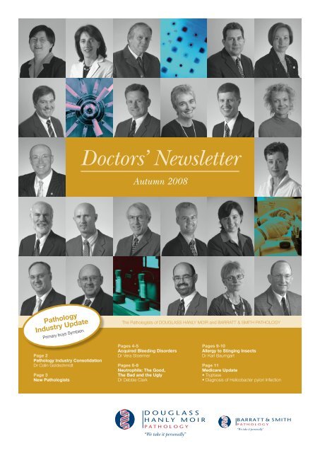
Doctors' Newsletter - Autumn 2008 - Douglass Hanly Moir Pathology
Doctors' Newsletter - Autumn 2008 - Douglass Hanly Moir Pathology
Doctors' Newsletter - Autumn 2008 - Douglass Hanly Moir Pathology

You also want an ePaper? Increase the reach of your titles
YUMPU automatically turns print PDFs into web optimized ePapers that Google loves.
Doctors’ <strong>Newsletter</strong><br />
<strong>Autumn</strong> <strong>2008</strong><br />
<strong>Pathology</strong><br />
Industry Update<br />
Primary buys Symbion<br />
Page 2<br />
<strong>Pathology</strong> Industry Consolidation<br />
Dr Colin Goldschmidt<br />
Page 3<br />
New Pathologists<br />
The Pathologists of DOUGLASS HANLY MOIR and BARRATT & SMITH PATHOLOGY<br />
Pages 4-5<br />
Acquired Bleeding Disorders<br />
Dr Vera Stoermer<br />
Pages 6-8<br />
Neutrophils: The Good,<br />
The Bad and the Ugly<br />
Dr Debbie Clark<br />
Pages 9-10<br />
Allergy to Stinging Insects<br />
Dr Karl Baumgart<br />
Page 11<br />
Medicare Update<br />
• Tryptase<br />
• Diagnosis of Helicobacter pylori Infection<br />
“We take it personally”<br />
“We take it personally”
<strong>Pathology</strong> Industry Consolidation<br />
Dr Colin Goldschmidt<br />
Chief Executive Officer<br />
You may be aware that, after a<br />
protracted takeover battle, Primary<br />
Healthcare has emerged as the new<br />
owner of Symbion Health. Primary<br />
Healthcare is the owner of SDS<br />
<strong>Pathology</strong> in Sydney, but it is perhaps<br />
better known for its medical centre<br />
operations under its founder, Ed<br />
Bateman. Symbion Health operates<br />
Symbion Laverty <strong>Pathology</strong> in NSW<br />
and other pathology companies<br />
around Australia – all of these will<br />
now come under the control of<br />
Primary Healthcare. It is expected<br />
that a physical merger of the SDS<br />
laboratory and Symbion Laverty<br />
<strong>Pathology</strong> will take place in the<br />
coming months.<br />
Corporatisation and consolidation<br />
of Australian pathology gathered<br />
momentum through the nineties and<br />
was principally driven by the need<br />
to achieve economies of scale and<br />
improved efficiencies in the face of<br />
declining fee levels.<br />
In order to avoid the potentially<br />
negative impacts of corporatisation<br />
and consolidation, Sonic Healthcare<br />
(the parent company of both<br />
<strong>Douglass</strong> <strong>Hanly</strong> <strong>Moir</strong> and Barratt &<br />
Smith <strong>Pathology</strong>) adopted a `Medical<br />
Leadership’ model to guide our<br />
progression in this new environment<br />
and to preserve our professional<br />
image and personalised service to<br />
you. It was this fervent desire for<br />
us to remain a Specialist Medical<br />
Practice – notwithstanding increasing<br />
size or corporate structure – which<br />
led me to relinquish my role as<br />
a histopathologist in favour of a<br />
management position in 1993!<br />
Our Medical Leadership model<br />
is formally characterised by a set<br />
of `Foundation Principles’, which<br />
specifically guide our behaviour and<br />
service to you, our referring doctors.<br />
They are depicted below for your<br />
interest.<br />
Our Foundation Principles<br />
These Foundation Principles reflect<br />
the essential medical core of the<br />
company and express features which<br />
are specifically appropriate for a<br />
healthcare company – and which we<br />
believe are most important to you and<br />
your patients. They are:<br />
• Technical excellence in our medical<br />
testing and reporting<br />
• Delivering localised and<br />
personalised services to you<br />
• The showcasing and sharing of our<br />
highly specialised medical<br />
knowledge<br />
• Providing our patients with the best<br />
possible service.<br />
The integral involvement of<br />
pathologists and other experienced<br />
medical professionals in this<br />
leadership model is essential.<br />
The CEO of <strong>Douglass</strong> <strong>Hanly</strong> <strong>Moir</strong><br />
<strong>Pathology</strong>, Barratt & Smith <strong>Pathology</strong><br />
and Sonic Healthcare is a pathologist!<br />
And the CEOs of almost all our other<br />
pathology practices are pathologists<br />
too.<br />
Our Medical Leadership model<br />
has served to differentiate us from<br />
competitors. I believe that we are<br />
generally well-recognised for our<br />
medically focussed culture and values<br />
and, if there is a `DHM difference’ or<br />
a `Barratt & Smith difference’, then<br />
it surely resides most fundamentally<br />
at this level. This difference is<br />
essentially a commitment to involve<br />
our pathologists at every level of our<br />
operations, so that the best interests<br />
of our referring clinicians are served.<br />
In the setting of pathology sector<br />
consolidation, we believe that a<br />
healthy future for Australian pathology<br />
will be ensured under such a<br />
medical model.<br />
So, despite the significant industry<br />
changes inherent in the Primary/<br />
Symbion consolidation, you can<br />
expect no change to our operations,<br />
style or vision at DHM or at Barratt<br />
& Smith <strong>Pathology</strong>. In particular, our<br />
Medical Leadership model, which<br />
sits proudly at the very heart of our<br />
organisation, remains steadfast.<br />
With my warm regards,<br />
Technical<br />
and<br />
Operational<br />
Excellence<br />
Medical Practice<br />
Medical Leadership<br />
Personalised<br />
Service for<br />
Doctors<br />
Professional<br />
and<br />
Academic<br />
Expertise<br />
Satisfying<br />
Patient<br />
Needs<br />
Dr Colin Goldschmidt<br />
MB BCh, FRCPA<br />
CEO <strong>Douglass</strong> <strong>Hanly</strong> <strong>Moir</strong> <strong>Pathology</strong>/<br />
Barratt & Smith <strong>Pathology</strong><br />
CEO Sonic Healthcare<br />
2
New Pathologists<br />
Dr Nick Taylor B.Med.Sc, MB, BS, MAACB, FRCPA<br />
Chemical Pathologist, Director of Chemical <strong>Pathology</strong>, Automated Laboratory<br />
Dr Nick Taylor is a graduate of the University of New South Wales. He trained<br />
in chemical pathology at Westmead and Royal Prince Alfred Hospitals. In 1991<br />
he was appointed as staff specialist in chemical pathology at Concord Hospital,<br />
before moving into private pathology practice in 1994. Most recently, Dr Taylor was<br />
employed by Laverty <strong>Pathology</strong> as Clinical Head of Chemical <strong>Pathology</strong> from 2002<br />
until December 2007.<br />
Dr Taylor’s areas of clinical speciality include general biochemistry, endocrinology<br />
and toxicology. His main laboratory interests involve the development of useful<br />
reference intervals and improved interpretive comments on pathology reports.<br />
Dr Taylor has served on the NSW Branch Committee of the Australian Association<br />
of Clinical Biochemists in different roles, from 1988 to 1992, and from 2002 to<br />
2004. He has also been a NATA medical laboratory assessor since 1992.<br />
Dr Taylor is available for consultation in all areas of biochemistry and endocrinology,<br />
while his main role in the laboratory will be in the area of automated testing.<br />
Dr Lye Lin Ho MB, BS (Hons), FRACP, FRCPA<br />
Haematologist<br />
Dr Lye Lin Ho is a graduate of the University of Sydney. She trained in both<br />
clinical and laboratory haematology at Royal Prince Alfred and St George<br />
hospitals. At present she is completing her thesis for a PhD in the field of<br />
cancer drug resistance, investigating the relationship between oncogenes and<br />
multidrug transporters. Dr Ho has joined <strong>Douglass</strong> <strong>Hanly</strong> <strong>Moir</strong> <strong>Pathology</strong> as a<br />
laboratory haematologist. Her particular interest is in malignant haematology and<br />
haemoglobinopathies.<br />
Dr Peter Kyle BScAgr, MB, BS, FRCPA, FRCPath<br />
Haematologist<br />
Dr Peter Kyle is a graduate of the University of Sydney. His residency at Prince of<br />
Wales Hospital included two haematology terms. This experience stimulated his<br />
interest in haematology, which he pursued by completing specialist haematology<br />
training at Charing Cross Hospital in London. Following his return to Australia in<br />
1986, he spent periods at Newcastle Mater, Royal Newcastle, Prince of Wales and<br />
St George hospitals. From 1990 to 2006, he was Staff Specialist in Haematology at<br />
St George Hospital. Dr Kyle’s particular interests are in blood banking, coagulation<br />
and haemoglobinopathies.<br />
3
Acquired Bleeding Disorders<br />
Dr Vera Stoermer<br />
Haematologist<br />
Acute and chronic acquired<br />
bleeding disorders in outpatients<br />
are common and often difficult<br />
to evaluate. They are initially<br />
dependent on subjective evaluation<br />
of an increased tendency to<br />
bleed, firstly by the patient and<br />
secondly by the clinician. What is<br />
considered significant bleeding by<br />
one patient may be disregarded by<br />
another. If a significant bleeding<br />
tendency is being considered,<br />
subsequent laboratory testing is<br />
guided by the clinical history and<br />
physical examination of the patient.<br />
A correct diagnosis is essential<br />
if appropriate therapy is to be<br />
instigated, particularly for those<br />
patients undergoing a diagnostic or<br />
invasive procedure.<br />
Picture 1:<br />
Ginko biloba<br />
leaf<br />
Initial assessment<br />
In the initial consultation, questions<br />
need to be asked to ascertain<br />
whether the patient has excessive,<br />
prolonged, recurrent, or delayed<br />
bleeding, or abnormal bruising. A<br />
family history of significant bleeding<br />
should be excluded, even if the<br />
onset of abnormal bleeding is<br />
recent. Any history of excessive<br />
bleeding after physical trauma, skin<br />
lacerations, dental extractions, or<br />
surgery, is likely to be significant.<br />
The presence of epistaxis, gum<br />
bleeding and menorrhagia should<br />
also be ascertained. Ecchymoses<br />
after trauma are normal; however,<br />
the sudden development of new or<br />
multiple ecchymoses may be due to<br />
an underlying medical condition.<br />
Questions should concern general<br />
health and possible underlying<br />
disease which may be associated<br />
with a bleeding tendency.<br />
Examples include chronic liver and<br />
renal disease, and myelodysplastic or<br />
myeloproliferative disorders.<br />
In addition, medication or<br />
nutritional supplements which affect<br />
platelet function will enhance bruising<br />
or bleeding due to an underlying<br />
bleeding tendency. A detailed history<br />
of all drugs and medications is<br />
essential, especially with regard to<br />
any 'over-the-counter' medication or<br />
herbal remedies which the patient<br />
may not consider relevant.<br />
The commonest cause of an<br />
acquired bleeding disorder is drugrelated,<br />
due to the increasing use of<br />
anticoagulant therapy, which includes<br />
antiplatelet therapy and warfarin.<br />
Antiplatelet agents, such as aspirin,<br />
clopidogrel and non-steroidal antiinflammatory<br />
drugs are in widespread<br />
use throughout the community.<br />
Individual response to these<br />
drugs is extremely variable<br />
and will produce a significant<br />
bleeding tendency in some<br />
patients. Herbal remedies<br />
and other over-the-counter<br />
medications may have antiplatelet<br />
or other haemostatic<br />
effects, and produce a bleeding<br />
tendency. This may especially be<br />
seen when these medications are<br />
taken in combination with other<br />
anticoagulants; for example, Gingko<br />
biloba in addition to warfarin.<br />
Another common cause of an<br />
acquired bleeding or bruising<br />
tendency is thrombocytopenia<br />
which may result from a wide<br />
variety of causes. Primary or<br />
secondary immune-mediated<br />
destruction, including drug-induced,<br />
viral infections, or autoimmune<br />
diseases, should be considered.<br />
Decreased production, sequestration,<br />
or increased consumption of platelets<br />
are all possible causes.<br />
A less common cause of a bleeding<br />
tendency includes inhibitors or<br />
antibodies directed against specific<br />
factors e.g. factor VIII or IX, which<br />
may arise spontaneously in elderly<br />
patients, in pregnancy or in patients<br />
with autoimmune disorders. These<br />
inhibitors, although rare, may result<br />
in catastrophic bleeding, especially<br />
in patients undergoing surgery.<br />
Paraproteins also act as inhibitors by<br />
interfering with platelet or coagulation<br />
factor function. Common medical<br />
conditions have a range of effects on<br />
the haemostatic system.<br />
4
Acquired Bleeding Disorders<br />
Chronic liver disease produces<br />
thrombocytopenia and reduced<br />
synthesis of almost all coagulation<br />
factors as well as abnormal<br />
fibrinogen synthesis. In severe liver<br />
disease, chronic disseminated<br />
intravascular coagulation (DIC) is<br />
often present. Vitamin K deficiency<br />
occurs especially with cholestatic<br />
jaundice and results in impaired<br />
synthesis of vitamin K-dependent<br />
coagulation factors. Other systemic<br />
disorders with increased bleeding<br />
include severe renal impairment<br />
resulting in platelet dysfunction and<br />
hypothyroidism with an acquired von<br />
Willebrand disorder.<br />
Conditions such as myeloproliferative<br />
disease and myelodysplasia<br />
are often associated with a<br />
bleeding tendency. Patients with<br />
myeloproliferative disorders may<br />
develop a range of defects, including<br />
acquired von Willebrand disorder.<br />
The bleeding tendency in these<br />
disorders is compounded by the<br />
use of aspirin to reduce the risk of<br />
thrombosis which also occurs in<br />
these conditions. In myelodysplasia,<br />
not only thrombocytopenia but<br />
also dysfunctional platelets are a<br />
common occurrence, and may cause<br />
significant bleeding if the patient<br />
undergoes surgery.<br />
A less common cause of a<br />
bleeding tendency is disseminated<br />
intravascular coagulation<br />
(DIC), which may arise with<br />
some malignancies, such as<br />
adenocarcinoma, or suddenly in<br />
acute promyelocytic leukaemia<br />
(APML), and may have catastrophic<br />
effects. Disseminated intravascular<br />
coagulation in association with<br />
cancer does not generally result<br />
in bleeding, unless the patient<br />
undergoes surgery.<br />
and blood film, platelet function<br />
analysis (PFA-100) if available,<br />
activated partial thromboplastin<br />
time (APTT), prothrombin time<br />
(PT), thrombin time (TT) and<br />
fibrinogen level are indicated.<br />
These tests, if abnormal, warrant<br />
further investigation, such as factor<br />
assays, von Willebrand and platelet<br />
aggregation studies, depending on<br />
the abnormality. If the haemostatic<br />
screening tests are normal, as may<br />
be the case for mild defects in<br />
haemostasis, further investigation<br />
may still be required if the clinical<br />
history is highly suspicious or the<br />
patient is to undergo a higher risk<br />
procedure, such as cardiovascular<br />
surgery.<br />
A recent onset of a bleeding<br />
tendency may be due to a previously<br />
undiagnosed mild inherited bleeding<br />
disorder. This may not become<br />
evident until the patient has<br />
undergone a haemostatic challenge,<br />
such as surgery, or complains of<br />
menorrhagia. In these cases, the<br />
haemostatic screening tests, which<br />
are insensitive to mild reductions in<br />
factor levels, may be within reference<br />
limits, so that further investigation<br />
is warranted.<br />
In the investigation of all bleeding<br />
disorders, the importance of a careful<br />
history cannot be overemphasised,<br />
as it is the key to directing both the<br />
initial tests and judging the extent to<br />
which further investigation will<br />
be required.<br />
If you have any questions regarding<br />
the investigation of a bleeding<br />
diathesis, Dr Vera Stoermer, Director<br />
of Coagulation at <strong>Douglass</strong> <strong>Hanly</strong><br />
<strong>Moir</strong> <strong>Pathology</strong>, or any of our<br />
Haematologists, will be pleased<br />
to advise.<br />
Suggested Initial Haematological<br />
Tests for an Acquired Bleeding<br />
Disorder<br />
FBC/platelet count and blood<br />
film examination<br />
PT, APTT, TT and fibrinogen<br />
PFA-100 (if available)<br />
Picture 2: Petechial rash due to DIC in patient with APML<br />
Investigations<br />
For those patients with a clinical<br />
history or physical examination<br />
suspicious of a bleeding tendency,<br />
haemostatic screening tests which<br />
include a FBC/platelet count<br />
If you have any enquiries, please contact Dr Vera Stoermer on (02) 98 555 312<br />
5
Neutrophils: The Good, The Bad and the Ugly<br />
Dr Debbie Clark<br />
Haematologist<br />
Neutrophilia<br />
In the normal adult full blood count,<br />
neutrophils form the majority<br />
cell percentage of the white cell<br />
differential count. Their main function<br />
is to fight bacterial infection and the<br />
majority of cases with neutrophilia will<br />
be due to infection, an association<br />
that doctors are universally familiar<br />
with.<br />
The absolute neutrophil count is<br />
of greater significance than the<br />
percentage. Reference range in a<br />
normal adult is: 2.0 – 7.5 x 10^9 /L.<br />
Commonly, a left shift is also<br />
reported at the time of a neutrophilia.<br />
This simply means that less mature<br />
cells of the same series, such as<br />
band forms or even myelocytes,<br />
are also present in the film.<br />
See diagram 1.<br />
Although most neutrophilias<br />
will be secondary to obvious<br />
infection, sometimes there is no<br />
clinical indication of this and then<br />
the question arises: what is this<br />
neutrophilia due to and should I be<br />
worried about it At the back of<br />
everyone’s mind is the possibility of<br />
an early myeloid leukaemia. There<br />
are, however, a substantial number of<br />
other causes of a neutrophilia which<br />
are more common than primary<br />
marrow disorders. Table 1 lists them.<br />
Physiological causes of a<br />
neutrophilia are a result of circadian<br />
rhythms and reaction to physiological<br />
processes. The neutrophil count<br />
rises after a meal and after physical<br />
exertion.<br />
Pregnancy is frequently associated<br />
with a neutrophilia and our quoted<br />
reference ranges on the full blood<br />
count report reflect these changes, if<br />
we have sufficient clinical information<br />
to that effect. However, the ranges<br />
are not so readily fixed as in the<br />
non-pregnant state and should be<br />
interpreted with a degree of flexibility.<br />
Myelocytes are a common finding.<br />
Many factors influence the neutrophil<br />
count in pregnancy: the stage of<br />
pregnancy and the presence of a<br />
multiple pregnancy, for example.<br />
Some drugs are often associated<br />
with a neutrophilia: steroids are<br />
a frequently observed cause of a<br />
neutrophilia and myelocytes are often<br />
seen in the film. Of course, patients<br />
on steroids may have an increased<br />
risk of infection, which complicates<br />
the interpretation of this finding.<br />
Other drugs include: lithium, and<br />
GCSF.<br />
Inflammation of tissue without<br />
bacterial infection may also give rise<br />
to a neutrophilia. Examples include<br />
myocardial infarction, tissue necrosis,<br />
trauma and vasculitis.<br />
Acute haemorrhage or<br />
haemolysis, neoplasia, as well as<br />
some acute metabolic disorders<br />
may all sometimes be associated<br />
with a neutrophilia. Lastly, a<br />
neutrophilia may be an early sign of<br />
a myeloproliferative disorder. In<br />
chronic myeloid leukaemia, it is<br />
the predominant feature of the blood<br />
count, with a white count often in the<br />
200-300 x 10^9 /L range. In its very<br />
early stages, there may be a milder<br />
neutrophilia, but there is often quite<br />
a marked left shift. The presence<br />
of a basophilia or eosinophilia, plus<br />
changes in other parameters, such<br />
as the platelets, may give a clue to<br />
the underlying disorder.<br />
Other myeloproliferative disorders,<br />
such as polycythaemia rubra<br />
vera and myelofibrosis, may<br />
also include a neutrophilia<br />
among their features. Chronic<br />
myelomonocytic leukaemia,<br />
a disorder of the elderly which<br />
includes both myeloproliferative and<br />
myelodysplastic features, usually<br />
shows a marked monocytosis with<br />
an accompanying neutrophilia and<br />
left shift.<br />
Table 1: Causes of a neutrophilia<br />
Bacterial infection<br />
Tissue inflammation / necrosis<br />
Drugs (e.g. steroids)<br />
Pregnancy<br />
Acute haemorrhage or haemolysis<br />
Neoplasia<br />
Severe metabolic disorders<br />
Myeloproliferative disease<br />
6
Neutrophils: The Good, The Bad and the Ugly<br />
Diagram 1: Maturation of neutrophils<br />
Neutropenia<br />
Another common abnormality in<br />
the full blood count is the finding<br />
of a neutropenia. The lower limit<br />
of the reference range for adults<br />
in our laboratory is 2.0 x 10^9 /L.<br />
However, it is generally accepted<br />
that in patients of Middle Eastern<br />
and African descent, the absolute<br />
neutrophil count may normally be as<br />
low as 1.5 x 10^9 /L. The underlying<br />
cause of a neutropenia is often<br />
more difficult to establish than for a<br />
neutrophilia.<br />
An increased tendency to infection<br />
may become significant in patients<br />
with an absolute neutrophil count<br />
below 1 x 10^9 /L, and a risk of<br />
severe or spontaneous infection is<br />
frequently seen below 0.5 x 10^9 /L.<br />
The underlying cause will influence<br />
the likelihood of this, infection being<br />
much commoner in patients with<br />
bone marrow failure disorders. Some<br />
causes of a neutropenia are listed in<br />
Table 2.<br />
Overall, and especially in children, a<br />
viral illness is by far the commonest<br />
cause and is, of course, usually selflimiting.<br />
Drug therapy is probably<br />
the second commonest cause of<br />
neutropenias seen in our laboratory<br />
and a very wide range of drugs are<br />
potentially implicated. A neutropenia<br />
is often seen in autoimmune disease<br />
and, interestingly, may precede its<br />
onset by months or even years.<br />
Sometimes, particularly in the<br />
elderly, a persisting neutropenia may<br />
be the only sign of an underlying<br />
bone marrow disease, such as<br />
myelodysplasia.<br />
Table 2: Causes of a neutropenia<br />
Infection (especially viral)<br />
Drug induced<br />
Autoimmune disease<br />
Hypersplenism<br />
Bone marrow disease<br />
Cyclical neutropenia<br />
Congenital neutropenia<br />
Idiopathic<br />
Morphological Changes<br />
When a blood film is examined, we may comment on morphological abnormalities. Although these are less common than an<br />
increase or decrease in neutrophil numbers, their presence may give important clues to underlying disease.<br />
Leukoerythroblastic film<br />
This term is used to indicate the presence of both immature white cells<br />
(usually myelocytes) and nucleated red blood cells. If persistent, it<br />
suggests an underlying bone marrow abnormality, particularly metastatic<br />
carcinoma in the marrow or a primary marrow disease. Occasionally, this<br />
change is seen in patients with severe sepsis, haemorrhage or haemolysis,<br />
and represents simply a reactive process in the marrow.<br />
Picture 5: Leukoerythroblastic film (A nucleated red cell and myelocyte are shown) ><br />
7
Neutrophils: The Good, The Bad and the Ugly<br />
Hypersegmented neutrophils or a ‘right shift’<br />
When the normal lobulation of the neutrophil nucleus becomes more<br />
pronounced, cells with five lobes or more may be readily seen in the film.<br />
Because the appearance is the opposite of the unsegmented band cells<br />
or myelocytes seen in a left shift, the term ‘right shift’ is sometimes used.<br />
Hypersegmented neutrophils are classically an early sign of B12 or folate<br />
deficiency, however they may also be seen in patients on some drugs<br />
(e.g. hydroxyurea) and occasionally in uraemia.<br />
Picture 2: Hypersegmented neutrophil ><br />
Hyposegmented neutrophils, hypogranulation and the pseudo-<br />
Pelger-Huet anomaly<br />
In myelodysplastic syndromes, neutrophils may become abnormally<br />
segmented, often with only two lobes connected by a thin strand of<br />
nuclear material; there may also be a loss of granulation. Typical cells<br />
are illustrated. The abnormality is known as the pseudo-Pelger-Huet<br />
abnormality and strongly suggests an underlying myelodysplasia. The term<br />
pseudo-Pelger-Huet derives from a benign and uncommon congenital<br />
abnormality of nuclear segmentation known as Pelger-Huet.<br />
Picture 3: Neutrophil in myelodysplasia (Pseudo-Pelger-Huet abnormality) ><br />
Döhle bodies and toxic granulation<br />
Neutrophil granules become enlarged, dark and prominent in severe<br />
infections. In these cases, we will comment on the FBC to this effect. In<br />
the most severe cases, a pale blue structure is seen within the cytoplasm,<br />
known as a Dohle body and, when seen in the context of possible<br />
infection, is often a marker of severe bacterial infection.<br />
Picture 4: Neutrophil with toxic gramilation and a Dohle body near lower edge ><br />
Blast cells<br />
Blast cells are very immature cells and may be of either the granulocyte<br />
series or lymphoid cells. Their presence raises the possibility of an<br />
underlying leukaemia, therefore a new finding would normally prompt<br />
further investigation. When these, or other less common abnormalities<br />
of the granulocyte series, are noted on the FBC report, the blood<br />
film will have been reviewed by a haematologist or one of our senior<br />
scientists. If you have any questions arising from one of our reports, our<br />
haematologists will always be happy to review the blood film further at<br />
your request and discuss the abnormalities present.<br />
Picture 5: Blast cells ><br />
Dr Debbie Clark recently retired from haematology practice, after a long and distinguished career at<br />
<strong>Douglass</strong> <strong>Hanly</strong> <strong>Moir</strong> <strong>Pathology</strong>. This article was written prior to her retirement.<br />
Discussion and enquiries may be directed to any of our other Haematologists on 98 555 312.<br />
8
Allergy to Stinging Insects<br />
Dr Karl Baumgart<br />
Director of Immunology<br />
How common is stinging<br />
insect allergy and what<br />
do these stings do<br />
Around 1% of adults have<br />
experienced an allergic reaction<br />
to a stinging insect. These allergic<br />
reactions have their onset within four<br />
hours of a sting. They encompass<br />
local but limited reactions, large local<br />
reactions and systemic reactions.<br />
Delayed reactions can also occur.<br />
These have their onset four hours<br />
after the sting, and can include<br />
serum sickness, Guillain-Barre<br />
syndrome, glomerulonephritis,<br />
myocarditis and sometimes a flu-like<br />
syndrome. Occasional patients with<br />
longstanding chronic inflammatory<br />
disorders have been reported to<br />
develop clinical remission after stings!<br />
We know little about mortality from<br />
insect sting allergy, as there are no<br />
specific autopsy findings.<br />
What stinging insects do<br />
we have in Australia<br />
Important stinging insects belong to<br />
the orders of:<br />
• Hymenoptera - apids (bees)<br />
• Vespids (wasps)<br />
• Formicids (ants)<br />
Allergy to “jumper ants” is very<br />
common in Tasmania and in<br />
Southern Australia. We have nonsocial,<br />
stingless native bees, as well<br />
as social native bees and European<br />
honeybees that can sting. We also<br />
have a wide range of wasps that can<br />
sting.<br />
What should I know<br />
about bees<br />
Apids (honeybees) have been<br />
domesticated by humans for<br />
thousands of years and most live<br />
in constructed hives, although<br />
feral colonies in trees do occur.<br />
Honeybee venom is bacteriostatic,<br />
so secondary infections are rare.<br />
The venom is a complex mixture<br />
of phospholipase, hyaluronidase,<br />
melittin, acid phosphatase, apamin<br />
and other peptides. There are some<br />
common components with vespid<br />
(wasp) venoms.<br />
Do we have “killer”<br />
or africanised bees in<br />
Australia<br />
NO. African bees are highly<br />
aggressive and in Brazil were<br />
crossbred with European bees,<br />
resulting in “africanised” or “killer”<br />
bees. They have gradually spread<br />
northward from South America and<br />
now occur in Southern states of<br />
the USA. These bees can attack<br />
individuals without clear provocation,<br />
which has resulted in a number<br />
of deaths each year in the United<br />
States. There are a large number of<br />
Websites dedicated to this issue.<br />
What should I know<br />
about wasps<br />
Yellow jackets (Vespula spp)<br />
scavenge for food in waste bins<br />
and rotting vegetation. Their stings<br />
commonly result in cellulitis. They are<br />
very aggressive and can sting with<br />
little provocation when food supplies<br />
are reduced.<br />
Yellow hornets (Dolichovespula<br />
arenaria) and white-faced hornets<br />
(D. maculata) build nests in trees,<br />
bushes and near roofs. They can<br />
become aggressive when exposed to<br />
vibrating tools or other disturbances<br />
of the plant where their nest is<br />
situated. Paper wasps (Polistes spp)<br />
are less aggressive. They make nests<br />
that have a paper like appearance<br />
under leaves and in other sheltered<br />
areas around buildings.<br />
Vespid venoms include<br />
phospholipase (with minimal<br />
cross-reactivity to that of bees),<br />
hyaluronidase, acid phosphatase,<br />
kinin and other peptides common<br />
to ant-venoms. Unlike honeybees,<br />
vespid venoms are not bacteriostatic.<br />
What should I remember<br />
when a patient has had a<br />
sting<br />
Remove the sting, if present<br />
If there has been a bee-sting,<br />
it can be removed by flicking it<br />
with a fingernail, credit card or<br />
pocketknife. Wasps do not leave<br />
stingers.<br />
Local reactions<br />
These may require no treatment,<br />
or may be treated (like large<br />
local reactions) with rest and ice<br />
compresses.<br />
Large local reactions<br />
These are commonly and, not<br />
unreasonably, treated with rest, ice<br />
compresses, antihistamines and<br />
glucocorticoids.<br />
9
Allergy to Stinging Insects<br />
Systemic or generalised reactions<br />
• Should be treated, like other<br />
forms of anaphylaxis, with<br />
adrenaline and iv fluids. Rapidacting<br />
antihistamines, steroids<br />
and H2-antihistamines are often<br />
used.<br />
• iv glucagon may be needed for<br />
refractory patients or those on<br />
beta blockers.<br />
• Local measures should include<br />
sting removal and ice<br />
compresses. Some authors<br />
recommend use of a tourniquet.<br />
• Appropriate observation in a<br />
supervised environment (e.g.<br />
emergency department) for<br />
several hours is usual. Further<br />
antihistamines and oral steroids<br />
may be needed over the next<br />
24-48 hours.<br />
• Antibiotics may be needed after<br />
wasp stings.<br />
What management<br />
is appropriate after a<br />
significant sting<br />
Follow up testing, first aid measures<br />
and consideration of desensitisation<br />
should be reviewed after significant<br />
stings. Mastocytosis should<br />
always be excluded by testing<br />
for Tryptase. Patients should be<br />
given advice on handling “a nexttime<br />
situation”. This may include<br />
access to injectable adrenaline such<br />
as Epi-Pen or Epi-Pen Jnr plus other<br />
medications suitable for use in such<br />
a setting.<br />
Patients must not be<br />
prescribed beta blockers.<br />
Anaphylaxis action plans and<br />
current contact details of Allergy<br />
Specialists are available online at<br />
www.allergy.org.au.<br />
How can I confirm my<br />
patient has stinging<br />
insect allergy<br />
Specific IgE antibodies to bee<br />
venom, wasp venom and other<br />
stinging insects may be detected by<br />
in-vitro assays (RAST or ELISA tests)<br />
on serum.<br />
Specimens for these tests should<br />
not be collected within one week<br />
of a sting that caused a significant<br />
reaction, as the levels may be<br />
depressed following consumption of<br />
the allergen-specific IgE antibodies<br />
after the sting. Note that the actual<br />
level of specific IgE antibodies to bee<br />
venom may not correlate very closely<br />
with the severity of the reactions<br />
experienced by the patient.<br />
A very small number of patients may<br />
have negative in-vitro tests for insect<br />
venom allergy but positive skin tests.<br />
If your patient has a very definite<br />
history of venom allergy but negative<br />
in-vitro tests, further discussion with<br />
your immunologist is recommended.<br />
Some of these patients may have<br />
IgG4 antibodies directed to bee<br />
venom. Skin tests with insect venom<br />
are another diagnostic option but<br />
carry some risk of generalised<br />
reactions. Tests for a range of ant<br />
venoms are available. Jumper<br />
ant venom tests are not routinely<br />
available but have been performed in<br />
a research laboratory environment.<br />
Sometimes it is not clear that the<br />
patient did, in fact, have a bite or a<br />
sting and in-vitro tests can provide<br />
strong circumstantial evidence that<br />
this was the case!<br />
Apart from first aid or<br />
next-time strategy, what<br />
else should I advise my<br />
patients<br />
Insect avoidance measures should<br />
be stressed, including:<br />
• Care with outdoor activities<br />
• Use of covered footwear<br />
• Avoidance of clothing with bright<br />
colours or floral patterns<br />
• Avoidance of scents. (This is<br />
particularly appropriate for<br />
bee-venom sensitive patients.)<br />
• When patients eat outdoors, some<br />
caution should be exercised with<br />
food or beverages that have fruity<br />
scents. Having a means to wash<br />
hands and face is also important.<br />
• When gardening, gloves and long<br />
trousers may provide additional<br />
protection.<br />
Products with activity against flies<br />
and mosquitoes have little value in<br />
discouraging bees, wasps or ants.<br />
Sprays that have activity against<br />
wasps and bees are not suitable for<br />
personal application.<br />
When is venom<br />
immunotherapy<br />
indicated<br />
Immunotherapy is indicated for<br />
patients with a systemic reaction,<br />
although there are certain issues<br />
to consider. These include safety,<br />
efficacy, relative risk of re-sting<br />
vs risk of immunotherapy, cost,<br />
convenience, as well as patient and<br />
doctor preferences.<br />
The risk of a second systemic<br />
reaction in adults has been estimated<br />
at 60-70%. In children this may be<br />
10-20% if their initial reaction was<br />
milder.<br />
Immunotherapy is not indicated for<br />
large local reactions, although most<br />
have demonstrable venom-specific<br />
IgE on RAST or skin prick tests.<br />
Varying practices exist for venom<br />
immunotherapy in regards to the<br />
reagents, the regime, duration and<br />
monitoring of therapy.<br />
Initial Evaluation Tests<br />
IgE<br />
Specific<br />
IgE<br />
(RAST)<br />
TRYPTASE<br />
Bee Venom I1<br />
Paper Wasp Venom I4<br />
Yellow Jacket Venom I3<br />
If you have any enquiries, please contact Dr Karl Baumgart on (02) 98 555 286<br />
10
Medicare Update<br />
Tryptase<br />
Measurement of tryptase is now<br />
reimbursed by Medicare for the<br />
following indications:<br />
• Exclusion of mastocytosis<br />
• Monitoring of known mastocytosis<br />
• Assessment of risk (by excluding<br />
mastocytosis) in persons with<br />
stinging insect anaphylaxis<br />
• Confirmation of anaphylaxis<br />
in persons with unexplained<br />
acute hypotension or suspected<br />
anaphylactic events.<br />
Specimen<br />
Serum (clotted or gel tube)<br />
Testing Frequency<br />
Tryptase estimations are performed<br />
weekly (occasionally more urgently<br />
following discussion with<br />
Dr Baumgart on 02 98 555 286).<br />
Reference Range<br />
Normal value: less than 13.5 ug/l<br />
Interpretation<br />
Anaphylaxis not due to parenteral<br />
agents (venom or drugs) will not<br />
raise Tryptase levels. Occasional<br />
persons with mastocytosis may have<br />
levels just within the normal range.<br />
Persons with elevated levels after<br />
‘reactions’ should be retested 1-2<br />
weeks later to document a return to<br />
normal and exclude mastocytosis.<br />
Direct mast cell degranulating agents<br />
should not be given to persons with<br />
mastocytosis. Allergic persons with<br />
mastocytosis are at much greater<br />
risk of anaphylaxis and should have<br />
access to self-injectable adrenaline.<br />
Diagnosis of Helicobacter pylori Infection<br />
The non-histological methods<br />
for the investigation of possible<br />
Helicobacter pylori infection<br />
include the Urea Breath Test (UBT),<br />
detection of H. pylori faecal antigen<br />
(HpAg) and H. pylori serology<br />
(HpIgG).<br />
Urea Breath Test<br />
Until recently, the Medicare rebate<br />
for UBT was restricted to patients<br />
known to have peptic ulcer<br />
disease. It can now be claimed<br />
when the test is done for the<br />
diagnosis of H. pylori infection<br />
in ANY patient and also in the<br />
assessment of response to<br />
treatment. The UBT is the test of<br />
choice, therefore, when the patient<br />
is old enough and cooperative<br />
enough to allow successful<br />
specimen collection.<br />
Because the amount of<br />
radioactivity present in the<br />
dose of 14C-labelled urea is<br />
a small fraction of our daily<br />
exposure to environmental<br />
radioactivity, the test is not<br />
contra-indicated in women<br />
who are pregnant or who are<br />
breastfeeding.<br />
H.pylori Faecal Antigen<br />
When age or disability make<br />
successful specimen collection<br />
for the UBT unlikely or impossible,<br />
the faecal HpAg test is a<br />
useful alternative. Its reliability<br />
approaches that of the UBT and<br />
specimen collection (a random<br />
stool sample) is straightforward.<br />
There is a Medicare rebate for this<br />
test.<br />
H.pylori Serology<br />
There are very few clinical<br />
situations where H. pylori<br />
serology is useful. Detectable<br />
HpIgG is not diagnostic of active<br />
infection, nor is post-treatment<br />
serology a useful assessment of<br />
the success or failure of therapy.<br />
One of the direct diagnostic<br />
methods (UBT or HpAg) is always<br />
preferable.<br />
11
DOUGLASS HANLY MOIR PATHOLOGY • ABN 80 003 332 858<br />
A subsidiary of SONIC HEALTHCARE LIMITED<br />
14 GIFFNOCK AVENUE • MACQUARIE PARK • NSW 2113 • AUSTRALIA<br />
TEL (02) 98 555 222 • FAX (02) 9878 5077<br />
MAIL ADDRESS • LOCKED BAG 145 • NORTH RYDE • NSW 1670 • AUSTRALIA<br />
BARRATT & SMITH PATHOLOGY<br />
A trading name of DOUGLASS HANLY MOIR PATHOLOGY PTY LTD • ABN 80 003 332 858<br />
A subsidiary of SONIC HEALTHCARE LIMITED<br />
31 LAWSON STREET • PENRITH • NSW 2750 • AUSTRALIA<br />
TEL (02) 4734 6500 • FAX (02) 4732 2503<br />
MAIL ADDRESS • PO BOX 443 • PENRITH • NSW 2751 • AUSTRALIA<br />
www.dhm.com.au<br />
www.bsp.com.au