Cell Biology Products
Cell Biology Products
Cell Biology Products

You also want an ePaper? Increase the reach of your titles
YUMPU automatically turns print PDFs into web optimized ePapers that Google loves.
1 <strong>Cell</strong> Analysis 9<br />
1.2 Live/Dead <strong>Cell</strong> Staining Kits<br />
Applications<br />
Ethidium homodimer III (EthD-III). Cal-<br />
Bacteria Live/Dead Staining Kit<br />
cein-AM penetrates the cell membrane<br />
Monitoring of viable and dead cells<br />
of living cells. Once inside, the cellular<br />
The Bacteria Live/Dead Staining Kit en-<br />
<strong>Cell</strong> counting by fluorescence micros-<br />
esterases present in living cells hydrolyze<br />
ables a two-color fluorescent staining<br />
copy<br />
Features<br />
it to cell membrane-impermeable, greenfluorescent<br />
Calcein (emission maximum<br />
at 515 nm).<br />
In contrast, EthD-III can only pass<br />
on both live bacteria (green) and dead<br />
bacteria (red) using the two nucleic<br />
acid dyes DMAO and EthD-III. DMAO<br />
is a green-fluorescent dye which stains<br />
1 <strong>Cell</strong> Analysis<br />
Simultaneous monitoring of viable<br />
through the disrupted membrane areas<br />
both live and dead bacteria with intact<br />
and dead cells<br />
of dead cells where it intercalates with<br />
and damaged cell membranes while<br />
High fluorescent intensity<br />
the DNA of the nucleus, emitting red<br />
the red-fluorescing EthD-III only stains<br />
Fast and convenient procedure<br />
light at 620 nm. Compared with PI,<br />
dead bacteria with damaged cell mem-<br />
Excitation and emission spectra are<br />
EthD-III is more stable and emits brighter<br />
branes. With an appropriate mixture of<br />
compatible with common excitation<br />
fluorescent signals. Since both Calcein<br />
both dyes, viable bacteria with intact cell<br />
sources and filter sets<br />
and EthD-III can be excited at 490 nm,<br />
membranes are stained green, whereas<br />
Live/Dead <strong>Cell</strong> Staining Kit II<br />
a simultaneous monitoring of viable and<br />
dead cells is possible using a fluorescence<br />
bacteria with damaged cell membranes<br />
fluoresce red. The kit is applicable to most<br />
microscope. At an excitation of 545 nm,<br />
bacteria types for viability and cytotoxic-<br />
The Live/Dead <strong>Cell</strong> Staining Kit II utilizes<br />
the number of dead cells can be moni-<br />
ity studies, and suitable for fluorescence<br />
two fluorescent dyes, Calcein-AM and<br />
tored.<br />
microscopy and flow cytometry.<br />
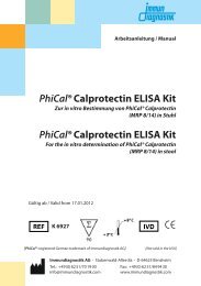
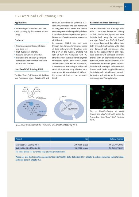
Calcein-AM<br />
R: acetoxymethyl<br />
EthD-III<br />
Esterase<br />
Viable cell<br />
DNA intercalation<br />
red fluorescence<br />
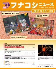
Fig. 4: Double-staining of viable<br />
Calcein<br />
green fluorescence<br />
(green) and dead (red) cells using the<br />
PromoKine Live/Dead <strong>Cell</strong> Staining<br />
Kit II.<br />
Fig. 3: Assay mechanism of the PromoKine Live/Dead <strong>Cell</strong> Staining Kit II.<br />
Product Size Catalog Number<br />
Live/Dead <strong>Cell</strong> Staining Kit II 300-1000 assays PK-CA707-30002<br />
Bacteria Live/Dead Staining Kit 100-1000 assays PK-CA707-30027<br />
For prices please see our online shop at www.promokine.info<br />
Please see also the PromoKine Apoptotic/Necrotic/Healthy <strong>Cell</strong>s Detection Kit in Chapter 2 and our individual stains for viable<br />
and dead cells in Chapter 1.6.