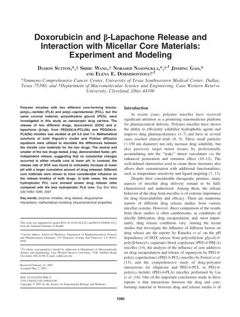
Doxorubicin and b-Lapachone Release and ... - UT Southwestern
Doxorubicin and b-Lapachone Release and ... - UT Southwestern
Doxorubicin and b-Lapachone Release and ... - UT Southwestern

You also want an ePaper? Increase the reach of your titles
YUMPU automatically turns print PDFs into web optimized ePapers that Google loves.
<strong>Doxorubicin</strong> <strong>and</strong> b-<strong>Lapachone</strong> <strong>Release</strong> <strong>and</strong><br />
Interaction with Micellar Core Materials:<br />
Experiment <strong>and</strong> Modeling<br />
DAMON S<strong>UT</strong>TON,*, SHIHU WANG, NORASED NASONGKLA,*, ,1 JINMING GAO,*<br />
AND ELENA E. DORMIDONTOVA ,2<br />
*Simmons Comprehensive Cancer Center, University of Texas <strong>Southwestern</strong> Medical Center, Dallas,<br />
Texas 75390; <strong>and</strong> Department of Macromolecular Science <strong>and</strong> Engineering, Case Western Reserve<br />
University, Clevel<strong>and</strong>, Ohio 44106<br />
Polymer micelles with two different core-forming blocks,<br />
poly(D,L-lactide) (PLA) <strong>and</strong> poly(e-caprolactone) (PCL), but the<br />
same coronal material, poly(ethylene glycol) (PEG), were<br />
investigated in this study as nanoscopic drug carriers. The<br />
release of two different drugs, doxorubicin (DOX) <strong>and</strong> b-<br />
lapachone (b-lap), from PEG(5k)-b-PCL(5k) <strong>and</strong> PEG(5k)-b-<br />
PLA(5k) micelles was studied at pH 5.0 <strong>and</strong> 7.4. Mathematical<br />
solutions of both Higuchi’s model <strong>and</strong> Fickian diffusion<br />
equations were utilized to elucidate the differences between<br />
the micelle core materials for the two drugs. The neutral <strong>and</strong><br />
smaller of the two drugs tested, b-lap, demonstrated faster, pHindependent<br />
release, suggesting that no substantial changes<br />
occurred in either micelle core at lower pH. In contrast, the<br />
release rate of DOX was found to noticeably increase at lower<br />
pH with a larger cumulative amount of drug released. Different<br />
core materials were shown to have considerable influence on<br />
the release kinetics of both drugs: in both cases, the more<br />
hydrophobic PCL core showed slower drug release rates<br />
compared with the less hydrophobic PLA core. Exp Biol Med<br />
232:1090–1099, 2007<br />
Key words: polymer micelles; drug release; drug-polymer<br />
interactions; mathematical modeling; physicochemical properties<br />
This work was supported by grants R21CA112436 (E.E.D.) <strong>and</strong> RO1CA90696 (J.G.)<br />
from the National Institutes of Health.<br />
1 Current address: School of Pharmacy, Department of Biopharmaceutical Sciences<br />
<strong>and</strong> Pharmaceutical Chemistry, 513 Parnassus Avenue, San Francisco, CA 94143-<br />
0446.<br />
2 To whom correspondence should be addressed at Department of Macromolecular<br />
Science <strong>and</strong> Engineering, Case Western Reserve University, 2100 Adelbert Road,<br />
Clevel<strong>and</strong>, OH 44106. E-mail: eed@case.edu<br />
Received February 14, 2007.<br />
Accepted May 2, 2007.<br />
DOI: 10.3181/0702-RM-31<br />
1535-3702/07/2328-1090$15.00<br />
Copyright Ó 2007 by the Society for Experimental Biology <strong>and</strong> Medicine<br />
Introduction<br />
In recent years, polymer micelles have received<br />
significant attention as a promising nanomedicine platform<br />
for pharmaceutical delivery. Polymer micelles have shown<br />
the ability to efficiently solubilize hydrophobic agents <strong>and</strong><br />
improve drug pharmacokinetics (1–7) <strong>and</strong> have in several<br />
cases reached clinical trials (8, 9). These small particles<br />
(,150 nm diameter) not only increase drug solubility, but<br />
also passively target tumor tissues by preferentially<br />
accumulating into the ‘‘leaky’’ tumor vasculature via the<br />
enhanced permeation <strong>and</strong> retention effect (10–12). The<br />
well-defined chemistries used to create these structures also<br />
allows their customization with additional functionalities,<br />
such as temperature sensitivity <strong>and</strong> lig<strong>and</strong> targeting (7, 13).<br />
Despite their considerable therapeutic promise, many<br />
aspects of micellar drug delivery remain to be fully<br />
characterized <strong>and</strong> understood. Among them, the release<br />
behavior of the drug from micelles is of extreme importance<br />
for drug bioavailability <strong>and</strong> efficacy. There are numerous<br />
reports of different drug release studies from various<br />
micellar systems. However, direct comparison of the results<br />
from these studies is often cumbersome, as conditions of<br />
micelle fabrication, drug encapsulation, <strong>and</strong>, most importantly,<br />
drug release conditions vary. Among the recent<br />
studies that investigate the influence of different factors on<br />
drug release are the reports by Kataoka et al. on the pH<br />
dependence of DOX release from poly(ethylene glycol)-bpoly(b-benzyl-L-aspartate)<br />
block copolymer (PEG-b-PBLA)<br />
micelles (14), the analysis of the influence of core additives<br />
on drug encapsulation <strong>and</strong> release of rapamycin by PEG-bpoly(e-caprolactone)<br />
(PEG-b-PCL) micelles by Forrest et al.<br />
(15), <strong>and</strong> the comprehensive study of drug-polymer<br />
interactions for ellipticine <strong>and</strong> PEG-b-PCL or PEG-bpoly(D,L-lactide)<br />
(PEG-b-PLA) micelles performed by Liu<br />
et al. (16). One of the important conclusions made in these<br />
reports is that interactions between the drug <strong>and</strong> coreforming<br />
material or between drug <strong>and</strong> release media is of<br />
1090
DOX AND b-LAP RELEASE FROM MICELLES 1091<br />
Figure 1. Chemical structures of PEG-b-PCL, PEG-b-PLA, b-lap,<br />
<strong>and</strong> DOX.<br />
great importance to drug release by polymeric micellar<br />
systems. Of course, there might be an influence of the<br />
release media on the polymeric material itself, so these two<br />
effects may be interconnected. In this report, we attempt to<br />
analyze these effects by studying release of two different<br />
anticancer drugs, doxorubicin (DOX) <strong>and</strong> b-lapachone (blap)<br />
from two polymer micelles, PEG-b-PCL <strong>and</strong> PEG-b-<br />
PLA (Fig. 1). Mathematical modeling of the release<br />
behavior is employed to quantify the importance of such<br />
factors as core hydrophobicity <strong>and</strong> drug solubility on drug<br />
release. This complex experimental-analytical approach will<br />
help to identify important factors that influence drug release.<br />
DOX (Fig. 1) was chosen as one of the most common<br />
chemotherapeutic drugs used in micellar formulations (8,<br />
17–19), with DOX-encapsulated micelles being one of the<br />
few systems that reached clinical trials (8). DOX is known<br />
to have a pH-dependent solubility (20), unlike the neutral b-<br />
lap (Fig. 1) (21). By comparing the pH dependences of<br />
release for these two compounds, we will gain insight into<br />
any pH dependence of the micelle release properties<br />
themselves. Both micelles, PEG-b-PCL <strong>and</strong> PEG-b-PLA,<br />
are comprised of nontoxic <strong>and</strong> biodegradable polyesters that<br />
have been explored previously as micelle cores (13, 16, 17,<br />
22–25). PCL is a semicrystalline polymer with a melting<br />
temperature (T m ) of around 558C (26), whereas PLA is fully<br />
amorphous, with a glass transition temperature (T g ) of<br />
around 508C (27). The semicrystalline <strong>and</strong> hydrophobic<br />
nature of PCL has been suggested to cause slow drug release<br />
(17), <strong>and</strong> we further hypothesize that an amorphous, less<br />
hydrophobic PLA core will result in faster release kinetics.<br />
Drug release from a polymer matrix involves a<br />
multitude of processes, including possible matrix swelling,<br />
erosion or degradation of the polymers, drug dissolution, as<br />
well as external or internal mass transport of the dissolved<br />
drugs. To describe it exactly is a mathematically challenging<br />
task. A large number of analytical models have been put<br />
forward that use different assumptions to describe a<br />
particular process (28–30). These models are normally case<br />
specific, but could be combined to simultaneously address<br />
several factors (such as the polydispersity <strong>and</strong> crystallinity<br />
of polymers, particle size distribution [31, 32], <strong>and</strong><br />
distribution of drugs in the matrix [31], as well as the<br />
entrapment of drugs in the matrix [32, 33], etc.). While such<br />
approaches could provide a complete picture of drug<br />
release, they require knowledge of multiple parameters of<br />
the system, which are often not readily available from<br />
experiments. As a result, the most commonly used models<br />
are those based on relatively simple mathematical approaches<br />
<strong>and</strong> that have the advantage of employing a small<br />
number of parameters. Among them, Higuchi’s model (34)<br />
<strong>and</strong> Fickian diffusion–based approaches (35, 36) are the<br />
most commonly used. In our current study, we attempt to<br />
ascertain the effects of drug hydrophobicity, pH sensitivity,<br />
<strong>and</strong> the influence of the polymeric core on drug release. To<br />
serve this purpose, we employ both the Fickian diffusion–<br />
based approach <strong>and</strong> Higuchi’s model, which allow us to<br />
take into account the difference in drug solubility <strong>and</strong><br />
provide a quantitative measure of the kinetics of drug<br />
release. The comparison of the release behavior of different<br />
drug-polymer systems using equivalent modeling approaches<br />
will assist our fundamental underst<strong>and</strong>ing of their<br />
differences.<br />
Materials <strong>and</strong> Methods<br />
Materials. D,L-lactide was purchased from Alfa Aesar<br />
(Ward Hill, MA) <strong>and</strong> was purified by recrystallization from<br />
dried ethyl acetate <strong>and</strong> thoroughly vacuum dried for 24 hrs<br />
before use. Stannous (II) octoate (Sn(Oct) 2 ; Aldrich, St.<br />
Louis, MO) was used as received. e-Caprolactone (e-CL;<br />
Aldrich) <strong>and</strong> purified by vacuum distillation over calcium<br />
hydride. DOX in aqueous solution (DOX-HCl, 2 mg/ml)<br />
was purchased from the Bedford Laboratories (Bedford,<br />
OH), <strong>and</strong> was deprotonated at pH 9.6 to obtain the<br />
hydrophobic DOX. b-Lap was synthesized by Dr. William<br />
G. Bornmann from M. D. Anderson Cancer Center<br />
(Houston, TX). All organic solvents were of analytical<br />
grade. Toluene (Aldrich) was dried by refluxing over<br />
sodium <strong>and</strong> distilled under dry argon.<br />
Synthesis of Methoxy-Terminated PEG-b-PLA
1092 S<strong>UT</strong>TON ET AL<br />
Copolymer. PEG-b-PLA was synthesized by ring-opening<br />
polymerization of D,L-lactide under dry argon at 1108C.<br />
Monomethyl ether hydroxyl (HO-PEG-OCH 3 ; number<br />
average molecular weight M n ¼ 5000 Da) was used as a<br />
macroinitiator. D,L-lactide was added as a monomer <strong>and</strong><br />
stannous (II) octoate (Sn(Oct) 2 ) was added as a catalyst.<br />
After reacting for 4 hrs at 1108C, the mixture was allowed to<br />
cool to room temperature. PEG-b-PLA was purified by<br />
redissolving in tetrahydrofuran (THF) <strong>and</strong> precipitating in<br />
hexane three times. The overall yield was 95%. The degree<br />
of polymerization of the PLA was calculated by comparing<br />
the integral intensity of the characteristic resonance of the<br />
PLA at 5.2 ppm ( C(¼O)-CH( CH 3 )) <strong>and</strong> PEG resonance<br />
at 3.64 ppm ( OCH 2 CH 2 ) in the 1 H nuclear magnetic<br />
resonance (NMR) spectrum in chloroform (CDCl 3 ). The<br />
molecular weight <strong>and</strong> polydispersity index (PDI) of PEG-b-<br />
PLA were also characterized by gel permeation chromatography<br />
(THF as eluent), <strong>and</strong> the results were found to be<br />
consistent with 1 H NMR data. PEG-b-PLA (M n ¼ 10.0 kD;<br />
PDI ¼ 1.2) was used in this study.<br />
Synthesis of PEG-b-PCL Copolymer. The PEGb-PCL<br />
copolymer was synthesized (with yields .95%) by<br />
ring-opening polymerization of e-caprolactone under dry<br />
argon at 1158C for 24 hrs using PEG as a macroinitiator <strong>and</strong><br />
Sn(Oct) 2 as a catalyst. The product was purified by<br />
precipitating twice into cold methanol from CH 2 Cl 2<br />
solution, <strong>and</strong> was then vacuum dried at 408C. The block<br />
copolymer was characterized by 1 H NMR in CDCl 3 at room<br />
temperature. The degree of polymerization of the PCL block<br />
was calculated by comparing the integrals of the 1 HNMR<br />
characteristic peaks of the PCL block at 2.31 ppm (triplet,<br />
C(¼O)-CH 2 ) <strong>and</strong> PEG block at 3.39 ppm (singlet,<br />
OCH 2 CH 2 ). The molecular weight <strong>and</strong> polydispersity of<br />
PEG-b-PCL were also characterized by gel permeation<br />
chromatography (THF as eluent), <strong>and</strong> the results were found<br />
to be consistent with 1 H NMR data. PEG-b-PCL (M n ¼ 10.0<br />
kD; PDI ¼ 1.3) was used in this study.<br />
Preparation of Drug-Loaded Micelles. Drug-containing<br />
polymer micelles were prepared as follows: 18 mg<br />
of PEG-b-PLA or PEG-b-PCL copolymer <strong>and</strong> 2 mg of drug<br />
(hydrophobic DOX was predissolved in 0.12 ml dimethylsulfoxide<br />
[DMSO]) were added to 1.08 ml THF in a glass<br />
vial. Next, the mixture was slowly added to 13 ml of water<br />
under sonication (60 Sonic Dismembrator; Fisher Scientific;<br />
Pittsburgh, PA). The mixture was vigorously stirred overnight<br />
to remove THF followed by filtration through a<br />
syringe filter (pore size 0.45 lm; Millipore, Billerica, MA)<br />
to remove large drug aggregates. Micelles were characterized<br />
by dynamic light scattering (see below). 1 H NMR was<br />
used to confirm the formation of core-shell structure. The<br />
strong resonance of methylene proton in PEG was detected,<br />
whereas all of the D,L-lactide or caprolactone proton<br />
resonances were hardly observed, demonstrating the coreshell<br />
structure of these micelles.<br />
Drug-Loading Content Determination. The drugloading<br />
content, defined as the weight percentage of DOX<br />
or b-lap based on the total micellar weight (i.e., weight of<br />
copolymer <strong>and</strong> drug) was quantified by UV-Vis analysis<br />
using a Lambda 20 spectrophotometer (Perkin-Elmer,<br />
Boston, MA). First, micelle solutions were frozen <strong>and</strong><br />
lyophilized to yield the solid micelle samples. Then, the<br />
dried samples were weighed <strong>and</strong> redissolved in CDCl 3 for<br />
the b-lap micelles or a mixture of CDCl 3 <strong>and</strong> DMSO (1:1, v/<br />
v) for the DOX micelles followed by ultraviolet-visible<br />
spectroscopy (UV-Vis) analysis. The amount of loaded drug<br />
was determined based on the absorbance at 480 nm for<br />
DOX <strong>and</strong> at 257 nm for b-lap.<br />
Dynamic Light Scattering (DLS). DLS was performed<br />
on a DLS Model 802 (Viscotek, Houston, TX).<br />
Scattered light was detected at an angle of 908 at room<br />
temperature <strong>and</strong> analyzed on an autocorrelator. Sample<br />
concentration during measurement was 1.4 mg/ml. The data<br />
for each sample was obtained in five independent measurements.<br />
The average hydrodynamic diameters <strong>and</strong> their<br />
st<strong>and</strong>ard deviations are provided in Table 1.<br />
In Vitro <strong>Release</strong> of Drugs from Polymer Micelles.<br />
The drug-loaded micelles were purified using<br />
Millipore centrifugal filters with a molecular weight cutoff<br />
of 100 kD to remove the free drug <strong>and</strong> to concentrate the<br />
samples in preparation for release studies. Approximately<br />
15 mg of DOX-loaded polymeric micelles were placed into<br />
a total of 2 ml of water inside dialysis tubing, resulting in a<br />
micelle concentration of 7.5 mg/ml. The tubing was placed<br />
into 13 ml phosphate-buffered saline (PBS; pH 7.4) or<br />
acetate-buffered saline (pH 5.0) solutions. For b-lap release,<br />
10 mg of drug-loaded micelles in water (2 ml), resulting in a<br />
micelle concentration of 5 mg/ml, were transferred into<br />
dialysis tubing (MW cutoff, 100 kDa). The tubing was<br />
placed into 8 ml PBS (pH 7.4) or acetate-buffered saline (pH<br />
5.0) solutions. <strong>Release</strong> studies were performed at 378C ina<br />
C24 Incubator Shaker (New Brunswick Scientific, Edison,<br />
NJ). At selected time intervals, all of the buffered solution<br />
outside the dialysis bag was removed for UV-Vis analysis<br />
<strong>and</strong> replaced with fresh buffer solution. The DOX <strong>and</strong> b-lap<br />
concentrations were calculated based on the absorbance<br />
intensity at 480 <strong>and</strong> 257 nm, respectively. Free drug<br />
transport from the dialysis tubing was studied under the<br />
same conditions as drug release from micelles, except the<br />
amount of the free drug was different: 1 ml solution of 2 mg/<br />
ml DOX-HCl at pH 5.0 <strong>and</strong> 2 ml of saturated deionized<br />
water solution (with 0.04 mg/ml of b-lap) were placed<br />
inside the dialysis tubing, respectively.<br />
Mathematical Modeling. To simulate drug release<br />
kinetics from PEG-b-PCL <strong>and</strong> PEG-b-PLA micelles, we<br />
applied Higuchi’s model (34). The advantage of this model<br />
over the Fickian diffusion model (35, 36) is that it accounts<br />
for the difference in solubility of the drugs in the buffer<br />
solution. As discussed below, this feature is especially<br />
important in underst<strong>and</strong>ing DOX release at different pH<br />
values.<br />
The cumulative amount of drug released (SdQ) per unit<br />
time (dt) is given by the following equation:
DOX AND b-LAP RELEASE FROM MICELLES 1093<br />
Table 1.<br />
Summary of the Main Characteristics of Micelles Composed of PEG 5000 -b-PCL 5000 or<br />
PEG 5000 -b-PLA 5000 With or Without DOX or b-Lap Loading a<br />
Drug Micelle Size without drug (nm) b Drug-loading content (wt %) Size with drug (nm) b,c<br />
DOX PEG-b-PCL 23.2 6 2.2 3.80 6 0.3 20.0 6 3.3<br />
PEG-b-PLA 17.4 6 1.2 2.32 6 0.4 23.3 6 4.5<br />
b-lap PEG-b-PCL 23.2 6 2.2 1.0 6 0.1 21.9 6 2.4<br />
PEG-b-PLA 17.4 6 1.2 0.7 6 0.1 19.1 6 2.1<br />
a DOX, doxorubicin; PEG, poly(ethylene glycol); PCL, poly(e-caprolactone); PLA, poly(D,L-lactide); b-lap, b-lapachone.<br />
b Hydrodynamic diameter from dynamic light scattering.<br />
c Average size (mean 6 SD) ¼ 21.075 6 1.61 nm.<br />
SdQ<br />
¼<br />
dt<br />
4pa 2 D dc<br />
da ;<br />
ð1Þ<br />
where S is the surface area of the sphere exposed to the<br />
release medium, D is the diffusion constant of the drugs in<br />
the polymer matrix, <strong>and</strong> c is the concentration of drugs at<br />
radial distance, a, from the center of the sphere.<br />
By assuming a pseudo–steady state at a moving front of<br />
the permeating fluids, the following equation is derived<br />
(34):<br />
c o ða 3 o þ 2a93 3a o a9 2 Þ<br />
<br />
þ c s 4a9 2 a o þ a 3 o ln a o<br />
a9<br />
<br />
a 3 o a 2 oa9 2a93 ¼ 6Dc s a o t;<br />
ð2Þ<br />
where a o is the radius of the spherical core of a micelle, a9 is<br />
the distance of the moving front from the center of the core<br />
at time t, c o is the drug-loading concentration, <strong>and</strong> c s is the<br />
solubility of drug in the permeating fluids. The fractional<br />
drug released, M(t)/M(‘), is given by:<br />
MðtÞ<br />
Mð‘Þ ¼ 1<br />
" <br />
a9 3<br />
þ 1 <br />
c s a9<br />
þ<br />
a9 2 <br />
2 a9 !#<br />
3<br />
;<br />
a o 2 c o a o a o a o<br />
ð3Þ<br />
where M(t) is the mass of drug released at time t <strong>and</strong> M(‘) is<br />
the amount of drug released as time approaches infinity.<br />
Eqs. 2 <strong>and</strong> 3 are utilized to fit the experimental data. We<br />
note that in the case when the fit of the whole range of<br />
experimental data to the mathematical model is not possible<br />
(as for DOX release), the matching is optimized based on<br />
the initial period of the release only.<br />
The effect of solubility of drugs on their release is<br />
reflected in Eqs. 2 <strong>and</strong> 3 by the ratio c s /c o . The drug-loading<br />
concentration, c o , was estimated from the weight fraction of<br />
loaded drug <strong>and</strong> assuming the density of polymer in the core<br />
of the micelles to be 1 g/cm 3 . The obtained values are listed<br />
in Table 2. The solubility of DOX at pH 7.4 was reported to<br />
be 0.0625 mg/ml (20). At lower pH, the degree of DOX<br />
protonation increases as does its solubility. The reported<br />
values range from 0.37 mg/ml at pH 5.0 in phosphate buffer<br />
(20) to 10–30 mg/ml for DOX-HCl (37, 38). The solubility<br />
of b-lap was measured to be 0.038 mg/ml <strong>and</strong> does not<br />
change with pH (21).<br />
Knowing c s /c o , the ratio D=a 2 o can be determined from<br />
fitting the experimental data. In order to obtain the diffusion<br />
coefficient, it is necessary to estimate a o , the size of the<br />
micelle core. For a known hydrodynamic diameter (d) of the<br />
micelle (obtained by DLS), the core size was estimated as<br />
(Fig. 2):<br />
a o ¼ d=2 R corona : ð4Þ<br />
The thickness of the micelle corona (R corona ) could be<br />
estimated from the radius of gyration R g of PEG (39):<br />
R corona } 2R g ¼ 2 3 0:215Mw<br />
0:58360:031 Å; ð5Þ<br />
where M w is the average molecular weight of the PEG. The<br />
Table 2.<br />
<strong>Release</strong> Characteristics of DOX <strong>and</strong> b-Lap from Micelles Composed of Diblock Copolymers of<br />
PEG 5000 -b-PCL 5000 or PEG 5000 -b-PLA 5000 at Different pHs a<br />
Drug Micelle c o (mg/ml) c s /c o D/a o 2 (1/sec) D (cm 2 /sec) b<br />
DOX PEG-b-PCL 79.0 pH 5.0 9.44 3 10 3 5.91 3 10 6 1.13 3 10 18<br />
pH 7.4 7.90 3 10 4<br />
PEG-b-PLA 47.5 pH 5.0 1.57 3 10 2 9.54 3 10 6 1.82 3 10 18<br />
pH 7.4 1.32 3 10 3<br />
b-lap PEG-b-PCL 20.2 1.88 3 10 3 7.78 3 10 5 1.49 3 10 17<br />
PEG-b-PLA 14.1 2.70 3 10 3 1.27 3 10 4 2.42 3 10 17<br />
a c s , solubility of the drug in the bulk liquid phase, as required by Higuchi’s model; c o , drug-loading concentration in the micelle; D, diffusion<br />
coefficient of the drug in the core matrix; DOX, doxorubicin; PEG, poly(ethylene glycol); PCL, poly(e-caprolactone); PLA, poly(D,L-lactide); b-lap,<br />
b-lapachone.<br />
b Using average micellar core size a o ¼ 4.37 6 0.33 nm (mean 6 SD) calculated using Eq. 4.
1094 S<strong>UT</strong>TON ET AL<br />
Figure 2. Schematic illustration of DOX or b-lap loaded diblock<br />
copolymer micelle with the same corona block PEG <strong>and</strong> two different<br />
core blocks poly (D,L-lactide) or poly(e-caprolactone). d, hydrodynamic<br />
diameter of the micelle; 2R g, PEG , thickness of corona.<br />
methoxy-terminated PEG used in this study has a molecular<br />
weight of 5000 Da. Therefore, the thickness of micelle<br />
corona is estimated to be around 6.16 nm (40).<br />
Results<br />
Drug Loading <strong>and</strong> Micelle Characterization.<br />
The DOX loading content was quantified by UV-<br />
Vis analysis <strong>and</strong> was found to be 2.32% for PEG-b-PLA<br />
micelles <strong>and</strong> 3.80% for PEG-b-PCL micelles. b-Lap loading<br />
content was noticeably lower than that for DOX, as shown<br />
in Table 1. We note that, in both cases of b-lap <strong>and</strong> DOX,<br />
larger loading content is achieved for PEG-b-PCL micelles,<br />
possibly due to the larger hydrophobicity of PCL (as<br />
discussed below).<br />
The micelle size with <strong>and</strong> without loaded drugs was<br />
studied by DLS <strong>and</strong> the results are listed in Table 1. The<br />
hydrodynamic diameter of PEG-b-PCL micelles without<br />
drug was found to be around 23.2 nm, while for PEG-b-<br />
PLA micelles, a smaller diameter around 17.4 nm was<br />
observed. The measured micelle sizes are comparable (PEGb-PCL)<br />
or somewhat smaller (PEG-b-PLA) compared with<br />
other reported results (15–17, 24, 25). For instance, in a<br />
recent study by Liu et al. of similar molecular weight<br />
polymers, PEG-b-PCL micelles (24 nm) were found to be<br />
smaller than PEG-b-PLA micelles (66 nm) (16). The<br />
difference in micelle sizes could have originated from<br />
different micelle preparation techniques. Parameters such as<br />
mixing rate have been known to affect particle size by 3- to<br />
4-fold (41), <strong>and</strong> the fast mixing resulting from the sonication<br />
mixer could be the cause of the small size of the micelles as<br />
compared with those made using a slow-mixing dialysis<br />
method.<br />
Drug loading did not noticeably affect micelle size, as<br />
shown in Table 1. Student’s t test analysis performed for the<br />
size comparison of loaded to unloaded micelles resulted in P<br />
values in the range of 0.24–0.70, indicating no significant<br />
difference in micelle sizes. Furthermore, comparing the<br />
Figure 3. Cumulative release of b-lap versus time from two different<br />
micelles composed of diblock copolymer of PEG 5000 -block-PLA<br />
(circle) or PEG 5000 -block-PCL 5000 (square) <strong>and</strong> for free b-lap (stars)<br />
in PBS (pH 7.4; open symbols) <strong>and</strong> acetate-buffered saline (pH 5.0;<br />
filled symbols) solutions at 378C. Experimental data points for release<br />
from micelles as fitted by Higuchi’s model <strong>and</strong> that for free b-lap as<br />
fitted by Fickian diffusion through a membrane are shown as solid<br />
lines.<br />
sizes of all drug-loaded micelles (Table 2), one can observe<br />
that they are nearly the same: the largest difference between<br />
average micelle sizes is less than the smallest uncertainty<br />
range, <strong>and</strong> t test analysis of any given pair shows no<br />
significant variance. Based on these observations, we<br />
consider all drug-loaded micelles to be of the same size,<br />
defined by the average of the four values (i.e., d ¼ 21.08 nm;<br />
st<strong>and</strong>ard deviation, 61.9 nm). Accordingly, the average<br />
core size calculated using Eq. 4 results in a o ¼ 4.37 nm,<br />
which we use to calculate the diffusion coefficients. It is also<br />
useful to estimate the average number of drug molecules per<br />
polymer micelle. Based on the drug-loading content <strong>and</strong> the<br />
estimated value of a o , the number of drug molecules per<br />
micelle varies from about 12 for b-lap in PEG-b-PLA<br />
micelles to about 30 for DOX in PEG-b-PCL micelle. We<br />
note that the experimentally recorded data for drug release<br />
are the cumulative result of drug release from an ensemble<br />
of micelles, so that variation in number of molecules per<br />
micelle or in drug distribution inside micelles may not be<br />
important, as long as all drug molecules on average<br />
experience the same surrounding in their release pattern.<br />
<strong>Release</strong> of b-Lap. The release profiles of b-lap from<br />
PEG-b-PLA <strong>and</strong> PEG-b-PCL micelles are shown in Figure<br />
3. As is seen, the influence of pH on b-lap release is rather<br />
weak, with the deviations being within the experimental<br />
error range (based on results of three experiments). Since<br />
the solubility of b-lap is practically independent of pH, the<br />
only difference in release could come from changes in<br />
polymer matrix. Since we observe no appreciable effect of<br />
pH on drug release, this implies that no substantial change
DOX AND b-LAP RELEASE FROM MICELLES 1095<br />
Figure 4. Cumulative release of DOX versus time from two different<br />
micelles composed of diblock copolymer of PEG 5000 -block-PLA 5000<br />
(circle) or PEG 5000 -block-PCL 5000 (square) <strong>and</strong> for free DOX (stars)<br />
in PBS (pH 7.4; open symbols) <strong>and</strong> acetate-buffered saline (pH 5.0;<br />
filled symbols) solutions at 378C. Experimental data points for shortterm<br />
(up to 100 hrs) release from micelles are fitted by Higuchi’s<br />
model (solid lines) <strong>and</strong> for long-term release are fitted by Fickian<br />
diffusion from a sphere (dashed lines).<br />
in the micelle core occurs with a decrease of pH for either<br />
PEG-b-PLA or PEG-b-PCL micelles.<br />
The release rate from PEG-b-PLA micelles is somewhat<br />
faster than for PEG-b-PCL micelles: 87% of b-lap was<br />
released from PEG-b-PLA micelles in 72 hrs, compared<br />
with 66% for PEG-b-PCL micelles during the same period<br />
of time. A similar trend was observed by Liu et al. (16) for<br />
ellipticine <strong>and</strong> PEG-b-PCL or PEG-b-PLA micelles of<br />
similar composition. Knowing the drug-loading concentration<br />
c o (Table 2) <strong>and</strong> solubility of b-lap in the bulk liquid<br />
phase (c s ¼ 0.038 mg/ml [21]), we fit the diffusion of b-lap<br />
from these different polymeric media using Higuchi’s model<br />
(34) for drug release from a sphere. Since the difference<br />
between the experimental data for pH 7.4 <strong>and</strong> pH 5.0 was<br />
very small for the same polymeric carriers, we used a single<br />
fit for both data sets. The results of fitting are shown as solid<br />
curves in Figure 3. The diffusion coefficient for b-lap<br />
release from PEG-b-PLA micelles was found to be 2.42 3<br />
10 17 cm 2 /sec, while that for b-lap release from PEG-b-PCL<br />
micelles was about 1.6-times smaller (1.49 3 10 17 cm 2 /sec)<br />
(Table 2).<br />
We note that in the control measurement of free b-lap<br />
transport across the dialysis membrane, the release occurs<br />
noticeably quicker. The corresponding diffusion coefficient<br />
obtained using a model of Fickian transport across a<br />
membrane (42) was 3.0 3 10 10 cm 2 /sec (see Fig. 3 <strong>and</strong><br />
supporting information), which is available in the on-line<br />
version, much larger than 2.42 3 10 17 cm 2 /sec estimated<br />
for release from PEG-b-PLA micelles. Thus, the slowest<br />
step of the drug release is from the dense polymer core,<br />
which is well described by Higuchi’s model (34). We were<br />
also able to obtain good fits to the b-lap micelle release data<br />
using a simple Fickian model of diffusion from a sphere<br />
(36). However, for the DOX release, discussed in the next<br />
section, the pH effects play an important role. As a result,<br />
we have chosen the Higuchi model (34) as a unifying<br />
approach for both b-lap <strong>and</strong> DOX-containing systems.<br />
Short-Term <strong>Release</strong> of DOX. The release of DOX<br />
from PEG-b-PCL <strong>and</strong> PEG-b-PLA micelles at pH 7.4 <strong>and</strong><br />
pH 5.0 is shown in Figure 4. There is a strong pH<br />
dependence of the release of DOX from different micelles:<br />
DOX release from the micelles at pH 5.0 is faster than at pH<br />
7.4, <strong>and</strong> the total amount released at the longest investigated<br />
time is greater at pH 5.0 than at pH 7.4. For DOX-loaded<br />
PEG-b-PLA micelles, the amount of release at pH 5.0 is<br />
about 50% larger than that at pH 7.4, while for DOX-loaded<br />
PEG-b-PCL micelles, the difference is more than 40%. A<br />
similar effect of pH on DOX release has also been observed<br />
by Kataoka et al. using PEG-b-PBLA copolymer micelles<br />
(14). Similar to b-lap release, different core materials have<br />
an influence on DOX release, as PEG-b-PLA micelles have<br />
a faster release rate <strong>and</strong> a greater cumulative amount of drug<br />
released than PEG-b-PCL micelles.<br />
The fitting of experimental data using Higuchi’s model<br />
(34) (over relatively short time scales) is shown in Figure 4<br />
as solid lines. For pH 7.4, using known drug-loading<br />
concentration, c o (Table 2), <strong>and</strong> solubility of DOX in the<br />
bulk liquid phase (c s ¼ 0.0625 mg/ml [20]), fitting was<br />
performed by varying D=a 2 o . The following diffusion<br />
coefficients for DOX release were obtained (using the<br />
average value for micelle core radius, a o ¼ 4.37 nm [Table<br />
2]): 1.82 3 10 18 cm 2 /sec for PEG-b-PLA micelles <strong>and</strong> 1.13<br />
3 10 18 cm 2 /sec for PEG-b-PCL micelles (a factor of 1.6<br />
difference). As shown by the release behavior of b-lap, the<br />
polymer in the micellar cores is unaffected by the pH.<br />
Therefore, the diffusion coefficient for DOX release from<br />
micelle cores is assumed to be independent of pH, <strong>and</strong> we<br />
fixed the diffusion coefficient to the value obtained for pH<br />
7.4 <strong>and</strong> varied c s /c o to obtain the best match with<br />
experimental data for pH 5.0. The best-fit ratio of c s /c o is<br />
1.57 3 10 2 for the DOX-loaded PEG-b-PLA micelle <strong>and</strong><br />
9.4 3 10 3 for DOX-loaded PEG-b-PCL micelle at pH 5.0<br />
(Table 2). As is also shown in Table 2, for both types of<br />
DOX-loaded micelles, the ratio, c s /c o , increases by approximately<br />
a factor 12 when the pH is changed from 7.4 to 5.0.<br />
In the control measurement of free DOX transport across the<br />
same membrane, the release occurs noticeably quicker—<br />
100% of DOX releases from the membrane in less than 10<br />
hrs (see Fig. 4 <strong>and</strong> supporting information). The corresponding<br />
diffusion coefficient obtained using the same<br />
transport across a membrane model discussed above was 1.3<br />
3 10 9 cm 2 /sec, much larger than the diffusion constant<br />
measured for micelles.<br />
Long-Term <strong>Release</strong> of DOX. DOX release from<br />
different polymer micelles displays a noticeable deviation<br />
from Higuchi’s model over longer periods of time. To
1096 S<strong>UT</strong>TON ET AL<br />
Table 3.<br />
Long-Term (over 100 Hrs) <strong>Release</strong> Characteristics of DOX from Micelles Composed of Diblock<br />
Copolymer of PEG 5000 -b-PCL 5000 or PEG 5000 -b-PLA 5000 at pH 5.0 <strong>and</strong> 7.4 a<br />
Drug Micelle pH D/a o 2 (1/sec) b D (cm 2 /sec) b p b<br />
DOX PEG-b-PCL 5.0 2.46 3 10 7 6 0.05 3 10 7 4.70 3 10 20 6 0.37 3 10 20 0.67 6 0.002<br />
7.4 2.46 3 10 7 6 0.28 3 10 7 4.70 3 10 20 6 0.64 3 10 20 0.218 6 0.005<br />
PEG-b-PLA 5.0 4.00 3 10 7 6 0.49 3 10 7 7.65 3 10 20 6 1.10 3 10 20 0.82 6 0.02<br />
7.4 2.46 3 10 7 6 0.43 3 10 7 4.70 3 10 20 6 0.90 3 10 20 0.34 6 0.01<br />
a D, diffusion coefficient of the drug in the core matrix; a 0 , radius of the micelle core; p, fraction of drug released at ‘‘infinite time’’ (longest time of<br />
measurements); DOX, doxorubicin; PEG, polyethylene glycol; PCL, poly(e-caprolactone); PLA, poly(D,L-lactide).<br />
b Values are mean 6 SD.<br />
describe this time regime, we applied the long-time<br />
approximation for Fickian diffusion from a sphere (35, 36):<br />
<br />
MðtÞ<br />
Mð‘Þ ¼ p 1 6<br />
p 2 exp p 2 <br />
Dt<br />
a 2 ; ð6Þ<br />
o<br />
where p is the fraction of drugs released at infinite time,<br />
which is approximated by the extrapolation of the fraction<br />
of drugs released at the longest times of the measurements<br />
(Table 3). These fits are shown as dashed lines in Figure 4.<br />
Of course, in reality, at even longer times, the additional<br />
mechanism of polymer decomposition will take over,<br />
resulting in release of the rest of the drug. Nonetheless,<br />
for this intermediate time scale, we obtained 7.65 3 10 20<br />
<strong>and</strong> 4.7 3 10 20 cm 2 /sec for the DOX diffusion coefficient<br />
from PEG-b-PLA micelles at pH 5.0 <strong>and</strong> pH 7.4,<br />
respectively, <strong>and</strong> 4.7 3 10 20 cm 2 /sec for PEG-b-PCL<br />
micelles at both pH 5.0 <strong>and</strong> pH 7.4. As is seen, these<br />
diffusion coefficients are more than 20-times smaller than<br />
that for short-term DOX release (cf., Tables 2 <strong>and</strong> 3).<br />
Discussion<br />
Effects of pH on Drug <strong>Release</strong>. As we have<br />
discussed, there is hardly any influence of pH on b-lap<br />
release from either type of micelles. Since the solubility of<br />
b-lap is independent of pH, this suggests that there is little<br />
pH effect on the micelles themselves. However, DOX<br />
release is a totally different case, especially in the short-time<br />
regime. In the fit to DOX release by the Higuchi model (34)<br />
there is a 12-fold increase in c s /c o when the pH is lowered<br />
from 7.4 to 5.0. This is qualitatively consistent with the<br />
observations by Fritze et al., who report a 6-fold increase of<br />
DOX solubility with a decrease of pH (from 0.0625 mg/ml<br />
at pH 7.4 to 0.37 mg/ml at pH 5.0 in phosphate buffer) (20).<br />
The solubility of protonated DOX (DOX-HCl) was reported<br />
to be even higher: 10–30 mg/ml (37, 38), leading to a<br />
solubility increase of more than 100-fold. As solubility of<br />
DOX in the buffer solution increases, its partition coefficient<br />
in polymeric media will decrease, leading to a greater<br />
amount of drug released. This is also reflected in the<br />
partition coefficient of DOX between the octanol/aqueous<br />
phases, decreasing from 1.20 at pH 7.4 to 0.23 at pH 5.0<br />
(43). However, the effect of pH on the long-time DOX<br />
release is much smaller (see Table 3), suggesting that the<br />
dissolution-diffusion mechanism described by Higuchi is no<br />
longer operative.<br />
Effects of Core Materials on Drug <strong>Release</strong>. From<br />
the release study, one can assume that the difference in the<br />
release rate from two different micelles could be partially<br />
caused by the drug-loading content, which is reflected in the<br />
ratio of c s /c o in Higuchi’s model (34). Aside from the<br />
difference in loading contents, the physical properties of the<br />
core-forming polymer are also different, as PLA is less<br />
hydrophobic <strong>and</strong> more swellable than PCL. For instance, the<br />
water/air contact angle of PLA is reported to be in the range<br />
of 638–698, while the contact angle of PCL is in the range of<br />
748–938 (44–47). Sharp et al. have shown that water uptake<br />
by PLA is about 3% for 10 kDa molecular weight <strong>and</strong><br />
increases with a decrease of molecular weight (48). The<br />
water uptake by PCL is very minor—it can be extrapolated to<br />
be not more than 1% (49). Another possible reason is the<br />
difference in physicochemical properties of PLA, an<br />
amorphous polymer, <strong>and</strong> PCL, a semicrystalline polymer<br />
(at least at large molecular weights). The crystalline structure<br />
of PCL could entrap drug inside the core, lowering the<br />
release rate of drug into the outside environment. The<br />
influences of the core materials on drug release could be<br />
observed for both b-lap <strong>and</strong> DOX. It is also interesting to<br />
note that the ratios of diffusion coefficients for DOX release<br />
from PEG-b-PLA <strong>and</strong> PEG-b-PCL (1.6) is essentially the<br />
same as that for b-lap release (1.6), even through the absolute<br />
values of the diffusion coefficient differ by about a factor of<br />
10 (see Table 2). The latter is not surprising, considering that<br />
b-lap is a smaller molecule.<br />
As discussed above, there is a noticeable deviation from<br />
Higuchi’s model (34) for times longer than 100 hrs for DOX<br />
release from different polymer micelles, <strong>and</strong> Fickian<br />
diffusion (35, 36) is used for fitting long-term release<br />
instead. There have been several hypotheses <strong>and</strong> experimental<br />
verifications for the entrapment of DOX inside the<br />
micelle core, which delays its release from the micelle (14).<br />
As was discussed by Kataoka et al. (14), DOX has a<br />
tendency to form a chemically bonded dimer. Reverse-phase<br />
high-performance liquid chromatography analysis is used to<br />
study the dimer fraction, <strong>and</strong> the results show that, in our<br />
micelles, the amount of chemically bonded DOX dimer <strong>and</strong><br />
higher-order complexes is around 10% for both PEG-b-PCL
DOX AND b-LAP RELEASE FROM MICELLES 1097<br />
Figure 5. Typical snapshots from molecular dynamic simulations (using Impact 4 by Schrödinger) showing (A) DOX surrounded by PLA<br />
oligomers <strong>and</strong> (B) DOX surrounded by PCL oligomers. Oligomers are shown as lines <strong>and</strong> DOX is depicted using a ‘‘balls <strong>and</strong> sticks’’<br />
representation. Hydrogen bonds are shown as dashed lines <strong>and</strong> their average number (based on 10 simulations each) is plotted in the middle<br />
plane.<br />
<strong>and</strong> PEG-b-PLA micelles (see the supporting information<br />
available in the on-line version). However, in most cases,<br />
the limiting fraction of DOX release is far less than 90%.<br />
Therefore, it seems that, aside from the chemically bonded<br />
DOX dimer, there are other factors influencing the drug<br />
release results. Among the possible reasons is formation of<br />
physical aggregates of DOX (50–54) inside the micelles due<br />
to the lipophilic nature of the DOX (43). DOX is also<br />
known to adhere to dialysis tubing (55) <strong>and</strong> is capable of<br />
degradation in PBS buffer (56).<br />
Another factor that may play some role in the<br />
entrapment of DOX is its specific interactions (hydrogen<br />
bonding) with the core-forming polymer. Hydrogen bonding<br />
between PCL <strong>and</strong> DOX has previously been detected<br />
experimentally using Fourier transform infrared spectroscopy<br />
(FTIR) (17). In this case, FTIR spectra of the PCL<br />
carbonyl b<strong>and</strong> at 1735 cm 1 demonstrated formation of<br />
hydrogen bonds with entrapped DOX in a micelle core of<br />
similar molecular weight PCL (M n ¼ 5000). We have<br />
explored hydrogen bonding using molecular dynamic<br />
simulations (Impact 4.0 <strong>and</strong> Macromodel 8.5 by Schrödinger<br />
[57]) <strong>and</strong> demonstrated that, on average, five to six<br />
hydrogen bonds are formed between a single DOX molecule<br />
<strong>and</strong> surrounding PCL chains (Fig. 5). In contrast, when the<br />
core is PLA, the number of formed hydrogen bonds with<br />
DOX is somewhat smaller (3–4), which is consistent with a<br />
larger fraction of DOX release from PEG-b-PLA micelles.<br />
In both cases, there is a large excess of lactide monomers to<br />
DOX molecules (over 50-fold), indicating that different<br />
drug molecules are probably as fully hydrogen bonded as<br />
polymer conformation allows. For a larger DOX loading<br />
content, physical aggregation of DOX inside of micelles<br />
could occur (51, 53) in addition to hydrogen bonding<br />
between drug <strong>and</strong> polymer, making release even more<br />
complex.<br />
Any of the above-mentioned factors could contribute to<br />
apparent slowing down of DOX release at longer time<br />
periods. At the moment, there is not sufficient experimental<br />
evidence to establish the dominant importance of any of<br />
these factors. More detailed comprehensive studies may be<br />
required to achieve this goal.<br />
Conclusions. The encapsulation <strong>and</strong> release behaviors<br />
of two different drugs, DOX <strong>and</strong> b-lap, from two<br />
different micellar systems, PEG-b-PLA <strong>and</strong> PEG-b-PCL,<br />
with the same corona-forming polymers but different cores,<br />
were studied experimentally <strong>and</strong> by mathematical modeling.<br />
We found that core-forming material plays an important role<br />
in drug release due to the differences in their physicochemical<br />
properties <strong>and</strong> interactions with the drugs (15, 16, 58).<br />
The stronger interactions between hydrophobic drugs <strong>and</strong><br />
PCL (a more hydrophobic, semicrystalline polymer) leads to<br />
a 1.6-fold slower release rate of both DOX <strong>and</strong> b-lap from<br />
PEG-b-PCL micelles compared with PLA (an amorphous,<br />
more swellable <strong>and</strong> hydrophilic polymer). The smaller of<br />
the two drugs, b-lap, has a more than 10-fold larger<br />
diffusion coefficient than DOX for both of the polymeric<br />
micelles considered. In contrast to pH-independent b-lap<br />
release from both PEG-b-PLA <strong>and</strong> PEG-b-PCL micelles,<br />
which demonstrated the absence of any pH effect on the<br />
core material, DOX release noticeably accelerated at lower<br />
pH, when it became protonated (14). This effect was well<br />
described by Higuchi’s model, which accounts for the<br />
difference of solubility of drugs in different media via the<br />
ratio of c s /c o . At longer time periods, however, the release of<br />
DOX was better described by the Fickian diffusion model,<br />
which yielded a much smaller diffusion coefficient than the<br />
Higuchi model. The possible reasons for slowing down of<br />
DOX release at longer time periods include formation of<br />
chemical <strong>and</strong> physical DOX aggregates (50–54), specific<br />
interactions with the core-forming polymer (e.g., H-bond-
1098 S<strong>UT</strong>TON ET AL<br />
ing), adhesion of DOX on the dialysis membrane (55), <strong>and</strong><br />
DOX degradation (56) during the course of measurements.<br />
We thank Jessica Kingsberg, Weiqun Li, <strong>and</strong> Kinnell Shah for their<br />
help with molecular dynamic simulations of polymer-DOX <strong>and</strong> DOX-DOX<br />
interactions.<br />
1. Soga O, van Nostrum CF, Fens M, Rijcken CJ, Schiffelers RM, Storm<br />
G, Hennink WE. Thermosensitive <strong>and</strong> biodegradable polymeric<br />
micelles for paclitaxel delivery. J Control <strong>Release</strong> 103:341–353, 2005.<br />
2. Zhang C, Ping QN, Zhang HJ. Self-assembly <strong>and</strong> characterization of<br />
paclitaxel-loaded N-octyl-O-sulfate chitosan micellar system. Colloids<br />
Surf B Biointerfaces 39:69–75, 2004.<br />
3. Liggins RT, Burt HM. Polyether-polyester diblock copolymers for the<br />
preparation of paclitaxel loaded polymeric micelle formulations. Adv<br />
Drug Deliv Rev 54:191–202, 2002.<br />
4. Zhang XC, Jackson JK, Burt HM. Development of amphiphilic diblock<br />
copolymers as micellar carriers of taxol. Int J Pharm 132:195–206,<br />
1996.<br />
5. Torchilin VP. Micellar nanocarriers: pharmaceutical perspectives.<br />
Pharm Res 24:1–16, 2007.<br />
6. Kwon GS. Polymeric micelles for delivery of poorly water-soluble<br />
compounds. Crit Rev Ther Drug Carrier Syst 20:357–403, 2003.<br />
7. Sutton D, Nasongkla N, Blanco E, Gao J. Functionalized micellar<br />
systems for cancer targeted drug delivery. Pharm Res 24:1029–1046,<br />
2007.<br />
8. Matsumura Y, Hamaguchi T, Ura T, Muro K, Yamada Y, Shimada Y,<br />
Shirao K, Okusaka T, Ueno H, Ikeda M, Watanabe N. Phase I clinical<br />
trial <strong>and</strong> pharmacokinetic evaluation of NK911, a micelle-encapsulated<br />
doxorubicin. Br J Cancer 91:1775–1781, 2004.<br />
9. Kim TY, Kim DW, Chung JY, Shin SG, Kim SC, Heo DS, Kim NK,<br />
Bang YJ. Phase I <strong>and</strong> pharmacokinetic study of Genexol-PM, a<br />
cremophor-free, polymeric micelle-formulated paclitaxel, in patients<br />
with advanced malignancies. Clin Cancer Res 10:3708–3716, 2004.<br />
10. Jain RK. Barriers to drug-delivery in solid tumors. Sci Am 271:58–65,<br />
1994.<br />
11. Yuan F, Dellian M, Fukumura D, Leunig M, Berk DA, Torchilin VP,<br />
Jain RK. Vascular-permeability in a human tumor xenograft:<br />
molecular-size dependence <strong>and</strong> cutoff size. Cancer Res 55:3752–<br />
3756, 1995.<br />
12. Hobbs SK, Monsky WL, Yuan F, Roberts WG, Griffith L, Torchilin<br />
VP, Jain RK. Regulation of transport pathways in tumor vessels: Role<br />
of tumor type <strong>and</strong> microenvironment. Proc Natl Acad Sci U S A 95:<br />
4607–4612, 1998.<br />
13. Torchilin VP. Structure <strong>and</strong> design of polymeric surfactant-based drug<br />
delivery systems. J Control <strong>Release</strong> 73:137–172, 2001.<br />
14. Kataoka K, Matsumoto T, Yokoyama M, Okano T, Sakurai Y,<br />
Fukushima S, Okamoto K, Kwon GS. <strong>Doxorubicin</strong>-loaded poly<br />
(ethylene glycol)-poly(beta-benzyl-l-aspartate) copolymer micelles:<br />
their pharmaceutical characteristics <strong>and</strong> biological significance. J<br />
Control <strong>Release</strong> 64:143–153, 2000.<br />
15. Forrest ML, Won CY, Malick AW, Kwon GS. In vitro release of the<br />
mTOR inhibitor rapamycin from poly(ethylene glycol)-b-poly(epsiloncaprolactone)<br />
micelles. J Control <strong>Release</strong> 110:370–377, 2006.<br />
16. Liu JB, Xiao YH, Allen C. Polymer-drug compatibility: A guide to the<br />
development of delivery systems for the anticancer agent, Ellipticine.<br />
J Pharm Sci 93:132–143, 2004.<br />
17. Shuai XT, Ai H, Nasongkla N, Kim S, Gao JM. Micellar carriers based<br />
on block copolymers of poly(e-caprolactone) <strong>and</strong> poly(ethylene glycol)<br />
for doxorubicin delivery. J Control <strong>Release</strong> 98:415–426, 2004.<br />
18. Yoo HS, Lee EA, Park TG. <strong>Doxorubicin</strong>-conjugated biodegradable<br />
polymeric micelles having acid-cleavable linkages. J Control <strong>Release</strong><br />
82:17–27, 2002.<br />
19. Lee ES, Na K, Bae YH. <strong>Doxorubicin</strong> loaded pH-sensitive polymeric<br />
micelles for reversal of resistant MCF-7 tumor. J Control <strong>Release</strong> 103:<br />
405–418, 2005.<br />
20. Fritze A, Hens F, Kimpfler A, Schubert R, Peschka-Suss R. Remote<br />
loading of doxorubicin into liposomes driven by a transmembrane<br />
phosphate gradient. BBA-Biomembranes 1758:1633–1640, 2006.<br />
21. Nasongkla N, Wiedmann AF, Bruening A, Beman M, Ray D,<br />
Bornmann WG, Boothman DA, Gao JM. Enhancement of solubility<br />
<strong>and</strong> bioavailability of beta-lapachone using cyclodextrin inclusion<br />
complexes. Pharm Res 20:1626–1633, 2003.<br />
22. Jones MC, Leroux JC. Polymeric micelles: a new generation of<br />
colloidal drug carriers. Eur J Pharm Biopharm 48:101–111, 1999.<br />
23. Forrest ML, Zhao A, Won CY, Malick AW, Kwon GS. Lipophilic<br />
prodrugs of Hsp90 inhibitor geldanamycin for nanoencapsulation in<br />
poly(ethylene glycol)-b-poly(epsilon-caprolactone) micelles. J Control<br />
<strong>Release</strong> 116:139–149, 2006.<br />
24. Kang N, Perron ME, Prud’homme RE, Zhang YB, Gaucher G, Leroux<br />
JC. Stereocomplex block copolymer micelles: core-shell nanostructures<br />
with enhanced stability. Nano Lett 5:315–319, 2005.<br />
25. Riley T, Stolnik S, Heald CR, Xiong CD, Garnett MC, Illum L, Davis<br />
SS, Purkiss SC, Barlow RJ, Gellert PR. Physicochemical evaluation of<br />
nanoparticles assembled from poly(lactic acid)-poly(ethylene glycol)<br />
(PLA-PEG) block copolymers as drug delivery vehicles. Langmuir 17:<br />
3168–3174, 2001.<br />
26. Engelberg I, Kohn J. Physicomechanical properties of degradable<br />
polymers used in medical applications: a comparative-study. Biomaterials<br />
12:292–304, 1991.<br />
27. Jamshidi K, Hyon SH, Ikada Y. Thermal characterization of<br />
polylactides. Polymer 29:2229–2234, 1988.<br />
28. Abdekhodaie MJ, Cheng YL. Diffusional release of a dispersed solute<br />
from a spherical polymer matrix. J Membr Sci 115:171–178, 1996.<br />
29. Baker R. Controlled <strong>Release</strong> of Biologically Active Agents. New York:<br />
John Wiley & Sons, pp39–83, 1987.<br />
30. Siepmann J, Peppas NA. Modeling of drug release from delivery<br />
systems based on hydroxypropyl methylcellulose (HPMC). Adv Drug<br />
Deliv Rev 48:139–157, 2001.<br />
31. Zhou Y, Chu JS, Wu XY. Theoretical analysis of drug release into a<br />
finite medium from sphere ensembles with various size <strong>and</strong> concentration<br />
distributions. Eur J Pharm Sci 22:251–259, 2004.<br />
32. Grassi M, Colombo I, Lapasin R. Drug release from an ensemble of<br />
swellable crosslinked polymer particles. J Control <strong>Release</strong> 68:97–113,<br />
2000.<br />
33. Kiil S, Dam-Johansen K. Controlled drug delivery from swellable<br />
hydroxypropylmethylcellulose matrices: model-based analysis of<br />
observed radial front movements. J Control <strong>Release</strong> 90:1–21, 2003.<br />
34. Higuchi T. Mechanism of sustained-action medication. J Pharm Sci 52:<br />
1145–1149, 1963.<br />
35. Ritger PL, Peppas NA. A simple equation for description of solute<br />
release I: Fickian <strong>and</strong> non-Fickian release from non-swellable devices<br />
in the form of slabs, spheres, cylinders or discs. J Control <strong>Release</strong> 5:23–<br />
36, 1987.<br />
36. Crank J. The Mathematics of Diffusion. New York: Oxford University<br />
Press, pp89–103, 1975.<br />
37. Lasic DD, Frederik PM, Stuart MCA, Barenholz Y, McIntosh TJ.<br />
Gelation of liposome interior: a novel method for drug encapsulation.<br />
FEBS Lett 312:255–258, 1992.<br />
38. Sigma-Aldrich. <strong>Doxorubicin</strong> hydrochloride. In: product detail. Available<br />
at: www.sigmaaldrich.com. Accessed March 2006.<br />
39. Devan<strong>and</strong> K, Selser JC. Asymptotic-behavior <strong>and</strong> long-range interactions<br />
in aqueous-solutions of poly(ethylene oxide). Macromolecules<br />
24:5943–5947, 1991.<br />
40. Siffert B, Li JF. Adsorbed polymer layer thickness determination at the
DOX AND b-LAP RELEASE FROM MICELLES 1099<br />
solid liquid interface by different techniques. Colloids Surf 62:307–<br />
314, 1992.<br />
41. Johnson BK, Prud’homme RK. Mechanism for rapid self-assembly of<br />
block copolymer nanoparticles. Phys Rev Let 91:1183021–1183024,<br />
2003.<br />
42. Crank J. The Mathematics of Diffusion. New York: Oxford University<br />
Press, p44–68, 1975.<br />
43. Marszalek M, Bartosz M, Bartosz G. There is no evidence for the<br />
existence of complex formation between doxorubicin <strong>and</strong> glutathione.<br />
Cell Mol Biol Lett 8:311–315, 2003.<br />
44. Cai KY, Yao KD, Lin SB, Yang ZM, Li XQ, Xie HQ, Qing TW, Gao<br />
LB. Poly(D,L-lactic acid) surfaces modified by silk fibroin: effects on<br />
the culture of osteoblast in vitro. Biomaterials 23:1153–1160, 2002.<br />
45. Garric X, Moles JP, Garreau H, Guilhou JJ, Vert M. Human skin cell<br />
cultures onto PLA(50) (PDLLA) bioresorbable polymers: influence of<br />
chemical <strong>and</strong> morphological surface modifications. J Biomed Mater<br />
Res A 72A:180–189, 2005.<br />
46. Tiaw KS, Goh SW, Hong M, Wang Z, Lan B, Teoh SH. Laser surface<br />
modification of poly(epsilon-caprolactone) (PCL) membrane for tissue<br />
engineering applications. Biomaterials 26:763–769, 2005.<br />
47. Tang ZG, Black RA, Curran JM, Hunt JA, Rhodes NP, Williams DF.<br />
Surface properties <strong>and</strong> biocompatibility of solvent-cast poly[epsiloncaprolactone]<br />
films. Biomaterials 25:4741–4748, 2004.<br />
48. Sharp JS, Forrest JA, Jones RAL. Swelling of poly(DL-lactide) <strong>and</strong><br />
polylactide-co-glycolide in humid environments. Macromolecules 34:<br />
8752–8760, 2001.<br />
49. Bei JZ, Wang WH, Wang ZF, Wang SG. Surface properties <strong>and</strong> drug<br />
release behavior of polycaprolactone polyether blend <strong>and</strong> copolymer.<br />
Polym Adv Technol 7:104–107, 1996.<br />
50. Hruby M, Konak C, Ulbrich K. Polymeric micellar pH-sensitive drug<br />
delivery system for doxorubicin. J Control <strong>Release</strong> 103:137–148, 2005.<br />
51. Li XG, Hirsh DJ, Cabral-Lilly D, Zirkel A, Gruner SM, Janoff AS,<br />
Perkins WR. <strong>Doxorubicin</strong> physical state in solution <strong>and</strong> inside<br />
liposomes loaded via a pH gradient. BBA-Biomembranes 1415:23–<br />
40, 1998.<br />
52. Menozzi M, Valentini L, Vannini E, Arcamone F. Self-association of<br />
doxorubicin <strong>and</strong> related-compounds in aqueous-solution. J Pharm Sci<br />
73:766–770, 1984.<br />
53. Gillies ER, Frechet JMJ. pH-responsive copolymer assemblies for<br />
controlled release of doxorubicin. Bioconjug Chem 16:361–368, 2005.<br />
54. Kitaeva MV, Melik-Nubarov NS, Menger FM, Yaroslavov AA.<br />
<strong>Doxorubicin</strong>-poly(acrylic acid) complexes: interaction with liposomes.<br />
Langmuir 20:6575–6579, 2004.<br />
55. Missirlis D, Kawamura R, Tirelli N, Hubbell JA. <strong>Doxorubicin</strong><br />
encapsulation <strong>and</strong> diffusional release from stable, polymeric, hydrogel<br />
nanoparticles. Eur J Pharm Sci 29:120–129, 2006.<br />
56. Janssen MJH, Crommelin DJA, Storm G, Hulshoff A. <strong>Doxorubicin</strong><br />
decomposition on storage: effect of pH, type of buffer <strong>and</strong> liposome<br />
encapsulation. Int J Pharm 23:1–11, 1985.<br />
57. Impact, version 4.0. User’s manual. New York: Schrödinger, LLC,<br />
2005.<br />
58. Soo PL, Luo LB, Maysinger D, Eisenberg A. Incorporation <strong>and</strong> release<br />
of hydrophobic probes in biocompatible polycaprolactone-block-poly<br />
(ethylene oxide) micelles: implications for drug delivery. Langmuir 18:<br />
9996–10004, 2002.