Buccal fat pad flap - Vula - University of Cape Town
Buccal fat pad flap - Vula - University of Cape Town
Buccal fat pad flap - Vula - University of Cape Town
Create successful ePaper yourself
Turn your PDF publications into a flip-book with our unique Google optimized e-Paper software.
OPEN ACCESS ATLAS OF OTOLARYNGOLOGY, HEAD &<br />
NECK OPERATIVE SURGERY<br />
BUCCAL FAT PAD FLAP<br />
Johan Fagan<br />
The buccal <strong>fat</strong> <strong>pad</strong> <strong>flap</strong> is an axial <strong>flap</strong> and<br />
may be used to fill small-to-medium sized<br />
s<strong>of</strong>t tissue and bony defects in the palate,<br />
superior and inferior alveoli and buccal<br />
mucosa. It is <strong>of</strong>ten encountered as it bulges<br />
into the surgical field during surgery in the<br />
pterygomandibular region.<br />
Relevant Anatomy<br />
<strong>Buccal</strong> <strong>fat</strong> <strong>pad</strong><br />
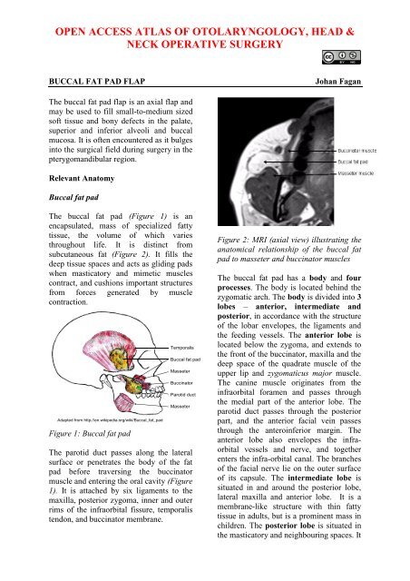
The buccal <strong>fat</strong> <strong>pad</strong> (Figure 1) is an<br />
encapsulated, mass <strong>of</strong> specialized <strong>fat</strong>ty<br />
tissue, the volume <strong>of</strong> which varies<br />
throughout life. It is distinct from<br />
subcutaneous <strong>fat</strong> (Figure 2). It fills the<br />
deep tissue spaces and acts as gliding <strong>pad</strong>s<br />
when masticatory and mimetic muscles<br />
contract, and cushions important structures<br />
from forces generated by muscle<br />
contraction.<br />
Adapted from http://en.wikipedia.org/wiki/<strong>Buccal</strong>_<strong>fat</strong>_<strong>pad</strong><br />
Figure 1: <strong>Buccal</strong> <strong>fat</strong> <strong>pad</strong><br />
Temporalis<br />
<strong>Buccal</strong> <strong>fat</strong> <strong>pad</strong><br />
Masseter<br />
Buccinator<br />
Parotid duct<br />
Masseter<br />
The parotid duct passes along the lateral<br />
surface or penetrates the body <strong>of</strong> the <strong>fat</strong><br />
<strong>pad</strong> before traversing the buccinator<br />
muscle and entering the oral cavity (Figure<br />
1). It is attached by six ligaments to the<br />
maxilla, posterior zygoma, inner and outer<br />
rims <strong>of</strong> the infraorbital fissure, temporalis<br />
tendon, and buccinator membrane.<br />
Figure 2: MRI (axial view) illustrating the<br />
anatomical relationship <strong>of</strong> the buccal <strong>fat</strong><br />
<strong>pad</strong> to masseter and buccinator muscles<br />
The buccal <strong>fat</strong> <strong>pad</strong> has a body and four<br />
processes. The body is located behind the<br />
zygomatic arch. The body is divided into 3<br />
lobes – anterior, intermediate and<br />
posterior, in accordance with the structure<br />
<strong>of</strong> the lobar envelopes, the ligaments and<br />
the feeding vessels. The anterior lobe is<br />
located below the zygoma, and extends to<br />
the front <strong>of</strong> the buccinator, maxilla and the<br />
deep space <strong>of</strong> the quadrate muscle <strong>of</strong> the<br />
upper lip and zygomaticus major muscle.<br />
The canine muscle originates from the<br />
infraorbital foramen and passes through<br />
the medial part <strong>of</strong> the anterior lobe. The<br />
parotid duct passes through the posterior<br />
part, and the anterior facial vein passes<br />
through the anteroinferior margin. The<br />
anterior lobe also envelopes the infraorbital<br />
vessels and nerve, and together<br />
enters the infra-orbital canal. The branches<br />
<strong>of</strong> the facial nerve lie on the outer surface<br />
<strong>of</strong> its capsule. The intermediate lobe is<br />
situated in and around the posterior lobe,<br />
lateral maxilla and anterior lobe. It is a<br />
membrane-like structure with thin <strong>fat</strong>ty<br />
tissue in adults, but is a prominent mass in<br />
children. The posterior lobe is situated in<br />
the masticatory and neighbouring spaces. It
extends up to the inferior orbital fissure<br />
and surrounds the temporalis muscle, and<br />
extends down to the superior rim <strong>of</strong> the<br />
mandibular body, and back to the anterior<br />
rim <strong>of</strong> the temporalis tendon and ramus. In<br />
doing so it forms the buccal,<br />
pterygopalatine and temporal processes.<br />
Four processes (buccal, pterygoid,<br />
superficial and deep temporal) extend from<br />
the body into surrounding spaces such as<br />
the pterygomandibular and infratemporal<br />
fossae.<br />
Coverage <strong>of</strong> exposed maxillary and<br />
mandibular bone or bone grafts and<br />
bone <strong>flap</strong>s<br />
Alternative or backup for failed buccal<br />
advancement <strong>flap</strong>s, palatal rotation and<br />
transposition <strong>flap</strong>s, tongue and<br />
nasolabial <strong>flap</strong>s, and radial free<br />
forearm <strong>flap</strong>s.<br />
Blood supply<br />
The buccal <strong>fat</strong> <strong>pad</strong> <strong>flap</strong> is an axial <strong>flap</strong>. The<br />
facial, transverse facial and internal<br />
maxillary arteries and their anastomosing<br />
branches enter the <strong>fat</strong> to form a<br />
subcapsular vascular plexus (Figure 3).<br />
Figure 4: The buccal <strong>fat</strong> <strong>pad</strong> can be<br />
rotated to cover a variety <strong>of</strong> defects<br />
Surgical Steps<br />
Figure 3: Blood supply to buccal <strong>fat</strong> <strong>pad</strong><br />
Indications<br />
Reconstruction <strong>of</strong> small to medium<br />
(
Figure 7: Flap placed over an oronasal<br />
defect<br />
Figure 5: Position <strong>of</strong> <strong>fat</strong> <strong>pad</strong> relative to<br />
parotid duct<br />
Gently deliver the required volume <strong>of</strong><br />
buccal <strong>fat</strong> tissue into oral cavity by<br />
gentle to-and-fro traction on the buccal<br />
<strong>fat</strong>, so as not to disrupt the blood<br />
supply and hence devascularise the <strong>flap</strong><br />
(Figure 6).<br />
Figure 8: Flap sutured to defect, and<br />
pedicle covered with mucosa<br />
Await epithelialisation <strong>of</strong> the <strong>flap</strong><br />
which usually occurs within 1 month<br />
(Figure 9)<br />
Figure 6: Careful delivery <strong>of</strong> <strong>fat</strong> <strong>pad</strong> after<br />
incising the capsule<br />
Take care not to injure the inferior<br />
buccinator branches <strong>of</strong> facial artery so<br />
as to avoid causing a haematoma<br />
Freshen the edges <strong>of</strong> the donor site<br />
Position the buccal <strong>fat</strong> <strong>pad</strong> <strong>flap</strong> in<br />
defect and secure it with absorbable<br />
sutures (Figures 7 & 8)<br />
Cover the <strong>flap</strong> with mucosa if feasible<br />
(Figure 8)<br />
Figure 8: Mucosalised <strong>flap</strong> approximately<br />
a month postoperatively<br />
3
Complications<br />
Complications rarely occur, and may<br />
include partial necrosis and excessive<br />
scarring. With large <strong>flap</strong>s used for buccal<br />
defects there is a risk <strong>of</strong> fibrosis and<br />
trismus.<br />
Summary<br />
The buccal <strong>fat</strong> <strong>pad</strong> is a simple, reliable <strong>flap</strong><br />
for repair <strong>of</strong> small-to-medium sized oral<br />
defects. It has an excellent blood supply<br />
and causes minimal donor site morbidity<br />
Author & Editor<br />
Johan Fagan MBChB, FCORL, MMed<br />
Pr<strong>of</strong>essor and Chairman<br />
Division <strong>of</strong> Otolaryngology<br />
<strong>University</strong> <strong>of</strong> <strong>Cape</strong> <strong>Town</strong><br />
<strong>Cape</strong> <strong>Town</strong><br />
South Africa<br />
johannes.fagan@uct.ac.za<br />
The Open Access Atlas <strong>of</strong> Otolaryngology, Head &<br />
Neck Operative Surgery by Johan Fagan (Editor)<br />
johannes.fagan@uct.ac.za is licensed under a Creative<br />
Commons Attribution - Non-Commercial 3.0 Unported<br />
License<br />
4


