Resonant nonlinear magneto-optical effects in atomsâ - The Budker ...
Resonant nonlinear magneto-optical effects in atomsâ - The Budker ...
Resonant nonlinear magneto-optical effects in atomsâ - The Budker ...

You also want an ePaper? Increase the reach of your titles
YUMPU automatically turns print PDFs into web optimized ePapers that Google loves.
6<br />
A <br />
n <br />
(a)<br />
Na D1<br />
(b)<br />
Na D2<br />
A <br />
Ω Ω <br />
Dichroism<br />
A A <br />
A A <br />
(a)<br />
gΜB/@ ⩵ <br />
(b)<br />
n <br />
n n <br />
n n <br />
Birefr<strong>in</strong>gence<br />
0<br />
0<br />
0<br />
220 C<br />
195 C<br />
177 C<br />
experiment<br />
theory<br />
155 C<br />
0<br />
0<br />
0<br />
220 C<br />
195 C<br />
177 C<br />
155 C<br />
gΜB/@ ⩵ <br />
(c)<br />
Frequency (Ω)<br />
0<br />
0<br />
140 C<br />
1<br />
(c)<br />
2<br />
Na D1<br />
3<br />
4<br />
5<br />
0<br />
0<br />
0<br />
140 C<br />
1 2 3<br />
(d) Na D2<br />
4<br />
5<br />
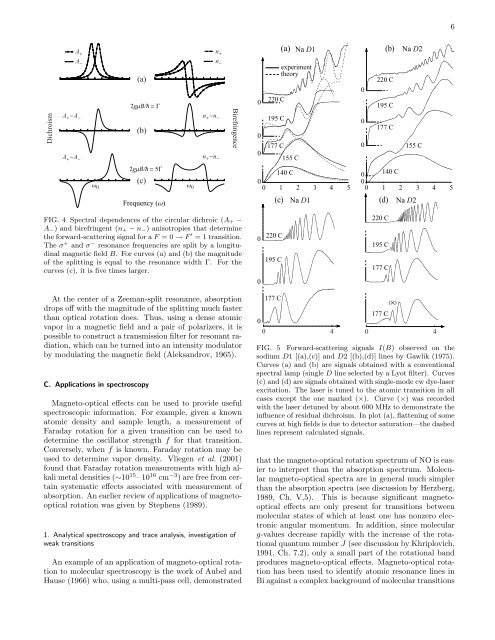
FIG. 4 Spectral dependences of the circular dichroic (A + −<br />
A − ) and birefr<strong>in</strong>gent (n + − n − ) anisotropies that determ<strong>in</strong>e<br />
the forward-scatter<strong>in</strong>g signal for a F = 0 → F ′ = 1 transition.<br />
<strong>The</strong> σ + and σ − resonance frequencies are split by a longitud<strong>in</strong>al<br />
magnetic field B. For curves (a) and (b) the magnitude<br />
of the splitt<strong>in</strong>g is equal to the resonance width Γ. For the<br />
curves (c), it is five times larger.<br />
0<br />
0<br />
220 C<br />
195 C<br />
220 C<br />
195 C<br />
177 C<br />
At the center of a Zeeman-split resonance, absorption<br />
drops off with the magnitude of the splitt<strong>in</strong>g much faster<br />
than <strong>optical</strong> rotation does. Thus, us<strong>in</strong>g a dense atomic<br />
vapor <strong>in</strong> a magnetic field and a pair of polarizers, it is<br />
possible to construct a transmission filter for resonant radiation,<br />
which can be turned <strong>in</strong>to an <strong>in</strong>tensity modulator<br />
by modulat<strong>in</strong>g the magnetic field (Aleksandrov, 1965).<br />
C. Applications <strong>in</strong> spectroscopy<br />
Magneto-<strong>optical</strong> <strong>effects</strong> can be used to provide useful<br />
spectroscopic <strong>in</strong>formation. For example, given a known<br />
atomic density and sample length, a measurement of<br />
Faraday rotation for a given transition can be used to<br />
determ<strong>in</strong>e the oscillator strength f for that transition.<br />
Conversely, when f is known, Faraday rotation may be<br />
used to determ<strong>in</strong>e vapor density. Vliegen et al. (2001)<br />
found that Faraday rotation measurements with high alkali<br />
metal densities (∼10 15 – 10 16 cm −3 ) are free from certa<strong>in</strong><br />
systematic <strong>effects</strong> associated with measurement of<br />
absorption. An earlier review of applications of <strong>magneto</strong><strong>optical</strong><br />
rotation was given by Stephens (1989).<br />
1. Analytical spectroscopy and trace analysis, <strong>in</strong>vestigation of<br />
weak transitions<br />
An example of an application of <strong>magneto</strong>-<strong>optical</strong> rotation<br />
to molecular spectroscopy is the work of Aubel and<br />
Hause (1966) who, us<strong>in</strong>g a multi-pass cell, demonstrated<br />
0<br />
177 C<br />
177 C<br />
0 4 0 4<br />
FIG. 5 Forward-scatter<strong>in</strong>g signals I(B) observed on the<br />
sodium D1 [(a),(c)] and D2 [(b),(d)] l<strong>in</strong>es by Gawlik (1975).<br />
Curves (a) and (b) are signals obta<strong>in</strong>ed with a conventional<br />
spectral lamp (s<strong>in</strong>gle D l<strong>in</strong>e selected by a Lyot filter). Curves<br />
(c) and (d) are signals obta<strong>in</strong>ed with s<strong>in</strong>gle-mode cw dye-laser<br />
excitation. <strong>The</strong> laser is tuned to the atomic transition <strong>in</strong> all<br />
cases except the one marked (×). Curve (×) was recorded<br />
with the laser detuned by about 600 MHz to demonstrate the<br />
<strong>in</strong>fluence of residual dichroism. In plot (a), flatten<strong>in</strong>g of some<br />
curves at high fields is due to detector saturation—the dashed<br />
l<strong>in</strong>es represent calculated signals.<br />
that the <strong>magneto</strong>-<strong>optical</strong> rotation spectrum of NO is easier<br />
to <strong>in</strong>terpret than the absorption spectrum. Molecular<br />
<strong>magneto</strong>-<strong>optical</strong> spectra are <strong>in</strong> general much simpler<br />
than the absorption spectra (see discussion by Herzberg,<br />
1989, Ch. V,5). This is because significant <strong>magneto</strong><strong>optical</strong><br />
<strong>effects</strong> are only present for transitions between<br />
molecular states of which at least one has nonzero electronic<br />
angular momentum. In addition, s<strong>in</strong>ce molecular<br />
g-values decrease rapidly with the <strong>in</strong>crease of the rotational<br />
quantum number J (see discussion by Khriplovich,<br />
1991, Ch. 7.2), only a small part of the rotational band<br />
produces <strong>magneto</strong>-<strong>optical</strong> <strong>effects</strong>. Magneto-<strong>optical</strong> rotation<br />
has been used to identify atomic resonance l<strong>in</strong>es <strong>in</strong><br />
Bi aga<strong>in</strong>st a complex background of molecular transitions<br />
(/\)