Build a Paper Model of DNA 1 2 3 4 5 6 7 8 9
Build a Paper Model of DNA 1 2 3 4 5 6 7 8 9
Build a Paper Model of DNA 1 2 3 4 5 6 7 8 9

Create successful ePaper yourself
Turn your PDF publications into a flip-book with our unique Google optimized e-Paper software.
www.rcsb.org • info@rcsb.org<br />
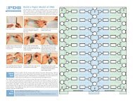
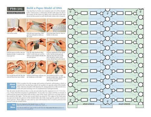
<strong>Build</strong> a <strong>Paper</strong> <strong>Model</strong> <strong>of</strong> <strong>DNA</strong><br />
Use this flyer to build one complete turn <strong>of</strong> a <strong>DNA</strong> double<br />
helix. Chose from: a schematic model with space to fill in<br />
the names <strong>of</strong> the bases (at right) and a detailed model that<br />
shows all the atoms in each nucleotide (other side <strong>of</strong> paper).<br />
1 2 3<br />
Cut out the model.<br />
Fold all long creases first. Solid<br />
grey lines should be visible on<br />
the crease.<br />
4 5 6<br />
Fold dotted grey lines so they are<br />
hidden in the crease.<br />
Fold the paper in half so that the<br />
backbone (with 3’ written at the<br />
top) pops out.<br />
Tuck the other backbone flaps<br />
(with 5’ at the top) one over the<br />
other, so your model looks like the<br />
on in the picture.<br />
7 8 9<br />
Fold the backbones so the model<br />
is flat. Fold the horizonal and<br />
diagonal lines like a fan (solid<br />
lines should be visible on the<br />
crease, dotted lines on the inside).<br />
Your model should look like this<br />
when all lines have been folded.<br />
Pull the model open, and pop out<br />
the backbones on the sides.<br />
Your finished model is a righthanded<br />
double helix. Use the tabs<br />
to connect several models to<br />
make longer strands.<br />
About<br />
<strong>DNA</strong><br />
<strong>DNA</strong> is perfect for the storage and readout <strong>of</strong> genetic information, which is<br />
stored in the way the bases match one another on opposite sides <strong>of</strong> the double<br />
helix. Adenine (A) pairs with thymine (T), and guanine (G) with cytosine (C),<br />
with each pair forming a set <strong>of</strong> complementary hydrogen bonds.<br />
The all-atom model (shown here on the second side) has the sequence C-G-C-T-T-A-A-G-<br />
C-G. Notice that this sequence is palindromic: if you take one chain and flip it around, it<br />
will form the proper base pairs with another copy <strong>of</strong> the chain. Add your own base pairs in<br />
the model to the right…but be sure to pair them up properly! The edges <strong>of</strong> the base pairs<br />
are exposed in the two grooves <strong>of</strong> the double helix: the wider major groove and the narrower<br />
minor groove. These edges are also used to carry information that is read by proteins that<br />
interact with the double helix.<br />
For<br />
More<br />
Read the Molecule <strong>of</strong> the Month feature on <strong>DNA</strong> at<br />
http://dx.doi.org/10.2210/rcsb_pdb/mom_2001_11<br />
Download additional copies <strong>of</strong> this model from the Educational Resources section <strong>of</strong><br />
http://www.rcsb.org/pdb101
www.rcsb.org/pdb101