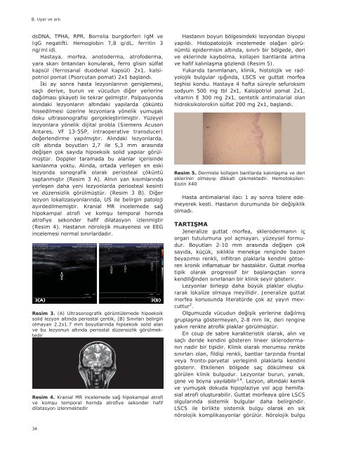
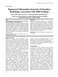
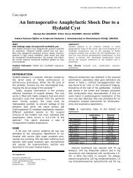
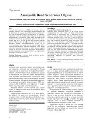
B. Uyar ve ark. dsDNA, TPHA, RPR, Borrelia burgdorferi IgM ve IgG negatifti. Hemoglobin 7,8 g/dL, ferritin 3 ng/ml idi. Hastaya, morfea, anotoderma, atrofoderma, kapsül (ferrosanal duodenal kapsül) 2x1, kalsipotriol pomat (Psorcutan pomat) 2x1 Palpasyonda lezyonlara yönelik dijital probla (Siemens Acuson Antares, VF 13-5SP, intraoperative transducer) Ai lezyonlarda, 3 - lezyonda sonografik olarak periosteal çöküntü sapt hipokampal atrof Resim 3. (A) Ultrasonografik görüntülemede hipoekoik tedir Resim 4. hafif dilatasyon izlenmektedir - ve eklerinde kaybolma, kollajen bantlarda artma di (Resim 5). Yu - du. Hastaya 4 hafta süreyle sefuroksim sodyum 500 mg tbl 2x1, Kalsipotriol pomat 2x1, vitamin E 300 mg 2x1, sentetik antimalarial olan hidroksikolorokin sülfat 200 Resim 5. - Eozin X40 - lik organ tutulumuna yol açmayan, yüzeysel formu- - götser rarak lokalize olmaya meyillidir. Jeneralize guttat morfea cuttur 2 . -8 mm lik, deri rengine teren lineer skleroderma- veya fronto- görülen klinik bulgudur. Lezyonlar burun, yanak, 3,4 - LSCS LSCS ile birlikte sistemik bulgu nörolojik komplikasyonlar görülür. Nörolojik bulgu 34
B. Uyar ve ark. - 5 . - 6 . Marzano ve ark. bildirdikleri 239 lokalize skleroderma hastas 17 hastada oral ve nörolojik tutulum mevcuttur 7 . <strong>De</strong>ri bulgular kaç ay sonra görülür. Fakat bir bulgularla seyreden vakalar 8,9 . - - 10 . Helene E. ve ark. hipokampal atrofi ve 6 Sistemik 9 . Nörolojik bulgusu olmayan, epilepsi nöbetleri tariflemeyen hastalarda bile anormal MRG ve 11,12 . - luklarla kendini gösterir. Öyküsünde tipik epilepsi anemnezi vermeyen olgumuzun nörolojik muaye- 8,13 . kimal kalsi- 1. Krieg T. Scleroderma. in: Braun-Falco O, Burgdorf WHC, Plewig G, Wolff HH, Landthaler M, editors. 3th ed. Braun Falco’s <strong>De</strong>rmatology. Italya: Springer Medizin Verlag Heidelberg; 2009. p. 701-15. 2. Blaya B, Gardeazabal J, Martínez de LagránZ, Díaz-Pérez JL. Patient With Generalized <strong>Guttat</strong>e Morphea and Lichen Sclerosus et Atrophicus. Actas <strong>De</strong>rmosifiliogr 2008;99:808-11. 3. Soma Y, Fujimoto M. Frontoparietal scleroderma (en coup de sabre) following Blaschko's lines. J Am Acad <strong>De</strong>rmatol 1998;38:366–8. 4. Itin PH, Schiller P. Double-lined frontoparietal scleroderma en coup de sabre. <strong>De</strong>rmatology 1999;199:185–6. 5. Flores-Alvarado DE, Esquivel-Valerio JA, Garza-Elizondo M, Espinoza LR. Linear scleroderma en coup de sabre and brain calcification: is there a pathologic relationship J Rheumatol 2003;30:193–5. 6. Verhelst HE, Beele H, Vanneuville RB, VanCoster RN. Hippocampal atrophy and developmental regressionas first sign of linear scleroderma ‘‘en coup de sabre’’. European Journal of Paediatric Neurology 2008;12:508–11. 7. Marzano AV, Menni S, Parodi A, Borghi A, Fuligni A, Fabbri P, et al. Localized scleroderma in adults and children: clinical and laboratory investigations of 239 cases. Eur J <strong>De</strong>rmatol 2003;13:171–6. 8. Stone J, Franks AJ, Guthrie JA, Johnson MH. Scleroderma “en coup de sabre”: pathological evidence of intracerebral inflammation. J Neurol Neurosurg Psychiatry 2001;70:382–5. 9. Menni S, Marzano AV, Passoni EP. Neurologic abnormalities in two patients with facial hemiatrophy and sclerosis coexisting with morphea. Pediatr <strong>De</strong>rmatol 1997;14:113–6. 10. Holland KE, Steffes B, Nocton JJ, Schwabe MJ, Jacobson RD, Drolet BA, et al. Linear Scleroderma en coup de sabre With Associated Neurologic Abnormalities. Pediatrics 2006;117: e132 11. Liu P, Uziel Y, Chuang S, Silverman E, Krafchik B, Laxer R. Localized scleroderma: imaging features. Pediatr Radiol 1994;24:207–9. REFERANSLAR radyolojik bulgular da izlenebilmektedir 8,11,12,14,15 . nik olarak düzelebilir. -sklerotik lezyonlardaki kemik defektini, yeni lezyonlardaki periostal kesilme ve inflamatuar periostiti göster- olgumuzun nörolojik muayenesinde patolojik bul- Sistemik, topikal ve lezyon içi kortizon tedavisi syonu azaltabilmekte- - aminobenzoat potasyum, penisilin, retinoidler, difenilhidantoin, interferon- unsupressan ilaçlar gibi sistemik ajanlar ve ultraviole-A tedavi- hydroxychloroquine, D-penisilamin, methotrexate, cyclosporine, and cyclophosphamide ekil -rekonstrüktif 13,16-20 . uzun süreli ve dikkatli takip etmek gerekir. Etyolojisi ve patogenezi tam olarak bilinmeyen bu 12. Appenzeller S, Montenegro MA, <strong>De</strong>rtkigil SS, Sampaio-Barros PD, Marques-Neto JF, Samara AM, et al. Neuroimaging findings in scleroderma en coup de sabre. Neurology 2004;62:1585–9. 13. Obermoser G, Pfaulser BE, Linder DM, Sepp NT. Scleroderma en coup de sabre with central nervous system and ophthalmologic involvement: treatment of ocular symptoms with interferon gamma. J Am Acad <strong>De</strong>rmatol 2003;49:543–6. 14. Unterberger I, Trinka E, <strong>En</strong>gelhardt K, Muigg A, Eller P, Wagner M, et al. Linear scleroderma “en coup de sabre” coexisting with plaquemorphea: neuroradiological manifestation and response to steroids. J Neurol Neurosurg Psychiatry 2003;74:661–4. 15. Heron E, Hernigou A, Chatellier G, Fornes P, Emmerich J, Fiessinger JN, et al. Intracerebral calcification in systemic sclerosis. Stroke 1999;30:2183–5. 16. Eubanks LE, McBurney EI, Galen W, Reed R. Linear scleroderma in children. Int J <strong>De</strong>rmatol 1996;35:330–6. 17. Cunningham BB, Landells ID, Langman C, Saliler DE, Paller AS. Topical calcipotriene for morphea/linear scleroderma. J Am Acad <strong>De</strong>rmatol 1998;39:211–5. 18. Hawk A, <strong>En</strong>glish JC 3rd. Localized and systemic scleroderma. Semin Cutan Med Surg 2001;20:27–37. 19. Elst EF, Van-Suijlekom-Smit LW, Oranje AP. Treatment of linear scleroderma with oral 1,25-dihydroxyvitamin D3 (calcitriol) in seven children. Pediatr <strong>De</strong>rmatol 1999;16:53–8. 20. Uziel Y, Feldman BM, Krafchik BR, Yeung RS, Laxer RM. Methotrexate and corticosteroid therapy for pediatric localized scleroderma. J Pediatr 2000;13:91–5. 35