Full range of dental X-ray units - Planmeca Oy
Full range of dental X-ray units - Planmeca Oy
Full range of dental X-ray units - Planmeca Oy

You also want an ePaper? Increase the reach of your titles
YUMPU automatically turns print PDFs into web optimized ePapers that Google loves.
ENGLISH<br />
<strong>Full</strong> <strong>range</strong> <strong>of</strong> <strong>dental</strong> X-<strong>ray</strong> <strong>units</strong>
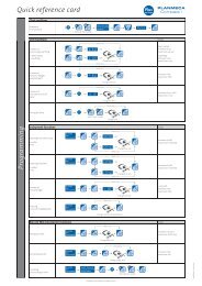
Standard Panoramic<br />
All-round <strong>dental</strong> X-<strong>ray</strong> unit<br />
<strong>Planmeca</strong> ProOne<br />
Lateral Double TMJ<br />
Cross-sections<br />
The fully digital <strong>Planmeca</strong> ProOne X-<strong>ray</strong> unit provides absolute ease <strong>of</strong> use with cutting<br />
edge technology. The wide selection <strong>of</strong> exposure programs and parameters on the unit<br />
graphical user interface ensures that all types <strong>of</strong> radiographic examinations are highly<br />
rapid and effortless to perform. With the side entry concept and open view for precise<br />
patient positioning, <strong>Planmeca</strong> ProOne truly inherits <strong>Planmeca</strong>’s expert knowledge<br />
and long traditions in <strong>dental</strong> imaging. Being small-sized, <strong>Planmeca</strong> ProOne brings the<br />
benefits <strong>of</strong> digital imaging within the reach <strong>of</strong> every dentist the world over.<br />
2 3
Standard Pediatric<br />
Leader in <strong>dental</strong> X-<strong>ray</strong> <strong>units</strong><br />
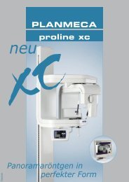
<strong>Planmeca</strong> Proline XC<br />
Standard Panoramic, segmented<br />
Lateral cephalometric<br />
The <strong>Planmeca</strong> Proline XC provides well-proven panoramic imaging capabilities and ease<br />
<strong>of</strong> use for any <strong>dental</strong> practice. The unit features a full colour TFT display and a graphical<br />
user interface, versatile imaging programs, as well as side entry and open patient<br />
positioning. Anatomically correct imaging geometry results in accurate and undistorted<br />
images every time.<br />
<strong>Planmeca</strong> Proline XC is available in two versions: film-based and fully digital. A film<br />
unit can be digitalised any time in the future. Both versions can be equipped with<br />
cephalometric imaging modality.<br />
4 5
3 longitudinal tomograms<br />
New era <strong>of</strong> <strong>dental</strong> imaging<br />
<strong>Planmeca</strong> ProMax<br />
4 cross-sectional tomograms<br />
Combined tomography: 1 cross-sectional and 1 longitudinal tomograms<br />
The revolutionary <strong>Planmeca</strong> ProMax <strong>of</strong>fers a complete <strong>range</strong> <strong>of</strong> extraoral X-<strong>ray</strong> imaging<br />
modalities for the needs <strong>of</strong> modern surgical dentistry. The state-<strong>of</strong>-the-art <strong>Planmeca</strong><br />
ProMax platform uses robotic SCARA technology to provide such utterly precise arm<br />
movements that are needed for rotational maxill<strong>of</strong>acial radiography. With this unique<br />
technology, any movement pattern required by existing or future exposure program can<br />
be produced; hence the unit mechanic design will never restrict its imaging capabilities.<br />
The <strong>Planmeca</strong> ProMax platform is unique in that it allows effortless upgradeability to 3D<br />
imaging; as this imaging modality is made possible simply with an upgrade <strong>of</strong> an existing<br />
digital <strong>Planmeca</strong> ProMax. The advanced design and functional concept ensure that the<br />
unit can perform superior maxill<strong>of</strong>acial radiography now and in decades to come.<br />
6 7
TMJ study<br />
Maximised 3D diagnostics<br />
<strong>Planmeca</strong> ProMax 3D<br />
<strong>Planmeca</strong> Romexis 3D Cross Sections Module<br />
Sinus study<br />
<strong>Planmeca</strong> ProMax 3D, a Cone Beam Volumetric Tomography (CBVT) unit, is expressly<br />
designed to obtain complete information on patient anatomy in the minutest detail. It<br />
supplies clear, dependable imaging in a three-dimensional format with limited patient<br />
radiation dose. The unit complies with a multitude <strong>of</strong> diagnostic requirements: those <strong>of</strong><br />
endodontics, periodontics, orthodontics, implantology, <strong>dental</strong> and maxill<strong>of</strong>acial surgery,<br />
and TMJ analysis.<br />
The <strong>Planmeca</strong> ProMax 3D provides digital panoramic, cephalometric, and threedimensional<br />
imaging, as well as advanced imaging s<strong>of</strong>tware tools for fulfilling every<br />
possible need in <strong>dental</strong> radiology. As a result, one intelligent X-<strong>ray</strong> unit can meet virtually<br />
any need in maxill<strong>of</strong>acial imaging.<br />
8 9
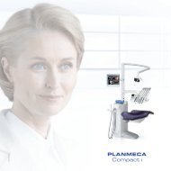
The premium intraoral X-<strong>ray</strong> unit<br />
<strong>Planmeca</strong> Intra<br />
Assured digital intraoral imaging<br />
<strong>Planmeca</strong> Dixi®3<br />
Advanced technology and practical design make the <strong>Planmeca</strong> Intra X-<strong>ray</strong> unit the<br />
premium choice for intraoral radiography. The freely selectable exposure parameters<br />
(kV, mA, exposure time) maximise the diagnostic value <strong>of</strong> intraoral radiography, and the<br />
pre-programmed quick settings make the operation fast and effortless. The unique nonsymmetric<br />
design <strong>of</strong> the X-<strong>ray</strong> tube head makes aiming exceptionally easy and precise.<br />
The <strong>Planmeca</strong> Dixi®3 interconnection cable is routed inside the X-<strong>ray</strong> unit arm, which<br />
results in a clear and clean working area with no interfering cables.<br />
The <strong>Planmeca</strong> Dixi®3 intraoral X-<strong>ray</strong> imaging system optimally supports fluent chairside<br />
workflow. The sensor is always ready for taking an image; no interaction with PC, keyboard, or<br />
mouse is required during the imaging procedure.<br />
All cabling runs inside the X-<strong>ray</strong> equipment and the sensor is conveniently placed at the X-<strong>ray</strong><br />
tube head. The flexible cable is connected to the backside <strong>of</strong> the sensor, which enables it<br />
to easily turn to allow any required position in the mouth. The hermetically sealed sensor<br />
encapsulation gives highly improved protection against shocks and bacteria.<br />
10 11
Comprehensive imaging s<strong>of</strong>tware<br />
<strong>Planmeca</strong> Dimaxis<br />
All <strong>Planmeca</strong>’s digital imaging products use the <strong>Planmeca</strong> Dimaxis imaging s<strong>of</strong>tware,<br />
which links together intraoral and extraoral X-<strong>ray</strong>, intraoral video, and still camera<br />
images. The s<strong>of</strong>tware includes powerful and easy-to-use image processing tools that<br />
facilitate image communications, bring visible significant image information and thus<br />
improve the diagnostic value <strong>of</strong> radiographs after the exposure. With these versatile<br />
tools, the s<strong>of</strong>tware supports optimal imaging workflow and usability at chairside.<br />
The <strong>Planmeca</strong> Dimaxis is delivered with a Solid EmbeddedEngine TM database that<br />
provides automated and secure storage <strong>of</strong> all image information. Automated backups<br />
meet the strictest requirements <strong>of</strong> data security and prevent all loss <strong>of</strong> clinical<br />
information. <strong>Planmeca</strong> Dimaxis is 100% DICOM compatible and fully ADA compliant.<br />
12 13
Complete clinical s<strong>of</strong>tware for 3D imaging<br />
<strong>Planmeca</strong> Romexis<br />
The <strong>Planmeca</strong> Romexis s<strong>of</strong>tware is used with the <strong>Planmeca</strong> ProMax 3D for image<br />
acquisition, viewing, and processing. The <strong>Planmeca</strong> Romexis 3D Explorer can be<br />
extended with an advanced 3D Panoramic Module, 3D Cross Sections Module, and 3D<br />
Implant Planning Module. With the optional DICOM functionality, 3D studies can be<br />
transferred to any other implant planning s<strong>of</strong>tware that receives images in DICOM<br />
format. <strong>Planmeca</strong> Romexis is also used to view and process 2D images captured with<br />
<strong>Planmeca</strong>’s digital devices. All patient’s digital images – such as photographs, intraoral<br />
and extraoral X-<strong>ray</strong> images, 3D images – are displayed in a single interface <strong>of</strong>fering a<br />
holistic view into the patient status.<br />
<strong>Planmeca</strong> Romexis is a pure JAVA based s<strong>of</strong>tware that runs in various operating systems<br />
and modern web environments. With specific features particularly designed for<br />
educational needs, the s<strong>of</strong>tware platform creates a unique learning environment for<br />
<strong>dental</strong> universities and teaching hospitals.<br />
14 15
<strong>Planmeca</strong> <strong>Oy</strong> | Asentajankatu 6 | 00880 Helsinki | Finland | tel. +358 20 7795 500 | fax +358 20 7795 555 | sales@planmeca.com | www.planmeca.com<br />
Images may contain optional items not included in standard delivery. Rights for changes reserved.<br />
10016132/0908/en