Basic Principles of Transcription and Translation - Computer ...
Basic Principles of Transcription and Translation - Computer ...
Basic Principles of Transcription and Translation - Computer ...

You also want an ePaper? Increase the reach of your titles
YUMPU automatically turns print PDFs into web optimized ePapers that Google loves.
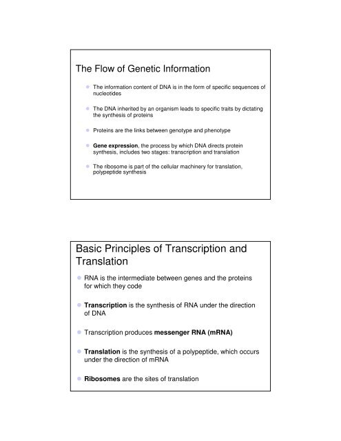
The Flow <strong>of</strong> Genetic Information<br />
The information content <strong>of</strong> DNA is in the form <strong>of</strong> specific sequences <strong>of</strong><br />
nucleotides<br />
The DNA inherited by an organism leads to specific traits by dictating<br />
the synthesis <strong>of</strong> proteins<br />
Proteins are the links between genotype <strong>and</strong> phenotype<br />
Gene expression, the process by which DNA directs protein<br />
synthesis, includes two stages: transcription <strong>and</strong> translation<br />
The ribosome is part <strong>of</strong> the cellular machinery for translation,<br />
polypeptide synthesis<br />
<strong>Basic</strong> <strong>Principles</strong> <strong>of</strong> <strong>Transcription</strong> <strong>and</strong><br />
<strong>Translation</strong><br />
RNA is the intermediate between genes <strong>and</strong> the proteins<br />
for which they code<br />
<strong>Transcription</strong> is the synthesis <strong>of</strong> RNA under the direction<br />
<strong>of</strong> DNA<br />
<strong>Transcription</strong> produces messenger RNA (mRNA)<br />
<strong>Translation</strong> is the synthesis <strong>of</strong> a polypeptide, which occurs<br />
under the direction <strong>of</strong> mRNA<br />
Ribosomes are the sites <strong>of</strong> translation
• In prokaryotes, mRNA produced by transcription is<br />
immediately translated without more processing<br />
• In a eukaryotic cell, the nuclear envelope separates<br />
transcription from translation<br />
• Eukaryotic RNA transcripts are modified through RNA<br />
processing to yield finished mRNA<br />
• A primary transcript is the initial RNA transcript from any<br />
gene<br />
• The central dogma is the concept that cells are governed<br />
by a cellular chain <strong>of</strong> comm<strong>and</strong>: DNA → RNA → protein<br />
• Eukaryotic RNA transcripts are modified through RNA<br />
processing to yield finished mRNA<br />
TRANSCRIPTION<br />
TRANSLATION<br />
Polypeptide<br />
DNA<br />
mRNA<br />
Ribosome<br />
a) Bacterial cell. In a bacterial cell which<br />
lacks a nucleus, mRNA produced by<br />
transcription is immediately translated<br />
without additional processing.<br />
(a) Bacterial cell<br />
TRANSCRIPTION<br />
DNA<br />
Nuclear<br />
envelope<br />
b) Eukaryotic cell. The nucleus provides a<br />
separate compartment for transcription.<br />
The original RNA transcript called pre<br />
mRNA is processed in various ways<br />
before leaving the nucleus as mRNA.<br />
RNA PROCESSING<br />
Pre-mRNA<br />
mRNA<br />
TRANSLATION Ribosome<br />
Polypeptide<br />
(b) Eukaryotic cell<br />
Overview: the roles <strong>of</strong> transcription <strong>and</strong><br />
translation in the flow <strong>of</strong> genetic<br />
information. In a cell inherited information<br />
flows from DNA to RNA to protein. The<br />
two main stages <strong>of</strong> information flow are<br />
transcription <strong>and</strong> translation.
TRANSCRIPTION<br />
DNA<br />
mRNA<br />
(a) Bacterial cell<br />
TRANSCRIPTION<br />
TRANSLATION<br />
Polypeptide<br />
DNA<br />
mRNA<br />
Ribosome<br />
Nuclear<br />
envelope<br />
TRANSCRIPTION<br />
DNA<br />
Pre-mRNA<br />
(b) Eukaryotic cell
Nuclear<br />
envelope<br />
TRANSCRIPTION<br />
DNA<br />
RNA PROCESSING<br />
Pre-mRNA<br />
mRNA<br />
(b) Eukaryotic cell<br />
Nuclear<br />
envelope<br />
TRANSCRIPTION<br />
DNA<br />
RNA PROCESSING<br />
Pre-mRNA<br />
mRNA<br />
TRANSLATION<br />
Ribosome<br />
Polypeptide<br />
(b) Eukaryotic cell
The Genetic Code<br />
How are the instructions for assembling amino acids into<br />
proteins encoded into DNA<br />
There are 20 amino acids, but there are only four<br />
nucleotide bases in DNA<br />
How many bases correspond to an amino acid<br />
Codons: Triplets <strong>of</strong> Bases<br />
The flow <strong>of</strong> information from gene to protein is based on a<br />
triplet code: a series <strong>of</strong> nonoverlapping, three-nucleotide<br />
words<br />
These triplets are the smallest units <strong>of</strong> uniform length that<br />
can code for all the amino acids<br />
Example: AGT at a particular position on a DNA str<strong>and</strong><br />
results in the placement <strong>of</strong> the amino acid serine at the<br />
corresponding position <strong>of</strong> the polypeptide to be produced
During transcription, one <strong>of</strong> the two DNA str<strong>and</strong>s called the<br />
template str<strong>and</strong> provides a template for ordering the<br />
sequence <strong>of</strong> nucleotides in an RNA transcript<br />
During translation, the mRNA base triplets, called codons,<br />
are read in the 5′ to 3′ direction<br />
Each codon specifies the amino acid to be placed at the<br />
corresponding position along a polypeptide<br />
Colons along an mRNA molecule are read by translation<br />
machinery in the 5′ to 3′ direction<br />
Each codon specifies the addition <strong>of</strong> one <strong>of</strong> 20 amino acids<br />
DNA<br />
molecule<br />
Gene 1<br />
Gene 2<br />
DNA<br />
template<br />
str<strong>and</strong><br />
TRANSCRIPTION<br />
mRNA<br />
Codon<br />
TRANSLATION<br />
Protein<br />
Amino acid<br />
Gene 3<br />
The triplet code. For each<br />
gene, one str<strong>and</strong> <strong>of</strong> DNA<br />
functions as a template for<br />
transcription. The base<br />
pairing rules for DNA<br />
synthesis also guide<br />
transcription, but uracil (U)<br />
takes the place <strong>of</strong> thymine<br />
(T) in RNA. During<br />
translation the mRNA is read<br />
as a sequence <strong>of</strong> base<br />
triplets called codons. Each<br />
codon specifies an amino<br />
acid to be added to the<br />
growing polypeptide chain.<br />
The mRNA is read in the<br />
5’⇒ 3’ direction.
Cracking the Code<br />
All 64 codons were deciphered by the mid-1960s<br />
Of the 64 triplets, 61 code for amino acids; 3 triplets are<br />
“stop” signals to end translation<br />
The genetic code is redundant but not ambiguous; no<br />
codon specifies more than one amino acid<br />
Codons must be read in the correct reading frame (correct<br />
groupings) in order for the specified polypeptide to be<br />
produced<br />
Second mRNA base<br />
First mRNA base (5′ end <strong>of</strong> codon)<br />
Third mRNA base (3′ end <strong>of</strong> codon)<br />
The dictionary <strong>of</strong> the genetic<br />
code. The three bases <strong>of</strong> an<br />
mRNA codon are designated<br />
here as the first, second <strong>and</strong><br />
third bases reading in the 5’<br />
⇒ 3’ direction along the<br />
mRNA. The codon AUG not<br />
only st<strong>and</strong>s for the amino<br />
acid methionine (Met) but<br />
also functions as a start<br />
signal for ribosomes to begin<br />
translating the mRNA at that<br />
point. Three <strong>of</strong> the 64<br />
codons function as “stop”<br />
signals marking the end <strong>of</strong> a<br />
genetic message
Evolution <strong>of</strong> the Genetic Code<br />
The genetic code is nearly universal, shared by the<br />
simplest bacteria to the most complex animals<br />
Genes can be transcribed <strong>and</strong> translated after being<br />
transplanted from one species to another<br />
Because diverse forms <strong>of</strong> life share a common genetic<br />
code, one species can be programmed to produce proteins<br />
characteristic <strong>of</strong> a second species by introducing DNA from<br />
the second species into the first<br />
<strong>Transcription</strong> is the DNA-directed synthesis <strong>of</strong><br />
RNA: a closer look<br />
<strong>Transcription</strong>, the first stage <strong>of</strong> gene expression, can be<br />
examined in more detail<br />
The three stages <strong>of</strong> transcription:<br />
Initiation<br />
Elongation<br />
Termination
Molecular Components <strong>of</strong> <strong>Transcription</strong><br />
RNA synthesis is catalyzed by RNA polymerase, which<br />
pries the DNA str<strong>and</strong>s apart <strong>and</strong> hooks together the RNA<br />
nucleotides<br />
RNA synthesis follows the same base-pairing rules as<br />
DNA, except uracil substitutes for thymine<br />
The DNA sequence where RNA polymerase attaches is<br />
called the promoter; in bacteria, the sequence signaling<br />
the end <strong>of</strong> transcription is called the terminator<br />
The stretch <strong>of</strong> DNA that is transcribed is called a<br />
transcription unit<br />
Promoter<br />
<strong>Transcription</strong> unit<br />
5′<br />
3′<br />
DNA<br />
Start point<br />
RNA polymerase<br />
1 Initiation<br />
5′<br />
3′<br />
Unwound<br />
DNA<br />
RNA<br />
transcript<br />
Template str<strong>and</strong><br />
<strong>of</strong> DNA<br />
2<br />
Elongation<br />
3′<br />
5′<br />
3′<br />
5′<br />
3′<br />
Elongation<br />
Nontemplate<br />
str<strong>and</strong> <strong>of</strong> DNA<br />
RNA<br />
polymerase<br />
3′ end<br />
RNA nucleotides<br />
5′<br />
3′<br />
5′<br />
3′<br />
Rewound<br />
DNA<br />
5′<br />
RNA<br />
transcript<br />
5′<br />
3′<br />
3 Termination<br />
Completed RNA transcript<br />
3′<br />
3′<br />
5′<br />
3′<br />
5′<br />
5′<br />
5′<br />
Newly made<br />
RNA<br />
Direction <strong>of</strong><br />
transcription<br />
(“downstream”)<br />
Template<br />
str<strong>and</strong> <strong>of</strong> DNA
Promoter<br />
5′<br />
3′<br />
Start point<br />
RNA polymerase<br />
5′<br />
3′<br />
Unwound<br />
DNA<br />
5′<br />
3′<br />
5′<br />
3′<br />
Rewound<br />
DNA<br />
5′<br />
RNA<br />
transcript<br />
5′<br />
<strong>Transcription</strong> unit<br />
RNA<br />
transcript<br />
DNA<br />
1<br />
Initiation<br />
Template str<strong>and</strong><br />
<strong>of</strong> DNA<br />
2 Elongation<br />
3′<br />
3 Termination<br />
Completed RNA transcript<br />
3′<br />
3′<br />
5′<br />
3′<br />
5′<br />
3′<br />
5′<br />
3′<br />
5′<br />
1) Initiation. After RNA polymerase binds<br />
to the promoter, the DNA str<strong>and</strong>s<br />
unwind <strong>and</strong> the polymerase initiates<br />
RNA synthesis at the start point on the<br />
template str<strong>and</strong>.<br />
2) 2) Elongation The polymerase moves<br />
downstream unwinding the DNA <strong>and</strong><br />
elongating the RNA transcript 5’ 3’ In<br />
the wake <strong>of</strong> transcription the DNA<br />
str<strong>and</strong>s re-form a double helix.<br />
3) 3) Termination Eventually the RNA<br />
transcript is released <strong>and</strong> the<br />
polymerase detaches from the DNA<br />
The stages <strong>of</strong> transcription: initiation<br />
elongation <strong>and</strong> termination. Thus<br />
general depiction <strong>of</strong> transcription<br />
applies to both bacteria <strong>and</strong><br />
eukaryotes but the details <strong>of</strong><br />
termination differ, as described in the<br />
text. Also in a bacterium the transcript<br />
is immediately usable as mRNA in a<br />
eukaryote the RNA transcript must first<br />
undergo processing.<br />
Elongation<br />
Nontemplate<br />
str<strong>and</strong> <strong>of</strong> DNA<br />
RNA<br />
polymerase<br />
RNA nucleotides<br />
3′<br />
3′ end<br />
5′<br />
5′<br />
Newly made<br />
RNA<br />
Direction <strong>of</strong><br />
transcription<br />
(“downstream”)<br />
Template<br />
str<strong>and</strong> <strong>of</strong> DNA
RNA Polymerase Binding <strong>and</strong> Initiation <strong>of</strong><br />
<strong>Transcription</strong><br />
Promoters signal the initiation <strong>of</strong> RNA synthesis<br />
<strong>Transcription</strong> factors mediate the binding <strong>of</strong> RNA<br />
polymerase <strong>and</strong> the initiation <strong>of</strong> transcription<br />
The completed assembly <strong>of</strong> transcription factors <strong>and</strong> RNA<br />
polymerase II bound to a promoter is called a<br />
transcription initiation complex<br />
A promoter called a TATA box is crucial in forming the<br />
initiation complex in eukaryotes<br />
5′<br />
3′<br />
5′<br />
3′<br />
1<br />
Promoter<br />
Template<br />
3′<br />
5′<br />
TATA box Start point Template<br />
DNA str<strong>and</strong><br />
<strong>Transcription</strong><br />
factors<br />
RNA polymerase II<br />
5′<br />
3′<br />
2<br />
3<br />
3′<br />
5′<br />
<strong>Transcription</strong> factors<br />
3′<br />
5′ 5′<br />
RNA transcript<br />
<strong>Transcription</strong> initiation complex<br />
1) A eukaryotic promoter<br />
commonly includes a TATA box a<br />
nucleotide sequence containing<br />
TATA about 25 nucleotides<br />
upstream from the transcription<br />
start point. By convention<br />
nucleotide sequences are given as<br />
they occur on the nontemplate<br />
str<strong>and</strong><br />
2) Several transcription factors one<br />
recognizing the TATA box must bind<br />
to the DNA before RNA polymerase<br />
II can do so.<br />
3) Additional transcription factors<br />
(purple) bind to the DNA along with<br />
RNA polymerase II forming the<br />
transcription initiation complex. The<br />
DNA double helix then unwinds <strong>and</strong><br />
RNA synthesis begins at the start<br />
point on the template str<strong>and</strong>.<br />
The initiation <strong>of</strong> transcription at a<br />
eukaryotic promoter. In eukaryotic<br />
cells proteins called transcription<br />
factors mediate the initiation <strong>of</strong><br />
transcription by RNA polymerase II
Elongation <strong>of</strong> the RNA Str<strong>and</strong><br />
As RNA polymerase moves along the DNA, it untwists the<br />
double helix, 10 to 20 bases at a time<br />
<strong>Transcription</strong> progresses at a rate <strong>of</strong> 40 nucleotides per<br />
second in eukaryotes<br />
A gene can be transcribed simultaneously by several RNA<br />
polymerases<br />
Termination <strong>of</strong> <strong>Transcription</strong><br />
The mechanisms <strong>of</strong> termination are different in<br />
bacteria <strong>and</strong> eukaryotes<br />
In bacteria, the polymerase stops transcription<br />
at the end <strong>of</strong> the terminator<br />
In eukaryotes, the polymerase continues<br />
transcription after the pre-mRNA is cleaved<br />
from the growing RNA chain; the polymerase<br />
eventually falls <strong>of</strong>f the DNA
Eukaryotic cells modify RNA after<br />
transcription<br />
Enzymes in the eukaryotic nucleus modify pre-mRNA<br />
before the genetic messages are dispatched to the<br />
cytoplasm<br />
During RNA processing, both ends <strong>of</strong> the primary transcript<br />
are usually altered<br />
Also, usually some interior parts <strong>of</strong> the molecule are cut<br />
out, <strong>and</strong> the other parts spliced together<br />
Alteration <strong>of</strong> mRNA Ends<br />
Each end <strong>of</strong> a pre-mRNA molecule is modified in a<br />
particular way:<br />
The 5′ end receives a modified nucleotide 5′ cap<br />
The 3′ end gets a poly-A tail<br />
These modifications share several functions:<br />
They seem to facilitate the export <strong>of</strong> mRNA<br />
They protect mRNA from hydrolytic enzymes<br />
They help ribosomes attach to the 5′ end
5′<br />
Protein-coding segment Polyadenylation signal<br />
3′<br />
G P P P<br />
AAUAAA AAA…<br />
AAA<br />
5′ Cap 5′ UTR Start codon Stop codon 3′ UTR Poly-A tail<br />
RNA processing addition <strong>of</strong> the 5’ cap <strong>and</strong> poly A tail. Enzymes modify the two<br />
ends <strong>of</strong> a eukaryotic pre mRNA molecule. The modified ends may promote the<br />
export <strong>of</strong> mRNA from the nucleus <strong>and</strong> they help protect the mRNA from<br />
degradation. When the mRNA reaches the cytoplasm the modified ends in<br />
conjunction with certain cytoplasmic proteins facilitate ribosome attachment.<br />
The 5’ cap <strong>and</strong> poly A tail are not translated into protein, nor are the regions<br />
called the 5’ untranslated regions (5’ UTR) <strong>and</strong> 3’ untranslated regions (3’ UTR)<br />
Split Genes <strong>and</strong> RNA Splicing<br />
• Most eukaryotic genes <strong>and</strong> their RNA transcripts have long<br />
noncoding stretches <strong>of</strong> nucleotides that lie between coding<br />
regions<br />
• These noncoding regions are called intervening<br />
sequences, or introns<br />
• The other regions are called exons because they are<br />
eventually expressed, usually translated into amino acid<br />
sequences<br />
• RNA splicing removes introns <strong>and</strong> joins exons, creating<br />
an mRNA molecule with a continuous coding sequence
5′ Exon Intron Exon Intron<br />
Pre-mRNA 5′ Cap<br />
1 30 31 104 105<br />
Exon 3′<br />
Poly-A tail<br />
146<br />
Coding<br />
segment<br />
Introns cut out <strong>and</strong><br />
exons spliced together<br />
mRNA<br />
5′ Cap<br />
Poly-A tail<br />
1 146<br />
5′ UTR 3′ UTR<br />
RNA processing: mRNA splicing. The RNA molecule shown here codes for β globin one<br />
<strong>of</strong> the polypeptides <strong>of</strong> hemoglobin. The numbers under the RNA refer to the codons. β<br />
globin is 146 amino acids long. The β globin gene <strong>and</strong> its pre mRNA transcript have<br />
three exons corresponding to sequences that will leave the nucleus as RNA. (The 5’<br />
UTR <strong>and</strong> 3’ UTR are parts <strong>of</strong> exons because they are included in the mRNA however<br />
they do not code for protein). During RNA processing the introns are cut out <strong>and</strong> the<br />
exons are spliced together. In many genes the introns are much larger relative to the<br />
exons than they are in the β globin gene. The mRNA is not drawn to scale.<br />
In some cases, RNA splicing is carried out by<br />
spliceosomes<br />
Spliceosomes consist <strong>of</strong> a variety <strong>of</strong> proteins <strong>and</strong> several<br />
small nuclear ribonucleoproteins (snRNPs) that recognize<br />
the splice sites
5′<br />
Protein<br />
snRNA<br />
RNA transcript (pre-mRNA)<br />
Exon 1 Intron Exon 2<br />
5′<br />
snRNPs<br />
Spliceosome<br />
components<br />
5′<br />
Spliceosome<br />
mRNA<br />
Exon 1 Exon 2<br />
Other<br />
proteins<br />
Cut-out<br />
intron<br />
The roles <strong>of</strong> snRNPS <strong>and</strong><br />
spliceosomes in pre mRNA<br />
splicing. The diagram shows only<br />
a portion <strong>of</strong> the pre mRNA<br />
transcript, additional introns <strong>and</strong><br />
exons lie downstream from the<br />
pictured ones here. 1) Small<br />
nuclear ribonucleoprotein<br />
(snRNPs) <strong>and</strong> other proteins form<br />
a molecular complex called a<br />
spliceosome on a pre mRNA<br />
molecules containing exons <strong>and</strong><br />
introns. 2) Within the spliceosome<br />
snRNA base pairs with nucleotides<br />
at specific sites along the intron. 3)<br />
The spliceosome cuts the pre<br />
mRNA releasing the intron <strong>and</strong> at<br />
the same time splices the exons<br />
together. The spliceosome then<br />
comes apart releasing mRNA<br />
which now contains only exons.<br />
Ribozymes<br />
Ribozymes are catalytic RNA molecules that function as<br />
enzymes <strong>and</strong> can splice RNA<br />
The discovery <strong>of</strong> ribozymes rendered obsolete the belief<br />
that all biological catalysts were proteins<br />
Three properties <strong>of</strong> RNA enable it to function as an<br />
enzyme<br />
It can form a three-dimensional structure because <strong>of</strong> its ability to<br />
base pair with itself<br />
Some bases in RNA contain functional groups<br />
RNA may hydrogen-bond with other nucleic acid molecules
The Functional <strong>and</strong> Evolutionary Importance<br />
<strong>of</strong> Introns<br />
Some genes can encode more than one kind <strong>of</strong><br />
polypeptide, depending on which segments are treated as<br />
exons during RNA splicing<br />
Such variations are called alternative RNA splicing<br />
Because <strong>of</strong> alternative splicing, the number <strong>of</strong> different<br />
proteins an organism can produce is much greater than its<br />
number <strong>of</strong> genes<br />
Proteins <strong>of</strong>ten have a modular architecture consisting <strong>of</strong><br />
discrete regions called domains<br />
In many cases, different exons code for the different<br />
domains in a protein<br />
Exon shuffling may result in the evolution <strong>of</strong> new proteins
DNA<br />
Gene<br />
Exon 1 Intron Exon 2 Intron Exon 3<br />
<strong>Transcription</strong><br />
RNA processing<br />
<strong>Translation</strong><br />
Correspondence<br />
between exons<br />
<strong>and</strong> protein<br />
domains.<br />
Domain 3<br />
Domain 2<br />
Domain 1<br />
Polypeptide<br />
<strong>Translation</strong> is the RNA-directed synthesis <strong>of</strong> a<br />
polypeptide: a closer look<br />
A cell translates an mRNA message into protein with the<br />
help <strong>of</strong> transfer RNA (tRNA)<br />
Molecules <strong>of</strong> tRNA are not identical:<br />
Each carries a specific amino acid on one end<br />
Each has an anticodon on the other end; the anticodon<br />
base-pairs with a complementary codon on mRNA
Polypeptide<br />
Trp<br />
Phe<br />
Ribosome<br />
Amino<br />
acids<br />
tRNA with<br />
amino acid<br />
attached<br />
Gly<br />
tRNA<br />
Anticodon<br />
<strong>Translation</strong>: the basic concept. As<br />
a molecule <strong>of</strong> mRNA is moved<br />
through a ribosome codons are<br />
translated into amino acids one by<br />
one. The interpreters are tRNA<br />
molecules, each type with a<br />
specific anticodon at one end <strong>and</strong><br />
a corresponding amino acid at the<br />
other end. A tRNA adds its amino<br />
acid cargo to a growing<br />
polypeptide chain when the<br />
anticodon hydrogen bonds to a<br />
complementary codon on the<br />
mRNA.<br />
5′<br />
Codons 3′<br />
mRNA<br />
The Structure <strong>and</strong> Function <strong>of</strong> Transfer RNA<br />
A tRNA molecule consists <strong>of</strong> a single RNA str<strong>and</strong> that is<br />
only about 80 nucleotides long<br />
A<br />
C<br />
C<br />
Flattened into one plane to reveal its base pairing, a tRNA<br />
molecule looks like a cloverleaf<br />
Because <strong>of</strong> hydrogen bonds, tRNA actually twists <strong>and</strong><br />
folds into a three-dimensional molecule<br />
tRNA is roughly L-shaped
Amino acid<br />
attachment site<br />
3′<br />
5′<br />
Anticodon<br />
(a) Two-dimensional structure<br />
Anticodon<br />
5′<br />
Hydrogen<br />
bonds<br />
3′<br />
Hydrogen<br />
bonds<br />
(b) Three-dimensional structure<br />
Amino acid<br />
attachment site<br />
3′ 5′<br />
Anticodon<br />
(c) Symbol used<br />
in this book<br />
Two dimensional structure. The four base<br />
paired regions <strong>and</strong> three loops are<br />
characteristic <strong>of</strong> all tRNAs as is the base<br />
sequence <strong>of</strong> the amino acid attachment<br />
site at the 3’ end. The anticodon triplet is<br />
unique to each tRNA type as are some<br />
sequences in the other two loops. (the<br />
asterisks mark bases that have been<br />
chemically modified a characteristic <strong>of</strong><br />
tRNA)<br />
The structure <strong>of</strong> transfer RNA (tRNA).<br />
Anticodons are conventionally written 3’∏<br />
5’ to align properly with codons written 5’<br />
∏3’. For base pairing RNA str<strong>and</strong>s must<br />
be antiparallel like DNA. For example<br />
anticodon 3’ AAG 5’ pairs with mRNA<br />
codon 5’ UUC 3’<br />
Accurate translation requires two steps:<br />
First: a correct match between a tRNA <strong>and</strong> an amino<br />
acid, done by the enzyme aminoacyl-tRNA synthetase<br />
Second: a correct match between the tRNA anticodon<br />
<strong>and</strong> an mRNA codon<br />
Flexible pairing at the third base <strong>of</strong> a codon is called<br />
wobble <strong>and</strong> allows some tRNAs to bind to more than one<br />
codon
Amino acid<br />
P P P<br />
ATP<br />
Adenosine<br />
Aminoacyl-tRNA<br />
synthetase (enzyme)<br />
1) Active site binds to the amino acid<br />
<strong>and</strong> ATP<br />
2) ATP loses two P groups <strong>and</strong> joins<br />
amino acids as AMP<br />
tRNA<br />
P Adenosine<br />
P P i<br />
P i P i<br />
tRNA<br />
Aminoacyl-tRNA<br />
synthetase<br />
3) Appropriate tRNA covalently bonds to<br />
amino acid displacing AMP<br />
4) The tRNA charged with amino acid is<br />
released by the enzyme<br />
P<br />
Adenosine<br />
AMP<br />
Aminoacyl-tRNA<br />
(“charged tRNA”)<br />
<strong>Computer</strong> model<br />
An aminoacyl tRNA synthethase joining<br />
a specific amino acid to a tRNA.<br />
Linkage <strong>of</strong> the tRNA <strong>and</strong> amino acid<br />
is an endergonic process that occurs<br />
at the expense <strong>of</strong> ATP. The ATP<br />
loses two phosphate groups<br />
becoming AMP (adenosine<br />
monophosphate)<br />
Ribosomes<br />
Ribosomes facilitate specific coupling <strong>of</strong> tRNA anticodons<br />
with mRNA codons in protein synthesis<br />
The two ribosomal subunits (large <strong>and</strong> small) are made <strong>of</strong><br />
proteins <strong>and</strong> ribosomal RNA (rRNA)
tRNA<br />
molecules<br />
Growing<br />
polypeptide<br />
E P A<br />
Exit tunnel<br />
Large<br />
subunit<br />
Small<br />
subunit<br />
5′<br />
mRNA 3′<br />
(a) <strong>Computer</strong> model <strong>of</strong> functioning ribosome<br />
P site (Peptidyl-tRNA<br />
binding site)<br />
E site<br />
(Exit site)<br />
mRNA<br />
binding site<br />
E P A<br />
A site (AminoacyltRNA<br />
binding site)<br />
Large<br />
subunit<br />
Small<br />
subunit<br />
(b) Schematic model showing binding sites<br />
Amino end<br />
mRNA<br />
E<br />
5′ Codons<br />
Growing polypeptide<br />
Next amino acid<br />
to be added to<br />
polypeptide chain<br />
tRNA<br />
3′<br />
(c) Schematic model with mRNA <strong>and</strong> tRNA<br />
a) <strong>Computer</strong> model <strong>of</strong> functioning ribosome. This<br />
is a model <strong>of</strong> a bacterial ribosome showing its<br />
overall shape. The eukaryotic ribosome is roughly<br />
similar. A ribosomal subunit is an aggregate <strong>of</strong><br />
ribosomal RNA molecules <strong>and</strong> proteins<br />
b) Schematic model showing binding sites. A<br />
ribosome has an mRNA binding site <strong>and</strong> three<br />
tRNA binding sites known as the A, P <strong>and</strong> E sites.<br />
c) Schematic model with mRNA <strong>and</strong> tRNA. A<br />
tRNA fits into a binding site when its anticodon<br />
base pairs with an mRNA codon. The P site holds<br />
the tRNA attached to the growing polypetide. The<br />
A site holds the tRNA carrying the next amino acid<br />
to be added to the polypeptide chain. Discharged<br />
tRNAs leaves from the E site.<br />
The anatomy <strong>of</strong> a functioning ribosome.<br />
A ribosome has three binding sites for tRNA:<br />
The P site holds the tRNA that carries the growing<br />
polypeptide chain<br />
The A site holds the tRNA that carries the next amino acid<br />
to be added to the chain<br />
The E site is the exit site, where discharged tRNAs leave<br />
the ribosome
Building a Polypeptide<br />
The three stages <strong>of</strong> translation:<br />
Initiation<br />
Elongation<br />
Termination<br />
All three stages require protein “factors” that aid in the<br />
translation process<br />
Ribosome Association <strong>and</strong> Initiation <strong>of</strong><br />
<strong>Translation</strong><br />
The initiation stage <strong>of</strong> translation brings together mRNA, a<br />
tRNA with the first amino acid, <strong>and</strong> the two ribosomal<br />
subunits<br />
First, a small ribosomal subunit binds with mRNA <strong>and</strong> a<br />
special initiator tRNA<br />
Then the small subunit moves along the mRNA until it<br />
reaches the start codon (AUG)<br />
Proteins called initiation factors bring in the large subunit<br />
that completes the translation initiation complex
Met<br />
3′ U A C5′<br />
5′ A U G3′<br />
P site<br />
Met<br />
Large<br />
ribosomal<br />
subunit<br />
Initiator<br />
tRNA<br />
mRNA<br />
5′<br />
Start codon<br />
mRNA binding site<br />
3′<br />
GTP<br />
Small<br />
ribosomal<br />
subunit<br />
GDP<br />
E A<br />
5′<br />
3′<br />
<strong>Translation</strong> initiation complex<br />
1) A small ribosomal subunit binds to a<br />
molecule <strong>of</strong> mRNA. In a bacterial cell the<br />
mRNA binding site on this subunit<br />
recognizes a specific nucleotide<br />
sequence on the mRNA just upstream <strong>of</strong><br />
the start codon. An initiator tRNA with the<br />
anticodon UAC base pairs with the start<br />
codon, AUG. This tRNA carries the amino<br />
acid methionine Met)<br />
2) The arrival <strong>of</strong> a large ribosomal subunit<br />
completes the initiation complex. Proteins<br />
called initiation factors (not shown) are<br />
required to bring all the translation<br />
components together GTP provides the<br />
energy for the assembly. The initiator tRNA<br />
is in the P site, the A site is available to the<br />
tRNA bearing the next amino acid.<br />
The initiation <strong>of</strong> translation<br />
Elongation <strong>of</strong> the Polypeptide Chain<br />
During the elongation stage, amino acids are added one by<br />
one to the preceding amino acid<br />
Each addition involves proteins called elongation factors<br />
<strong>and</strong> occurs in three steps: codon recognition, peptide bond<br />
formation, <strong>and</strong> translocation
The elongation cycle <strong>of</strong> translation.<br />
The hydrolysis <strong>of</strong> GTP plays an<br />
important role in the elongation<br />
process<br />
Ribosome ready for<br />
next aminoacyl tRNA<br />
Amino end<br />
<strong>of</strong> polypeptide<br />
mRNA<br />
5′<br />
E<br />
P<br />
site site<br />
A<br />
3′<br />
GTP<br />
1) Codon recognition. The<br />
anticodon <strong>of</strong> an incoming<br />
aminoacyl tRNA base pairs with<br />
the complementary mRNA codon<br />
in the A site. Hydrolysis <strong>of</strong> GTP<br />
increases the accuracy <strong>and</strong><br />
efficiency <strong>of</strong> this step<br />
GDP<br />
E<br />
E<br />
P A<br />
P<br />
A<br />
3) Translocation The ribosome<br />
translocates the tRNA in the A to the<br />
P site. The empty tRNA in the P site<br />
is moved to the E site where it is<br />
released. The mRNA moves along<br />
with its bound tRNAs bringing the<br />
next codon to be translated into the<br />
A site<br />
GDP<br />
GTP<br />
E<br />
P A<br />
2) Peptide bond formation. An<br />
rRNA molecule <strong>of</strong> the large<br />
ribosomal subunit catalyses<br />
the formation <strong>of</strong> a peptide<br />
bond between the new amino<br />
acid in the A site <strong>and</strong> the<br />
carboxyl end <strong>of</strong> the growing<br />
polypeptide in the P site. This<br />
step removes the polypeptide<br />
from the tRNA in the P site<br />
<strong>and</strong> attaches it to the amino<br />
acid on the tRNA in the A site<br />
Termination <strong>of</strong> <strong>Translation</strong><br />
Termination occurs when a stop codon in the mRNA<br />
reaches the A site <strong>of</strong> the ribosome<br />
The A site accepts a protein called a release factor<br />
The release factor causes the addition <strong>of</strong> a water molecule<br />
instead <strong>of</strong> an amino acid<br />
This reaction releases the polypeptide, <strong>and</strong> the translation<br />
assembly then comes apart
Release<br />
factor<br />
Free<br />
polypeptide<br />
5′<br />
5′<br />
3′<br />
5′<br />
3′<br />
3′<br />
Stop codon<br />
(UAG, UAA, or UGA)<br />
When a ribosome reaches a stop<br />
codon on mRNA, the A site <strong>of</strong> the<br />
ribosome accepts a release factor<br />
a protein shaped like a tRNA instead<br />
<strong>of</strong> an aminoacyl tRNA,<br />
The release factor hydrolyzes the<br />
bond between the tRNA in the<br />
P site <strong>and</strong> the last amino acid <strong>of</strong> the<br />
polypeptide chain. The polypeptide<br />
is thus freed from the ribosome.<br />
The two ribosomal subunits<br />
<strong>and</strong> the other components<br />
<strong>of</strong> the assembly dissociate.<br />
The termination <strong>of</strong> translation. Like elongation, termination requires GTP<br />
hydrolysis as well as additional factors which are not shown here<br />
Polyribosomes<br />
A number <strong>of</strong> ribosomes can translate a single mRNA<br />
simultaneously, forming a polyribosome (or polysome)<br />
Polyribosomes enable a cell to make many copies <strong>of</strong> a<br />
polypeptide very quickly
Growing<br />
polypeptides<br />
Completed<br />
polypeptides<br />
Incoming<br />
ribosomal<br />
subunits<br />
Start <strong>of</strong><br />
mRNA<br />
(5′ end)<br />
Polyribosome<br />
End <strong>of</strong><br />
mRNA<br />
(3′ end)<br />
An mRNA molecule is generally translated simultaneously<br />
by several ribosomes in clusters called polyribosomes.<br />
Ribosomes<br />
mRNA<br />
0.1 µm<br />
This micrograph shows a large polyribosome in a prokaryotic cell (TEM).<br />
Completing <strong>and</strong> Targeting the Functional<br />
Protein<br />
Often translation is not sufficient to make a functional<br />
protein<br />
Polypeptide chains are modified after translation<br />
Completed proteins are targeted to specific sites in the cell
Protein Folding <strong>and</strong> Post-<strong>Translation</strong>al<br />
Modifications<br />
During <strong>and</strong> after synthesis, a polypeptide chain<br />
spontaneously coils <strong>and</strong> folds into its three-dimensional<br />
shape<br />
Proteins may also require post-translational modifications<br />
before doing their job<br />
Some polypeptides are activated by enzymes that cleave<br />
them<br />
Other polypeptides come together to form the subunits <strong>of</strong> a<br />
protein<br />
Targeting Polypeptides to Specific Locations<br />
Two populations <strong>of</strong> ribosomes are evident in cells: free<br />
ribsomes (in the cytosol) <strong>and</strong> bound ribosomes (attached<br />
to the ER)<br />
Free ribosomes mostly synthesize proteins that function in<br />
the cytosol<br />
Bound ribosomes make proteins <strong>of</strong> the endomembrane<br />
system <strong>and</strong> proteins that are secreted from the cell<br />
Ribosomes are identical <strong>and</strong> can switch from free to bound
Polypeptide synthesis always begins in the cytosol<br />
Synthesis finishes in the cytosol unless the polypeptide<br />
signals the ribosome to attach to the ER<br />
Polypeptides destined for the ER or for secretion are<br />
marked by a signal peptide<br />
A signal-recognition particle (SRP) binds to the signal<br />
peptide<br />
The SRP brings the signal peptide <strong>and</strong> its ribosome to the<br />
ER<br />
1) Polypetide<br />
synthesis<br />
begins on a<br />
free<br />
ribosomo in<br />
the cytosol.<br />
2) An SRP binds<br />
to the signal<br />
peptide.<br />
halting<br />
synthesis<br />
momentarily<br />
3) The SRP binds to a receptor<br />
protein in the ER<br />
membrane. This receptor<br />
is part <strong>of</strong> a protein<br />
complex (a translocation<br />
complex) that has a<br />
membrane pore <strong>and</strong> a<br />
signal cleaving enzyme<br />
4) The SRP leaves <strong>and</strong> 5) The signal<br />
polypetides synthesis cleaving<br />
resumes with enzyme<br />
simultaneous cuts <strong>of</strong>f the<br />
translocation across the signal<br />
membrane (The signal peptide<br />
peptide stays attached to<br />
the translocation<br />
complex)<br />
6) The rest <strong>of</strong><br />
the completed<br />
polypeptide<br />
leaves the<br />
ribosome <strong>and</strong><br />
folds into its<br />
final<br />
conformation<br />
Ribosome<br />
Signal<br />
peptide<br />
Signalrecognition<br />
particle (SRP)<br />
CYTOSOL<br />
ER LUMEN<br />
mRNA<br />
Translocation<br />
complex<br />
SRP<br />
receptor<br />
protein<br />
Signal<br />
peptide<br />
removed<br />
ER<br />
membrane<br />
Protein<br />
The signal mechanism for targeting proteins to the ER. A polypeptide destined for the endomembrane<br />
system or for secretion from the cell begins with a signal peptide a series <strong>of</strong> amino acids that targets it<br />
for the ER. This figure shows the synthesis <strong>of</strong> a secretory protein <strong>and</strong> its simultaneous import into the<br />
ER. In the ER <strong>and</strong> then in the Golgi, the protein will be processed further. Finally a transport vesicle will<br />
convey it to the plasma membrane for release from the cell
RNA plays multiple roles in the cell: a review<br />
Type <strong>of</strong> RNA<br />
Messenger RNA<br />
(mRNA)<br />
Transfer RNA<br />
(tRNA)<br />
Ribosomal RNA<br />
(rRNA)<br />
Functions<br />
Carries information specifying amino<br />
acid sequences <strong>of</strong> proteins from DNA<br />
to ribosomes<br />
Serves as adapter molecule in protein<br />
synthesis; translates mRNA codons<br />
into amino acids<br />
Plays catalytic (ribozyme) roles <strong>and</strong><br />
structural roles in ribosomes<br />
Type <strong>of</strong> RNA<br />
Primary<br />
transcript<br />
Small nuclear<br />
RNA (snRNA)<br />
SRP RNA<br />
Functions<br />
Serves as a precursor to mRNA,<br />
rRNA, or tRNA, before being<br />
processed by splicing or<br />
cleavage<br />
Plays structural <strong>and</strong> catalytic<br />
roles in spliceosomes<br />
Is a component <strong>of</strong> the the signalrecognition<br />
particle (SRP)
Type <strong>of</strong> RNA<br />
Small<br />
nucleolar RNA<br />
(snoRNA)<br />
Small<br />
interfering<br />
RNA (siRNA)<br />
<strong>and</strong> microRNA<br />
(miRNA)<br />
Functions<br />
Aids in processing pre-rRNA<br />
transcripts for ribosome subunit<br />
formation in the nucleolus<br />
Are involved in regulation <strong>of</strong><br />
gene expression<br />
• RNA’s diverse functions range from structural to<br />
informational to catalytic<br />
Properties that enable RNA to perform many<br />
different functions:<br />
Can hydrogen-bond to other nucleic acids<br />
Can assume a three-dimensional shape<br />
Has functional groups that allow it to act as a catalyst<br />
(ribozyme)
While gene expression differs among the<br />
domains <strong>of</strong> life, the concept <strong>of</strong> a gene is<br />
universal<br />
Archaea are prokaryotes, but share many features <strong>of</strong> gene<br />
expression with eukaryotes<br />
Comparing Gene Expression in Bacteria,<br />
Archaea, <strong>and</strong> Eukarya<br />
Bacteria <strong>and</strong> eukarya differ in their RNA polymerases,<br />
termination <strong>of</strong> transcription <strong>and</strong> ribosomes; archaea tend to<br />
resemble eukarya in these respects<br />
Bacteria can simultaneously transcribe <strong>and</strong> translate the same<br />
gene<br />
In eukarya, transcription <strong>and</strong> translation are separated by the<br />
nuclear envelope. In addition extensive RNA processing<br />
occurs in the nucleus<br />
In archaea, transcription <strong>and</strong> translation are likely coupled
RNA polymerase<br />
RNA<br />
polymerase<br />
Polyribosome<br />
Polypeptide<br />
(amino end)<br />
DNA<br />
Polyribosome<br />
Direction <strong>of</strong><br />
transcription<br />
Ribosome<br />
mRNA (5′ end)<br />
mRNA<br />
0.25 µm<br />
DNA<br />
Coupled transcription<br />
<strong>and</strong> translation in<br />
bacteria. In bacterial<br />
cells, the translation<br />
<strong>of</strong> mRNA can begin<br />
as soon as the<br />
leading (5’) end <strong>of</strong> the<br />
mRNA molecule<br />
peels away from the<br />
DNA template. The<br />
micrograph (TEM)<br />
shows a str<strong>and</strong> <strong>of</strong> E<br />
coli DNA being<br />
transcribed by RNA<br />
polymerase<br />
molecules. Attached<br />
to each RNA<br />
polymerase molecule<br />
is a growing str<strong>and</strong> <strong>of</strong><br />
mRNA which is<br />
already being<br />
translated by<br />
ribosomes. The<br />
newly synthesized<br />
polypeptides are not<br />
visible in the<br />
micrograph but are<br />
shown in the<br />
diagram.<br />
What Is a Gene Revisiting the Question<br />
The idea <strong>of</strong> the gene itself is a unifying concept <strong>of</strong> life<br />
We have considered a gene as:<br />
A discrete unit <strong>of</strong> inheritance<br />
A region <strong>of</strong> specific nucleotide sequence in a<br />
chromosome<br />
A DNA sequence that codes for a specific polypeptide<br />
chain<br />
In summary, a gene can be defined as a region <strong>of</strong> DNA<br />
that can be expressed to produce a final functional product,<br />
either a polypeptide or an RNA molecule
A summary <strong>of</strong> transcription <strong>and</strong><br />
translation in a eukaryotic cell<br />
1) <strong>Transcription</strong>- RNA is transcribed from a DNA template.<br />
2) RNA processing- In eukaryotes the RNA transcript (pre mRNA) is<br />
spliced <strong>and</strong> modified to produce mRNA, which moves from the<br />
nucleus to the cytoplasm<br />
3) The mRNA leaves the nucleus <strong>and</strong> attaches to a ribosome<br />
4) Amino acid activation- Each amino acid attaches to its proper<br />
tRNA with the help <strong>of</strong> a specific enzyme <strong>and</strong> ATP.<br />
5) <strong>Translation</strong>- A succession <strong>of</strong> tRNAs add their amino acids to the<br />
polypeptide chain as the mRNA is moved through the ribosome<br />
one codon at a time (When completed the polypeptide is released<br />
from the ribosome<br />
Conducting the Genetic Orchestra<br />
Prokaryotes <strong>and</strong> eukaryotes alter gene expression in<br />
response to their changing environment<br />
In multicellular eukaryotes, gene expression regulates<br />
development <strong>and</strong> is responsible for differences in cell types<br />
RNA molecules play many roles in regulating gene<br />
expression in eukaryotes
Individual bacteria respond to environmental<br />
change by regulating their gene expression<br />
A bacterium can tune its metabolism to the changing<br />
environment <strong>and</strong> food sources<br />
This metabolic control occurs on two levels:<br />
Adjusting activity <strong>of</strong> metabolic enzymes<br />
Regulating genes that encode metabolic enzymes<br />
Bacteria <strong>of</strong>ten respond to environmental<br />
change by regulating transcription<br />
Natural selection has favored bacteria that produce only<br />
the products needed by that cell<br />
A cell can regulate the production <strong>of</strong> enzymes by feedback<br />
inhibition or by gene regulation<br />
Gene expression in bacteria is controlled by the operon<br />
model
Operons: The <strong>Basic</strong> Concept<br />
An operon is the entire stretch <strong>of</strong> DNA that includes the<br />
operator, the promoter, <strong>and</strong> the genes that they control<br />
In bacteria, genes are <strong>of</strong>ten clustered into operons,<br />
composed <strong>of</strong><br />
An operator, an “on-<strong>of</strong>f” switch<br />
A promoter<br />
Genes for metabolic enzymes<br />
A cluster <strong>of</strong> functionally related genes can be under<br />
coordinated control by a single on-<strong>of</strong>f “switch”<br />
The regulatory “switch” is a segment <strong>of</strong> DNA called an<br />
operator usually positioned within the promoter<br />
The operon can be switched <strong>of</strong>f by a protein repressor<br />
The repressor prevents gene transcription by binding to<br />
the operator <strong>and</strong> blocking RNA polymerase<br />
The repressor is the product <strong>of</strong> a separate regulatory<br />
gene<br />
The repressor can be in an active or inactive form,<br />
depending on the presence <strong>of</strong> other molecules<br />
A corepressor is a molecule that cooperates with a<br />
repressor protein to switch an operon <strong>of</strong>f
Eukaryotic gene expression can be regulated<br />
at any stage<br />
All organisms must regulate which genes are expressed at<br />
any given time<br />
In multicellular organisms gene expression is essential for<br />
cell specialization<br />
Differential Gene Expression<br />
Almost all the cells in an organism are genetically identical<br />
Differences between cell types result from differential gene<br />
expression, the expression <strong>of</strong> different genes by cells with the same<br />
genome<br />
In each type <strong>of</strong> differentiated cell, a unique subset <strong>of</strong> genes is<br />
expressed<br />
Many key stages <strong>of</strong> gene expression can be regulated in eukaryotic<br />
cells<br />
Errors in gene expression can lead to diseases including cancer<br />
Gene expression is regulated at many stages
All our cells start <strong>of</strong>f with the same set <strong>of</strong> genes<br />
A small percentage <strong>of</strong> these genes are expressed in all our<br />
cells- housekeeping genes like for example for glycolysis<br />
Through development <strong>and</strong> our lives different cells selectively<br />
express different genes<br />
For example RBCs transcribe hemoglobin genes whereas the<br />
eye does not transcribe this gene <strong>and</strong> instead it expresses<br />
crystalline<br />
Signal<br />
DNA<br />
RNA<br />
Cap<br />
Gene<br />
NUCLEUS<br />
Chromatin<br />
Chromatin modification<br />
DNA unpacking involving<br />
histone acetylation <strong>and</strong><br />
DNA demethylation<br />
Exon<br />
Intron<br />
Tail<br />
Gene available<br />
for transcription<br />
<strong>Transcription</strong><br />
Primary transcript<br />
RNA processing<br />
mRNA in nucleus<br />
Transport to cytoplasm<br />
CYTOPLASM<br />
Stages in gene expression that<br />
can be regulated in eukaryotic<br />
cells. In this diagram the colored<br />
boxes indicate the processes<br />
most <strong>of</strong>ten regulated; each color<br />
indicates the type pf molecule<br />
that is affected (blue=DNA,<br />
orange= RNA, purple= protein).<br />
The nuclear envelope<br />
separating transcription from<br />
translation in eukaryotic cells<br />
<strong>of</strong>fers an opportunity for post<br />
transcriptional control in the<br />
form <strong>of</strong> RNA processing that is<br />
absent in prokaryotes. In<br />
addition eukaryotes have a<br />
greater variety <strong>of</strong> control<br />
mechanisms operating before<br />
transcription <strong>and</strong> after<br />
translation. The expression <strong>of</strong><br />
any given gene, however does<br />
not necessarily involve every<br />
stage shown; for example not<br />
every polypeptide is cleaved.
CYTOPLASM<br />
mRNA in cytoplasm<br />
Degradation<br />
<strong>of</strong> mRNA<br />
<strong>Translation</strong><br />
Polypeptide<br />
Protein processing such<br />
as cleavage <strong>and</strong> chemical<br />
modification<br />
Degradation<br />
<strong>of</strong> protein<br />
Active protein<br />
Transport to cellular<br />
destination<br />
Cellular function (such as<br />
enzymatic activity,<br />
structural support etc.)<br />
Controls before transcription<br />
Promoters<br />
Enhancers<br />
Methylation <strong>and</strong> acetylation<br />
Rearrangement- multiplication- for example polytene<br />
chromosomes contain several copies <strong>of</strong> genes allowing<br />
more RNA <strong>and</strong> subsequently more proteins to get<br />
produced
Regulation <strong>of</strong> Chromatin Structure<br />
Genes within highly packed heterochromatin are usually<br />
not expressed<br />
Chemical modifications to histones <strong>and</strong> DNA <strong>of</strong> chromatin<br />
influence both chromatin structure <strong>and</strong> gene expression<br />
Histone Modifications<br />
In histone acetylation, acetyl groups are attached to positively<br />
charged lysines in histone tails<br />
This process loosens chromatin structure, thereby promoting the<br />
initiation <strong>of</strong> transcription<br />
The addition <strong>of</strong> methyl groups (methylation) can condense chromatin;<br />
the addition <strong>of</strong> phosphate groups (phosphorylation) next to a<br />
methylated amino acid can loosen chromatin<br />
The histone code hypothesis proposes that specific<br />
combinations <strong>of</strong> modifications help determine chromatin<br />
configuration <strong>and</strong> influence transcription
Histone<br />
tails<br />
DNA<br />
double helix<br />
Amino<br />
acids<br />
available<br />
for chemical<br />
modification<br />
(a) Histone tails protrude outward from a nucleosome.<br />
This is an end view <strong>of</strong> a nucleosome. The amino acids<br />
in the Ntermincal tails are accessible for chemical<br />
modification<br />
Unacetylated histones<br />
Acetylated histones<br />
A simple model <strong>of</strong> histone<br />
tails <strong>and</strong> the effect <strong>of</strong><br />
histone acetylation. In<br />
addition to acetylation<br />
histones can undergo<br />
several other types <strong>of</strong><br />
modifications that also<br />
help determine the<br />
chromatin configuration in<br />
a region.<br />
(b) Acetylation <strong>of</strong> histone tails promotes loose chromatin<br />
structure that permits transcription. A region <strong>of</strong><br />
chromatin in which nucleosomes are unacetylated forms<br />
a compact structure (left) in which the DNA is not<br />
transcribed. When nucleosomes are highly acetylated<br />
(right) the chromatin becomes less compact, <strong>and</strong> the<br />
DNA is accessible for transcription<br />
DNA Methylation<br />
DNA methylation, the addition <strong>of</strong> methyl groups to certain<br />
bases in DNA, is associated with reduced transcription in<br />
some species<br />
DNA methylation can cause long-term inactivation <strong>of</strong> genes<br />
in cellular differentiation<br />
In genomic imprinting, methylation regulates expression<br />
<strong>of</strong> either the maternal or paternal alleles <strong>of</strong> certain genes at<br />
the start <strong>of</strong> development
Chemical Modifications<br />
Methylation <strong>of</strong> DNA can<br />
inactivate genes<br />
Acetylation <strong>of</strong> histones<br />
allows DNA unpacking<br />
<strong>and</strong> transcription<br />
Methylation <strong>of</strong> histone or <strong>of</strong> DNA usually turns a gene <strong>of</strong>f.<br />
Acetylation <strong>of</strong> histone usually turns a gene on.
Epigenetic Inheritance<br />
Although the chromatin modifications just discussed do not<br />
alter DNA sequence, they may be passed to future<br />
generations <strong>of</strong> cells<br />
The inheritance <strong>of</strong> traits transmitted by mechanisms not<br />
directly involving the nucleotide sequence is called<br />
epigenetic inheritance<br />
Regulation <strong>of</strong> <strong>Transcription</strong> Initiation<br />
Chromatin-modifying enzymes provide initial control <strong>of</strong><br />
gene expression by making a region <strong>of</strong> DNA either<br />
more or less able to bind the transcription machinery
Organization <strong>of</strong> a Typical Eukaryotic Gene<br />
Associated with most eukaryotic genes are control<br />
elements, segments <strong>of</strong> noncoding DNA that help regulate<br />
transcription by binding certain proteins<br />
Control elements <strong>and</strong> the proteins they bind are critical to<br />
the precise regulation <strong>of</strong> gene expression in different cell<br />
types<br />
Enhancer<br />
(distal control elements)<br />
DNA<br />
Upstream<br />
Proximal<br />
control elements<br />
Primary RNA<br />
Transcript<br />
(pre-mRNA)<br />
mRNA<br />
Promoter<br />
5′<br />
Intron RNA<br />
5′ Cap<br />
Exon<br />
Intron<br />
Exon<br />
Exon Intron Exon Intron Exon<br />
5’ UTR<br />
untranslated<br />
region<br />
Coding segment<br />
Start<br />
codon<br />
Poly-A signal<br />
sequence<br />
Termination<br />
region<br />
Intron Exon<br />
<strong>Transcription</strong><br />
RNA processing<br />
Cap <strong>and</strong> tail added<br />
introns excised<br />
<strong>and</strong> exons spliced<br />
together<br />
Downstream<br />
Cleaved 3′ end<br />
<strong>of</strong> primary<br />
transcript<br />
Poly-A<br />
signal<br />
3′<br />
Stop<br />
codon 3′ UTR Poly-A<br />
tail<br />
A eukaryotic gene <strong>and</strong> its transcript. Each eukaryotic gene has a promoter a DNA sequence where<br />
RNA polymerase binds <strong>and</strong> starts transcription proceeding downstream. A number <strong>of</strong> control<br />
elements (gold) are involved in regulating the initiation <strong>of</strong> transcription; these are DNA sequences<br />
located near (proximal to) or far from (distal to) the promoter. Distal control elements can be<br />
grouped together as enhancers one <strong>of</strong> which is shown for this gene. A polyadenylation (poly-A)<br />
signal sequence in the last exon <strong>of</strong> the gene is transcribed into an RNA sequence that signals<br />
where the transcript is cleaved <strong>and</strong> the poly A tail added. <strong>Transcription</strong> may continue for hundreds<br />
<strong>of</strong> nucleotides beyond the poly A signal before terminating. RNA processing <strong>of</strong> the primary<br />
transcript into a functional mRNA involves three steps: addition <strong>of</strong> the 5’ cap addition <strong>of</strong> the poly A<br />
tail <strong>and</strong> splicing. In the cell the 5’ cap is added soon after transcription is initiated splicing <strong>and</strong> poly<br />
A tail addition may also occur while transcription is till under way.
The Roles <strong>of</strong> <strong>Transcription</strong> Factors<br />
To initiate transcription, eukaryotic RNA polymerase<br />
requires the assistance <strong>of</strong> proteins called transcription<br />
factors<br />
General transcription factors are essential for the<br />
transcription <strong>of</strong> all protein-coding genes<br />
In eukaryotes, high levels <strong>of</strong> transcription <strong>of</strong> particular<br />
genes depend on control elements interacting with specific<br />
transcription factors<br />
Enhancers <strong>and</strong> Specific <strong>Transcription</strong> Factors<br />
Proximal control elements are located close to the<br />
promoter<br />
Distal control elements, groups <strong>of</strong> which are called<br />
enhancers, may be far away from a gene or even located<br />
in an intron<br />
An activator is a protein that binds to an enhancer <strong>and</strong><br />
stimulates transcription <strong>of</strong> a gene<br />
Bound activators cause mediator proteins to interact with<br />
proteins at the promoter
DNA<br />
Enhancer<br />
Activators<br />
1) Activator proteins bind to distal control elements<br />
grouped as an enhancer in the DNA. This<br />
enhancer has three binding sites.<br />
2) A DNA bending protein brings the bound<br />
activators closer to the promoter. General<br />
transcription factors mediator proteins <strong>and</strong> RNA<br />
polymerase are nearby.<br />
3) The activators bind to certain mediator proteins<br />
<strong>and</strong> general transcription factors, helping them<br />
form an active transcription initiation complex on<br />
the promoter.<br />
Distal control<br />
element<br />
DNA-bending<br />
protein<br />
Promoter<br />
TATA<br />
box<br />
General<br />
transcription<br />
factors<br />
Gene<br />
Group <strong>of</strong><br />
mediator proteins<br />
A model for the action <strong>of</strong> enhancers <strong>and</strong><br />
transcription activators: Bending <strong>of</strong> the DNA by a<br />
proteins enables enhancers the influence a<br />
promoter hundreds or even thous<strong>and</strong>s <strong>of</strong><br />
nucleotides away. Specific transcription factors<br />
called activators bind to the enhancer DNA<br />
sequences <strong>and</strong> then to a group <strong>of</strong> mediator<br />
proteins, which in turn bind to general<br />
transcription factors assembling the transcription<br />
initiation complex. These protein protein<br />
interactions facilitate the correct positioning <strong>of</strong><br />
the complex on the promoter <strong>and</strong> the initiation <strong>of</strong><br />
RNA synthesis. Only one enhancer (with three<br />
orange control elements) is shown here, but a<br />
gene may have several enhancers that act at<br />
different times or on different cell types<br />
<strong>Transcription</strong><br />
initiation complex<br />
RNA<br />
polymerase II<br />
RNA<br />
polymerase II<br />
RNA synthesis<br />
Coordinately Controlled Genes in Eukaryotes<br />
Unlike the genes <strong>of</strong> a prokaryotic operon, each <strong>of</strong> the<br />
coordinately controlled eukaryotic genes has a promoter<br />
<strong>and</strong> control elements<br />
These genes can be scattered over different<br />
chromosomes, but each has the same combination <strong>of</strong><br />
control elements<br />
Copies <strong>of</strong> the activators recognize specific control<br />
elements <strong>and</strong> promote simultaneous transcription <strong>of</strong> the<br />
genes
Mechanisms <strong>of</strong> Post-<strong>Transcription</strong>al<br />
Regulation<br />
<strong>Transcription</strong> alone does not account for gene expression<br />
Regulatory mechanisms can operate at various stages<br />
after transcription<br />
Such mechanisms allow a cell to fine-tune gene expression<br />
rapidly in response to environmental changes<br />
Control <strong>of</strong> RNA processing<br />
In alternative RNA splicing, different mRNA molecules<br />
are produced from the same primary transcript,<br />
depending on which RNA segments are treated as<br />
exons <strong>and</strong> which as introns<br />
Alternative splicing- For example different muscle cells<br />
express slightly different forms <strong>of</strong> the troponin gene by<br />
utilizing alternative splicing. This generates proteins with<br />
somewhat unique functions<br />
Nuclear envelope- UTRs contain zipcodes which allow<br />
the RNA to exit the nucleus with the aid <strong>of</strong> proteins which<br />
recognize it <strong>and</strong> selectively bind to it. In addition these<br />
unique zipcodes specify to which cytoplasmic location<br />
these RNA must move. This is crucial during embryonic<br />
development.
Exons<br />
DNA<br />
Troponin T gene<br />
Primary<br />
RNA<br />
transcript<br />
mRNA<br />
RNA splicing<br />
Alternative RNA splicing <strong>of</strong> the troponin T gene. The primary transcript <strong>of</strong> this gene can be<br />
spliced in more than one way, generating different mRNA molecules. Notice that one mRNA<br />
molecule has ended up with exon 3 (green) <strong>and</strong> the other with exon 4 (purple). These two<br />
mRNAs are translated into different but related muscle proteins.<br />
or<br />
mRNA Degradation<br />
The life span <strong>of</strong> mRNA molecules in the cytoplasm is a key<br />
to determining protein synthesis<br />
Eukaryotic mRNA is more long lived than prokaryotic<br />
mRNA<br />
The mRNA life span is determined in part by sequences in<br />
the leader <strong>and</strong> trailer regions
Initiation <strong>of</strong> <strong>Translation</strong><br />
The initiation <strong>of</strong> translation <strong>of</strong> selected<br />
mRNAs can be blocked by regulatory proteins that<br />
bind to sequences or structures <strong>of</strong> the mRNA<br />
Alternatively, translation <strong>of</strong> all mRNAs<br />
in a cell may be regulated simultaneously<br />
For example, translation initiation factors are<br />
simultaneously activated in an egg following<br />
fertilization<br />
Protein Processing <strong>and</strong> Degradation<br />
After translation, various types <strong>of</strong> protein processing,<br />
including cleavage <strong>and</strong> the addition <strong>of</strong> chemical<br />
groups, are subject to control<br />
Proteasomes are giant protein complexes that bind<br />
protein molecules <strong>and</strong> degrade them
1) Multiple ubiquitin<br />
molecules are attached to a<br />
protein by enzymes in the<br />
cytosol<br />
Ubiquitin<br />
2) The ubiquitin tagged protein is<br />
recognized by a proteasome which<br />
unfolds the protein <strong>and</strong> sequesters<br />
it within a central cavity<br />
Proteasome<br />
3) Enzymatic components<br />
<strong>of</strong> the proteasome cut the<br />
protein into small peptides<br />
which can be further<br />
degraded by other<br />
enzymes in the cytosol.<br />
Proteasome<br />
<strong>and</strong> ubiquitin<br />
to be recycled<br />
Protein to<br />
be degraded<br />
Ubiquitinated<br />
protein<br />
Protein entering a<br />
proteasome<br />
Protein<br />
fragments<br />
(peptides)<br />
Degradation <strong>of</strong> a protein by a proteasome. A proteasome, an enormous protein<br />
complex shaped like a trash can, chops up unneeded proteins in the cell. In<br />
most cases the proteins attacked by a proteasome have been tagged with short<br />
chains <strong>of</strong> ubquitin a small protein. Steps 1 <strong>and</strong> 3 require ATP. Eukaryotic<br />
proteasomes are as massive as ribosomal subunits <strong>and</strong> are distributed<br />
throughout the cell. Their shape somewhat resembles that <strong>of</strong> chaperone<br />
proteins, which protect protein structure rather than destroy it<br />
Control after translation (post-translational)<br />
Example: phosphorylation <strong>and</strong> other protein<br />
modifications which occur after the protein has been<br />
synthesized can change their activity
Noncoding RNAs play multiple roles in<br />
controlling gene expression<br />
Only a small fraction <strong>of</strong> DNA codes for proteins, rRNA,<br />
<strong>and</strong> tRNA<br />
A significant amount <strong>of</strong> the genome may be transcribed<br />
into noncoding RNAs<br />
Noncoding RNAs regulate gene expression at two points:<br />
mRNA translation <strong>and</strong> chromatin configuration<br />
Effects on mRNAs by MicroRNAs <strong>and</strong> Small<br />
Interfering RNAs<br />
MicroRNAs (miRNAs) are small single-str<strong>and</strong>ed RNA<br />
molecules that can bind to mRNA<br />
These can degrade mRNA or block its translation
(a)<br />
(b)<br />
5′<br />
3′<br />
Primary miRNA transcript.<br />
This RNA molecule is<br />
transcribed from a gene in a<br />
nematode worm. Each<br />
double str<strong>and</strong>ed region that<br />
ends in a loop is called a<br />
hairpin <strong>and</strong> generates one<br />
miRNA (shown in orange)<br />
Regulation <strong>of</strong> gene<br />
expression by miRNAs. RNA<br />
transcripts are processed<br />
into miRNAs which prevent<br />
expression <strong>of</strong> mRNAs<br />
containing complementary<br />
sequences.<br />
Hairpin<br />
miRNA<br />
miRNA<br />
Hydrogen<br />
bond<br />
Dicer<br />
1) An enzyme cuts<br />
each hairpin from<br />
the primary miRNA<br />
transcript<br />
2) A second enzyme<br />
called Dicer, trims<br />
the loop <strong>and</strong> the<br />
single str<strong>and</strong>ed<br />
ends from the<br />
hairpin, cutting at<br />
the arrows.<br />
3) One str<strong>and</strong> <strong>of</strong> the<br />
double str<strong>and</strong>ed<br />
RNA is degraded;<br />
the other str<strong>and</strong><br />
(miRNA) then forms<br />
miRNAprotein<br />
a complex with one<br />
complex<br />
or more proteins<br />
4) The miRNA in the<br />
complex can bind to<br />
any target mRNA<br />
that contains at<br />
least 6 bases <strong>of</strong><br />
complementary<br />
sequence.<br />
5) If miRNA <strong>and</strong><br />
mRNA are<br />
complementary all<br />
along their length,<br />
the mRNA is<br />
mRNA degraded<br />
<strong>Translation</strong> blocked degraded (left); if<br />
the match is less<br />
(b) Generation <strong>and</strong> function <strong>of</strong> miRNAs<br />
complete translation<br />
is blocked (right)<br />
The phenomenon <strong>of</strong> inhibition <strong>of</strong> gene expression by RNA<br />
molecules is called RNA interference (RNAi).<br />
This is caused by single-str<strong>and</strong>ed small interfering RNAs<br />
(siRNAs) <strong>and</strong> can lead to degradation <strong>of</strong> an mRNA or block<br />
its translation<br />
siRNAs <strong>and</strong> miRNAs are similar but form from different<br />
RNA precursors
Chromatin Remodeling <strong>and</strong> Silencing <strong>of</strong><br />
<strong>Transcription</strong> by Small RNAs<br />
siRNAs play a role in heterochromatin formation <strong>and</strong> can<br />
block large regions <strong>of</strong> the chromosome<br />
Small RNAs may also block transcription <strong>of</strong> specific<br />
genes