

Base of tongue cancer resection - Vula - University of Cape Town
Base of tongue cancer resection - Vula - University of Cape Town
Base of tongue cancer resection - Vula - University of Cape Town

Create successful ePaper yourself
Turn your PDF publications into a flip-book with our unique Google optimized e-Paper software.
OPEN ACCESS ATLAS OF OTOLARYNGOLOGY, HEAD &<br />
NECK OPERATIVE SURGERY<br />
RESECTION OF CANCERS OF THE BASE OF TONGUE<br />
Johan Fagan<br />
Cancers <strong>of</strong> the base <strong>of</strong> <strong>tongue</strong> (BOT) may<br />
be treated with primary surgery, and/or<br />
irradiation, and/or chemoradiation therapy.<br />
Both the oncology team and patient need<br />
to carefully weigh up morbidity vs. cure <strong>of</strong><br />
surgical and nonsurgical options, both <strong>of</strong><br />
which may cause significant morbidity.<br />
Patients need to be carefully assessed relating<br />
to their ability to deal with a measure<br />
<strong>of</strong> aspiration, access to speech and swallowing<br />
services and to PEG feeding should<br />
they not resume oral feeding.<br />
Surgical approaches<br />
Surgeons have to be au fait with the full<br />
range <strong>of</strong> surgical approaches and reconstructive<br />
options so as to ensure complete<br />
<strong>resection</strong>, minimise morbidity, and optimise<br />
speech and swallowing function.<br />
Surgical approaches include the following:<br />
Transoral<br />
o Electrocautery (Bovie)<br />
o CO 2 laser<br />
o Transoral robotic (TORS)<br />
External<br />
o Mandibulotomy with mandibular<br />
swing<br />
o Suprahyoid<br />
o Lateral pharyngotomy<br />
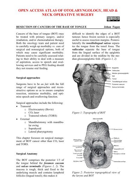
difficult to identify the edges <strong>of</strong> a BOT<br />
tumour; hence frozen section is especially<br />
useful to assess <strong>resection</strong> margins. Posterolaterally<br />
the tonsillolingual sulcus separates<br />
the <strong>tongue</strong> from the tonsil fossa. The<br />
valleculae separate the base <strong>of</strong> <strong>tongue</strong><br />
from the lingual surface <strong>of</strong> the epiglottis<br />
and are divided in the midline by the median<br />
glossoepiglottic fold. (Figures 1, 2)<br />
Figure 1: Topography <strong>of</strong> BOT<br />
Epiglottis<br />
Valleculae<br />
Median glossoepiglottic<br />
fold<br />
Tonsil<br />
Tonsillolingual sulcus<br />
Foramen caecum<br />
Sulcus terminalis<br />
This chapter focusses on surgical management<br />
<strong>of</strong> BOT <strong>cancer</strong> other than CO 2 laser<br />
and TORS.<br />
Surgical Anatomy<br />
The BOT comprises the posterior 1/3 <strong>of</strong><br />
the <strong>tongue</strong> behind the foramen caecum<br />
and sulcus terminalis (Figures 1, 2). The<br />
mucosa is rough, thick and fixed to the<br />
underlying muscle and contains lymphoid<br />
follicles (lingual tonsil); this makes it<br />
Figure 2: Posterior topographical view <strong>of</strong><br />
the larynx and BOT
The <strong>tongue</strong> has eight muscles; four<br />
extrinsic muscles (genioglossus, hyoglossus,<br />
styloglossus, palatoglossus) control<br />
the position <strong>of</strong> the <strong>tongue</strong> and are attached<br />
to bone (Figure 3); four intrinsic muscles<br />
modulate the shape <strong>of</strong> the <strong>tongue</strong>, and are<br />
not attached to bone. Below the <strong>tongue</strong> are<br />
the geniohyoid and mylohoid muscles; the<br />
mylohyoid muscle serves as the diaphragm<br />
<strong>of</strong> the mouth and separates the <strong>tongue</strong> and<br />
FOM from the submental and submandibular<br />
triangles <strong>of</strong> the neck (Figures 3, 4).<br />
preserved when using a suprahyoid surgical<br />
approach (Figures 5, 6). Most <strong>of</strong> the<br />
<strong>tongue</strong> muscles attach to the hyoid bone<br />
(Figures 3, 4); the lingual artery and<br />
hypoglossal nerve (XIIn) pass medial to<br />
the greater cornu <strong>of</strong> the hyoid (Figures 7,<br />
8). The hyoid bone forms the anterior<br />
boundary <strong>of</strong> the preepiglottic space; the<br />
superior boundary is the hyoepiglottic<br />
ligament (floor <strong>of</strong> vallecula), and the<br />
posterior border is formed by the petiole <strong>of</strong><br />
the epiglottis (Figures 5, 6).<br />
<strong>Base</strong> <strong>of</strong> <strong>tongue</strong><br />
Vallecula<br />
Hyoid bone<br />
Hyoepiglottic ligament<br />
Preepiglottic space<br />
Petiole <strong>of</strong> epiglottis<br />
Thyrohyoid membrane<br />
Thyroid cartilage<br />
Figure 3: Extrinsic <strong>tongue</strong> muscles<br />
(palatoglossus not shown)<br />
Figure 5: Sagittal view <strong>of</strong> posterior<br />
relations <strong>of</strong> BOT and preepiglottic space<br />
(yellow)<br />
Genioglossus<br />
Foramen caecum<br />
Epiglottis<br />
Vallecula<br />
Hyoepiglottic ligament<br />
Preepiglottic fat<br />
Hyoid<br />
Figure 6: Midline sagittal view <strong>of</strong> <strong>tongue</strong><br />
Figure 4: Geniohyoid and mylohyoid<br />
muscles<br />
Posteriorly, the hyoid bone and preepiglottic<br />
space are important anatomical<br />
structures, an understanding <strong>of</strong> which is<br />
key to assessing whether the larynx can be<br />
The arterial supply <strong>of</strong> the BOT is derived<br />
from the paired lingual arteries and its<br />
posterior (dorsalis linguae) branches<br />
(Figures 7, 8). Additional supply to the<br />
BOT emanates from the tonsillar branch<br />
<strong>of</strong> the facial artery and the ascending<br />
pharyngeal artery. In practice the lingual<br />
arteries are the only vessels that have to<br />
2
e looked for during BOT <strong>resection</strong> as<br />
they are easily injured; it is important to<br />
preserve at least one lingual artery to<br />
avoid infarction <strong>of</strong> the <strong>tongue</strong>. (The author<br />
has had one case where sacrifice <strong>of</strong> a<br />
single lingual artery led to necrosis <strong>of</strong> half<br />
the <strong>tongue</strong>; this is however unusual as<br />
there is usually adequate cross-flow).<br />
Figure 7: XIIn accompanied by the ranine<br />
veins<br />
Ranine artery<br />
Sublingual artery<br />
Tonsillar<br />
artery<br />
artery (pr<strong>of</strong>unda linguae); it lies to either<br />
side <strong>of</strong> the genioglossus, and is accompanied<br />
by the lingual nerve. Two or three<br />
small dorsalis linguæ arteries arise<br />
beneath the hyoglossus and ascend to the<br />
posterior part <strong>of</strong> the dorsum <strong>of</strong> the <strong>tongue</strong><br />
and also supply the mucous membrane <strong>of</strong><br />
the posterior floor <strong>of</strong> mouth (FOM) and<br />
oropharynx (Figures 7, 8).<br />
Venous drainage is via lingual and ranine<br />
veins (Figure 7). The lingual veins originnate<br />
on the dorsum, sides, and undersurface<br />
<strong>of</strong> the <strong>tongue</strong> and accompany the lingual<br />
artery and join the internal jugular vein.<br />
The ranine veins originate below the tip <strong>of</strong><br />
the <strong>tongue</strong> and are visible on its ventral<br />
surface; they accompany the XIIn as venae<br />
comitantes and either join the lingual vein<br />
or pass lateral to hyoglossus to join the<br />
common facial vein.<br />
An understanding <strong>of</strong> the nerve supply is<br />
important to preserve oral function. All<br />
intrinsic and extrinsic muscles are innerveted<br />
by the XIIn except for palatoglossus<br />
which is innervated by Xn. The IXn provides<br />
somatic afferent and taste sensation<br />
to the posterior 1/3 <strong>of</strong> the <strong>tongue</strong>. The lingual<br />
nerve provides general somatic sensetion<br />
to the anterior 2/3 <strong>of</strong> the mouth and<br />
FOM; taste is provided by the chorda tympani<br />
branch <strong>of</strong> VIIn via the lingual nerve.<br />
Figure 8: Lingual artery<br />
Lingual artery<br />
Dorsalis linguae artery<br />
Lingual nerve<br />
Sublingual gland<br />
The lingual artery originates from the<br />
external carotid artery between the superior<br />
thyroid and facial arteries and courses<br />
obliquely forwards and medial to the<br />
greater cornu <strong>of</strong> the hyoid (Figures 7, 8). It<br />
then loops downward and anteriorly and<br />
passes medial to XIIn and the stylohyoid<br />
muscle. It then courses directly anteriorly<br />
and deep to the hyoglossus and finally<br />
ascends submucosally on the undersurface<br />
<strong>of</strong> the <strong>tongue</strong> as far as its tip as the ranine<br />
Submandibular duct<br />
Submandibular gland<br />
Mylohyoid muscle<br />
Geniohyoid muscle<br />
Figure 9: Superior view <strong>of</strong> FOM, submandibular<br />
gland and duct, lingual nerve<br />
and mylohyoid and geniohyoid muscles<br />
The lingual nerve is at risk when using a<br />
mandibular swing approach to resect<br />
3
tumours <strong>of</strong> the BOT. The nerve crosses<br />
deep to the submandibular duct in the<br />
lateral FOM; in the anterior FOM it is located<br />
posterior to the duct (Figures 9, 10).<br />
Figure 10: Intraoral view <strong>of</strong> left sublingual<br />
gland with ducts <strong>of</strong> Rivinus, submandibular<br />
gland and duct, lingual nerve and<br />
mylohyoid muscles<br />
The remainder <strong>of</strong> this chapter focuses on<br />
surgical <strong>resection</strong> <strong>of</strong> <strong>cancer</strong>s <strong>of</strong> the BOT.<br />
Surgical Objectives<br />
Adequate tumour <strong>resection</strong> margins;<br />
this requires a surgical approach that<br />
permits good exposure<br />
Surgical morbidity should be kept to a<br />
minimum relating to swallowing, aspiration,<br />
speech, mandibulotomy, lingual<br />
nerve function and voice (laryngectomy<br />
is required in selected cases to<br />
avoid aspiration)<br />
Control occult cervical metastasis;<br />
both sides <strong>of</strong> the neck are electively<br />
treated because <strong>of</strong> a significant risk <strong>of</strong><br />
having bilateral occult cervical<br />
metastases; the author generally does<br />
bilateral elective neck dissection<br />
(END) levels I-IV for squamous cell<br />
carcinoma <strong>of</strong> the BOT<br />
Key surgical points<br />
Lingual nerve<br />
Submandibular duct<br />
Sublingual gland<br />
Submandibular gland<br />
Mylohyoid muscle<br />
Oral or transnasal intubation may be<br />
difficult or impossible<br />
Select a surgical approach(es) that ensures<br />
adequate exposure; transoral<br />
access is <strong>of</strong>ten inadequate<br />
The surface <strong>of</strong> the BOT is firm and<br />
irregular making it difficult to determine<br />
margins; employ frozen section<br />
to control <strong>resection</strong> margins<br />
Preserve <strong>tongue</strong> function<br />
o Preserve at least one XIIn;<br />
sacrificing both nerves is<br />
crippling from speech and<br />
swallowing perspectives<br />
o Preserve at least one lingual<br />
artery to avoid infarction <strong>of</strong> the<br />
<strong>tongue</strong><br />
o Preserve sensation with the<br />
mandibular swing approach by<br />
preserving the lingual nerve<br />
Avoid/minimise aspiration; this may<br />
necessitate total laryngectomy in<br />
selected cases<br />
Preoperative evaluation<br />
1. Are there synchronous primaries,<br />
cervical or distant metastases CXR<br />
or CT chest, and panendoscopy<br />
2. Is the tumour resectable BOT <strong>cancer</strong><br />
may be irresectable if it invades the<br />
parapharyngeal space and carotid<br />
sheath, or extends anteriorly to necessitate<br />
total glossectomy in patients not<br />
prepared to undergo such surgery. It<br />
may be difficult to assess the extent <strong>of</strong><br />
the primary due to pain, tenderness or<br />
trismus. If in doubt, a patient requires<br />
imaging such as CT/MRI, or examination<br />
under anaesthesia.<br />
3. Is laryngectomy required Invasion <strong>of</strong><br />
the preepiglottic space or epiglottis<br />
generally indicates a need for laryngectomy<br />
to permit swallowing without<br />
aspiration (Figure 11)<br />
4
Figure 11: BOT <strong>cancer</strong> invading vallecula,<br />
preepiglottic space and epiglottis<br />
and requiring total laryngectomy<br />
Invasion <strong>of</strong> the preepiglottic space is<br />
best assessed with MRI / CT scan in a<br />
sagittal plane (Figure 12).<br />
Figure 12: BOT <strong>cancer</strong> invading and<br />
replacing the preepiglottic space (x)<br />
Invasion <strong>of</strong> the pre-epiglottic space can<br />
also be assessed intraoperatively by<br />
palpating the pre-epiglottic space between<br />
a finger placed in the vallecula<br />
and a finger placed on the skin <strong>of</strong> the<br />
neck just above/below the hyoid bone;<br />
fullness is indicative <strong>of</strong> tumour in the<br />
pre-epiglottic space (Figure 13)<br />
4. Is reconstruction required Restoring<br />
BOT bulk with s<strong>of</strong>t tissue flaps may<br />
improve speech; however, having an<br />
insensate flap may impair swallowing.<br />
Therefore filling the defect with a flap<br />
should only be done in selected cases<br />
X<br />
Figure 13: Digital palpation for involvement<br />
<strong>of</strong> pre-epiglottic space<br />
5. Can the patient tolerate aspiration<br />
Physical fitness, pulmonary reserve,<br />
and cognitive function should be considered<br />
when selecting patients for<br />
major <strong>resection</strong>s and reconstructions as<br />
such surgery may be complicated by a<br />
variable measure dysphagia and aspiration,<br />
especially if followed by chemoradiation.<br />
6. Status <strong>of</strong> dentition Carious teeth<br />
should be removed at the time <strong>of</strong> the<br />
surgery to avoid osteoradionecrosis.<br />
7. Is a tracheostomy required Almost<br />
all patients require temporary tracheostomy<br />
Anaesthesia<br />
The surgeon always stands by the patient<br />
during induction <strong>of</strong> anaesthesia as it may<br />
be difficult or impossible to intubate a<br />
patient with a bulky BOT tumour that<br />
precludes elevation <strong>of</strong> the <strong>tongue</strong> to visualise<br />
the larynx. Should the anaesthetist be<br />
unable to intubate the larynx, the surgeon<br />
may be able to intubate with a laryngoscope,<br />
or do an emergency tracheostomy<br />
or cricothyroidotomy; it is prudent to inject<br />
the tracheostomy or cricothyroidotomy site<br />
with local anaesthetic with adrenaline prior<br />
to induction. Nasal intubation facilitates<br />
5
esection <strong>of</strong> BOT tumours, and is converted<br />
to a tracheostomy during the course <strong>of</strong><br />
the operation.<br />
Perioperative antibiotics are prescribed for<br />
24hrs.<br />
Surgical access<br />
Good surgical access is essential in order<br />
to attain adequate <strong>resection</strong> margins, to<br />
control bleeding, and for reconstruction. A<br />
combination <strong>of</strong> open surgical approaches<br />
can be used, and will now be discussed.<br />
Level 1 <strong>of</strong> the neck should first be<br />
dissected if neck dissection is indicated<br />
before proceeding to address the primary<br />
tumour.<br />
Figure 14: Laterally located BOT <strong>cancer</strong><br />
suited to transoral <strong>resection</strong><br />
http://www.tobacc<strong>of</strong>acts.info/images/20071112-oral-<strong>cancer</strong>.jpg<br />
Transoral access with electrocautery<br />
(Bovie) <strong>resection</strong><br />
The adequacy <strong>of</strong> transoral access to the<br />
BOT varies considerably. A useful way to<br />
predict whether transoral <strong>resection</strong> is likely<br />
to be possible is to pull on the anterior<br />
<strong>tongue</strong> with a cotton swab during preoperative<br />
clinical evaluation and to then<br />
see how accessible the tumour is. Note that<br />
tumours becomes more visible and<br />
accessible as the <strong>resection</strong> proceeds,<br />
especially once the thick BOT lining has<br />
been incised around the tumour. Laterally<br />
placed BOT <strong>cancer</strong>s, especially in edentulous<br />
patients, are more amenable to transoral<br />
access (Figure 14).<br />
The mouth is kept wide open either with a<br />
dental bite block (Figure 15) or with a selfretaining<br />
retractor, taking care to protect<br />
the teeth from injury (Figure 16). Apply<br />
traction to <strong>tongue</strong> and tumour with silk<br />
traction sutures or with tissue forceps<br />
(Figure 16).<br />
Figure 15: Dental bite block is interposed<br />
between lateral teeth to keep mouth open<br />
Figure 16: Self-retaining retractor; lateral<br />
oral and BOT tumour being resected<br />
Resect the tumour with at least a 1cm margin<br />
<strong>of</strong> normal tissue using electrocautery<br />
(Bovie). As the <strong>resection</strong> proceeds posteriorly,<br />
insert new silk traction sutures or<br />
move the tissue forceps more posteriorly to<br />
facilitate delivery <strong>of</strong> the tumour into the<br />
mouth.<br />
6
Transoral access with median glossotomy<br />
Midline BOT tumours may be exposed by<br />
bisecting the <strong>tongue</strong> along the median<br />
raphe with electrocautery (Figure 17); it is<br />
a relatively avascular plane as the vessels<br />
and nerves all course laterally, and results<br />
in little, if any, functional deficits. The<br />
incision can be carried posteriorly up to the<br />
hyoid bone if needed.<br />
cuff <strong>of</strong> s<strong>of</strong>t tissue on the bone to facilitate<br />
subsequent s<strong>of</strong>t tissue closure (Figure 20).<br />
Strip s<strong>of</strong>t tissue <strong>of</strong>f the mandible with<br />
monopolar cautery or with a periosteal<br />
elevator up to the mental foramen, taking<br />
care not to injure the mental nerve where it<br />
exits the foramen (Figure 20).<br />
Figure 18: Scoring vermillion border for<br />
accurate closure<br />
Figure 17: Median glossotomy for lingual<br />
thyroid<br />
Mandibulotomy with mandibular swing<br />
This affords excellent access to the BOT. It<br />
is especially well suited to <strong>cancer</strong>s that<br />
extend anteriorly to involve the oral <strong>tongue</strong><br />
(Figure 14), or onto the s<strong>of</strong>t palate. It does<br />
however leave a facial scar; it may cause<br />
deformity <strong>of</strong> the lower lip; there is a risk <strong>of</strong><br />
complications relating to the mandibulotomy<br />
and dental malocclusion; and the<br />
lingual nerve is at risk <strong>of</strong> injury.<br />
The vermillion border is scored or marked<br />
so as to ensure an accurate repair (Figure<br />
18). The lower lip is divided vertically in<br />
the midline (Figure 19). Bleeding from the<br />
labial artery is controlled with cautery.<br />
Incise the gingivolabial and gingivobuccal<br />
mucosae >0.5cms from the bone leaving a<br />
Figure 19: Lip-split incision down to bone<br />
Figure 20: Note preserved mental nerve<br />
and cuff <strong>of</strong> s<strong>of</strong>t tissue<br />
Cuff <strong>of</strong> s<strong>of</strong>t tissue<br />
on mandible<br />
Mental nerve<br />
7
The mandible is divided just anterior to the<br />
mental foramen with a Gigli or a powered<br />
saw (Figure 21). The osteotomy may be<br />
made vertically or alternatively in a stepor<br />
V-shaped fashion so as to achieve a<br />
more stable repair (Figure 22). It is advisable<br />
to extract a tooth and make the osteotomy<br />
through the dental socket so as avoid<br />
devitalising adjacent teeth. In dentate<br />
patients the mandible is preplated with titanium<br />
miniplates contoured to the mandible<br />
so as to ensure perfect dental alignment.<br />
Two 4-holed, non-compression, 2mm mandibular<br />
plates, one placed along the inferior<br />
border <strong>of</strong> the mandible, and the other<br />
placed more superiorly are used. Once the<br />
plates have been contoured and the holes<br />
drilled, they are removed and the bone cut<br />
is made.<br />
The surgeon then distracts the cut ends <strong>of</strong><br />
the mandible to gain access to the oral<br />
cavity and proceeds to divide the floor <strong>of</strong><br />
mouth (FOM) mucosa and mylohyoid<br />
muscle about 1cm from, and parallel to,<br />
the mandible, so as to leave s<strong>of</strong>t tissue on<br />
the mandible for the subsequent FOM<br />
repair at conclusion <strong>of</strong> surgery (Figure 23).<br />
Figure 23: Divide FOM mucosa and<br />
mylohyoid muscle keeping about 1cm from<br />
and parallel to the mandible<br />
Figure 21: Gigli saw<br />
Continue the incision posteriorly along the<br />
FOM until the tumour comes into view;<br />
the lingual artery (medial to the hyoglossus<br />
muscle) and the XIIn course medial to the<br />
FOM incision are not at risk <strong>of</strong> injury at<br />
this stage <strong>of</strong> the dissection. The tumour is<br />
resected using electrocautery. The lingual<br />
artery may have to be ligated (Figure 24).<br />
Figure 22: Examples <strong>of</strong> osteotomies<br />
Figure 24: Lingual artery<br />
8
Posteriorly the lingual nerve extends from<br />
the skull base and crosses the line <strong>of</strong> the<br />
incision from lateral to medial to course<br />
along the lateral FOM; preserve the nerve<br />
if possible.<br />
At conclusion <strong>of</strong> surgery the FOM is<br />
closed with a running vicryl suture, and the<br />
osteotomy is plated; when plating sets are<br />
not available, opposing holes are drilled on<br />
either side <strong>of</strong> the osteotomy and the<br />
mandible is wired together with stainless<br />
steel wiring (Figure 25). The lip is carefully<br />
repaired in layers to approximate the<br />
muscles as well as mucosa and skin.<br />
Because the pharynx is entered through the<br />
vallecula, it is not suited to <strong>cancer</strong>s that<br />
involve the apex <strong>of</strong> the vallecula, preepiglottic<br />
space or epiglottis (Figure 11). Following<br />
completion <strong>of</strong> Levels 1a and b <strong>of</strong><br />
the neck dissection(s), the superior aspect<br />
<strong>of</strong> the body <strong>of</strong> the hyoid bone is skeletonised<br />
with electrocautery, keeping the dissection<br />
between the lesser cornua <strong>of</strong> the<br />
hyoid bone. The dissection is carried posteriorly<br />
above the hyoepiglottic ligament,<br />
which forms the ro<strong>of</strong> <strong>of</strong> the preepiglottic<br />
fat (Figure 27). The vallecula is entered at<br />
its apex (Figure 27).<br />
Figure 25: Mandible wired together with<br />
stainless steel wire<br />
Suprahyoid approach<br />
This is one <strong>of</strong> the external approaches<br />
favoured by the author. It is similar to the<br />
suprahyoid dissection step during total<br />
laryngectomy. It is well suited to most<br />
BOT <strong>cancer</strong>s (Figure 26), although access<br />
is limited with <strong>cancer</strong>s that extend far<br />
anteriorly into the <strong>tongue</strong>.<br />
Figure 27: Red arrow indicates suprahyoid<br />
access to BOT (hyoepiglottic ligament<br />
coloured green)<br />
The tumour now comes into view. The<br />
<strong>cancer</strong> is resected with electrocautery<br />
(Figures 28-30). Take care to identify and<br />
preserve the lingual arteries and, if<br />
dissecting even more laterally, the XIIn<br />
(Figures 29, 30). Tumour exposure improves<br />
as the thick BOT lining is incised to<br />
either side <strong>of</strong> the tumour making the<br />
tumour more mobile (Figure 30).<br />
Orientate the specimen for the pathologist<br />
with a suture before removing the specimen<br />
so as not to lose orientation. The<br />
adequacy <strong>of</strong> the <strong>resection</strong> margins is<br />
checked with frozen section if available.<br />
Figure 26: BOT <strong>cancer</strong> with cystic<br />
metastasis suited to suprahyoid approach<br />
Primary closure can almost always be<br />
done by approximating the BOT <strong>resection</strong><br />
margin to the vallecula with a running<br />
9
vicryl suture. The supra- and infrahyoid<br />
muscles are then sutured together and the<br />
neck is closed in the usual fashion.<br />
A temporary tracheostomy is done and a<br />
nasogastric feeding tube is inserted. Once<br />
the airway appears adequate the tracheostomy<br />
tube is corked for 24hrs before<br />
being removed.<br />
Lateral pharyngotomy combined with<br />
suprahyoid approach<br />
Cancer <strong>of</strong> BOT<br />
Body <strong>of</strong> hyoid<br />
Additional exposure can be obtained by<br />
extending the incision along the greater<br />
cornu <strong>of</strong> the hyoid bone and along the<br />
posterior margin <strong>of</strong> the thyroid cartilage;<br />
take special care to identify and not to<br />
injure the XIIn, the lingual artery or the<br />
superior laryngeal nerve (Figure 31).<br />
Figure 28: Initial entry into vallecula<br />
Cancer <strong>of</strong> BOT<br />
Lingual artery<br />
Figure 29: Note wide access to epiglottis,<br />
BOT and posterior wall <strong>of</strong> pharynx<br />
Figure 31: Additional access by lateral<br />
pharyngotomy extension (Yellow arrow)<br />
Pull-through approach<br />
Figure 30: BOT <strong>cancer</strong> <strong>resection</strong><br />
Lingual artery<br />
This may be employed when tumour stops<br />
some distance from the inner aspect <strong>of</strong> the<br />
mandible. The mucosa <strong>of</strong> the lateral and<br />
anterior FOM is divided >0.5cm from the<br />
inner aspect <strong>of</strong> the mandible (so as to<br />
facilitate later repair), taking care not to<br />
injure the lingual and XIIns, or the<br />
submandibular ducts.<br />
Following bilateral neck dissections <strong>of</strong><br />
Levels 1a and 1b, the mandibular attachments<br />
<strong>of</strong> the anterior bellies <strong>of</strong> digastric,<br />
mylohyoid, geniohyoids and genioglossus<br />
are divided with electrocautery working<br />
10
from inferiorly (Figure 32). This permits<br />
the surgeon to drop the FOM and <strong>tongue</strong><br />
into the neck and to proceed with the<br />
<strong>resection</strong>. At conclusion <strong>of</strong> surgery the<br />
FOM mucosa and muscle are<br />
reapproximated with vicryl sutures.<br />
filled. However it is less pliable than the<br />
radial free forearm flap and is only suitable<br />
for oral reconstruction in patients with thin<br />
thighs.<br />
Pectoralis major flap: This is a good<br />
option to use (See chapter: Pectoralis<br />
major flap)<br />
Buccinator flap: A posteriorly based<br />
buccinator flap can be used to close a small<br />
orocervical communication following <strong>resection</strong><br />
<strong>of</strong> a lateral BOT <strong>cancer</strong>. (See chapter:<br />
Buccinator myomucosal flap)<br />
Final comments<br />
Figure 32: Incision for pull-through<br />
approach (Yellow line)<br />
Repair / Reconstruction<br />
Obtain meticulous haemostasis using ties,<br />
monopolar and bipolar cautery before<br />
closing the defect. Primary closure <strong>of</strong> BOT<br />
defects gives the best swallowing function<br />
although inadequate bulk impairs speech if<br />
the BOT cannot approximate the s<strong>of</strong>t<br />
palate. Simply shaping a flap to match the<br />
resected tissue may well restore form but<br />
may have a poor functional result. Therefore<br />
one has to carefully assess the defect<br />
to determine how best to optimise swallowing<br />
and speech.<br />
Radial free forearm flap: This is a thin<br />
and pliable flap and preserves mobility <strong>of</strong><br />
the <strong>tongue</strong>, but has limited bulk.<br />
Anterolateral free thigh flap: Muscle<br />
harvested with the flap can be tailored to<br />
conform to the volume <strong>of</strong> the defect to be<br />
Resecting tumours <strong>of</strong> the BOT is challenging<br />
particularly in terms <strong>of</strong> access and<br />
maintaining swallowing function. One<br />
should not compromise <strong>resection</strong> margins<br />
for function. The surgical team has to master<br />
an array <strong>of</strong> surgical approaches and reconstructive<br />
techniques so as to ensure the<br />
best oncological and functional outcomes.<br />
Author & Editor<br />
Johan Fagan MBChB, FCORL, MMed<br />
Pr<strong>of</strong>essor and Chairman<br />
Division <strong>of</strong> Otolaryngology<br />
<strong>University</strong> <strong>of</strong> <strong>Cape</strong> <strong>Town</strong><br />
<strong>Cape</strong> <strong>Town</strong>, South Africa<br />
johannes.fagan@uct.ac.za<br />
THE OPEN ACCESS ATLAS OF<br />
OTOLARYNGOLOGY, HEAD &<br />
NECK OPERATIVE SURGERY<br />
www.entdev.uct.ac.za<br />
The Open Access Atlas <strong>of</strong> Otolaryngology, Head &<br />
Neck Operative Surgery by Johan Fagan (Editor)<br />
johannes.fagan@uct.ac.za is licensed under a Creative<br />
Commons Attribution - Non-Commercial 3.0 Unported<br />
License<br />
11


