Emdogain® and PrefGelTM
Emdogain® and PrefGelTM
Emdogain® and PrefGelTM

Create successful ePaper yourself
Turn your PDF publications into a flip-book with our unique Google optimized e-Paper software.
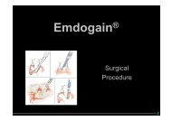
Emdogain ® <strong>and</strong> PrefGel TM<br />
Instructions for use
Straumann is the exclusive industrial<br />
partner of the ITI (International Team for<br />
Implantology) in the areas of research,<br />
development, <strong>and</strong> education.<br />
Contents<br />
Introduction 2<br />
PrefGel instructions for use 3<br />
Application in intrabony defects 4–5<br />
Application in recession defects 6–7<br />
Application in furcation defects 8–9<br />
For additional information on Emdogain ® , please refer to:<br />
• Emdogain ® the reliable solution for periodontal treatment (USLIT 199)<br />
• Emdogain ® referral booklet (USBIO 015)<br />
• Emdogain ® list of publications (152.287)<br />
• Emdogain ® treating recession defects flyer (USBIO 016)<br />
1
Introduction<br />
This brochure presents step-by-step instructions for the use of Emdogain <strong>and</strong><br />
PrefGel. Further information can be found in the package inserts enclosed in<br />
each product package.<br />
Emdogain ® is a resorbable, implantable material consisting of enamel<br />
matrix proteins that is intended as an adjunct to periodontal surgery for<br />
topical application onto exposed root surfaces. Once applied to the cleaned<br />
root surface, Emdogain forms an insoluable protein matrix that initiates the<br />
process of periodontal tissue regrowth.<br />
PrefGel is a pH neutral, 24 % EDTA root surface conditioner, which offers<br />
an effective, yet gentle removal of the “smear layer” during periodontal<br />
surgical procedures.<br />
2
PrefGel – instructions for use<br />
PrefGel is contained in a 1 ml sterilized, single-use pipette. It is available<br />
in a package of 10 pipettes or co-packaged with Emdogain ® (3 units of<br />
Emdogain <strong>and</strong> 3 pipettes of PrefGel).<br />
1. The pipette is opened by twisting the wing.<br />
2. Press out the contents into a sterile dappen dish <strong>and</strong> apply the gel using<br />
a gauze, a small sterile instrument (e.g. a plastic instrument) or a sterile<br />
syringe with a blunt ended cannula. Alternatively withdraw the contents<br />
directly from the pipette into a syringe.<br />
3. Remove remaining plaque <strong>and</strong>/or calculus as well as blood from<br />
root surfaces exposed during periodontal surgery.<br />
4. Apply PrefGel onto exposed root surfaces <strong>and</strong> leave for 2 minutes.<br />
Active rubbing (“burnishing”) is not recommended.<br />
5. Rinse thoroughly with sterile saline.<br />
6. Avoid recontamination of the conditioned root surfaces after the final rinse<br />
<strong>and</strong> prior to treatment with Emdogain.<br />
7. After application, discard any residual gel.<br />
Debridement of root surface Application of PrefGel Rinsing with sterile saline<br />
before Emdogain ® use<br />
3
Application in intrabony defects<br />
1. Anesthetize the area selected for surgery by block <strong>and</strong>/or infiltration<br />
anesthesia. Avoid injection of local anesthetic with a vasoconstrictor into<br />
the interdental papilla or marginal gingiva.<br />
1<br />
2. Make intra-crevicular incisions. Then, if judged appropriate, make<br />
one or two vertical releasing incisions extending out into the alveolar<br />
mucosa. Raise full-thickness (mucoperiosteal) flaps on the buccal <strong>and</strong><br />
palatal/lingual surfaces of the teeth. Preserve as much of the gingival<br />
connective tissue in the flap as possible. Maintain viability of<br />
periondontal cells by hydration of the soft tissue with saline.<br />
2<br />
3. Only remove the granulation tissue adherent to the alveolar bone <strong>and</strong><br />
any associated osseous defects necessary to provide full access <strong>and</strong><br />
visibility to the root surfaces. Remove subgingival plaque <strong>and</strong> calculus.<br />
3<br />
4. Remove the “smear layer” by conditioning the root surface with PrefGel <br />
for two minutes. Rinse thoroughly with sterile saline solution. Avoid<br />
contaminating the cleaned <strong>and</strong> conditioned root surface with blood or<br />
saliva after the final rinse.<br />
4<br />
4
5. Apply Emdogain ® immediately on the exposed root surface. Start at<br />
the most apical bone level <strong>and</strong> apply Emdogain so that it covers the<br />
whole root surface.<br />
5<br />
6. Complete coverage of the interproximal area <strong>and</strong> optimal soft<br />
tissue adaptation are essential. If deemed appropriate, a periosteal<br />
fenestration at the base of the flap may be used to facilitate coronal<br />
repositioning of the soft tissue. Suture materials appropriate for extended<br />
stable closure are preferred.<br />
6<br />
7. To maintain the stability of the healing wound, do not probe<br />
surgically treated areas for 6 months after Emdogain treatment.<br />
7<br />
5
Application in recession defects<br />
1. Anesthetize the operative area. Scale <strong>and</strong> plane the exposed root<br />
surface to remove plaque, calculus, root surface irregularities <strong>and</strong>, if<br />
judged appropriate, to reduce prominence.<br />
1<br />
Extensive root planing is recommended when (i) root caries lesion has been diagnosed or<br />
(ii) when a reduced root prominence has been judged beneficial for tissue regeneration.<br />
The presence of a restoration does not preclude the possibility for root coverage, but<br />
restoration should ideally be removed prior to covering the root with the soft tissue flap.<br />
2. Make a sulcular incision at the site of the recession. Extend the incision<br />
horizontally into the adjacent interdental area slightly coronal to the level of<br />
the soft tissue margin of the recession.<br />
2<br />
Make two vertical divergent releasing incisions at the mesial <strong>and</strong> distal line<br />
angle connected to the horizontal incision.<br />
Raise a full-thickness (mucoperiosteal) flap until the mucogingival junction<br />
is passed.<br />
3. Make a cut through the periosteum <strong>and</strong> continue to raise a split-thickness flap<br />
by means of a blunt dissection. The aim is to eliminate any muscle tension on<br />
the flap margins <strong>and</strong> allow for a passive <strong>and</strong> tension-free coronal positioning<br />
of the flap at the level of the CEJ.<br />
3<br />
4. De-epithelialize the buccal aspect of the interdental papillae to create<br />
a connective tissue bed for suturing the coronally advanced flap.<br />
4<br />
6
5. Remove the “smear layer”by conditioning the root surface with PrefGel <br />
for two minutes. Rinse thoroughly with sterile saline solution. Avoid<br />
contaminating the cleaned <strong>and</strong> conditioned root surface. Try to minimize<br />
bleeding.<br />
5<br />
6. Apply Emdogain ® immediately on the exposed <strong>and</strong> conditioned root surface.<br />
6<br />
7. Advance the flap coronally <strong>and</strong> secure it at the level of the cementum<br />
enamel junction (CEJ) by suturing the flap into the recipient bed, i.e. the deepithelialized<br />
papillae. Also close the vertical incisions with lateral sutures.<br />
7<br />
Use suture materials for extended <strong>and</strong> stable closure. No pressure should be applied<br />
to the flap after suturing.<br />
7
Application in Class II m<strong>and</strong>ibular furcations<br />
1. Class ll m<strong>and</strong>ibular furcation defect with minimal interproximal<br />
bone loss.<br />
1<br />
Anesthetize the operative area.<br />
2. Make intra-crevicular incisions, <strong>and</strong> if judged appropriate, make<br />
one or two vertical releasing incisions extending out into the alveolar<br />
mucosa. Raise full-thickness (mucoperiosteal) flaps on the buccal <strong>and</strong><br />
palatal/ lingual surfaces of the teeth. Preserve as much of the gingival<br />
connective tissue in the flap as possible. Maintain viability of periodontal<br />
cells by hydration of the soft tissue with sterile saline.<br />
2<br />
3. Reflect soft tissue to expose the periodontal defect.<br />
3<br />
4. Remove only the granulation tissue adherent to the alveolar bone<br />
<strong>and</strong> any associated osseous defects necessary to provide full access <strong>and</strong><br />
visibility to the root surfaces. Remove subgingival plaque <strong>and</strong> calculus.<br />
4<br />
8
5. Remove the “smear layer” by conditioning the root surface with PrefGel <br />
for two minutes. Rinse thoroughly with sterile saline solution. Avoid<br />
contaminating the cleaned <strong>and</strong> conditioned root surface with blood or<br />
saliva after the final rinse.<br />
5<br />
6. Apply Emdogain ® immediately on the exposed root surface. Start at the<br />
most apical bone level <strong>and</strong> apply Emdogain so that it covers the whole<br />
root surface.<br />
6<br />
7. Complete coverage of the interproximal area <strong>and</strong> optimal soft tissue<br />
adaptation are essential. If deemed appropriate, a periosteal fenestration<br />
at the base of the flap may be used to facilitate coronal repositioning of<br />
the soft tissue. Suture materials appropriate for extended stable closure<br />
are preferred. Overflow of surplus material during flap closure <strong>and</strong><br />
suturing should occur.<br />
7<br />
9
North American Distributors<br />
Straumann USA, LLC<br />
60 Minuteman Road<br />
Andover, MA 01810<br />
Phone 800/448 8168<br />
978/747 2500<br />
Fax 978/747 2490<br />
www.straumannusa.com<br />
Straumann Canada Limited<br />
4145 North Service Road<br />
Suite 303<br />
Burlington, ON L7L6A3<br />
Phone 800/363 4024<br />
905/319 2900<br />
Fax 905/319 2911<br />
www.straumann.ca<br />
International Headquarters<br />
Institut Straumann AG<br />
Peter-Merian-Weg 12<br />
Postfach<br />
CH-4002 Basel<br />
Switzerl<strong>and</strong><br />
Phone +41 (0) 61 965 11 11<br />
Fax +41 (0) 61 965 11 01<br />
www.straumann.com<br />
Straumann products are CE marked<br />
USLIT 200 5/06 Printed in USA