Viscin Cells in the Dwarf Mistletoe Arceuthobium ... - Davidsonia
Viscin Cells in the Dwarf Mistletoe Arceuthobium ... - Davidsonia
Viscin Cells in the Dwarf Mistletoe Arceuthobium ... - Davidsonia
- No tags were found...

Create successful ePaper yourself
Turn your PDF publications into a flip-book with our unique Google optimized e-Paper software.
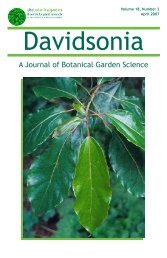
<strong>Davidsonia</strong> 17:3 79<br />
Image: Cynthia M. Ross<br />
Figure 1. The <strong>Arceuthobium</strong> americanum fruit, seed, and visc<strong>in</strong> tissue. Mature recurved fruit<br />
photographed two days prior to explosive discharge. scale bar = 4 mm.<br />
1988), <strong>in</strong>volved a spiral coil<strong>in</strong>g of <strong>the</strong> cellulose fibrils of <strong>the</strong> primary<br />
cell wall along <strong>the</strong> long axis of <strong>the</strong> cell. This has not been previously<br />
described <strong>in</strong> any o<strong>the</strong>r plant cells and requires fur<strong>the</strong>r study.<br />
The resemblance of <strong>the</strong>se visc<strong>in</strong> cells to “spr<strong>in</strong>gs” may reflect a<br />
function <strong>in</strong> explosive seed discharge, adhesion, or both. The elaters<br />
of fern sporangia are helical cells with a role <strong>in</strong> spore discharge but <strong>the</strong>