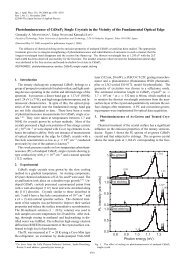
![J. Magn. Magn. Mater.304 [1]](https://img.yumpu.com/36362486/1/500x640/j-magn-magn-mater304-1.jpg)
J. Magn. Magn. Mater.304 [1]
J. Magn. Magn. Mater.304 [1]
J. Magn. Magn. Mater.304 [1]

Create successful ePaper yourself
Turn your PDF publications into a flip-book with our unique Google optimized e-Paper software.
Journal of <strong>Magn</strong>etism and <strong>Magn</strong>etic Materials 00 (2004) 000--000<br />
MFM observation of spin structures in nano magnetic dot arrays<br />
fabricated by damascene technique<br />
K. Sato a , T. Yamamoto a , T. Tezuka a , T. Ishibashi a , Y. Morishita a , A. Koukitu a ,<br />
K. Machida a,b , T. Yamaoka c<br />
a Tokyo University of Agriculture and Technolog, Koganei, Tokyo 184-8588, Japan<br />
b Science and Technology Research Laboratory of NHK, Setagaya-ku, Tokyo 157-8510, Japan<br />
c SII Nanotechnology Ltd., Matsudo, Chiba 270-2222, Japan<br />
Elsevier use only: Received date here; revised date here; accepted date here<br />
Abstract<br />
Regularly aligned array of magnetic nano dots buried in silicon wafers have been fabricated using damascene technique with a<br />
help of electron beam lithography. Arrays of square, rectangular, cross-shaped and Y-shaped structures of submicron size<br />
have been obtained. Spin distributions have been observed by means of magnetic force microscopy (MFM) and analyzed by a<br />
micromagnetic simulation with Landau-Lifshitz-Gilbert (LLG) equations. Importance of magnetostatic interactions working<br />
between adjacent dots has been elucidated.<br />
© 2005 Elsevier B.V. All rights reserved<br />
68.37.Rt; 75.60.Ch; 75.75.+a<br />
Keywords: nano magnetic dots; magneticforce microscopy; Landau-Lifshitz-Gilbert equation; magnetostatic interaction<br />
damascene technique. solving the Landau-Lifshitz-Gilbert equation. 2<br />
Arrays of square, rectangular, cross-shaped and Y-<br />
shaped structures of submicron size have been<br />
1. Introduction<br />
obtained. Isolated square dot of 1 μm in size shows a<br />
closure-domain image consisting of four domains<br />
Studies of spin distributions in nano or submicron<br />
scale magnetic dots with different pattern shapes are<br />
separated by 90°-domain walls. In a system where the<br />
distance between adjacent dots is as close as a few<br />
attracting interest since the state-of-the-art hundred nanometers, spin-structures become strongly<br />
technology for high density magnetic storages is<br />
based on magnetic structures as small as a few tens of<br />
nanometer in size. We have been working with<br />
fabrication of arrays of submicron permalloy<br />
(Fe 20 Ni 80 ) patterns buried in a silicon wafer using a<br />
influenced by a magnetostatic interaction. We<br />
experimentally observed that the chirality of the spin<br />
flow in the closure domain pattern is reversed<br />
between two adjacent dots, the result having been<br />
explained in terms of the magnetostatic interaction by
2 Katsuaki Sato / Journal of <strong>Magn</strong>etism and <strong>Magn</strong>etic Materials 00 (2005) 000–0001<br />
In the cross-shaped pattern were observed the<br />
magnetic poles as two pairs of bright and dark spots<br />
at the ends of the cross-bars, as well as the<br />
complicated spin structure at the crossing region.<br />
The force gradient distributions were simulated based<br />
on micromagnetic calculations taking into account<br />
the stray field from the MFM tip using the integral<br />
equation method. 3<br />
We recently prepared two different arrangements<br />
of Y-shaped patterns of submicron size. The spin<br />
distribution in the three-arm structure is of current<br />
interest since in such a system the geometric<br />
frustration effect plays an important role. 4<br />
In this paper we briefly review our previous<br />
results and describe our recent results on the Y-<br />
shaped patterns.<br />
2. Experiments<br />
The specimens used in this study were prepared<br />
by the damascene technique as described in the<br />
following: We employed a Si(100) wafer as a<br />
substrate to fabricate magnetic structures. An EBresist<br />
(ZEP-520 supplied by Nippon Zeon Co. Ltd.)<br />
which has an excellent dry-etching resistance was<br />
spin-coated at a rotation speed of 5000 rpm for 90 s,<br />
followed by baking at 160°C for 20 min before EB<br />
exposure. The thickness of the resist was about 35<br />
nm. Arrays of square, rectangular dots as well as<br />
cross-shaped and Y-shaped dots as shown in Fig. 1<br />
were patterned on the resist using an electron beam<br />
pattern generator (JEOL type JBX-5000SH in TUAT<br />
or ELIONIX type ELS-7300ULH in NHK). The<br />
patterned resist was developed using a ZED-N50 (n-<br />
Amyl acetate) developer for 2 min. The patterned<br />
area was as large as 3 mm × 3 mm.<br />
Using the patterned resist as a mask, the Si<br />
substrate was processed by a plasma-etching using<br />
CF 4 gas with an RF power of 400 W. The optimum<br />
etching rate was found to be about 0.1μm per minute.<br />
The remained resist was finally removed in an<br />
ultrasonic bath using acetone.<br />
By this method, arrays of pits of approximately<br />
150 nm in depth were uniformly formed over the<br />
patterned area on the Si wafer. Permalloy films were<br />
deposited using an electron beam (EB) evaporator.<br />
Thickness was monitored during evaporation by a<br />
quartz thickness monitor to adjust the evaporation<br />
period to adjust the thickness equal to the pit depth.<br />
1μm<br />
200nm<br />
1μm 300nm<br />
(a)<br />
3μm 3μm<br />
400nm<br />
(c)<br />
300nm<br />
100nm<br />
300nm<br />
1.4μm<br />
300nm<br />
(b)<br />
6μm<br />
(d)<br />
300 nm<br />
Fig. 1 Designed mask patterns of (a) square, (b)<br />
rectangular, (c) cross, (d) Y-shaped (linear<br />
well-spaced alignment) and (e) Y-shaped<br />
(honeycomb alignment) dot arrays.<br />
Typical deposition rate was 1.0 Å/s.<br />
The magnetic film outside the pit was polished<br />
out to obtain a flat surface by means of the chemical<br />
mechanical polishing (CMP) using a pH-controlled<br />
chemical solution including a polishing slurry<br />
(Glanzox SP-15, Fujimi Corp.). The polishing rate<br />
was optimized by adjusting combination of a<br />
mechanical polishing and an etching for chemical<br />
reaction. The typical rms roughness after the CMP<br />
process was less than 10 nm.<br />
Microscopic spin structures were observed using<br />
an SII Nanotechnology type SPI-4000/SPA300HV
MFM with a low-moment tip (coated with 25 nmthick<br />
CoCrPt) using a specially designed Q-control in<br />
the high vacuum environment. The measurements<br />
using the low-moment tips were carried out at SII-<br />
Nanotechnology Inc.<br />
3. Micromagnetic simulations<br />
Micromagnetic simulations using the Landau–<br />
Lifshitz–Gilbert equation were carried out to explain<br />
the observed MFM images of the cross-shaped nanoscale<br />
patterns. The micromagnetic simulator 5 was<br />
modified to correspond to a complicated threedimensional<br />
(3D) pattern, which was created using a<br />
generic 3D-CAD system. The equation was<br />
numerically solved using the fourth order Runge–<br />
Kutta method for high accuracy.<br />
To demonstrate the MFM images, the interaction<br />
between the magnetization of the tip and the stray<br />
field of the sample was evaluated. The MFM output<br />
signals are proportional to the force gradient between<br />
the tip and the sample. The force gradient is given by<br />
2<br />
∂F<br />
∂ H<br />
z<br />
tip<br />
3 ∂Htip<br />
δM sample<br />
= ∫ ⋅ M sampled<br />
r +<br />
⋅<br />
∂z<br />
2<br />
∫<br />
tip sample ∂z<br />
sample ∂z<br />
δztip<br />
where H tip is the stray field from the tip at the sample<br />
volume element, M sample is the magnetization of a<br />
sample volume element at equilibrium, and z tip is the<br />
tip–sample distance. 6 The values of H tip and M sample<br />
are considered to change corresponding to the tip–<br />
sample interaction.<br />
4. Results and discussion<br />
The MFM image of the square pattern (1 μm×1<br />
μm) shown in Fig. 2(a) clearly demonstrates a closure<br />
domain structure with 90°-domain walls. The<br />
Fig. 2 MFM images of (a) square and (b)<br />
rectangular shaped dot arrays<br />
Author name / Journal of <strong>Magn</strong>etism and <strong>Magn</strong>etic Materials 00 (2005) 000–0001 3<br />
propeller-like distortion of the domain wall pattern,<br />
which was ascribed to the stray field from the tip by<br />
Miltat, is clearly observed even with a low-moment<br />
tip. This means the propeller-like distortion is not<br />
necessarily due to the stray field of the MFM tip. In<br />
addition, a careful observation suggests that the<br />
direction of rotation (chirality) in the propellershaped<br />
domain walls of adjacent square dots shows a<br />
mirror-reflection of each other, suggesting a strong<br />
effect of magnetostatic interactions between dots in<br />
the array structure.<br />
(a)<br />
(b)<br />
Fig. 3 (a) Simulated spin structures by the micromagnetic<br />
calculation using LLG equation (b) force-gradient image<br />
calculated taking into account the tip-sample interaction.<br />
Simulation was carried out in the model structure<br />
consisting of four square dots with a dimension of<br />
200 nm×200 nm×20 nm with 50nm separation<br />
between dots. The calculated spin structure is<br />
illustrated in Fig. 3(a), in which a closure domain<br />
structure with the 90°-wall appears. The chirality of<br />
the spin direction in adjacent dots is opposite to each<br />
other as shown by white arrows. Figure 3(b) is the Z-<br />
component force-gradient image taking into account<br />
the tip-sample interaction, providing a good<br />
agreement with the MFM patterns shown in Fig. 2(a).<br />
On the other hand the MFM image of the<br />
rectangular dot (100 nm×300 nm) shows a checker<br />
pattern as shown in Fig. 2(b), in contrast with the<br />
case of circular dots of 100 nm in diameter, for which<br />
we confirmed single domain spin structure. The spin<br />
structure as observed in Fig. 2(b) was similar to that<br />
described in the literature. 7 The black and white<br />
contrast seems randomly aligned between adjacent<br />
dots, suggesting that the magnetostatic interaction has<br />
no strong influence in the present case.<br />
(a) (b) Figure 4 shows the MFM images of the crossshaped<br />
dot array, in which figure (a) shows a widearea-scan<br />
image and (b) a narrow-area-scan image
4 Katsuaki Sato / Journal of <strong>Magn</strong>etism and <strong>Magn</strong>etic Materials 00 (2005) 000–0001<br />
around the crossing region. Regularly aligned<br />
magnetic poles are observed in the MFM image<br />
shown in Fig. 4(a). Dark spots appear on the left and<br />
lower ends of the crossed bars, whereas bright spots<br />
on the right and upper ends. At the crossing point of<br />
cross-bars are observed a complicated MFM image at<br />
the crossing region as illustrated in Fig. 4(b).<br />
The results of the LLG simulation are illustrated in<br />
Fig. 5. Figure 5(a) shows a force-gradient image, and<br />
is found to show a remarkable agreement with the<br />
experimental MFM image of Fig. 4(a): Both<br />
theoretical and experimental images show dark or<br />
bright images of magnetic poles at the end of the<br />
cross-bars. From a three dimensional illustration of<br />
the spin structure at the end portion of the bar, an<br />
inclination of spins with vortex structure exists only<br />
at the end portion of the bar. The formation of the<br />
vortex may be consequence of fact that the height<br />
(150 nm) of the buried magnetic pattern is<br />
comparable to the width (200 nm) of the cross-bar.<br />
As shown in Fig. 5(b) the spin flows continuously<br />
from the lower left to the upper right in the figure at<br />
the crossing region with a vertical inclination along<br />
the diagonal line. Thus the complicated spin<br />
structures observed in Fig. 4(b) at the crossing point<br />
have been explained by the simulation. Evaluation of<br />
magnetostatic interactions between crosses is the<br />
issue for future investigations.<br />
A SEM micrograph of the linearly aligned array<br />
of Y-shaped magnetic dots with spacing as long as 6<br />
μm is shown in Fig. 6.<br />
4μm<br />
Fig. 6 SEM micrograph of the linearly<br />
aligned array of Y-shaped pattern<br />
(design: Fig. 1(d)) after CMP process<br />
(a)<br />
(a)<br />
(b)<br />
Fig. 4 MFM observation of cross-shaped dot<br />
array (a) included 4 dots (b) zoom up<br />
around the crossing region<br />
0.5μm<br />
(b)<br />
Fig. 7 MFM image of (a) the Y-shaped dot and<br />
(b) the magnified pattern in the arm<br />
without poles<br />
(a)<br />
(b)<br />
Fig. 5 Result of LLG simulation. (a) Forcegradient<br />
image taking into account tip-sample<br />
interaction, (b) spin flow image at the crossing<br />
region<br />
An MFM image in the Y-shaped dot is illustrated<br />
in Fig. 7(a). <strong>Magn</strong>etic poles are observed at the end<br />
of the single domain images in two of the three arms<br />
of the Y-shapes pattern. On the other hand, a multidomain<br />
structure is observed on the remaining arm,<br />
for which the magnified MFM image is given in Fig.<br />
7(b). A preliminary result of the LLG simulation<br />
reproduces a multi-domain spin structure in one of<br />
the three arms of the Y-pattern.
Author name / Journal of <strong>Magn</strong>etism and <strong>Magn</strong>etic Materials 00 (2005) 000–0001 5<br />
4μm<br />
(a)<br />
(b)<br />
Fig. 8 SEM micrograph of the honeycomb<br />
arrangement of Y-shaped patterns<br />
5μm<br />
Closely-spaced honeycomb arrangement of Y-<br />
shape pattern is successfully fabricated as shown in<br />
the SEM micrograph of Fig. 8(a). An MFM image is<br />
illustrated in Fig. 8(b), in which regularly arranged<br />
black and white spots are observed. The direction of<br />
magnetization in each arm is found to be determined<br />
to satisfy the “two-in, one-out” or the “one-in, twoout”<br />
rule around the vertex, similar to the case<br />
described in ref. 4.<br />
<strong>Magn</strong>eto-optical image of the same specimen has<br />
recently been obtained using a specially-designed<br />
high sensitivity magneto-optical Kerr microscope. 8<br />
The pattern is much different from the MFM image<br />
shown in Fig. 8(b). Difference may be caused by the<br />
fact that magneto-optical Kerr effect does not detect a<br />
magnetic flux as does the MFM, but detects only a<br />
vertical component of the sample magnetization.<br />
Detailed analysis is underway and will be published<br />
in our later publications.<br />
5. Conclusion<br />
Regularly aligned magnetic patterns with<br />
different shapes have been fabricated by a damascene<br />
technique. MFM observation revealed a closure<br />
domain for the square dot array, a checker pattern for<br />
the rectangular dot array, and magnetic poles for<br />
cross-shaped pattern. For separated Y-shaped pattern<br />
magnetic poles appear on two of the three arms and<br />
multi domain on the rest. For honeycomb<br />
arrangement regularly-aligned magnetic poles are<br />
observed. Importance of magnetostatic interaction is<br />
elucidated.<br />
Acknowledgments<br />
This work has been carried out under the 21 stcentury<br />
COE program of TUAT on “Future Nano<br />
Materials”.<br />
References<br />
1<br />
T. Matsumoto, T. Tezuka, T. Ishibashi, Y.<br />
Morishita, A. Koukitu and K. Sato, Trans. <strong>Magn</strong>. Soc.<br />
Jpn. 3 (2003) 103<br />
2 T. Tezuka, T. Yamamoto, K. Machida, S. Shimizu,<br />
T. Ishibashi, Y, Morishita, A. Koukitu and K. Sato,<br />
Trans. <strong>Magn</strong>. Soc. Jpn. 4 (2004) 241.<br />
3 K. Machida, T. Tezuka, T. Yamamoto, T. Ishibashi,<br />
Y. Morishita, A. Koukitu, K. Sato, J. <strong>Magn</strong>. <strong>Magn</strong>.<br />
Mater. 290-291 (2005) 779.<br />
4 E. Saitoh, M. Tanaka and H. Miyajima, J. Appl.<br />
Phys. 93 (2003) 7444.<br />
5 K. Machida, N. Hayashi, Y. Yoneda, J. Numazawa,<br />
M. Kohro, T. Tanabe, J. <strong>Magn</strong>. <strong>Magn</strong>. Mater. 226-<br />
230 (2001) 2054.<br />
6 J.M. Garcia, A. Thiaville, J. Miltat, K.J. Kirk, J.N.<br />
Chapman, F. Alouges, Appl. Phys. Lett. 79 (2001)<br />
656.<br />
7 A. Hubert, R. Schäfer, <strong>Magn</strong>etic Domains—The<br />
Analysis of <strong>Magn</strong>etic Microstructures, Springer, New<br />
York, 1998.<br />
8 T. Ishibashi, Z. Kuang, Y. Konishi, K. Akahane, X.<br />
R. Zhao, T. Hasegawa and K. Sato, Trans. <strong>Magn</strong>. Soc.<br />
Jpn. 4 (2004) 278.<br />
Fig.9 <strong>Magn</strong>eto-optical image of the<br />
honeycomb arrangement of Y-shaped<br />
patterns