Cases from private practices - Papimi
Cases from private practices - Papimi
Cases from private practices - Papimi

Create successful ePaper yourself
Turn your PDF publications into a flip-book with our unique Google optimized e-Paper software.
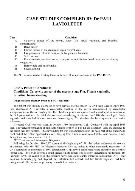
CASE STUDIES COMPILED BY Dr PAUL<br />
LAVIOLETTE<br />
Case<br />
Condition:<br />
A Ca-cervix cancer of the uterus, stage IVa, tistula vaginalis, and intestinal<br />
hemorrhaging.<br />
B Bone cancer.<br />
C Fibroid tumors of the uterus and digestive problems.<br />
D Lymphoma and chronic nonspecific lymphocytic leukemia.<br />
E Scleroderma.<br />
F Endometriosis, ovarian cancer, staphylococcus infection, hand burn, and respiratory<br />
congestion.<br />
G Hemorrhoid and mild bursitis.<br />
H Severe asthma<br />
.<br />
The PEC device, used in treating <strong>Cases</strong> A through D, is a predecessor of the PAP IMI.<br />
Case A Patient: Christina K<br />
Condition: Ca-cervix cancer of the uterus, stage IVa, Tistula vaginalis,<br />
Intestinal hemorrhaging.<br />
Diagnosis and Therapy Prior to PEC Treatment:<br />
The patient was initially diagnosed to have cervical uterine cancer. A CAT scan taken in April 1989<br />
[see attachment A-1] revealed a remarkable swelling of the cervix accompanied by remarkable<br />
amorphousness of the surrounding fat. The bladder appeared compressed and a small cyst was evident in<br />
the left parametrium. In 1989 she received radiotherapy treatment. In 1990 she developed fistula<br />
vaginalis and also had intense intestinal hemorrhaging. To alleviate the latter symptom she had a<br />
colostomy.<br />
A second CAT scan was taken in October 1990 [attachment A-2]. Compared with the April 1989<br />
scan, it revealed the presence of para-aortic nodes swollen to 1 to 1.5 cm diameter. Also the entrance to<br />
the cervix was less swollen. The surrounding fat was still amorphous and the back part of the bladder and<br />
front part of the rectum appeared unclear. Judging <strong>from</strong> a similar case treated at the same hospital, it was<br />
thought she only had months left to live.<br />
PEC Treatment and Subsequent Diagnosis:<br />
Following the October 1990 CAT scan until the beginning of 1991 the patient underwent six months<br />
of treatment with the PEC Ion Magnetic Induction Device, taking no other therapeutic treatments. A<br />
CAT scan taken in September of 1991 [attachment A-3] showed that the nodes had shrunken back to their<br />
former size. The cervix showed a small increase in its dimensions possibly indicating local damage. By<br />
February 1992, the condition in the patient's pelvis had significantly improved [attachment A-4]. Her<br />
intestinal hemorrhaging had stopped, her infection had ceased, and her fistula vaginalis had been<br />
extinguished. She was no longer using pain relief medicines.<br />
33