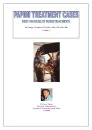
Cases from private practices - Papimi
Cases from private practices - Papimi
Cases from private practices - Papimi

You also want an ePaper? Increase the reach of your titles
YUMPU automatically turns print PDFs into web optimized ePapers that Google loves.
Case B Patient: Georgia S.Age: 49<br />
Condition: Bone cancer<br />
Georgia had bone scan on October 24, 1991 to check the condition of her bone cancer. She then used<br />
the PEC for about three months (late 1991 to early 1992) to expose certain affected areas which included<br />
the left frontal jugale, sacro-iliae, and the pubic dextra. Other areas, including the left scapula were<br />
exposed to a lesser extent. A bone scan performed on March 2, 1992 showed that in the intervening 4<br />
months the areas that had received primary exposure had improved. The cancer in the lesser exposed<br />
areas was found either to have remained stationary or to have slightly developed. In addition, several new<br />
cancer foci were noticed. These had not received any exposure.<br />
Case C Patient: Diane L. Age: 47<br />
Condition: Fibroid tumors of the uterus, digestive problems<br />
Since 1975, Diane has had endometriosis on the ligament behind her left ovary which had caused her<br />
severe pain. In 1990 the diagnosis was changed to large fibroid tumors. One of the tumors was pressing<br />
against her bladder and giving her difficulty with urination. Surgery was recommended. At this point the<br />
pain <strong>from</strong> her groin was so intense that it periodically woke her <strong>from</strong> a sound sleep. Before undergoing<br />
surgery she decided to try some treatments with the PEC Device at a clinic on Nassau. Prior to departing<br />
for Nassau in mid 1991, she had a sonogram exam.<br />
On Nassau, she underwent ten, five-minute treatments on the PEC Device over a period of three<br />
consecutive days. The treatments were painless and relaxing. After the third session she noticed that her<br />
urination was unhampered and that her pain was gone. She then had a second sonogram after returning<br />
<strong>from</strong> Nassau. Surprisingly this showed that the size of the tumors was drastically reduced. On June 30th<br />
she menstruated normally with no discomfort.<br />
A few weeks later, in July, she returned to Nassau and had 12 six-minute treatments over three<br />
consecutive days. She then returned home and had a third sonogram. The sonogram indicated that the<br />
tumors had totally disappeared. Also she had no pain during a subsequent menstruation.<br />
A secondary therapeutic effect of the treatment was the cure of Diane's digestive system malady. She<br />
had been diagnosed as having a hiatal hernia and a blockage in her duodenum. Also as a result of one GI<br />
exam doctors suggested that she have a gall bladder operation. Even after ceasing to eat foods that gave<br />
her a problem, she experienced a radiating pain <strong>from</strong> underneath her right ribs toward her neck. Since she<br />
had her 22 treatments with the PEC, she has had no pain in that area, no gas attacks, and has ceased<br />
taking digestive aids. She is even able to eat foods that bothered her before without a problem.<br />
Case D Patient: I.B. Age: 53<br />
Condition: Lymphoma, Chronic nonspecific lymphocytic leukemia<br />
Diagnosis and Therapy Prior to PEC Treatment:<br />
In September, 1987 I. B., then 48 years of age, was being checked to investigate her first-time heart<br />
fibrillation. At that time she was diagnosed with 3rd or 4th stage lymphoma. There are two main tumors<br />
in her abdomen, one attached to the vena cava, the other to the aorta, both inoperable, as well as several<br />
other smaller tumors. In 1988, I.B. had 7 months of chemotherapy treatment with Leukeran. However,<br />
the treatment had little effect. She refused further, more complicated chemotherapy and began turning to<br />
"holistic" treatments.<br />
Abdominal CAT scans were taken in June and July of 1991 [see attachments D-2, D-3 & D-4] and<br />
compared with scans taken in July 1989 and October 1990. These showed that the para-aortic adenopathy<br />
had progressively worsened since 1989 and 1990, with increases both in size and in number. The largest<br />
lymph nodes measured up to 3 cm in diameter (left of mid abdominal aorta).<br />
34