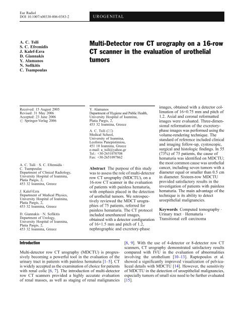
Multi-Detector row CT urography on a 16-row CT scanner in the ...
Multi-Detector row CT urography on a 16-row CT scanner in the ...
Multi-Detector row CT urography on a 16-row CT scanner in the ...

You also want an ePaper? Increase the reach of your titles
YUMPU automatically turns print PDFs into web optimized ePapers that Google loves.
Eur Radiol<br />
DOI 10.1007/s00330-006-0383-2<br />
UROGENITAL<br />
A. C. Tsili<br />
S. C. Efremidis<br />
J. Kalef-Ezra<br />
D. Giannakis<br />
Y. Alamanos<br />
N. Sofikitis<br />
C. Tsampoulas<br />
<str<strong>on</strong>g>Multi</str<strong>on</strong>g>-<str<strong>on</strong>g>Detector</str<strong>on</strong>g> <str<strong>on</strong>g>row</str<strong>on</strong>g> <str<strong>on</strong>g>CT</str<strong>on</strong>g> <str<strong>on</strong>g>urography</str<strong>on</strong>g> <strong>on</strong> a <strong>16</strong>-<str<strong>on</strong>g>row</str<strong>on</strong>g><br />
<str<strong>on</strong>g>CT</str<strong>on</strong>g> <strong>scanner</strong> <strong>in</strong> <strong>the</strong> evaluati<strong>on</strong> of uro<strong>the</strong>lial<br />
tumors<br />
Received: 15 August 2005<br />
Revised: 31 May 2006<br />
Accepted: 23 June 2006<br />
# Spr<strong>in</strong>ger-Verlag 2006<br />
A. C. Tsili . S. C. Efremidis .<br />
C. Tsampoulas<br />
Department of Cl<strong>in</strong>ical Radiology,<br />
University Hospital of Ioann<strong>in</strong>a,<br />
Platia Pargis, 2,<br />
453 32 Ioann<strong>in</strong>a, Greece<br />
J. Kalef-Ezra<br />
Department of Medical Physics,<br />
University Hospital of Ioann<strong>in</strong>a,<br />
Platia Pargis, 2,<br />
453 32 Ioann<strong>in</strong>a, Greece<br />
D. Giannakis . N. Sofikitis<br />
Department of Urology,<br />
University Hospital of Ioann<strong>in</strong>a,<br />
Platia Pargis, 2,<br />
453 32 Ioann<strong>in</strong>a, Greece<br />
Y. Alamanos<br />
Department of Hygiene and Public Health,<br />
University Hospital of Ioann<strong>in</strong>a,<br />
Platia Pargis, 2,<br />
453 32 Ioann<strong>in</strong>a, Greece<br />
A. C. Tsili (*)<br />
Medical School,<br />
University of Ioann<strong>in</strong>a,<br />
Leoforos Panepistimiou,<br />
451 10 Ioann<strong>in</strong>a, Greece<br />
e-mail: a_tsili@yahoo.gr<br />
Tel.: +30-2651070708<br />
Fax: +30-2651097862<br />
Abstract The purpose of this study<br />
was to assess <strong>the</strong> role of multi-detector<br />
<str<strong>on</strong>g>row</str<strong>on</strong>g> <str<strong>on</strong>g>CT</str<strong>on</strong>g> <str<strong>on</strong>g>urography</str<strong>on</strong>g> (MD<str<strong>on</strong>g>CT</str<strong>on</strong>g>U), <strong>on</strong> a<br />
<strong>16</strong>-<str<strong>on</strong>g>row</str<strong>on</strong>g> <str<strong>on</strong>g>CT</str<strong>on</strong>g> <strong>scanner</strong> <strong>in</strong> <strong>the</strong> evaluati<strong>on</strong><br />
of patients with pa<strong>in</strong>less hematuria,<br />
with emphasis placed <strong>in</strong> <strong>the</strong> detecti<strong>on</strong><br />
of uro<strong>the</strong>lial tumors. We retrospectively<br />
reviewed <strong>the</strong> MD<str<strong>on</strong>g>CT</str<strong>on</strong>g> urographies<br />
of 75 patients, referred for<br />
pa<strong>in</strong>less hematuria. The <str<strong>on</strong>g>CT</str<strong>on</strong>g> protocol<br />
<strong>in</strong>cluded unenhanced images,<br />
obta<strong>in</strong>ed with a detector c<strong>on</strong>figurati<strong>on</strong><br />
of <strong>16</strong>×1.5 mm and pitch of 1.2,<br />
nephrographic and excretory-phase<br />
images, obta<strong>in</strong>ed with a detector collimati<strong>on</strong><br />
of <strong>16</strong>×0.75 mm and pitch of<br />
1.2. Axial and cor<strong>on</strong>al reformatted<br />
images were evaluated. Three-dimensi<strong>on</strong>al<br />
reformati<strong>on</strong> of <strong>the</strong> excretoryphase<br />
images was performed us<strong>in</strong>g <strong>the</strong><br />
volume-render<strong>in</strong>g technique. The<br />
standard of reference <strong>in</strong>cluded cl<strong>in</strong>ical<br />
and imag<strong>in</strong>g follow-up, cystoscopic,<br />
surgical and histologic f<strong>in</strong>d<strong>in</strong>gs. In 55<br />
(73%) of 75 patients, <strong>the</strong> cause of<br />
hematuria was identified <strong>on</strong> MD<str<strong>on</strong>g>CT</str<strong>on</strong>g>U;<br />
<strong>the</strong> most comm<strong>on</strong> cause was uro<strong>the</strong>lial<br />
cancer, <strong>in</strong>clud<strong>in</strong>g seven tumors with a<br />
diameter equal or smaller than 0.5 cm<br />
<strong>in</strong> diameter. Sixteen-<str<strong>on</strong>g>row</str<strong>on</strong>g> MD<str<strong>on</strong>g>CT</str<strong>on</strong>g>U<br />
provided satisfactory results <strong>in</strong> <strong>the</strong><br />
<strong>in</strong>vestigati<strong>on</strong> of patients with pa<strong>in</strong>less<br />
hematuria. The ma<strong>in</strong> advantage of <strong>the</strong><br />
technique is its ability to detect<br />
uroepi<strong>the</strong>lial malignancies.<br />
Keywords Computed tomography .<br />
Ur<strong>in</strong>ary tract . Hematuria .<br />
Transiti<strong>on</strong>al cell carc<strong>in</strong>oma<br />
Introducti<strong>on</strong><br />
<str<strong>on</strong>g>Multi</str<strong>on</strong>g>-detector <str<strong>on</strong>g>row</str<strong>on</strong>g> <str<strong>on</strong>g>CT</str<strong>on</strong>g> <str<strong>on</strong>g>urography</str<strong>on</strong>g> (MD<str<strong>on</strong>g>CT</str<strong>on</strong>g>U) is progressively<br />
becom<strong>in</strong>g a powerful tool <strong>in</strong> <strong>the</strong> evaluati<strong>on</strong> of <strong>the</strong><br />
ur<strong>in</strong>ary tract <strong>in</strong> patients with pa<strong>in</strong>less hematuria [1–5]. <str<strong>on</strong>g>CT</str<strong>on</strong>g><br />
is widely accepted as <strong>the</strong> exam<strong>in</strong>ati<strong>on</strong> of choice for patients<br />
with renal colic [6, 7]. The <strong>in</strong>troducti<strong>on</strong> of multi-detector<br />
<str<strong>on</strong>g>row</str<strong>on</strong>g> <str<strong>on</strong>g>CT</str<strong>on</strong>g> <strong>scanner</strong>s provided a highly accurate evaluati<strong>on</strong><br />
of renal masses, as well as stag<strong>in</strong>g of renal malignancies<br />
[8, 9]. With <strong>the</strong> use of 4-detector or 8-detector <str<strong>on</strong>g>row</str<strong>on</strong>g> <str<strong>on</strong>g>CT</str<strong>on</strong>g><br />
<strong>scanner</strong>s, <str<strong>on</strong>g>CT</str<strong>on</strong>g> <str<strong>on</strong>g>urography</str<strong>on</strong>g> dem<strong>on</strong>strated satisfactory results<br />
compared with IVU <strong>in</strong> <strong>the</strong> evaluati<strong>on</strong> of abnormalities<br />
<strong>in</strong>volv<strong>in</strong>g <strong>the</strong> uro<strong>the</strong>lium [10–13]. Raptopoulos et al.<br />
showed a significantly improved visualizati<strong>on</strong> of pelvicaliceal<br />
details with MD<str<strong>on</strong>g>CT</str<strong>on</strong>g>U [14]. However, <strong>the</strong> sensitivity<br />
of MD<str<strong>on</strong>g>CT</str<strong>on</strong>g>U <strong>in</strong> <strong>the</strong> detecti<strong>on</strong> of uroepi<strong>the</strong>lial malignancies,<br />
especially tumors of small size need to be fur<strong>the</strong>r evaluated<br />
[15].
With <strong>the</strong> <strong>in</strong>troducti<strong>on</strong> of <strong>16</strong>-<str<strong>on</strong>g>row</str<strong>on</strong>g> <str<strong>on</strong>g>CT</str<strong>on</strong>g> <strong>scanner</strong>s, <strong>the</strong><br />
radiologic evaluati<strong>on</strong> of <strong>the</strong> ur<strong>in</strong>ary tract is chang<strong>in</strong>g<br />
rapidly. The advantages of a <strong>16</strong>-<str<strong>on</strong>g>row</str<strong>on</strong>g> <str<strong>on</strong>g>CT</str<strong>on</strong>g> <strong>scanner</strong> are <strong>the</strong><br />
follow<strong>in</strong>g: fast scann<strong>in</strong>g (coverage of at least 48 mm l<strong>on</strong>g<br />
body secti<strong>on</strong>s per sec<strong>on</strong>d), acquisiti<strong>on</strong> of th<strong>in</strong> collimati<strong>on</strong><br />
and creati<strong>on</strong> of high-resoluti<strong>on</strong> multiplanar and threedimensi<strong>on</strong>al<br />
(3D) reformatted images, with excellent<br />
anatomic details [<strong>16</strong>, 17]. The purpose of our study was<br />
to assess <strong>the</strong> role of multi-detector <str<strong>on</strong>g>row</str<strong>on</strong>g> <str<strong>on</strong>g>CT</str<strong>on</strong>g> <str<strong>on</strong>g>urography</str<strong>on</strong>g>,<br />
us<strong>in</strong>g a <strong>16</strong>-<str<strong>on</strong>g>row</str<strong>on</strong>g> <str<strong>on</strong>g>CT</str<strong>on</strong>g> <strong>scanner</strong> <strong>in</strong> <strong>the</strong> evaluati<strong>on</strong> of patients<br />
present<strong>in</strong>g with pa<strong>in</strong>less hematuria, with emphasis placed<br />
<strong>in</strong> <strong>the</strong> detecti<strong>on</strong> of uro<strong>the</strong>lial tumors.<br />
Materials and methods<br />
This study was a retrospective review of <str<strong>on</strong>g>CT</str<strong>on</strong>g> urographies<br />
performed <strong>on</strong> a <strong>16</strong>-<str<strong>on</strong>g>row</str<strong>on</strong>g> <str<strong>on</strong>g>CT</str<strong>on</strong>g> <strong>scanner</strong> for <strong>the</strong> evaluati<strong>on</strong> of<br />
pa<strong>in</strong>less hematuria. The study <strong>in</strong>cluded 75 c<strong>on</strong>secutive<br />
patients (47 men and 28 women) with a mean age of 63<br />
years (age range: 21–87 years), who underwent multidetector<br />
<str<strong>on</strong>g>row</str<strong>on</strong>g> <str<strong>on</strong>g>CT</str<strong>on</strong>g> <str<strong>on</strong>g>urography</str<strong>on</strong>g> between March 2004 and<br />
January 2005, referred to <strong>the</strong> department of Radiology for<br />
pa<strong>in</strong>less hematuria. Five patients were younger than 40<br />
years old. Informed c<strong>on</strong>sent for this study was obta<strong>in</strong>ed<br />
from each patient.<br />
The cl<strong>in</strong>ical evaluati<strong>on</strong> of <strong>the</strong> patients <strong>in</strong>cluded a careful<br />
history and cl<strong>in</strong>ical exam<strong>in</strong>ati<strong>on</strong>. The laboratory analysis<br />
began with a comprehensive exam<strong>in</strong>ati<strong>on</strong> of <strong>the</strong> ur<strong>in</strong>e and<br />
ur<strong>in</strong>e sediment and cytologic analysis. All patients had an<br />
ultrasound exam<strong>in</strong>ati<strong>on</strong> of <strong>the</strong> abdomen, some performed<br />
at an outpatient cl<strong>in</strong>ic and <strong>the</strong> rest at our department.<br />
Patients with hematuria and a s<strong>on</strong>ographic exam<strong>in</strong>ati<strong>on</strong><br />
dem<strong>on</strong>strat<strong>in</strong>g a kidney mass were not <strong>in</strong>cluded <strong>in</strong> this<br />
study, s<strong>in</strong>ce <strong>the</strong>y were evaluated with a different protocol<br />
tailored for <strong>the</strong> <strong>in</strong>vestigati<strong>on</strong> of renal masses. Cystoscopic<br />
exam<strong>in</strong>ati<strong>on</strong> was carried out <strong>in</strong> all patients.<br />
In patients <strong>in</strong> whom cl<strong>in</strong>ical and imag<strong>in</strong>g evaluati<strong>on</strong><br />
revealed no cause for <strong>the</strong> hematuria, a follow-up was<br />
recommended. This <strong>in</strong>cluded <strong>the</strong> repeat of <strong>the</strong> ur<strong>in</strong>ary<br />
analysis and ur<strong>in</strong>e cytology every m<strong>on</strong>th for <strong>the</strong> first 4<br />
m<strong>on</strong>ths and <strong>the</strong>reafter at 6, 12, 24 and 36 m<strong>on</strong>ths.<br />
Immediate reevaluati<strong>on</strong> with repeat of <strong>the</strong> <str<strong>on</strong>g>CT</str<strong>on</strong>g> exam<strong>in</strong>ati<strong>on</strong><br />
and/or cystoscopy and ur<strong>in</strong>e cytology was recommended if<br />
any of <strong>the</strong> follow<strong>in</strong>g occurred: development of gross<br />
hematuria, abnormal ur<strong>in</strong>ary cytologic f<strong>in</strong>d<strong>in</strong>gs or irritat<strong>in</strong>g<br />
void<strong>in</strong>g symptoms <strong>in</strong> <strong>the</strong> absence of <strong>in</strong>fecti<strong>on</strong> [18].<br />
Technique<br />
Exam<strong>in</strong>ati<strong>on</strong>s were performed <strong>on</strong> a <strong>16</strong>-<str<strong>on</strong>g>row</str<strong>on</strong>g> <str<strong>on</strong>g>CT</str<strong>on</strong>g> <strong>scanner</strong><br />
with 24 mm scann<strong>in</strong>g span per rotati<strong>on</strong> (Mx8000 IDT,<br />
Philips) and a three-phase protocol was used. Unenhanced<br />
images were obta<strong>in</strong>ed from <strong>the</strong> kidneys through <strong>the</strong> ur<strong>in</strong>ary<br />
bladder us<strong>in</strong>g a slice collimati<strong>on</strong> of <strong>16</strong>×1.5 mm and a slice<br />
thickness of 3 mm. These images were scrut<strong>in</strong>ized to<br />
identify ur<strong>in</strong>ary tract calculi or calcificati<strong>on</strong>s, as well as to<br />
assist <strong>in</strong> <strong>the</strong> characterizati<strong>on</strong> of any detected renal masses.<br />
Intravenous n<strong>on</strong>-i<strong>on</strong>ic iod<strong>in</strong>ated c<strong>on</strong>trast material (120 ml,<br />
320 mg I/ml) was subsequently adm<strong>in</strong>istered at a rate of<br />
3 ml/sec, via mechanical <strong>in</strong>jector, followed by 250 ml of<br />
normal sal<strong>in</strong>e 0.9%, rapidly <strong>in</strong>fused by <strong>in</strong>travenous drip.<br />
The sec<strong>on</strong>d phase was <strong>the</strong> nephrographic phase obta<strong>in</strong>ed at<br />
100 sec delay, cover<strong>in</strong>g <strong>the</strong> area from <strong>the</strong> diaphragm to <strong>the</strong><br />
iliac crests and <strong>the</strong> third phase was <strong>the</strong> excretory phase<br />
start<strong>in</strong>g with 10-m<strong>in</strong>ute delay, cover<strong>in</strong>g <strong>the</strong> kidneys, ureters<br />
and <strong>the</strong> ur<strong>in</strong>ary bladder. The nephrographic phase was<br />
chosen for <strong>the</strong> detecti<strong>on</strong> and characterizati<strong>on</strong> of renal<br />
masses, as well as for stag<strong>in</strong>g, while <strong>the</strong> excretory phase<br />
was utilized for <strong>the</strong> evaluati<strong>on</strong> of abnormalities <strong>in</strong>volv<strong>in</strong>g<br />
<strong>the</strong> uro<strong>the</strong>lium. For <strong>the</strong>se two phases a secti<strong>on</strong> collimati<strong>on</strong><br />
of <strong>16</strong>×0.75 mm and a slice thickness of 1 mm were<br />
employed. The nephrographic and excretory phase images<br />
were rec<strong>on</strong>structed at 0.5 mm <strong>in</strong>tervals (50% overlap). All<br />
<strong>the</strong> scans were performed with a pitch of 1.2, at 120 kV,<br />
us<strong>in</strong>g a rotati<strong>on</strong> time of 0.5 sec. Both dose modulati<strong>on</strong><br />
(DOM) and automatic current sett<strong>in</strong>g (DoseRight) were<br />
used [19]. No oral c<strong>on</strong>trast material was given, <strong>in</strong> order to<br />
facilitate 3D reformatt<strong>in</strong>g. The entire <str<strong>on</strong>g>CT</str<strong>on</strong>g> <str<strong>on</strong>g>urography</str<strong>on</strong>g><br />
exam<strong>in</strong>ati<strong>on</strong> lasted 15 m<strong>in</strong>utes per patient <strong>on</strong> <strong>the</strong> average<br />
and every scan lasted 7–12 sec<strong>on</strong>ds <strong>on</strong> <strong>the</strong> average. Our <str<strong>on</strong>g>CT</str<strong>on</strong>g><br />
protocol is illustrated <strong>on</strong> Table 1.<br />
<str<strong>on</strong>g>CT</str<strong>on</strong>g> data <strong>in</strong>terpretati<strong>on</strong><br />
Image <strong>in</strong>terpretati<strong>on</strong> was d<strong>on</strong>e <strong>on</strong> a workstati<strong>on</strong> (MxView,<br />
Philips) and <strong>in</strong>cluded <strong>the</strong> study of <strong>the</strong> axial images of <strong>the</strong><br />
unenhanced scan. The evaluati<strong>on</strong> of <strong>the</strong> axial source<br />
images of <strong>the</strong> nephrographic and <strong>the</strong> excretory phase was<br />
Table 1 <str<strong>on</strong>g>CT</str<strong>on</strong>g> protocol for <strong>the</strong> <strong>in</strong>vestigati<strong>on</strong> of hematuria<br />
Unenhanced<br />
scan<br />
Nephrographic<br />
phase,<br />
100 sec<br />
Excretory<br />
phase,<br />
10 m<strong>in</strong><br />
kV, rotati<strong>on</strong> time (sec) 120/0.5 120/0.5 120/0.5<br />
mAsnom/mAssel 200/170 200/175 200/<strong>16</strong>0<br />
<str<strong>on</strong>g>Detector</str<strong>on</strong>g> collimati<strong>on</strong> <strong>16</strong>×1.5 <strong>16</strong>×0.75 <strong>16</strong>×0.75<br />
(mm)<br />
Pitch 1.2 1.2 1.2<br />
Slice thickness (mm) 3 1 1<br />
Rec<strong>on</strong>structi<strong>on</strong> 3 0.5 0.5<br />
<strong>in</strong>terval (mm)<br />
Area<br />
Kidneysur<strong>in</strong>ary<br />
bladder<br />
Diaphragmiliac<br />
crests<br />
Kidneysur<strong>in</strong>ary<br />
bladder<br />
C<strong>on</strong>trast material<br />
(iv)<br />
120 ml (320), 3 ml/sec and 250 ml<br />
sal<strong>in</strong>e 0.9%
difficult, due to <strong>the</strong> large number (approximately 400–800)<br />
of noisy images. For <strong>the</strong>se reas<strong>on</strong>s reformatted images, of<br />
4 mm slice thickness and at 3 mm <strong>in</strong>tervals, usually <strong>in</strong> <strong>the</strong><br />
transverse and cor<strong>on</strong>al planes (us<strong>in</strong>g <strong>the</strong> Extended<br />
Brilliance V.1.0.1.1. software) were used for <strong>in</strong>terpretati<strong>on</strong><br />
of <strong>the</strong> nephrographic and excretory phases. The time to<br />
generate <strong>the</strong>se images was less than <strong>on</strong>e m<strong>in</strong>ute. Wide<br />
w<strong>in</strong>dow sett<strong>in</strong>gs (b<strong>on</strong>e w<strong>in</strong>dow sett<strong>in</strong>gs, W: 1500, L: 500)<br />
were also used for <strong>the</strong> excretory phase, to better visualize<br />
caliceal details, as well as fill<strong>in</strong>g defects <strong>in</strong> <strong>the</strong> ur<strong>in</strong>ary<br />
collect<strong>in</strong>g system (Fig. 1). F<strong>in</strong>ally, three-dimensi<strong>on</strong>al (3D)<br />
rec<strong>on</strong>structi<strong>on</strong>s of <strong>the</strong> excretory phase data were performed,<br />
us<strong>in</strong>g <strong>the</strong> volume render<strong>in</strong>g technique and <strong>the</strong><br />
same software, obta<strong>in</strong>ed <strong>in</strong> <strong>the</strong> cor<strong>on</strong>al and/or oblique<br />
planes. Volume render<strong>in</strong>g technique was c<strong>on</strong>sidered ideal<br />
for <strong>the</strong> creati<strong>on</strong> of <strong>the</strong> 3D rec<strong>on</strong>structi<strong>on</strong>s [3], especially<br />
because it does not obscure superimposed structures, such<br />
as <strong>the</strong> pelvis and <strong>the</strong> ureters. The time required to create <strong>the</strong><br />
3D reformatted images was less than <strong>on</strong>e m<strong>in</strong>ute. The ma<strong>in</strong><br />
advantage of <strong>the</strong>se images was that <strong>the</strong>y were more familiar<br />
to our cl<strong>in</strong>ical colleagues, because <strong>the</strong>y closely resemble<br />
those of <strong>in</strong>travenous <str<strong>on</strong>g>urography</str<strong>on</strong>g>. Additi<strong>on</strong>ally, <strong>the</strong>se images<br />
could be more effectively used for preoperative plann<strong>in</strong>g,<br />
whenever it was necessary.<br />
An important issue <strong>on</strong> <str<strong>on</strong>g>CT</str<strong>on</strong>g> <str<strong>on</strong>g>urography</str<strong>on</strong>g> is <strong>the</strong> degree of<br />
ureteral opacificati<strong>on</strong>. In cases, where excreti<strong>on</strong> of <strong>the</strong><br />
c<strong>on</strong>trast material was detected <strong>on</strong> <str<strong>on</strong>g>CT</str<strong>on</strong>g> <str<strong>on</strong>g>urography</str<strong>on</strong>g>, <strong>the</strong> extent<br />
of ureteral opacificati<strong>on</strong> was evaluated. The ureter was<br />
divided <strong>in</strong>to proximal, middle and distal thirds. The<br />
proximal third was def<strong>in</strong>ed as <strong>the</strong> porti<strong>on</strong> of <strong>the</strong> ureter<br />
that extended from <strong>the</strong> renal pelvis to <strong>the</strong> superior iliac<br />
crests. The middle third was def<strong>in</strong>ed as <strong>the</strong> porti<strong>on</strong> of <strong>the</strong><br />
ureter between <strong>the</strong> superior iliac crests and <strong>the</strong> anterior<br />
<strong>in</strong>ferior iliac sp<strong>in</strong>es. The lower third was def<strong>in</strong>ed as <strong>the</strong><br />
segment of <strong>the</strong> ureter located between <strong>the</strong> anterior <strong>in</strong>ferior<br />
iliac sp<strong>in</strong>es and <strong>the</strong> ureterovesical juncti<strong>on</strong>. An opacificati<strong>on</strong><br />
score from 1 to 3 was assigned for each ureter based <strong>on</strong><br />
<strong>the</strong> length of <strong>the</strong> ureteral porti<strong>on</strong> opacified. A score of 1<br />
<strong>in</strong>dicated opacificati<strong>on</strong> of less than <strong>on</strong>e third of <strong>the</strong> ureter; a<br />
score of 2 <strong>in</strong>dicated opacificati<strong>on</strong> of less than two thirds of<br />
<strong>the</strong> ureter; a score of 3, opacificati<strong>on</strong> of more than two<br />
thirds of <strong>the</strong> ureteral segments up to <strong>the</strong> entire length of <strong>the</strong><br />
ureter.<br />
<str<strong>on</strong>g>CT</str<strong>on</strong>g> data correlati<strong>on</strong><br />
F<strong>in</strong>d<strong>in</strong>gs of MD<str<strong>on</strong>g>CT</str<strong>on</strong>g>U were retrospectively correlated with<br />
<strong>the</strong> results of cl<strong>in</strong>ical and imag<strong>in</strong>g follow-up, cystoscopy<br />
and/or ureteroscopy, as well as with surgical and histological<br />
f<strong>in</strong>d<strong>in</strong>gs, <strong>in</strong> surgical cases. More specifically, <strong>in</strong><br />
patients with ur<strong>in</strong>ary tract lithiasis c<strong>on</strong>firmati<strong>on</strong> of <strong>the</strong> <str<strong>on</strong>g>CT</str<strong>on</strong>g><br />
diagnosis was documented <strong>on</strong> <strong>the</strong> basis of o<strong>the</strong>r imag<strong>in</strong>g<br />
exam<strong>in</strong>ati<strong>on</strong>s (pla<strong>in</strong> films and ultrasound). C<strong>on</strong>venti<strong>on</strong>al<br />
cystoscopy and histologic results were used as standard of<br />
reference <strong>in</strong> patients with ur<strong>in</strong>ary bladder carc<strong>in</strong>omas.<br />
C<strong>on</strong>venti<strong>on</strong>al cystoscopy was also used to c<strong>on</strong>firm <strong>the</strong><br />
presence of benign prostatic hyperplasia (BPH) as <strong>the</strong><br />
cause of hematuria. In patients with transiti<strong>on</strong>al cell<br />
carc<strong>in</strong>oma of <strong>the</strong> pelvicaliceal system, as well as ureteral<br />
lesi<strong>on</strong>s, c<strong>on</strong>firmati<strong>on</strong> of <strong>the</strong> <str<strong>on</strong>g>CT</str<strong>on</strong>g> diagnosis was obta<strong>in</strong>ed<br />
based <strong>on</strong> o<strong>the</strong>r imag<strong>in</strong>g exam<strong>in</strong>ati<strong>on</strong>s (IVU, retrograde<br />
pyelography), ureteroscopic f<strong>in</strong>d<strong>in</strong>gs and f<strong>in</strong>ally <strong>the</strong> surgical<br />
and histologic f<strong>in</strong>d<strong>in</strong>gs. Surgery and histologic diagnosis<br />
c<strong>on</strong>firmed <strong>the</strong> presence of o<strong>the</strong>r malignancies (renal<br />
cancer, pancreatic and prostate cancer). For patients with<br />
ur<strong>in</strong>ary tract c<strong>on</strong>genital anomalies, <strong>the</strong> f<strong>in</strong>d<strong>in</strong>gs of IVU<br />
were used for c<strong>on</strong>firm<strong>in</strong>g <strong>the</strong> <str<strong>on</strong>g>CT</str<strong>on</strong>g> diagnosis.<br />
The <str<strong>on</strong>g>CT</str<strong>on</strong>g> images were <strong>in</strong>terpreted by <strong>on</strong>e radiologist<br />
(A. C. T), bl<strong>in</strong>ded to <strong>the</strong> results of <strong>the</strong> o<strong>the</strong>r exam<strong>in</strong>ati<strong>on</strong>s.<br />
Dosimetry<br />
For dosimetry <strong>the</strong> free-<strong>in</strong>-air Computed Tomography Dose<br />
Index was measured al<strong>on</strong>g <strong>the</strong> axis of rotati<strong>on</strong> us<strong>in</strong>g a<br />
pencil-shaped i<strong>on</strong> chamber, type 10×5 10.3 <str<strong>on</strong>g>CT</str<strong>on</strong>g> (Radcal<br />
Corp., California, USA). The effective dose was calculated<br />
us<strong>in</strong>g <strong>the</strong> ImPA<str<strong>on</strong>g>CT</str<strong>on</strong>g> <str<strong>on</strong>g>CT</str<strong>on</strong>g> Patient Dosimetry Calculator<br />
(versi<strong>on</strong> 0.99v) [20]. The software used is based <strong>on</strong> <strong>the</strong><br />
Fig. 1 A 70-year-old woman<br />
with fibroepi<strong>the</strong>lial polyp of <strong>the</strong><br />
right ureter. Cor<strong>on</strong>al (a) and<br />
transverse (b) reformatted<br />
images of <strong>the</strong> excretory phase<br />
dem<strong>on</strong>strate an el<strong>on</strong>gated fill<strong>in</strong>g<br />
defect (ar<str<strong>on</strong>g>row</str<strong>on</strong>g>), smoothly marg<strong>in</strong>ated,<br />
<strong>in</strong>volv<strong>in</strong>g <strong>the</strong> <strong>in</strong>travesical<br />
part of <strong>the</strong> right ureter and<br />
prolaps<strong>in</strong>g <strong>in</strong>to <strong>the</strong> ur<strong>in</strong>ary<br />
bladder. The lesi<strong>on</strong> is clearly<br />
seen with b<strong>on</strong>e w<strong>in</strong>dow sett<strong>in</strong>gs
Table 2 MD<str<strong>on</strong>g>CT</str<strong>on</strong>g>U diagnoses for <strong>the</strong> hematuria<br />
<str<strong>on</strong>g>CT</str<strong>on</strong>g> diagnoses<br />
Number of patients<br />
St<strong>on</strong>e disease<br />
Ureter 4<br />
Kidney 7<br />
Ur<strong>in</strong>ary bladder 2<br />
Passed st<strong>on</strong>e 1<br />
Cancer<br />
Ur<strong>in</strong>ary bladder <strong>16</strong><br />
Kidney 7<br />
Ureter 1<br />
Prostate 1<br />
Pancreas 1<br />
Benign tumors<br />
Kidney 1<br />
Ureter 1<br />
Ur<strong>in</strong>ary tract <strong>in</strong>flammati<strong>on</strong> 2<br />
Benign prostatic hyperplasia 9<br />
Pyeloureteric juncti<strong>on</strong> obstructi<strong>on</strong> 1<br />
C<strong>on</strong>genital anomalies 4<br />
M<strong>on</strong>te Carlo c<strong>on</strong>versi<strong>on</strong> coefficients obta<strong>in</strong>ed by J<strong>on</strong>es and<br />
Shrimpt<strong>on</strong> [21]. C<strong>on</strong>sider<strong>in</strong>g <strong>the</strong> fact that <strong>the</strong> tube current is<br />
modulated across <strong>the</strong> patient’s body, based <strong>on</strong> plann<strong>in</strong>g<br />
scan projecti<strong>on</strong> radiograph (PSPR) and body asymmetry<br />
(dose modulati<strong>on</strong> functi<strong>on</strong>), <strong>the</strong> mean mAs per rotati<strong>on</strong> at<br />
each phase for each <strong>in</strong>dividual patient was recorded and <strong>the</strong><br />
mean mAs per rotati<strong>on</strong> am<strong>on</strong>g all patients at each phase<br />
was calculated. To assess <strong>the</strong> probability for <strong>in</strong>ducti<strong>on</strong> of<br />
determ<strong>in</strong>istic effects, two groups of LiF:Mg,Ti <strong>the</strong>rmolum<strong>in</strong>escent<br />
dosimeters were fixed with hypo-allergic tape<br />
<strong>on</strong> <strong>the</strong> sk<strong>in</strong> at <strong>the</strong> abdomen of six c<strong>on</strong>secutive patients, over<br />
a two-week period (<strong>on</strong>e group at umbilicus and <strong>the</strong> o<strong>the</strong>r at<br />
midaxillary l<strong>in</strong>e <strong>in</strong> <strong>the</strong> same level).<br />
Results<br />
In 18 of 75 patients (24%) no ur<strong>in</strong>ary tract abnormality was<br />
seen <strong>on</strong> MD<str<strong>on</strong>g>CT</str<strong>on</strong>g>U. In <strong>16</strong> of <strong>the</strong>se patients <strong>the</strong> cl<strong>in</strong>ical<br />
evaluati<strong>on</strong>, <strong>in</strong>clud<strong>in</strong>g cystoscopy and ur<strong>in</strong>alysis revealed<br />
no cause for <strong>the</strong> hematuria. These cases have been<br />
followed-up for a period ranged from 12 to 20 m<strong>on</strong>ths<br />
and rema<strong>in</strong>ed negative. These cases were c<strong>on</strong>sidered as<br />
true-negative. We had two false-negative cases. One with a<br />
patient, <strong>in</strong> whom <strong>the</strong> hematuria was attributed to carc<strong>in</strong>oma<br />
of <strong>the</strong> prostate gland, not shown <strong>on</strong> MD<str<strong>on</strong>g>CT</str<strong>on</strong>g>U and a sec<strong>on</strong>d<br />
case, <strong>in</strong> which <strong>the</strong> ur<strong>in</strong>ary bladder was filled with blood<br />
clots. In this case it was impossible for <strong>the</strong> <str<strong>on</strong>g>CT</str<strong>on</strong>g> exam<strong>in</strong>ati<strong>on</strong><br />
to recognize a small tumor, with a diameter less than<br />
0.5 cm, as seen with c<strong>on</strong>venti<strong>on</strong>al cystoscopy. This tumor<br />
could probably be detected, if <strong>the</strong>re were no blood clots <strong>on</strong><br />
<strong>the</strong> ur<strong>in</strong>ary bladder dur<strong>in</strong>g <str<strong>on</strong>g>CT</str<strong>on</strong>g> scann<strong>in</strong>g, as it was detected<br />
<strong>on</strong> s<strong>on</strong>ography. This lesi<strong>on</strong> was characterized as transiti<strong>on</strong>al<br />
cell carc<strong>in</strong>oma (TCC) <strong>on</strong> histology.<br />
In 57 of 75 patients (76%) <strong>the</strong> cause of hematuria was<br />
identified. Table 2 dem<strong>on</strong>strates <strong>the</strong> different etiologies of<br />
hematuria, as shown <strong>on</strong> MD<str<strong>on</strong>g>CT</str<strong>on</strong>g>U.<br />
In 12 patients, <strong>the</strong> cause of hematuria was ur<strong>in</strong>ary tract<br />
lithiasis. Four patients had ureteral st<strong>on</strong>es, rang<strong>in</strong>g from 3.5<br />
to 7 mm <strong>in</strong> diameter, with no signs of obstructi<strong>on</strong> <strong>in</strong> three.<br />
These four patients were followed-up with pla<strong>in</strong> films and<br />
ultrasound. In <strong>on</strong>e patient renal calculi were also seen. In<br />
six patients <strong>the</strong> calculi were located <strong>in</strong> <strong>the</strong> pelvicaliceal<br />
system, c<strong>on</strong>firmed also <strong>on</strong> pla<strong>in</strong> films and s<strong>on</strong>ography and<br />
<strong>in</strong> two <strong>in</strong> <strong>the</strong> ur<strong>in</strong>ary bladder, c<strong>on</strong>firmed by cystoscopy. In<br />
six of <strong>the</strong>se cases ur<strong>in</strong>ary tract lithiasis was detected as <strong>the</strong><br />
<strong>on</strong>ly possible cause for <strong>the</strong> hematuria, s<strong>in</strong>ce both MD<str<strong>on</strong>g>CT</str<strong>on</strong>g>U<br />
and cl<strong>in</strong>ical evaluati<strong>on</strong> revealed no o<strong>the</strong>r cause. In <strong>the</strong> o<strong>the</strong>r<br />
six patients BPH was also detected <strong>on</strong> <str<strong>on</strong>g>CT</str<strong>on</strong>g>U, but<br />
c<strong>on</strong>venti<strong>on</strong>al cystoscopy did not c<strong>on</strong>firm this f<strong>in</strong>d<strong>in</strong>g as<br />
<strong>the</strong> cause for <strong>the</strong> hematuria. In <strong>on</strong>e additi<strong>on</strong>al patient <strong>on</strong>ly<br />
signs of obstructi<strong>on</strong> were detected, probably due to a<br />
Fig. 2 A 67-year old man<br />
with TCCs of <strong>the</strong> ur<strong>in</strong>ary<br />
bladder, <strong>the</strong> larger tumor<br />
extend<strong>in</strong>g <strong>in</strong>to <strong>the</strong> perivesical<br />
space. (a) Cor<strong>on</strong>al and<br />
sagittal (b) reformati<strong>on</strong>s of<br />
<strong>the</strong> excretory phase show a<br />
small polypoid lesi<strong>on</strong> (short<br />
ar<str<strong>on</strong>g>row</str<strong>on</strong>g>) of <strong>the</strong> ur<strong>in</strong>ary bladder<br />
wall and a larger tumor (l<strong>on</strong>g<br />
ar<str<strong>on</strong>g>row</str<strong>on</strong>g>) extend<strong>in</strong>g <strong>in</strong>to <strong>the</strong><br />
perivesical fat
Fig. 3 A 73-year old man with TCC of <strong>the</strong> ur<strong>in</strong>ary bladder with<br />
<strong>in</strong>volvement of <strong>the</strong> perivesical fat. (a, b) Cor<strong>on</strong>al reformatted images<br />
of <strong>the</strong> excretory phase show left hydr<strong>on</strong>ephrosis and ureteral<br />
dilatati<strong>on</strong>. A mass <strong>in</strong>volv<strong>in</strong>g <strong>the</strong> left lateral wall of <strong>the</strong> ur<strong>in</strong>ary<br />
passed st<strong>on</strong>e, as it was <strong>in</strong>dicated by his past history of renal<br />
colic.<br />
In <strong>16</strong> patients MD<str<strong>on</strong>g>CT</str<strong>on</strong>g>U dem<strong>on</strong>strated carc<strong>in</strong>oma of <strong>the</strong><br />
ur<strong>in</strong>ary bladder. There were two false-positive cases. In <strong>on</strong>e<br />
case a small tumor of <strong>the</strong> ur<strong>in</strong>ary bladder seen <strong>on</strong> MD<str<strong>on</strong>g>CT</str<strong>on</strong>g>U<br />
was not c<strong>on</strong>firmed by cystoscopy and was c<strong>on</strong>sidered to<br />
represent a blood clot. In <strong>the</strong> sec<strong>on</strong>d case, <str<strong>on</strong>g>CT</str<strong>on</strong>g> <str<strong>on</strong>g>urography</str<strong>on</strong>g><br />
showed thicken<strong>in</strong>g of <strong>the</strong> right anterolateral wall of <strong>the</strong><br />
ur<strong>in</strong>ary bladder, as well as enhancement after c<strong>on</strong>trast<br />
material adm<strong>in</strong>istrati<strong>on</strong>. The patient underwent c<strong>on</strong>venti<strong>on</strong>al<br />
cystoscopy and no abnormality of <strong>the</strong> ur<strong>in</strong>ary<br />
bladder wall was seen. In 14 patients MD<str<strong>on</strong>g>CT</str<strong>on</strong>g>U detected<br />
bladder with perivesical fat <strong>in</strong>filtrati<strong>on</strong> is detected. (c) Threedimensi<strong>on</strong>al<br />
VR image c<strong>on</strong>firms <strong>the</strong> presence of <strong>the</strong> ur<strong>in</strong>ary bladder<br />
mass, as well as <strong>the</strong> f<strong>in</strong>d<strong>in</strong>gs of ur<strong>in</strong>ary tract obstructi<strong>on</strong><br />
20 ur<strong>in</strong>ary bladder tumors, c<strong>on</strong>firmed at c<strong>on</strong>venti<strong>on</strong>al<br />
cystoscopy, of <strong>the</strong>se patients three had multiple lesi<strong>on</strong>s<br />
(<strong>on</strong>e had two lesi<strong>on</strong>s, <strong>on</strong>e three and <strong>on</strong>e four lesi<strong>on</strong>s),<br />
(Fig. 2). Histologic exam<strong>in</strong>ati<strong>on</strong> dem<strong>on</strong>strated TCCs <strong>in</strong> 12<br />
patients and undifferentiated carc<strong>in</strong>oma of <strong>the</strong> ur<strong>in</strong>ary<br />
bladder <strong>in</strong> <strong>on</strong>e case, with <strong>in</strong>volvement of <strong>the</strong> perivesical fat.<br />
In two patients with TCCs <strong>the</strong> tumor <strong>in</strong>filtrated <strong>the</strong><br />
perivesical fat (Fig. 3), while <strong>in</strong> <strong>on</strong>e patient <strong>the</strong>re was<br />
Fig. 4 A 69-year old man with TCC of <strong>the</strong> ur<strong>in</strong>ary bladder.<br />
Transverse reformatted image of <strong>the</strong> excretory phase shows a sessile<br />
mass (ar<str<strong>on</strong>g>row</str<strong>on</strong>g>) of <strong>the</strong> left anterolateral wall of <strong>the</strong> ur<strong>in</strong>ary bladder. In<br />
this patient <strong>the</strong> first c<strong>on</strong>venti<strong>on</strong>al cystoscopy was negative and <strong>the</strong><br />
lesi<strong>on</strong> was c<strong>on</strong>firmed <strong>on</strong> a repeat exam<strong>in</strong>ati<strong>on</strong><br />
Fig. 5 A 69-year old man with transiti<strong>on</strong>al cell carc<strong>in</strong>oma of <strong>the</strong><br />
ur<strong>in</strong>ary bladder. Sagittal reformatted image of <strong>the</strong> excretory phase<br />
shows a small polypoid lesi<strong>on</strong> (ar<str<strong>on</strong>g>row</str<strong>on</strong>g>) of posterior wall of <strong>the</strong><br />
ur<strong>in</strong>ary bladder. The lesi<strong>on</strong> was seen <strong>on</strong>ly <strong>on</strong> sagittal reformati<strong>on</strong>s
Fig. 6 A 62-year old man with TCC of <strong>the</strong> renal pelvis. (a)<br />
Transverse unenhanced image shows a soft-tissue mass (ar<str<strong>on</strong>g>row</str<strong>on</strong>g>) of<br />
<strong>the</strong> right renal pelvis. (b) Transverse reformatted image of <strong>the</strong><br />
nephrographic phase dem<strong>on</strong>strates enhancement of <strong>the</strong> mass<br />
Fig. 7 A 65-year old woman with TCC of <strong>the</strong> right renal collect<strong>in</strong>g<br />
system. Sagittal reformati<strong>on</strong> of <strong>the</strong> excretory phase depicts a mass<br />
(ar<str<strong>on</strong>g>row</str<strong>on</strong>g>) <strong>in</strong>volv<strong>in</strong>g <strong>the</strong> lower calyx<br />
(ar<str<strong>on</strong>g>row</str<strong>on</strong>g>). (c) Cor<strong>on</strong>al reformatted image of <strong>the</strong> excretory phase<br />
shows <strong>the</strong> lesi<strong>on</strong> as a fill<strong>in</strong>g defect with irregular marg<strong>in</strong>s, <strong>in</strong>volv<strong>in</strong>g<br />
<strong>the</strong> right renal pelvis (ar<str<strong>on</strong>g>row</str<strong>on</strong>g>)<br />
metastatic <strong>in</strong>volvement of <strong>the</strong> retroperit<strong>on</strong>eal lymph nodes,<br />
shown by MD<str<strong>on</strong>g>CT</str<strong>on</strong>g>U. In a third patient with TCC <strong>the</strong> tumor<br />
<strong>in</strong>filtrated <strong>the</strong> retrovesical fat and <strong>the</strong>re was also retroperit<strong>on</strong>eal<br />
lymhadenopathy. This patient had a previous<br />
hysterectomy, performed at an outpatient cl<strong>in</strong>ic, reported<br />
for benign causes. F<strong>in</strong>ally <strong>in</strong> <strong>on</strong>e patient c<strong>on</strong>venti<strong>on</strong>al<br />
cystoscopy was negative, while MD<str<strong>on</strong>g>CT</str<strong>on</strong>g>U showed a sessile<br />
mass of <strong>the</strong> ur<strong>in</strong>ary bladder wall (Fig. 4). The patient<br />
underwent a sec<strong>on</strong>d cystoscopy, which c<strong>on</strong>firmed <strong>the</strong> <str<strong>on</strong>g>CT</str<strong>on</strong>g><br />
f<strong>in</strong>d<strong>in</strong>gs and histology revealed a TCC. The size of ur<strong>in</strong>ary<br />
bladder carc<strong>in</strong>omas detected <strong>on</strong> MD<str<strong>on</strong>g>CT</str<strong>on</strong>g>U ranged from 0.4<br />
to 7 cm <strong>in</strong> diameter (mean size: 1.8 cm), <strong>in</strong>cluded were six<br />
lesi<strong>on</strong>s with a diameter equal or smaller than 0.5 cm,<br />
(Fig. 5).<br />
In six patients carc<strong>in</strong>oma was located <strong>in</strong> <strong>the</strong> pelvicaliceal<br />
system, characterized as TCCs histologically (Figs. 6, 7)<strong>in</strong><br />
five, and as TCC, with sarcomatous elements <strong>in</strong> <strong>on</strong>e patient<br />
(Fig. 8). These lesi<strong>on</strong>s measured 0.7–5 cm <strong>in</strong> maximal<br />
diameter (mean diameter: 2 cm). In two more patients<br />
MD<str<strong>on</strong>g>CT</str<strong>on</strong>g>U revealed a ureteral lesi<strong>on</strong>, measur<strong>in</strong>g 0.3 cm <strong>in</strong><br />
transverse diameter, proved histologically to be a TCC <strong>in</strong><br />
<strong>on</strong>e patient (Fig. 9) and a fibroepi<strong>the</strong>lial polyp, <strong>in</strong>volv<strong>in</strong>g<br />
Fig. 8 A 68-year old man with<br />
TCC of <strong>the</strong> left renal collect<strong>in</strong>g<br />
system with sarcomatous<br />
elements, which <strong>in</strong>vades renal<br />
parenchyma. (a) Transverse<br />
reformati<strong>on</strong> of <strong>the</strong> nephrographic<br />
phase depicts a left atrophic<br />
kidney with a mass (ar<str<strong>on</strong>g>row</str<strong>on</strong>g>)<br />
<strong>in</strong>volv<strong>in</strong>g <strong>the</strong> lower calyx and<br />
<strong>in</strong>vad<strong>in</strong>g renal parenchyma. The<br />
patient underwent left nephrectomy<br />
and ureterectomy. Six<br />
m<strong>on</strong>ths later, local recurrence<br />
and retroperit<strong>on</strong>eal lymphadenopathy<br />
was detected <strong>on</strong> a<br />
follow-up <str<strong>on</strong>g>CT</str<strong>on</strong>g> exam<strong>in</strong>ati<strong>on</strong> (b)
Fig. 9 A 70-year old man with TCC of <strong>the</strong> right ureter. Cor<strong>on</strong>al<br />
reformatted image of <strong>the</strong> excretory phase dem<strong>on</strong>strates a mass<br />
(ar<str<strong>on</strong>g>row</str<strong>on</strong>g>s) <strong>in</strong>volv<strong>in</strong>g part of <strong>the</strong> upper and middle third of <strong>the</strong> right<br />
ureter. The lesi<strong>on</strong> causes dilatati<strong>on</strong> of <strong>the</strong> right ureter, as well as right<br />
hydr<strong>on</strong>ephrosis and delay of <strong>the</strong> excreti<strong>on</strong> from <strong>the</strong> right kidney<br />
<strong>the</strong> <strong>in</strong>travesical part of <strong>the</strong> ureter (Fig. 1) <strong>in</strong> <strong>the</strong> o<strong>the</strong>r. In <strong>the</strong><br />
last case, <strong>the</strong> lesi<strong>on</strong> ma<strong>in</strong>ly detected with wide w<strong>in</strong>dow<br />
sett<strong>in</strong>gs, measured 0.2 cm <strong>in</strong> transverse diameter and did<br />
not cause ureteral dilatati<strong>on</strong>.<br />
In three patients renal cell carc<strong>in</strong>oma, pancreatic cancer<br />
and carc<strong>in</strong>oma of <strong>the</strong> prostate gland were <strong>the</strong> causes of<br />
hematuria, respectively. The first patient was presented<br />
with a kidney mass, metastatic to <strong>the</strong> retroperit<strong>on</strong>eal lymph<br />
nodes and liver and <strong>the</strong> sec<strong>on</strong>d patient with a carc<strong>in</strong>oma of<br />
<strong>the</strong> pancreatic tail <strong>in</strong>filtrat<strong>in</strong>g <strong>the</strong> left adrenal gland, <strong>the</strong><br />
per<strong>in</strong>ephric space and <strong>the</strong> left renal pelvis. In <strong>the</strong>se cases<br />
<strong>the</strong> diagnosis was c<strong>on</strong>firmed histologically follow<strong>in</strong>g<br />
diagnostic laparotomy <strong>in</strong> two and by biopsy of <strong>the</strong> prostate<br />
gland <strong>in</strong> <strong>the</strong> third patient.<br />
Benign prostatic hyperplasia was <strong>the</strong> cause of hematuria<br />
<strong>in</strong> n<strong>in</strong>e patients, c<strong>on</strong>firmed by c<strong>on</strong>venti<strong>on</strong>al cystoscopy,<br />
seven of <strong>the</strong>m had surgery and <strong>the</strong> o<strong>the</strong>r two were<br />
followed-up for at least <strong>on</strong>e year under medical treatment.<br />
Ur<strong>in</strong>ary tract <strong>in</strong>flecti<strong>on</strong> was detected as <strong>the</strong> cause of<br />
hematuria <strong>in</strong> two patients, a case of acute pyel<strong>on</strong>ephritis<br />
and a case of cystitis, proved by <strong>the</strong> cl<strong>in</strong>ical and laboratory<br />
data.<br />
C<strong>on</strong>genital anomalies identified <strong>on</strong> MD<str<strong>on</strong>g>CT</str<strong>on</strong>g>U such as<br />
pelvic kidney, two partial ureteral duplicati<strong>on</strong>s, <strong>on</strong>e<br />
complete ureteral duplicati<strong>on</strong> and <strong>on</strong>e case with c<strong>on</strong>genital<br />
obstructi<strong>on</strong> at <strong>the</strong> ureteropelvic juncti<strong>on</strong> were c<strong>on</strong>sidered<br />
<strong>the</strong> cause of hematuria <strong>in</strong> five patients. F<strong>in</strong>ally, <strong>in</strong> <strong>on</strong>e<br />
patient MD<str<strong>on</strong>g>CT</str<strong>on</strong>g>U showed a small renal angiomyolipoma.<br />
The <str<strong>on</strong>g>CT</str<strong>on</strong>g> diagnoses for patients younger than 40 years, <strong>in</strong><br />
our study, were <strong>the</strong> follow<strong>in</strong>g: ureteral calculus (n=2),<br />
c<strong>on</strong>genital anomalies (n=2) and a negative exam<strong>in</strong>ati<strong>on</strong><br />
(n=1).<br />
In 72 of 75 <str<strong>on</strong>g>CT</str<strong>on</strong>g> exam<strong>in</strong>ati<strong>on</strong>s <strong>the</strong>re was excreti<strong>on</strong> of <strong>the</strong><br />
c<strong>on</strong>trast material from <strong>the</strong> kidneys. In 50 (70%) of <strong>the</strong> 72<br />
<str<strong>on</strong>g>CT</str<strong>on</strong>g> urographies (100 ureteral segments out of 144)<br />
complete distenti<strong>on</strong> and opacificati<strong>on</strong> of <strong>the</strong> ureters was<br />
achieved (score 3). In 22 (30%) of <strong>the</strong> 72 exam<strong>in</strong>ati<strong>on</strong>s (44<br />
out of 144), <strong>the</strong>re were ureteral segments not completely<br />
distended or unopacified. These <strong>in</strong>cluded n<strong>on</strong>-opacificati<strong>on</strong><br />
of less than <strong>on</strong>e third of <strong>the</strong> ureter (score 1) for 20<br />
(14%) ureteral units, <strong>in</strong>volv<strong>in</strong>g <strong>the</strong> distal parts and n<strong>on</strong>complete<br />
opacificati<strong>on</strong> of less than two thirds of <strong>the</strong> ureters<br />
(score 2) for 24 (<strong>16</strong>%) ureteral segments, <strong>in</strong>volv<strong>in</strong>g <strong>the</strong><br />
middle and lower parts. In <strong>the</strong>se cases, cl<strong>in</strong>ical follow-up<br />
revealed no cause for <strong>the</strong> hematuria.<br />
The mean mAs and length of <strong>the</strong> irradiated regi<strong>on</strong> were<br />
<strong>16</strong>5 mAs and 40.8 cm dur<strong>in</strong>g <strong>the</strong> first phase, 175 mAs and<br />
19.2 cm dur<strong>in</strong>g <strong>the</strong> sec<strong>on</strong>d, <strong>16</strong>0 mAs and 39.6 cm dur<strong>in</strong>g<br />
<strong>the</strong> third phase, respectively. The effective dose of <strong>the</strong><br />
entire exam<strong>in</strong>ati<strong>on</strong> (scann<strong>in</strong>g <strong>in</strong> three phases and PSPR)<br />
was 29 mSv. The stomach and g<strong>on</strong>ads were <strong>the</strong> ma<strong>in</strong><br />
c<strong>on</strong>tributors to <strong>the</strong> effective dose, 28% and 17% respectively<br />
and <strong>the</strong> kidneys <strong>the</strong> organ with <strong>the</strong> highest dose of<br />
75 mGy, neglect<strong>in</strong>g current modulati<strong>on</strong>. Us<strong>in</strong>g <strong>the</strong> c<strong>on</strong>servative<br />
risk factor of 5.6% per Sv for <strong>the</strong> studied populati<strong>on</strong>,<br />
<strong>the</strong> nom<strong>in</strong>al probability for stochastic effects was estimated<br />
to be 1.6 10 −3 . The mean sk<strong>in</strong> dose assessed by <strong>in</strong> vivo<br />
dosimetry was 58±9 mGy.<br />
Discussi<strong>on</strong><br />
Pa<strong>in</strong>less hematuria is a comm<strong>on</strong> urological problem with a<br />
wide range of causes <strong>in</strong>clud<strong>in</strong>g <strong>in</strong>fecti<strong>on</strong>, st<strong>on</strong>e disease,<br />
tumors of <strong>the</strong> kidney and <strong>the</strong> ur<strong>in</strong>ary tract, drug toxicity and<br />
coagulopathy [22]. Traditi<strong>on</strong>al imag<strong>in</strong>g evaluati<strong>on</strong> of <strong>the</strong>se<br />
patients <strong>in</strong>cluded a cross-secti<strong>on</strong>al study of <strong>the</strong> upper<br />
ur<strong>in</strong>ary tract (s<strong>on</strong>ography, <str<strong>on</strong>g>CT</str<strong>on</strong>g>, or MR imag<strong>in</strong>g) and IVU<br />
[18, 23, 24]. The results of our study dem<strong>on</strong>strated that a<br />
wide variety of causes for <strong>the</strong> hematuria could be<br />
successfully detected with multi-detector <str<strong>on</strong>g>CT</str<strong>on</strong>g> <str<strong>on</strong>g>urography</str<strong>on</strong>g><br />
<strong>on</strong> a <strong>16</strong>-<str<strong>on</strong>g>row</str<strong>on</strong>g> <str<strong>on</strong>g>CT</str<strong>on</strong>g> <strong>scanner</strong>. Us<strong>in</strong>g a very th<strong>in</strong> collimati<strong>on</strong>, we<br />
were able to depict <strong>the</strong> entire renal collect<strong>in</strong>g system, <strong>the</strong><br />
ureters and <strong>the</strong> ur<strong>in</strong>ary bladder <strong>in</strong> <strong>on</strong>e acquisiti<strong>on</strong>, obta<strong>in</strong>ed<br />
<strong>in</strong> 7–12 sec<strong>on</strong>ds <strong>on</strong> <strong>the</strong> average, with a resoluti<strong>on</strong> we<br />
c<strong>on</strong>sider very close to that of IVU, although no direct<br />
comparis<strong>on</strong> of <strong>the</strong>se two studies was performed. <str<strong>on</strong>g>Multi</str<strong>on</strong>g>planar<br />
reformati<strong>on</strong>s and 3D rec<strong>on</strong>structi<strong>on</strong>s provided<br />
images with excellent anatomic details, as well as IVUlike<br />
images. MD<str<strong>on</strong>g>CT</str<strong>on</strong>g>U has been well accepted by our<br />
cl<strong>in</strong>ical colleagues and, <strong>in</strong> our <strong>in</strong>stituti<strong>on</strong> we have g<strong>on</strong>e<br />
from perform<strong>in</strong>g IVU several times a week to perhaps <strong>on</strong>ce<br />
a week.<br />
The most comm<strong>on</strong> cause of hematuria <strong>in</strong> our study<br />
populati<strong>on</strong> was uro<strong>the</strong>lial cancer, which was <strong>the</strong> cause of<br />
hematuria <strong>in</strong> 21 (28%) of 75 patients. Although our series
is small, we successfully detected seven (100%) of seven<br />
foci of upper tract malignancies and n<strong>in</strong>e (90%) of 10<br />
ur<strong>in</strong>ary bladder malignancies, <strong>in</strong>clud<strong>in</strong>g seven tumors with<br />
a diameter equal or smaller than 0.5 cm. MD<str<strong>on</strong>g>CT</str<strong>on</strong>g>U has<br />
dem<strong>on</strong>strated satisfactory results <strong>in</strong> <strong>the</strong> detecti<strong>on</strong> of<br />
uroepi<strong>the</strong>lial malignancies [5, 13, 15, 25]. Caoili et al. [5]<br />
detected 15 out of <strong>16</strong> foci of TCCs, <strong>in</strong>clud<strong>in</strong>g <strong>on</strong>e lesi<strong>on</strong> of<br />
5 mm <strong>in</strong> maximal diameter. The same group of<br />
<strong>in</strong>vestigators [15] retrospectively review<strong>in</strong>g 370 MD<str<strong>on</strong>g>CT</str<strong>on</strong>g><br />
<str<strong>on</strong>g>urography</str<strong>on</strong>g> exam<strong>in</strong>ati<strong>on</strong>s of patients suspected of ur<strong>in</strong>ary<br />
tract disease, identified 24 of 27 upper tract cancers,<br />
<strong>in</strong>clud<strong>in</strong>g five masses with a diameter equal or smaller than<br />
5 mm. In ano<strong>the</strong>r series, McCarthy and Cowan [25]<br />
perform<strong>in</strong>g MD<str<strong>on</strong>g>CT</str<strong>on</strong>g>U and retrograde pyelography <strong>in</strong> 106<br />
high-risk patients correctly identified all upper tract<br />
malignancies, with MD<str<strong>on</strong>g>CT</str<strong>on</strong>g>U prov<strong>in</strong>g more sensitive than<br />
retrograde pyelography (sensitivity: 98% versus 79%). In<br />
<strong>the</strong> same study <strong>on</strong>ly <strong>on</strong>e bladder cancer was not detected.<br />
In ano<strong>the</strong>r multi-<strong>in</strong>stituti<strong>on</strong>al study [13] of 350 patients<br />
with hematuria four patients had bladder TCCs, <strong>on</strong>e had an<br />
urachal adenocarc<strong>in</strong>oma and <strong>on</strong>ly <strong>on</strong>e patient had TCC of<br />
<strong>the</strong> upper tact, all detected <strong>on</strong> MD<str<strong>on</strong>g>CT</str<strong>on</strong>g>U. The above groups<br />
of <strong>in</strong>vestigators used ei<strong>the</strong>r a 4- or an 8-detector <str<strong>on</strong>g>row</str<strong>on</strong>g> <str<strong>on</strong>g>CT</str<strong>on</strong>g><br />
<strong>scanner</strong>. Our study dem<strong>on</strong>strated that <strong>16</strong>-<str<strong>on</strong>g>row</str<strong>on</strong>g> <str<strong>on</strong>g>CT</str<strong>on</strong>g> <str<strong>on</strong>g>urography</str<strong>on</strong>g><br />
could detect uroepi<strong>the</strong>lial malignancies, <strong>in</strong>clud<strong>in</strong>g<br />
lesi<strong>on</strong>s of small size, even though a larger number of<br />
patients are needed to support <strong>the</strong> above data. The<br />
satisfactory results of <strong>16</strong>-<str<strong>on</strong>g>row</str<strong>on</strong>g> <str<strong>on</strong>g>CT</str<strong>on</strong>g> <str<strong>on</strong>g>urography</str<strong>on</strong>g> <strong>in</strong> <strong>the</strong><br />
detecti<strong>on</strong> of ur<strong>in</strong>ary bladder neoplasms <strong>in</strong> this study,<br />
perhaps <strong>the</strong>y would obviate <strong>the</strong> need of perform<strong>in</strong>g<br />
c<strong>on</strong>venti<strong>on</strong>al cystoscopy <strong>in</strong> patients present<strong>in</strong>g with hematuria<br />
and positive results <strong>on</strong> MD<str<strong>on</strong>g>CT</str<strong>on</strong>g> <str<strong>on</strong>g>urography</str<strong>on</strong>g>.<br />
Of major c<strong>on</strong>cern with <str<strong>on</strong>g>CT</str<strong>on</strong>g> <str<strong>on</strong>g>urography</str<strong>on</strong>g>, as well as with<br />
IVU, is <strong>the</strong> necessity for complete distenti<strong>on</strong> and opacificati<strong>on</strong><br />
of <strong>the</strong> renal collect<strong>in</strong>g systems and ureters. There is<br />
always a <strong>the</strong>oretical possibility that small uro<strong>the</strong>lial lesi<strong>on</strong>s<br />
located <strong>in</strong> <strong>the</strong>se unopacified segments could be missed.<br />
O<strong>the</strong>r <strong>in</strong>vestigators, however, believe that <strong>the</strong> necessity for<br />
ureteral distenti<strong>on</strong> is simply a radiological myth and that<br />
uro<strong>the</strong>lial neoplasms are manifested at imag<strong>in</strong>g as fill<strong>in</strong>g<br />
defects or obstructi<strong>on</strong> and not as underfill<strong>in</strong>g of <strong>the</strong> uretes<br />
[4]. Even <strong>in</strong> cases of n<strong>on</strong>-complete distenti<strong>on</strong> and opacificati<strong>on</strong><br />
of <strong>the</strong> uretes <strong>the</strong> evaluati<strong>on</strong> of <strong>the</strong> unopacified<br />
segments <strong>on</strong> axial images may be satisfactory [3]. The high<br />
negative predictive value (100%) of <strong>the</strong> n<strong>on</strong>-opacified<br />
ureter for <strong>the</strong> presence of uroepi<strong>the</strong>lial malignancies, as<br />
showed <strong>in</strong> this study, perhaps justifies <strong>the</strong> above statement.<br />
As with o<strong>the</strong>r multiphasic abdom<strong>in</strong>al <str<strong>on</strong>g>CT</str<strong>on</strong>g> protocols, <str<strong>on</strong>g>CT</str<strong>on</strong>g><br />
<str<strong>on</strong>g>urography</str<strong>on</strong>g> exposes <strong>the</strong> patient to <strong>in</strong>creased radiati<strong>on</strong><br />
exposure, which has to be weighted aga<strong>in</strong>st <strong>the</strong> expected<br />
benefit from <strong>the</strong> exam<strong>in</strong>ati<strong>on</strong>. We estimated an effective<br />
dose of 29 mSv is required for our entire protocol<br />
(11.1 mSv for <strong>the</strong> unenhanced scan, 5.9 mSv for <strong>the</strong><br />
nephrographic phase, 11.3 mSv for <strong>the</strong> excretory phase and<br />
1 mSv for PSPR). This value is with<strong>in</strong> <strong>the</strong> range of 25–<br />
35 mSv for an average size male, given by Caoili et al. [5],<br />
for a four-phase protocol, <strong>on</strong> a 4-detector <str<strong>on</strong>g>row</str<strong>on</strong>g> <str<strong>on</strong>g>CT</str<strong>on</strong>g> <strong>scanner</strong>.<br />
The same group of <strong>in</strong>vestigators found that MD<str<strong>on</strong>g>CT</str<strong>on</strong>g>U<br />
exposure for <strong>the</strong> same protocol was similar to <strong>the</strong> radiati<strong>on</strong><br />
exposure result<strong>in</strong>g from <strong>the</strong> comb<strong>in</strong>ati<strong>on</strong> of IVU and<br />
c<strong>on</strong>venti<strong>on</strong>al s<strong>in</strong>gle-phase <str<strong>on</strong>g>CT</str<strong>on</strong>g> exam<strong>in</strong>ati<strong>on</strong>. McTavish et al.<br />
[26] reported a total effective dose of 22.6 mGy, us<strong>in</strong>g a<br />
three-phase protocol, <strong>on</strong> a 4-detector <str<strong>on</strong>g>row</str<strong>on</strong>g> <str<strong>on</strong>g>CT</str<strong>on</strong>g> <strong>scanner</strong>. Sk<strong>in</strong><br />
dose should be also taken <strong>in</strong>to account. In <strong>the</strong> present study<br />
<strong>the</strong> mean sk<strong>in</strong> dose of 58 mGy was similar to <strong>the</strong> 54.6 mGy<br />
mean dose reported by Nawfel et al. [27] and lower than<br />
that of 74.1 mGy reported by McTavish et al. [26].<br />
In c<strong>on</strong>clusi<strong>on</strong>, multidetector <str<strong>on</strong>g>row</str<strong>on</strong>g> <str<strong>on</strong>g>CT</str<strong>on</strong>g> <str<strong>on</strong>g>urography</str<strong>on</strong>g> <strong>on</strong> a <strong>16</strong>-<br />
<str<strong>on</strong>g>row</str<strong>on</strong>g> <str<strong>on</strong>g>CT</str<strong>on</strong>g> <strong>scanner</strong> dem<strong>on</strong>strated satisfactory results <strong>in</strong> <strong>the</strong><br />
<strong>in</strong>vestigati<strong>on</strong> of pa<strong>in</strong>less hematuria, be<strong>in</strong>g able to dem<strong>on</strong>strate<br />
a wide spectrum of diseases affect<strong>in</strong>g <strong>the</strong> ur<strong>in</strong>ary<br />
tract. An important advantage of <strong>the</strong> technique was its<br />
ability to detect uroepi<strong>the</strong>lial malignancies, although a<br />
larger series is needed to c<strong>on</strong>firm <strong>the</strong> effectiveness of <strong>16</strong>-<br />
<str<strong>on</strong>g>row</str<strong>on</strong>g> <str<strong>on</strong>g>CT</str<strong>on</strong>g>U <strong>in</strong> <strong>the</strong> detecti<strong>on</strong> of small-sized tumors.<br />
References<br />
1. Noroozian M, Cohan RH, Caoli EM,<br />
Cowan NC, Ellis JH (2004) <str<strong>on</strong>g>Multi</str<strong>on</strong>g>slice<br />
<str<strong>on</strong>g>CT</str<strong>on</strong>g> <str<strong>on</strong>g>urography</str<strong>on</strong>g>: state of <strong>the</strong> art. Br J Rad<br />
77:S74–S86<br />
2. Kawashima A, Vrtiska TJ, Hartman RP,<br />
McCollough CH, K<strong>in</strong>g BF (2004) <str<strong>on</strong>g>CT</str<strong>on</strong>g><br />
<str<strong>on</strong>g>urography</str<strong>on</strong>g>. Radiographics 24:S35–S54<br />
3. Joffe SA, Servaes S, Ok<strong>on</strong> S, Ho<str<strong>on</strong>g>row</str<strong>on</strong>g>itz<br />
M (2003) <str<strong>on</strong>g>Multi</str<strong>on</strong>g>-detector <str<strong>on</strong>g>row</str<strong>on</strong>g> <str<strong>on</strong>g>CT</str<strong>on</strong>g> <str<strong>on</strong>g>urography</str<strong>on</strong>g><br />
<strong>in</strong> <strong>the</strong> evaluati<strong>on</strong> of hematuria.<br />
Radiographics 23:1441–1455<br />
4. Coakley FV, Yeh BM (2003) Invited<br />
commentary. Radiographics 23:1455–<br />
1456<br />
5. Caoli EM, Cohan RH, Korobk<strong>in</strong> M,<br />
Platt J, Francis I, Faerber G, M<strong>on</strong>tie J,<br />
Ellis J (2002) Ur<strong>in</strong>ary tract abnormalities:<br />
<strong>in</strong>itial experience with multi-detector<br />
<str<strong>on</strong>g>row</str<strong>on</strong>g> <str<strong>on</strong>g>CT</str<strong>on</strong>g> <str<strong>on</strong>g>urography</str<strong>on</strong>g>. Radiology<br />
222:353–360<br />
6. Smith RC, Rosenfield AT, Choe KA,<br />
Essenmacher KR, Verga M, Clickman<br />
MG, Lange RC (1995) Acute flank<br />
pa<strong>in</strong>: comparis<strong>on</strong> of n<strong>on</strong>-c<strong>on</strong>trast enhanced<br />
<str<strong>on</strong>g>CT</str<strong>on</strong>g> and <strong>in</strong>travenous <str<strong>on</strong>g>urography</str<strong>on</strong>g>.<br />
Radiology 194:789–794<br />
7. Wang LJ, Ng CJ, Chen JC, Chiu TF,<br />
W<strong>on</strong>g YC (2004). Diagnosis of acute<br />
flank a<strong>in</strong> caused by ureteral st<strong>on</strong>es:<br />
value of comb<strong>in</strong>ed direct and <strong>in</strong>direct<br />
signs <strong>on</strong> IVU and unenhanced helical<br />
<str<strong>on</strong>g>CT</str<strong>on</strong>g>. Eur Rad 14:<strong>16</strong>34–<strong>16</strong>40<br />
8. Sheth S, Scatarige JC, Hort<strong>on</strong> KM,<br />
Corl FM, Fishman EK (2001) Current<br />
c<strong>on</strong>cepts <strong>in</strong> <strong>the</strong> diagnosis and management<br />
of renal cell carc<strong>in</strong>oma: role of<br />
multidetector <str<strong>on</strong>g>CT</str<strong>on</strong>g> and three-dimensi<strong>on</strong>al<br />
<str<strong>on</strong>g>CT</str<strong>on</strong>g>. Radiographics 21:S237–S254
9. Catalano C, Fraoli F, Laghi A, Napoli<br />
A, Pedic<strong>on</strong>i F, Danti M, Nardis P,<br />
Passariello R (2003) High-resoluti<strong>on</strong><br />
multidetector <str<strong>on</strong>g>CT</str<strong>on</strong>g> <strong>in</strong> <strong>the</strong> preoperative<br />
evaluati<strong>on</strong> of patients with renal cell<br />
carc<strong>in</strong>oma. AJR 180:1271–1277<br />
10. McNicholas MM, Raptopoulos VD,<br />
Schwartz RK, Sheiman RG, Zormpala<br />
A, Prassopoulos PK, Ernst RD,<br />
Pearlman JD (1998) Excretory phase<br />
<str<strong>on</strong>g>CT</str<strong>on</strong>g> <str<strong>on</strong>g>urography</str<strong>on</strong>g> for opacificati<strong>on</strong> of<br />
<strong>the</strong> ur<strong>in</strong>ary collect<strong>in</strong>g system.<br />
AJR 170:1261–1267<br />
11. Christ<strong>in</strong>e L, Sears G, Ward JF, Sears<br />
ST, Puckett MF, Kane CJ, Aml<strong>in</strong>g CL<br />
(2002) Prospective comparis<strong>on</strong> of<br />
computerized tomography and excretory<br />
<str<strong>on</strong>g>urography</str<strong>on</strong>g> <strong>in</strong> <strong>the</strong> <strong>in</strong>itial evaluati<strong>on</strong><br />
of asymptomatic microhematuria.<br />
J Urol <strong>16</strong>8:2457–2460<br />
12. O’ Malley ME, Hahn PF, Yoder IC,<br />
Gazelle GS, McGovern FJ, Mueller PR<br />
(2003) Comparis<strong>on</strong> of excretory phase,<br />
helical computed tomography with <strong>in</strong>travenous<br />
<str<strong>on</strong>g>urography</str<strong>on</strong>g> <strong>in</strong> patients with<br />
pa<strong>in</strong>less hematuria. Cl<strong>in</strong> Rad 58<br />
(4):294–300<br />
13. Lang EK, Macchia RJ, Thomas R,<br />
Wats<strong>on</strong> RA, Marberger M, Lechner G,<br />
Gayle B, Richter F (2002) Computerized<br />
tomography tailored for <strong>the</strong> assessment<br />
of microscopic hematuria.<br />
J Urol <strong>16</strong>7:547–554<br />
14. Raptopoulos V, McNamara A (2005)<br />
Improved pelvicaliceal visualizati<strong>on</strong><br />
with multidetector computed tomography<br />
<str<strong>on</strong>g>urography</str<strong>on</strong>g>; comparis<strong>on</strong> with helical<br />
computed tomography. Eur Rad 15<br />
(9):1834–1840<br />
15. Caoili EM, Cohan RH, Inampudi P,<br />
Ellis JH, Shah RB, Faerber GJ, M<strong>on</strong>tie<br />
JE (2005) MD<str<strong>on</strong>g>CT</str<strong>on</strong>g> Urography of upper<br />
tract uro<strong>the</strong>lial neoplasms. AJR<br />
184:1873–1881<br />
<strong>16</strong>. Prokop M (2003) General pr<strong>in</strong>ciples of<br />
MD<str<strong>on</strong>g>CT</str<strong>on</strong>g>. Eur J Rad 45:S4–S10<br />
17. Flohr TG, Schaller S, Stierstorfer K,<br />
Bruder H, Ohnesorge BM, Schoepf UJ<br />
(2005) <str<strong>on</strong>g>Multi</str<strong>on</strong>g>-detector <str<strong>on</strong>g>row</str<strong>on</strong>g> <str<strong>on</strong>g>CT</str<strong>on</strong>g> systems<br />
and image-rec<strong>on</strong>structi<strong>on</strong> techniques.<br />
Radiology 235:756–773<br />
18. Grossfeld GD, Litw<strong>in</strong> MS, Wolf S,<br />
Hricak H, Shuler C, Agerter D, Carroll<br />
P (2001) Evaluati<strong>on</strong> of asymptomatic<br />
microscopic hematuria <strong>in</strong> adults: The<br />
American Urological Associati<strong>on</strong> best<br />
practice policy- Part II: patient evaluati<strong>on</strong>,<br />
cytology, voided markers, imag<strong>in</strong>g,<br />
cystoscopy, nephrology evaluati<strong>on</strong><br />
and follow-up. Urology 57:604–610<br />
19. Karla MK, Maher MM, Toth TL,<br />
Schmidt B, Westerman BL, Morgan<br />
HT, Sa<strong>in</strong>i (2004) Techniques and applicati<strong>on</strong>s<br />
of automatic tube current<br />
modulati<strong>on</strong> for <str<strong>on</strong>g>CT</str<strong>on</strong>g>. Radiology<br />
233:649–657<br />
20. CRP (1991) 1990 Recommendati<strong>on</strong>s of<br />
<strong>the</strong> Internati<strong>on</strong>al Commissi<strong>on</strong> <strong>on</strong> Radiological<br />
Protecti<strong>on</strong>, ICRP rpublicati<strong>on</strong><br />
60 Annals of <strong>the</strong> ICRP 21<br />
21. J<strong>on</strong>es DG, Shrimpt<strong>on</strong> PC (1991) Survey<br />
of <str<strong>on</strong>g>CT</str<strong>on</strong>g> practice <strong>in</strong> UK Part 3:<br />
Normalized organ doses calculated<br />
us<strong>in</strong>g M<strong>on</strong>te Carlo techniques.<br />
NRPB-R2<br />
22. Grossfeld GD, Litw<strong>in</strong> MS, Wolf S,<br />
Hricak H, Shuler C, Agerter D, Carroll<br />
P (2001) Evaluati<strong>on</strong> of asymptomatic<br />
microscopic hematuria <strong>in</strong> adults: The<br />
American Urological Associati<strong>on</strong> best<br />
practice policy- Part I: def<strong>in</strong>iti<strong>on</strong>,<br />
detecti<strong>on</strong>, prevalence and etiology.<br />
Urology 57:599–603<br />
23. Webb JAW (1997) Editorial imag<strong>in</strong>g <strong>in</strong><br />
hematuria. Cl<strong>in</strong> Radiol 52:<strong>16</strong>7–171<br />
24. Amis ES (1999) Epitaph for <strong>the</strong> urogram.<br />
Radiology 213:639–640<br />
25. McCarthy CL, Cowan NC (2002)<br />
<str<strong>on</strong>g>Multi</str<strong>on</strong>g>detector <str<strong>on</strong>g>CT</str<strong>on</strong>g> <str<strong>on</strong>g>urography</str<strong>on</strong>g><br />
(MD-<str<strong>on</strong>g>CT</str<strong>on</strong>g>U) for uro<strong>the</strong>lial imag<strong>in</strong>g.<br />
Radiology 225(P):237<br />
26. McTavish JD, J<strong>in</strong>zaki M, Zou KH,<br />
Nawfel RD, Silverman SG (2002)<br />
<str<strong>on</strong>g>Multi</str<strong>on</strong>g>-detector <str<strong>on</strong>g>row</str<strong>on</strong>g> <str<strong>on</strong>g>CT</str<strong>on</strong>g> <str<strong>on</strong>g>urography</str<strong>on</strong>g>:<br />
comparis<strong>on</strong> of strategies for depict<strong>in</strong>g<br />
<strong>the</strong> normal ur<strong>in</strong>ary collect<strong>in</strong>g system.<br />
Radiology 225:783–790<br />
27. Nawfel RD, Judy PF, Schleipman AR,<br />
Silveramn SG (2004) Patient radiati<strong>on</strong><br />
dose at <str<strong>on</strong>g>CT</str<strong>on</strong>g> <str<strong>on</strong>g>urography</str<strong>on</strong>g> and c<strong>on</strong>venti<strong>on</strong>al<br />
<str<strong>on</strong>g>urography</str<strong>on</strong>g>. Radiology 232:126–132