Hypoxyprobeâ„¢-1 Plus Kit for the Detection of Tissue ... - Millipore
Hypoxyprobeâ„¢-1 Plus Kit for the Detection of Tissue ... - Millipore
Hypoxyprobeâ„¢-1 Plus Kit for the Detection of Tissue ... - Millipore
You also want an ePaper? Increase the reach of your titles
YUMPU automatically turns print PDFs into web optimized ePapers that Google loves.
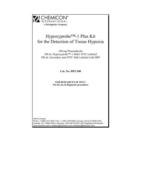
Hypoxyprobe-1 <strong>Plus</strong> <strong>Kit</strong><br />
<strong>for</strong> <strong>the</strong> <strong>Detection</strong> <strong>of</strong> <strong>Tissue</strong> Hypoxia<br />
100 mg Pimonidazole<br />
500 uL Hypoxyprobe-1 Mab1 FITC-Labeled<br />
500 uL Secondary anti-FITC Mab Labeled with HRP<br />
Cat. No. HP2-100<br />
FOR RESEARCH USE ONLY<br />
Not <strong>for</strong> use in diagnostic procedures<br />
USA & Canada<br />
Phone: +1(800) 437-7500 • Fax: +1 (951) 676-9209 • Europe +44 (0) 23 8026 2233<br />
Australia +61 3 9839 2000 • Germany +49-6192-207300 • ISO Registered Worldwide<br />
www.chemicon.com • custserv@chemicon.com • techserv@chemicon.com
This page left blank intentionally.<br />
1
Introduction<br />
Oxygen gradients exist in normal and tumor tissue. These gradients affect gene<br />
expression and are important in normal and pathological conditions. Until<br />
recently it was difficult to measure <strong>the</strong>se gradients at <strong>the</strong> cellular level; however,<br />
with <strong>the</strong> development <strong>of</strong> 2-nitroimidazole hypoxia markers, it is now possible to<br />
accurately measure oxygen gradient at <strong>the</strong> cellular level. There are a number <strong>of</strong><br />
ways that hypoxia markers can be detected. The immunohistochemical<br />
technique is particularly attractive because oxygen gradients can be visualized<br />
and compared with underlying hierarchical structures in tissues and with gene<br />
expression on a cell-by-cell basis.<br />
This insert describes <strong>the</strong> Hypoxyprobe system and its application to studies <strong>of</strong><br />
tissue hypoxia under both normal and pathological conditions. The hypoxia<br />
marker that has received most attention in <strong>the</strong> Hypoxyprobe system <strong>of</strong><br />
markers is Hypoxyprobe-1, which is also known as pimonidazole<br />
hydrochloride.<br />
Hypoxyprobe-1 (pimonidazole hydrochloride)<br />
The kit contains reagents <strong>for</strong> an improved two-step immunohistochemical<br />
procedure <strong>for</strong> detecting hypoxia in rodent tissues using a primary fluorescein<br />
(FITC)-conjugated mouse monoclonal antibody (Mab) directed against<br />
pimonidazole protein adducts and a secondary mouse anti-FITC Mab conjugated<br />
to horseradish peroxidase (HRP). These reagents produce slides that are<br />
virtually free <strong>of</strong> <strong>the</strong> non-specific background staining that is frequently observed<br />
when rodent antibodies are utilized to detect antigens in rodent tissues (see Ref<br />
50: J. Histochem Cytochem 52:837-839, 2004)<br />
1
Background<br />
In 1976 , Varghese et al. reported that 14 C-labelled misonidazole <strong>for</strong>med adducts<br />
in hypoxic cells in vitro and in vivo (1) . It was subsequently found that adducts<br />
<strong>for</strong>m with thiol groups in proteins, peptides and amino acids in a way that all<br />
atoms <strong>of</strong> <strong>the</strong> ring and side-chain <strong>of</strong> <strong>the</strong> 2-nitroimidazole are retained (2-5) .<br />
Hypoxia (pO2 < 10 mmHg) is required <strong>for</strong> binding but binding is not dependent<br />
on <strong>the</strong> presence <strong>of</strong> specialized redox enzymes such as P450 nitroreductases.<br />
Fur<strong>the</strong>rmore, wide variations in NADH and NADPH levels do not change <strong>the</strong><br />
oxygen dependence <strong>of</strong> binding (6, 7) .<br />
Chapman et al. showed that <strong>the</strong> oxygen dependence <strong>of</strong> binding was <strong>for</strong>tuitously<br />
close to that <strong>for</strong> radiation resistance and suggested that misonidazole binding<br />
might be used as a hypoxia marker in solid tumors (8) . The clinical feasibility <strong>of</strong><br />
<strong>the</strong> hypoxia marker idea was demonstrated by means <strong>of</strong> autoradiographic<br />
analyses <strong>of</strong> 3 H-misonidazole binding in a variety <strong>of</strong> human tumors (9) . While <strong>the</strong><br />
3<br />
H-misonidazole approach had limited clinical utility, it spurred <strong>the</strong><br />
development <strong>of</strong> a variety <strong>of</strong> non-invasive assays <strong>for</strong> tissue hypoxia based on 2nitroimidazoles.<br />
These included single photon electron capture tomography,<br />
positron emission tomography, nuclear medicine analysis and magnetic<br />
resonance spectroscopy <strong>of</strong> suitably labeled 2-nitroimidazole analogues (<strong>for</strong><br />
review see ref. (10) ).<br />
During 19 F MRS investigations <strong>of</strong> tumor hypoxia with <strong>the</strong> hexafluorinated 2nitroimidazole,<br />
CCI-103F, it became clear that an histological assessment <strong>of</strong><br />
hypoxia would be a useful complement to non-invasive assays (11-13) . This led to<br />
<strong>the</strong> invention <strong>of</strong> <strong>the</strong> immunochemical hypoxia marker technique based on<br />
monoclonal and polyclonal antibodies raised against protein adducts <strong>of</strong><br />
reductively activated 2-nitroimidazoles (14, 15) . Preclinical testing <strong>of</strong> immunochemical<br />
reagents in spontaneous canine tumors showed that immunochemical<br />
hypoxia markers would be useful in <strong>the</strong>ir own right (16-21) . In addition to<br />
providing a quantitative measure <strong>of</strong> hypoxia, immunohistochemical markers<br />
provide insights into microregional relationships between hypoxia and factors<br />
such as necrosis, proliferation, differentiation, apoptosis, and oxygen regulated<br />
protein expression. A variety <strong>of</strong> immunochemical hypoxia markers have now<br />
been used in clinical (22-27) and preclinical studies (16, 28-33) <strong>of</strong> such relationships.<br />
An example <strong>of</strong> <strong>the</strong> unique value <strong>of</strong> <strong>the</strong> immunohistochemical marker approach<br />
is <strong>the</strong> observation that nei<strong>the</strong>r metallothionein nor vascular endo<strong>the</strong>lial growth<br />
factor are expressed in <strong>the</strong> majority <strong>of</strong> hypoxic cells in human squamous cell<br />
carcinomas (24, 25) even though in vitro studies would have predicted o<strong>the</strong>rwise<br />
(34, 35)<br />
.<br />
With respect to quantifying hypoxia by <strong>the</strong> immunochemical technique, image<br />
analysis (24-27) or flow cytometric analysis (36, 37) appear most promising.<br />
Preclinical studies <strong>of</strong> sampling error showed that stratification <strong>of</strong> patients is<br />
2
feasible with <strong>the</strong> immunohistochemical approach if 3-4 biopsies are obtained<br />
from geographically separate regions <strong>of</strong> each tumor. Precision can be increased<br />
by increasing <strong>the</strong> number <strong>of</strong> sections analyzed per biopsy from 1 to 3 but<br />
analysis <strong>of</strong> multiple biopsies is <strong>the</strong> most important factor. Interestingly, <strong>the</strong><br />
accuracy <strong>of</strong> <strong>the</strong> immunochemical analysis increases as <strong>the</strong> amount <strong>of</strong> hypoxia<br />
decreases (19,21) .<br />
Protein adducts <strong>of</strong> reductively-activated pimonidazole are effective immunogens<br />
<strong>for</strong> <strong>the</strong> production <strong>of</strong> both polyclonal and monoclonal antibodies. The antibodies<br />
have been used <strong>for</strong> immunoperoxidase analysis <strong>of</strong> <strong>for</strong>malin-fixed, paraffinembedded<br />
sections (6, 16, 23-26, 32, 40) ; <strong>for</strong> immun<strong>of</strong>luorescence analysis <strong>of</strong> frozen<br />
fixed sections (27, 41-43) ; and, <strong>for</strong> flow cytometry with directly labeled or<br />
secondary fluorescent antibodies (36) .The antibodies have also been used in<br />
enzyme linked immunosorbent assays (6, 16, 40) . One final attractive feature <strong>of</strong><br />
pimonidazole is <strong>the</strong> fact that pimonidazole adducts in vivo are long-lived (16) .<br />
This provides flexibility in <strong>the</strong> timing <strong>of</strong> biopsy taking which is an advantage in<br />
a clinical setting. In summary Hypoxyprobe-1 and associated antibodies <strong>for</strong>m<br />
a very attractive basis on which to develop a low tech, low cost kit <strong>for</strong><br />
measuring normal and tumor tissue hypoxia.<br />
Application<br />
The rationale <strong>for</strong> developing pimonidazole hydrochloride (Hypoxyprobe-1) as<br />
a hypoxia marker <strong>for</strong> experimental and clinical use was based on its chemical<br />
stability, water solubility, wide tissue distribution and, in <strong>the</strong> case <strong>of</strong> clinical<br />
studies, <strong>the</strong> fact that human toxicity data were available from earlier<br />
radiosensitizer trials. This facilitated early clinical application and<br />
Hypoxyprobe kits have now been used in many experimental studies and<br />
clinical trials worldwide. Solid Hypoxyprobe-1 is very stable being<br />
unchanged after storage <strong>for</strong> 2 years at room temperature in subdued light. Saline<br />
solutions <strong>of</strong> Hypoxyprobe-1 used <strong>for</strong> clinical studies (34 millimolar in 0.9%<br />
saline, pH 3.9 ± 0.1) are extremely stable being unchanged after 1.5 years at 4 o C<br />
in subdued light as determined by high per<strong>for</strong>mance liquid chromatography and<br />
ultraviolet spectroscopy. In addition to chemical stability, Hypoxyprobe-1 has<br />
high water solubility (400 millimolar; 116 grams per 1000 mL) that facilitates<br />
intravenous marker infusion and produces a short plasma half-life <strong>of</strong> 5.1 ± 0.8<br />
hours.<br />
In spite <strong>of</strong> <strong>the</strong> water solubility <strong>of</strong> its hydrochloride salt (pKa 8.7), pimonidazole<br />
itself has an octanol-water partition coefficient <strong>of</strong> 8.5 (38) and diffuses readily<br />
into tumors and normal tissues including brain (39) . Consistent with a large, 155<br />
liter volume <strong>of</strong> distribution, pimonidazole concentrates approximately 3 fold<br />
above plasma levels in tumors and normal tissues (39) <strong>the</strong>reby increasing <strong>the</strong><br />
sensitivity <strong>of</strong> hypoxia marking. At <strong>the</strong> Hypoxyprobe-1 dose <strong>of</strong> 0.5 g/m 2 used<br />
3
in hypoxia marking, pimonidazole causes nei<strong>the</strong>r central nervous system toxicity<br />
nor sensation (e.g., flushing) (39). .Central nervous system toxicity was <strong>of</strong><br />
particular interest because this was <strong>the</strong> dose limiting toxicity <strong>for</strong><br />
Hypoxyprobe-1 at <strong>the</strong> higher, multiple doses used in radiosensitizer trials. In<br />
addition to <strong>the</strong> absence <strong>of</strong> central nervous system effects, <strong>the</strong> overall procedure<br />
from Hypoxyprobe-1 infusion to tumor biopsy is well-tolerated in both<br />
inpatient and outpatient settings.<br />
Currently, rodent (mouse and rat) animal models are frequently used to study<br />
hypoxia in normal and tumor tissues, including human-to-rodent<br />
xenotransplantation experiments. Because <strong>the</strong> Hypoxyprobe-1 Mab1, which<br />
recognizes pimonidazole adducts in hypoxic tissue proteins, is <strong>of</strong> mouse origin,<br />
<strong>the</strong> presence <strong>of</strong> normal mouse or highly homologous rat immunoglobulins<br />
makes it difficult <strong>for</strong> <strong>the</strong> secondary antibody reagent to selectively bind to<br />
Mab1. There<strong>for</strong>e, even when special blocking procedures are employed, <strong>the</strong><br />
immunostaining <strong>of</strong> rodent tissues with Mab1 may result in a strong non-specific<br />
background staining that can obscure <strong>the</strong> specific binding pattern <strong>of</strong> this primary<br />
antibody. The Hypoxyprobe-1 <strong>Plus</strong> <strong>Kit</strong> <strong>for</strong> <strong>the</strong> <strong>Detection</strong> <strong>of</strong> <strong>Tissue</strong> Hypoxia<br />
was developed to address this obstacle and ensure fast (two-step) and accurate<br />
detection <strong>of</strong> hypoxia gradients in mouse and rat animal models. In this detection<br />
system, <strong>the</strong> primary monoclonal antibody Mab1 is directly labeled with FITC,<br />
while <strong>the</strong> secondary Mab (directed against FITC) is labeled with HRP. Since <strong>the</strong><br />
secondary Mab does not cross-react with antigen epitopes present in rodent<br />
tissues, no non-specific background staining is generated using this set <strong>of</strong><br />
reagents.<br />
For Research Use Only; Not <strong>for</strong> use in diagnostic procedures<br />
4
<strong>Kit</strong> Components<br />
1. Hypoxyprobe-1: (Part No.: 90203) One vial containing 100 mg <strong>of</strong><br />
Pimonidazole Hydrochloride<br />
2. Hypoxyprobe-1 Mab1: (Part No.: 90531) One vial containing 500 µL <strong>of</strong><br />
purified Mab1 (clone 4.3.11.3) conjugated with FITC.<br />
3. Anti-FITC secondary Mab: (Part No.: 90532) One vial containing 500 µL<br />
<strong>of</strong> purified Mab against FITC (clone 5D6.2) conjugated with HRP.<br />
Storage<br />
The FITC-labeled Hypoxyprobe 1-Mab1 and <strong>the</strong> HRP-labeled Secondary Anti-<br />
FITC monoclonal antibody can be stored at 2-8°C until <strong>the</strong> expiration date<br />
indicated on <strong>the</strong> label. Keep dark.<br />
Hypoxyprobe-1 (Pimonidazole Hydrochloride) has great chemical stability in<br />
solid <strong>for</strong>m and in aqueous solutions and requires no stabilizer. Solid<br />
Hypoxyprobe-1 has been stored <strong>for</strong> two years at room temperature in subdued<br />
light without detectable degradation as assessed by UV and HPLC analyses.<br />
Hypoxyprobe-1 solutions in 0.9% saline have been stored at a concentration<br />
<strong>of</strong> 10 g/liter (34 millimolar) at 4°C in subdued light <strong>for</strong> 1.5 years without<br />
detectable degradation (UV and HPLC analyses). When exposed to laboratory<br />
light, Hypoxyprobe-1 slowly turns yellow.<br />
Assay Instructions<br />
1. Although doses <strong>of</strong> Hypoxyprobe-1 up to 400 mg/kg have been used<br />
without measurable toxicity in mice (36) , a dose <strong>of</strong> 60 mg/kg body weight is<br />
routinely used in studies <strong>of</strong> tissue hypoxia in rodents. The high water<br />
solubility <strong>of</strong> Hypoxyprobe-1 permits small volume injections to be made<br />
which is convenient <strong>for</strong> studies with small animals. Intravenous injection or<br />
intraperitoneal injection can be used. The plasma half-life <strong>of</strong><br />
Hypoxyprobe-1 is 0.5 hours in C3H/He mice. Hypoxyprobe-1 is<br />
distributed to all tissues in <strong>the</strong> body including brain but binds only to cells<br />
that have oxygen concentrations less than 14 micromolar, which is<br />
equivalent to a pO2 <strong>of</strong> 10 mm Hg at 37 o C. Tumors and normal liver, kidney<br />
and skin have cells at, or below, this pO2. For dogs, whole body doses <strong>of</strong><br />
0.28 gm/m 2 are recommended (16) . The plasma half-life <strong>for</strong> Hypoxyprobe-<br />
1 in dogs is 1.5 ± 1.0 hours.<br />
5
In addition to animal studies, Hypoxyprobe kits can be used <strong>for</strong> cells in<br />
tissue culture (6, 44) . Typically, cell suspensions are incubated under hypoxia<br />
<strong>for</strong> 1 to 2 hours in <strong>the</strong> presence <strong>of</strong> 100 to 200 micromolar Hypoxyprobe-<br />
1. The cells are harvested by cytospin, fixed and immunostained with an<br />
antibody <strong>for</strong> pimonidazole adducts. Sufficient concentrations <strong>of</strong><br />
pimonidazole adducts are <strong>for</strong>med on <strong>the</strong> surface <strong>of</strong> cells to elicit a response<br />
to complement or activated cytotoxic lymphocytes (44) . Hypoxyprobe-1<br />
<strong>Plus</strong> <strong>Kit</strong> has enough hypoxia marker to administer to 67 (25 gram) mice or<br />
6-7 (250 gram) rats.<br />
2. The primary antibody reagent is a conjugate consisting <strong>of</strong> Hypoxyprobe-<br />
1 MAb1 (mouse IgG1) covalently bound to FITC and is intended to detect<br />
protein adducts <strong>of</strong> Hypoxyprobe-1 in hypoxic cells. The conjugate is<br />
provided as a purified preparation at a concentration <strong>of</strong> 1 mg/mL. Fifteen to<br />
ninety minutes after <strong>the</strong> injection <strong>of</strong> Hypoxyprobe-1, tumor or normal<br />
tissue is excised or biopsied. The tissue <strong>of</strong> interest is <strong>the</strong>n studied as frozen<br />
sections, <strong>for</strong>malin-fixed paraffin-embedded sections, or as disaggregated<br />
tissues in flow cytometry assays. In <strong>the</strong> case <strong>of</strong> <strong>for</strong>malin-fixed, paraffinembedded<br />
tissue sections, 100 µL <strong>of</strong> a 1:50 dilution <strong>of</strong> <strong>the</strong> conjugate is<br />
added to each tissue section. Hypoxyprobe-1 <strong>Plus</strong> <strong>Kit</strong> has enough<br />
primary conjugate to analyze 250 tissue sections at a 1:50 dilution. Optimal<br />
working dilutions must be determined by <strong>the</strong> end user.<br />
3. The secondary antibody reagent is a conjugate consisting <strong>of</strong> a mouse anti-<br />
FITC covalently bound to HRP and is intended to detect Mab1-FITC<br />
stained structures in target slides. The conjugate is provided as a purified<br />
preparation at a concentration <strong>of</strong> 1 mg/mL. In <strong>the</strong> case <strong>of</strong> <strong>for</strong>malin-fixed,<br />
paraffin-embedded tissue sections, 100 µL <strong>of</strong> a 1:50 dilution <strong>of</strong> <strong>the</strong><br />
conjugate is added to each tissue section. We recommend utilizing standard<br />
colorimetric systems used in visualizing HRP-stained tissue sections in<br />
routine IHC protocols. Hypoxyprobe-1 <strong>Plus</strong> <strong>Kit</strong> has enough secondary<br />
conjugate to analyze 250 tissue sections at a 1:50 dilution. Optimal<br />
working dilutions must be determined by <strong>the</strong> end user.<br />
6
Peroxidase Immunohistochemistry Technique<br />
Recommended procedure <strong>for</strong> immunostaining Hypoxyprobe-1 adducts in<br />
<strong>for</strong>malin-fixed, paraffin-embedded tissues from a variety <strong>of</strong> animal species<br />
including mice and rats. O<strong>the</strong>r tissue fixation methods will require<br />
optimization <strong>of</strong> <strong>the</strong> suggested protocol.<br />
Improved immunohistochemical method <strong>for</strong> detecting hypoxia gradients in<br />
mouse tissues and tumors. Samoszuk MK, Walter J, Mechetner E. J. Histochem.<br />
Cytochem. 52(6):837-839, 2004.<br />
Step<br />
1<br />
2<br />
3<br />
4<br />
5<br />
6<br />
7<br />
8<br />
Procedure<br />
Heat Fix tissue<br />
slides<br />
Deparaffinize<br />
tissue<br />
Deparaffinize<br />
tissue<br />
Deparaffinize<br />
tissue<br />
Clear slides <strong>of</strong><br />
Xylene<br />
Clear slides <strong>of</strong><br />
Xylene<br />
Begin hydrating<br />
tissue<br />
Continue to<br />
hydrate<br />
Time,<br />
min.<br />
Temp.<br />
7<br />
Reagents<br />
60 60 o C None 1<br />
3 RT Xylene 2<br />
3 RT Xylene<br />
3 RT Xylene<br />
1 RT 100% Ethanol<br />
1 RT 100% Ethanol<br />
1 RT 70% Aqueous ethanol<br />
1 RT 30% Aqueous ethanol<br />
9 Hydrate tissue 1 RT Deionized water<br />
10 Hydrate tissue 1 RT Deionized water<br />
11<br />
Transfer slides to<br />
rinse buffer<br />
2 RT 1x TBS + Tween 20 3<br />
Notes
Step<br />
12<br />
Procedure<br />
Quench tissue<br />
peroxidase<br />
Time,<br />
min.<br />
5 RT<br />
Temp.<br />
13 Antigen retrieval 20 Steamer<br />
14<br />
15<br />
16<br />
Transfer slides to<br />
rinse buffer<br />
Block non-specific<br />
binding<br />
Incubate in Primary<br />
conjugate<br />
8<br />
Reagents<br />
3% H2O2 in distilled<br />
water<br />
10mM Citrate, pH<br />
6.0<br />
2 RT 1x TBS + Tween 20 3<br />
5 RT Blocking reagent 7<br />
30 RT<br />
Hypoxyprobe-1<br />
MAb1 conjugated<br />
with FITC (1:50)<br />
17 Wash in rinse buffer 2 RT 1x TBS + Tween 20 3<br />
18<br />
Incubate in<br />
Secondary conjugate<br />
30 RT<br />
Anti-FITC Mab<br />
conjugated with<br />
HRP (1:50)<br />
19 Wash in rinse buffer 2 RT 1x TBS + Tween 20 3<br />
20<br />
Peroxidase<br />
chromogen<br />
10 RT DAB 10<br />
21 Stop DAB reaction 1 RT Distilled water<br />
22 Wash in rinse buffer 1 RT 1x TBS + Tween 20 3<br />
23 Counterstaining 1 RT Hematoxylin 11<br />
24 Wash in rinse buffer 1 RT 1x TBS + Tween 20 3<br />
25<br />
Dehydration &<br />
Clearing<br />
See<br />
notes RT<br />
100% alcohol and<br />
Xylene<br />
26 Coverslip N/a RT Permount 13<br />
Notes<br />
4<br />
8<br />
9<br />
12
Technical Notes<br />
1. This step may be per<strong>for</strong>med in microwave oven on high <strong>for</strong> 2 minutes. If<br />
<strong>the</strong> sections are still wet, <strong>the</strong> water will turn to steam and separate <strong>the</strong> tissue<br />
from slides. Caution, glass can get very hot.<br />
2. Per xylene incubation use clean xylene. Clear-Rite 3 is a non-toxic<br />
alternative to xylene and is available from Richard-Allan Scientific,<br />
Kalamazoo, MI (Cat. No. 6901). If using this alternative, increase <strong>the</strong><br />
incubation periods accordingly.<br />
3. 20x TBS is Tris buffered saline, 10 mM (Chemicon Cat. No. 20845). All<br />
rinses were done with 1x TBS working solution.<br />
4. Make sure to rinse <strong>of</strong>f <strong>the</strong> 3% H2O2 since it can interfere with results. Store<br />
brand hydrogen peroxide may be utilized.<br />
5. Antigen retrieval solution is 10x Citrate Buffer (Chemicon Cat. No. 21545)<br />
used at a 1x working solution.<br />
6. HIER method is at <strong>the</strong> end users discretion.<br />
7. Blocking reagent (Chemicon Cat. No. 20773). 1% BSA or 1% normal<br />
mouse serum in TBS may also be used.<br />
8. The primary antibody reagent is a conjugate between Hypoxyprobe-1<br />
MAb1 (mouse IgG1) and FITC. The conjugate is provided as a purified<br />
preparation at a concentration <strong>of</strong> 1 mg/ml. Dilute <strong>the</strong> primary conjugate<br />
solution 1:50 in Antibody Diluent (Chemicon Cat. No. 21544). Typically,<br />
100 µL <strong>of</strong> diluted primary conjugate solution is applied to each tissue<br />
section.<br />
9. The secondary antibody reagent is a conjugate between a monoclonal<br />
antibody against FITC and HRP provided as a purified preparation at a<br />
concentration <strong>of</strong> 1 mg/mL. Dilute <strong>the</strong> secondary antibody solution 1:50 in<br />
Antibody Diluent (Chemicon Cat. No. 21544). Typically, 100 µL <strong>of</strong><br />
diluted secondary conjugate is added to each tissue section.<br />
10. Liquid 3,3'-diaminobenzidine reagent (DAB). DAB Chromogen-A<br />
(Chemicon Cat. No. 71895) and DAB Chromogen-B (Chemicon Cat. No.<br />
71896) mix at 1:25 ratio respectively.<br />
11. Hematoxylin counter staining reagent (Chemicon Cat. No. 20844).<br />
12. Use clean and fresh solutions to incubate slides <strong>for</strong> 1 minute in 4 changes <strong>of</strong><br />
100% alcohol and <strong>the</strong>n to three changes <strong>of</strong> clean xylene.<br />
13. Permount solution is available from Fisher Scientific (Cat. No. SP15-500).<br />
9
Frequently Asked Questions<br />
Q: What is <strong>the</strong> best solvent <strong>for</strong> Hypoxyprobe-1?<br />
A: Hypoxyprobe-1 is <strong>the</strong> hydrochloride salt <strong>of</strong> a weak base and, as such, is<br />
very soluble in aqueous solutions including neutral buffered saline (116<br />
mg/mL or 400 millimolar).<br />
Q. What dose <strong>of</strong> Hypoxyprobe-1 should be used <strong>for</strong> hypoxia marking<br />
experiments?<br />
A: For small animals <strong>of</strong> uni<strong>for</strong>m size such as laboratory rats and mice, a dose<br />
<strong>of</strong> Hypoxprobe-1 <strong>of</strong> 60 mg/kg body weight is recommended. Doses<br />
ranging from 30 mg/kg (47) to 400 mg/kg (36) have been used in mice and rats<br />
without toxicity or altered oxygen levels due to blood flow effects.<br />
Never<strong>the</strong>less, significant effects on blood flow at doses above 100 mg/kg <strong>of</strong><br />
Hypoxyprobe-1 have been observed in tumors implanted in <strong>the</strong> hind legs<br />
<strong>of</strong> mice. Caution must be taken, <strong>the</strong>re<strong>for</strong>e, when doses > 100 mg/kg are used.<br />
For larger animals with non-uni<strong>for</strong>m body size, <strong>the</strong> dose is calculated on <strong>the</strong><br />
basis <strong>of</strong> surface area. For humans, <strong>the</strong> recommended dose is 0.5 gm/m2 (23)<br />
while <strong>for</strong> dogs a dose <strong>of</strong> 0.28 gm/m2 has been used (16) .<br />
Q. Does Hypoxyprobe-1 penetrate hypoxic brain and brain tumor<br />
tissue?<br />
A: Although Hypoxyprobe-1 is water soluble, its corresponding free base<br />
has an octanol water coefficient <strong>of</strong> 8.5 and, as a result, <strong>the</strong> marker freely<br />
penetrates into both brain and brain tumor tissue (48, 49) .<br />
10
Q. Is Hypoxyprobe-1 <strong>the</strong> best probe <strong>for</strong> detecting hypoxia in vivo?<br />
A: Although o<strong>the</strong>r markers are available <strong>for</strong> detecting hypoxia in vivo,<br />
Hypoxyprobe-1 has some real advantages. Foremost is its high solubility in<br />
aqueous solution (400 millimolar; 116 mg/mL <strong>of</strong> saline), which allows <strong>the</strong><br />
marker to be administered to rodents as small volume (= 0.1 mL) injections <strong>of</strong><br />
saline ei<strong>the</strong>r intraperitoneally or intravenously. O<strong>the</strong>r markers such as <strong>the</strong><br />
hexafluorinated CCI-103F have aqueous solubilities <strong>of</strong> 10 millimolar or less.<br />
Although Hypoxyprobe-1, <strong>the</strong> hydrochloride salt <strong>of</strong> pimonidazole, is very<br />
water soluble, pimonidazole itself has an octanol-water partition coefficient <strong>of</strong><br />
8.5 and penetrates all tissues including brain.<br />
Ano<strong>the</strong>r advantage is that pimonidazole binding can be detected by<br />
immun<strong>of</strong>luorescence in frozen fixed tissue sections; by immunoperoxidase in<br />
<strong>for</strong>malin-fixed paraffin-embedded tissue sections; by ELISA or by flow<br />
cytometry.<br />
Q. Can <strong>the</strong> monoclonal antibody to Hypoxyprobe-1 adducts be used on<br />
mouse or rat tissues?<br />
A: Yes. The described procedure results in a clean IHC staining that is free <strong>of</strong><br />
non-specific background caused by mouse or rat immunoglobulins<br />
impregnating <strong>the</strong> surrounding tissue (50) .<br />
11
References<br />
1. Varghese, A. J., Gulyas, S., and Mohindra, J. K. (1976). Hypoxia-dependent<br />
reduction <strong>of</strong> 1-(2-nitro-1-imidazolyl)-3-methoxy-2- propanol by Chinese hamster<br />
ovary cells and KHT tumor cells in vitro and in vivo, Cancer Res. 36:3761-3765.<br />
2. Chacon, E., Morrow, C. J., Leon, A. A., Born, J. L., and Smith, B. R. (1988).<br />
Regioselective <strong>for</strong>mation <strong>of</strong> a misonidazole-glutathione conjugate as a function <strong>of</strong><br />
pH during chemical reduction., Biochem. Pharmacol. 37:361-363.<br />
3. Raleigh, J. A., Franko, A. J., Koch, C. J., and Born, J. L. (1985). Binding <strong>of</strong><br />
misonidazole to hypoxic cells in monolayer and spheroid culture: evidence that a<br />
side-chain label is bound as efficiently as a ring label. Br. J. Cancer. 51:229-235.<br />
4. Raleigh, J. A. and Koch, C. J. (1990). Importance <strong>of</strong> thiols in <strong>the</strong> reductive<br />
binding <strong>of</strong> 2-nitroimidazoles to macromolecules. Biochem Pharmacol. 40:2457-<br />
2464.<br />
5. Varghese, A. J. (1983). Glutathione conjugates <strong>of</strong> misonidazole. Biochem Biophys<br />
Res Commun. 112:1013-1020.<br />
6. Arteel, G. E., Thurman, R. G., Yates, J. M., and Raleigh, J. A. (1995). Evidence that<br />
hypoxia markers detect oxygen gradients in liver: pimonidazole and retrograde<br />
perfusion <strong>of</strong> rat liver. Br J Cancer. 72:889-895.<br />
7. Arteel, G. E., Thurman, R. G., and Raleigh, J. A. (1988). Reductive metabolism <strong>of</strong><br />
<strong>the</strong> hypoxia marker pimonidazole is regulated by oxygen tension independent <strong>of</strong> <strong>the</strong><br />
pyridine nucleotide redox state. Eur. J. Biochem. 253:743-750.<br />
8. Chapman, J. D., Franko, A. J., and Sharplin, J. A (1981). marker <strong>for</strong> hypoxic cells<br />
in tumours with potential clinical applicability. Br. J. Cancer. 43:546-550.<br />
9. Urtasun, R. C., Koch, C. J., Franko, A. J., Raleigh, J. A., and Chapman, J. D.<br />
(1986). A novel technique <strong>for</strong> measuring human tissue pO2 at <strong>the</strong> cellular level. Br.<br />
J. Cancer. 54:453-457.<br />
10. Raleigh, J. A., Dewhirst, M. W., and Thrall, D. E. Measuring tumor hypoxia., Sem.<br />
Radiat. Oncol. 6:37-45, 1996.<br />
11. Jin, G. Y., Li, S. J., Moulder, J. E., and Raleigh, J. A. Dynamic measurements <strong>of</strong><br />
hexafluoromisonidazole (CCI-103F) retention in mouse tumours by 1H/19F<br />
magnetic resonance spectroscopy, Int. J. Radiat. Biol. 58: 1025-1034, 1990.<br />
12. Raleigh, J. A., Franko, A. J., Treiber, E. O., Lunt, J. A., and Allen, P. S. Covalent<br />
binding <strong>of</strong> a fluorinated 2-nitroimidazole to EMT-6 tumors in BALB/C mice:<br />
<strong>Detection</strong> by F-19 nuclear magnetic resonance at 2.35T, Int. J. Radiat. Oncol. Biol.<br />
Phys. 12: 1243-1245, 1986.<br />
12
13. Raleigh, J., Franko, A., Kelly, D., Trimble, L., and Allen, P. (1991). Development<br />
<strong>of</strong> an in vivo 19F magnetic resonance method <strong>for</strong> measuring oxygen deficiency in<br />
tumors. Magn. Res. Med. 22:451-466.<br />
14. Raleigh, J. A., Miller, G. G., Franko, A. J., Koch, C. J., Fuciarelli, A. F., and<br />
Kelly, D. A. (1987). Fluorescence immunohistochemical detection <strong>of</strong> hypoxic cells<br />
in spheroids and tumours. Br J Cancer 56:395-400.<br />
15. Raleigh, J. A., Miller, G. G., Franko, A. J., and Chapman, J. D. Immunochemical<br />
detection <strong>of</strong> hypoxia in normal and tumor tissue. USA patent 5,086,068, February,<br />
1992.<br />
16. Azuma, C., Raleigh, J. A., and Thrall, D. E. (1997). Longevity <strong>of</strong> pimonidazole<br />
adducts in spontaneous canine tumors as an estimate <strong>of</strong> hypoxic cell lifetime.<br />
Radiat. Res. 148:35-42.<br />
17. Cline, J. M., Thrall, D. E., Page, R. L., Franko, A. J., and Raleigh, J. A. (1990).<br />
Immunohistochemical detection <strong>of</strong> a hypoxia marker in spontaneous canine<br />
tumours. Br. J. Cancer 62:925-931.<br />
18. Cline, J. M., Thrall, D. E., Rosner, G. L., and Raleigh, J. A. (1994). Distribution<br />
<strong>of</strong> <strong>the</strong> hypoxia marker CCI-103F in canine tumors. Int J Radiat Oncol Biol Phys.<br />
28:921-933.<br />
19. Cline, J., Rosner, G., Raleigh, J., and Thrall, D. (1997). Quantification <strong>of</strong> CCI-<br />
103F labeling heterogeneity in canine solid tumors. Int. J. Radiat. Oncol. Biol.<br />
Phys. 37:655-662.<br />
20. Thrall, D. E., McEntee, M. C., Cline, J. M., and Raleigh, J. A. (1994). ELISA<br />
quantification <strong>of</strong> CCI-103F binding in canine tumors prior to and during<br />
irradiation. Int. J. Radiat. Oncol. Biol. Phys. 28:649-659.<br />
21. Thrall, D. E., Rosner, G. L., Azuma, C., McEntee, M. C., and Raleigh, J. A.<br />
(1997). Hypoxia marker labeling in tumor biopsies: quantification <strong>of</strong> labeling<br />
variation and criteria <strong>for</strong> biopsy sectioning. Radio<strong>the</strong>r Oncol. 44:171-176.<br />
22. Evans, S. M., Hahn, S., Pook, D. R., Jenkins, W. T., Chalian, A. A., Zhang, P.,<br />
Stevens, C., Weber, R., Weinstein, G., Benjamin, I., Mirza, N., Morgan, M.,<br />
Rubin, S., McKenna, W. G., Lord, E. M., and Koch, C. J. (2000). <strong>Detection</strong> <strong>of</strong><br />
hypoxia in human squamous cell carcinoma by EF5 binding. Cancer Res. 60:<br />
2018-2024.<br />
23. Kennedy, A. S., Raleigh, J. A., Perez, G. M., Calkins, D. P., Thrall, D. E.,<br />
Novotny, D. B., and Varia, M. A. (1997). Proliferation and hypoxia in human<br />
squamous cell carcinoma <strong>of</strong> <strong>the</strong> cervix: first report <strong>of</strong> combined<br />
immunohistochemical assays. Int J Radiat Oncol Biol Phys. 37:897-905..<br />
13
24. Raleigh, J. A., Calkins-Adams, D. P., Rinker, L. H., Ballenger, C. A., Weissler,<br />
M. C., Fowler, W. C., Jr., Novotny, D. B., and Varia, M. A. (1998). Hypoxia and<br />
vascular endo<strong>the</strong>lial growth factor expression in human squamous cell carcinomas<br />
using pimonidazole as a hypoxia marker. Cancer Res. 58:3765-3768.<br />
25. Raleigh, J., Chou, S.-C., Calkins-Adams, D., Ballenger, C., Rinker, L., Novotny,<br />
D., and Varia, M. (2000). A clinical study <strong>of</strong> hypoxia and metallothionein protein<br />
expression in squamous cell carcinomas. Cl. Cancer Res. 6:855-862.<br />
26. Varia, M. A., Calkins-Adams, D. P., Rinker, L. H., Kennedy, A. S., Novotny, D.<br />
B., Fowler, W. C., Jr., and Raleigh, J. A. (1998). Pimonidazole : a novel hypoxia<br />
marker <strong>for</strong> complementary study <strong>of</strong> tumor hypoxia and cell proliferation in<br />
cervical carcinoma. Gynecol. Oncol. 71:270-277.<br />
27. Wijffels, K., Kaanders, J., Rijken, P., Bussink, J., Van den Hoogen, F., Marres,<br />
H., de Wilde, P., Raleigh, J., and Van der Kogel, A. (2000). Vascular architecture<br />
and hypoxic pr<strong>of</strong>iles in human head and neck squamous cell carcinomas. Br. J.<br />
Cancer. 83:674-683.<br />
28. Carmeliet, P., Dor, Y., Herbert, J. M., Fukumura, D., Brusselmans, K.,<br />
Dewerchin, M., Neeman, M., Bono, F., Abramovitch, R., Maxwell, P., Koch, C.<br />
J., Ratcliffe, P., Moons, L., Jain, R. K., Collen, D., and Keshet, E. (1998). Role <strong>of</strong><br />
HIF-1alpha in hypoxia-mediated apoptosis, cell proliferation and tumour<br />
angiogenesis. Nature 394:485-490.<br />
29. Hodgkiss, R. J. (1998). Use <strong>of</strong> 2-nitroimidazoles as bioreductive markers <strong>for</strong><br />
tumour hypoxia. Anticancer Drug Des. 13:687-702.<br />
30. Koch, C. J., Chasan, J. E., Jenkins, W. T., Chan, C. Y., Laughlin, K. M., and<br />
Evans, S. M. (1998). Co-localization <strong>of</strong> hypoxia and apoptosis in irradiated and<br />
untreated HCT116 human colon carcinoma xenografts. Adv Exp Med Biol. 454:<br />
611-618..<br />
31. Raleigh, J. A., Zeman, E. M., Calkins, D. P., McEntee, M. C., and Thrall, D. E.<br />
(1995). Distribution <strong>of</strong> hypoxia and proliferation associated markers in spontaneous<br />
canine tumors. Acta Oncol. 34:345-349.<br />
32. Raleigh, J. A., Chou, S.-C., Tables, L., Suchindran, S., Varia, M. A., and Horsman,<br />
M. R. (1998). Relationship <strong>of</strong> hypoxia to metallothionein expression in murine<br />
tumors. Int. J. Radiat. Oncol. Biol. Phys. 42:727-730.<br />
33. Waleh, N. S., Brody, M. D., Knapp, M. A., Mendonca, H. L., Lord, E. M., Koch, C.<br />
J., Laderoute, K. R., and Su<strong>the</strong>rland, R. M. (1995). Mapping <strong>of</strong> <strong>the</strong> vascular<br />
endo<strong>the</strong>lial growth factor-producing hypoxic cells in multicellular tumor spheroids<br />
using a hypoxia-specific marker. Cancer Res. 55:6222-6226.<br />
14
34. Leith, J. T. and Michelson, S. (1995). Secretion rates and levels <strong>of</strong> vascular<br />
endo<strong>the</strong>lial growth factor in clone A or HCT-8 human colon tumour cells as a<br />
function <strong>of</strong> oxygen concentration. Cell Prolif. 28:415-430.<br />
35. Murphy, B. J., Andrews, G. K., Bittel, D., Discher, D. J., McCue, J., Green, C. J.,<br />
Yanovsky, M., Giaccia, A., Su<strong>the</strong>rland, R. M., Laderoute, K. R., and Webster, K. A.<br />
(1999). Activation <strong>of</strong> metallothionein gene expression by hypoxia involves metal<br />
response elements and MTF-1. Cancer Res. 59:1315-1322.<br />
36. Durand, R. E. and Raleigh, J. A. (1998). Identification <strong>of</strong> nonproliferating but viable<br />
hypoxic cells in vivo. Cancer Res. 58:3547-3550.<br />
37. Webster, L., Hodgkiss, R. J., and Wilson, G. D. (1995). Simultaneous triple staining<br />
<strong>for</strong> hypoxia, proliferation, and DNA content in murine tumours. Cytometry 21:344-<br />
351.<br />
38. Smi<strong>the</strong>n, C., Clarke, E., Dale, J., Jacobs, R., Wardman, P., Watts, M., and<br />
Woodcock, M. Novel (nitro-1-imidazoyl)-alkanolamines as potential<br />
radiosensitizers with improved <strong>the</strong>rapeutic properties. In: L. Brady (ed.) Radiation<br />
Sensitizers. Their Use in <strong>the</strong> Clinical Management <strong>of</strong> Cancer., pp. 22-32. New York:<br />
Masson, 1980.<br />
39. Saunders, M. I., Anderson, P. J., Bennett, M. H., Dische, S., Minchinton, A. I., and<br />
Strat<strong>for</strong>d, M. R. L. (1984). The clinical testing <strong>of</strong> Ro 03-8799 -- pharmacokinetics,<br />
toxicology, tissue and tumor concentrations. Int. J. Radiat. Oncol. Biol. Phys. 10:<br />
1759-1763.<br />
40. Arteel, G. E., Iimuro, Y., Yin, M., Raleigh, J. A., and Thurman, R. G. (1997).<br />
Chronic enteral ethanol treatment causes hypoxia in rat liver tissue in vivo.<br />
Hepatology 25: 920-926.<br />
41. Bussink, J., Kaanders, J. H., Rijken, P. F., Raleigh, J. A., and Van Der Kogel, A. J.<br />
(2000). Changes in blood perfusion and hypoxia after irradiation <strong>of</strong> a human<br />
squamous cell carcinoma xenograft tumor line. Radiat. Res. 153:398-404.<br />
42. Rijken, P., Bernsen, H., Peters, J., Hodgkiss, R., Raleigh, J., and van der Kogel, A.<br />
(2000). Spatial relationship between hypoxia and <strong>the</strong> (perfused) vascular network in<br />
human glioma xenografts: a quantitative multi-parameter analysis. Int. J. Radiat.<br />
Oncol. Biol. Phys. 48:571-582.<br />
43. Bernsen, H. J., Rijken, P. F., Peters, H., Raleigh, J. A., Jeuken, J. W., Wesseling, P.,<br />
and van der Kogel, A. J. (2000). Hypoxia in a human intracerebral glioma model. J<br />
Neurosurg. 93:449-454.<br />
44. Chou, S. C., Flood, P. M., and Raleigh, J. A. (1996). Marking hypoxic cells <strong>for</strong><br />
complement and cytotoxic T lymphocyte- mediated lysis: using pimonidazole. Br J<br />
Cancer Suppl. 27:S213-S216.<br />
15
45. Ljungkvist,ASE, Bussink, J, Raleigh, JA, Denekemp, J and van der Kogel, AJ.<br />
(2000). Changes in tumor hypoxia measured with a double hypoxic marker<br />
technique. Int. J. Radiat. Oncol. Biol. Phys. 48:1529-1538.<br />
46. Wyk<strong>of</strong>f, CC, Beasley, NJ, Watson, PH, Turner, KJ, Pastorek, J, Sibtain, A, Wilson,<br />
GD, Turley, H, Talks, KL, Maxwell, PH, Pugh, CW, Ratcliffe, PJ and Harris, AL.<br />
(2000). Hypoxia-inducible expression <strong>of</strong> tumor-associated carbonic anhydrases.<br />
Cancer Res. 60:7075-7083.<br />
47. R<strong>of</strong>stad, EK and Maseide, K. (1999). Radiobiological and immunohistochemical<br />
assessment <strong>of</strong> hypoxia in human melanoma xenografts: acute and chronic hypoxia in<br />
individual tumours. Int. J. Radiat. Biol. 75:1377-93.<br />
48. Newman, HFV, Bleehen, NM, Ward, R and Workman, P. (1988). Hypoxic cell<br />
radiosensitizers in <strong>the</strong> treatment <strong>of</strong> high grade gliomas: A new direction using<br />
combined Ro 03-8799 (pimonidazole) and SR 2508 (etanidazole). Int. J. Radiat.<br />
Oncol. Biol. Phys. 15:677-684.<br />
49. Roberts, JT, Bleehen, NM, Walton, MI, and Workman, P. (1986). A clinical phase I<br />
toxicity study <strong>of</strong> Ro 03-8799: plasma, urine, tumour and normal brain<br />
pharmacokinetics. Br. J. Radiology 59:107-116.<br />
50. Samoszuk MK, Walter J, and Mechetner E. (2004). Improved immunohistochemical<br />
method <strong>for</strong> detecting hypoxia gradients in mouse tissues and tumors. J Histochem<br />
Cytochem. 52:837-839.<br />
Warranty<br />
These products are warranted to per<strong>for</strong>m as described in <strong>the</strong>ir labeling and in<br />
CHEMICON ® literature when used in accordance with <strong>the</strong>ir instructions.<br />
THERE ARE NO WARRANTIES, WHICH EXTEND BEYOND THIS<br />
EXPRESSED WARRANTY AND CHEMICON ® DISCLAIMS ANY<br />
IMPLIED WARRANTY OF MERCHANTABILITY OR WARRANTY OF<br />
FITNESS FOR PARTICULAR PURPOSE. CHEMICON ® ’s sole obligation<br />
and purchaser’s exclusive remedy <strong>for</strong> breach <strong>of</strong> this warranty shall be, at <strong>the</strong><br />
option <strong>of</strong> CHEMICON ® , to repair or replace <strong>the</strong> products. In no event shall<br />
CHEMICON ® be liable <strong>for</strong> any proximate, incidental or consequential damages<br />
in connection with <strong>the</strong> products.<br />
©2004: CHEMICON<br />
16<br />
® International, Inc. - By CHEMICON ® International, Inc.<br />
All rights reserved. No part <strong>of</strong> <strong>the</strong>se works may be reproduced in any <strong>for</strong>m<br />
without permissions in writing.
Cat. No. HP2-100<br />
September 2004<br />
Revision B; 41631


