Caribbean Acropora Restoration Guide - The Florida Reef ...
Caribbean Acropora Restoration Guide - The Florida Reef ...
Caribbean Acropora Restoration Guide - The Florida Reef ...

You also want an ePaper? Increase the reach of your titles
YUMPU automatically turns print PDFs into web optimized ePapers that Google loves.
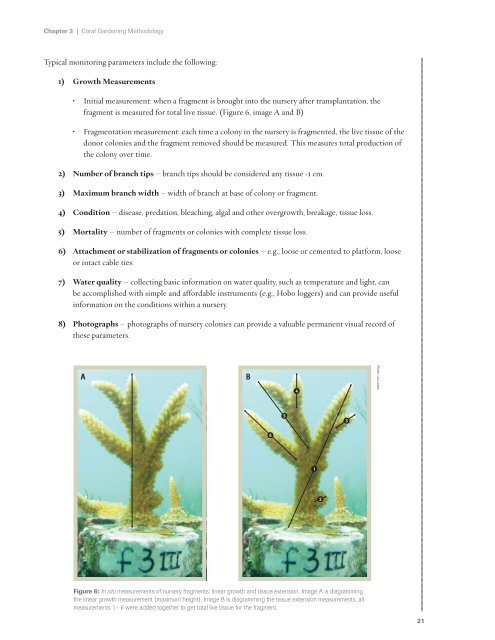
Chapter 3 | Coral Gardening Methodology<br />
Typical monitoring parameters include the following:<br />
1) Growth Measurements<br />
• Initial measurement: when a fragment is brought into the nursery after transplantation, the<br />
fragment is measured for total live tissue. (Figure 6, image A and B)<br />
• Fragmentation measurement: each time a colony in the nursery is fragmented, the live tissue of the<br />
donor colonies and the fragment removed should be measured. This measures total production of<br />
the colony over time.<br />
2) Number of branch tips – branch tips should be considered any tissue ›1 cm.<br />
3) Maximum branch width – width of branch at base of colony or fragment.<br />
4) Condition – disease, predation, bleaching, algal and other overgrowth, breakage, tissue loss.<br />
5) Mortality – number of fragments or colonies with complete tissue loss.<br />
6) Attachment or stabilization of fragments or colonies – e.g., loose or cemented to platform, loose<br />
or intact cable ties.<br />
7) Water quality – collecting basic information on water quality, such as temperature and light, can<br />
be accomplished with simple and affordable instruments (e.g., Hobo loggers) and can provide useful<br />
information on the conditions within a nursery.<br />
8) Photographs – photographs of nursery colonies can provide a valuable permanent visual record of<br />
these parameters.<br />
A<br />
B<br />
4<br />
Photo: Liz Larson<br />
5<br />
3<br />
6<br />
1<br />
2<br />
Figure 6: In situ measurements of nursery fragments; linear growth and tissue extension. Image A is diagramming<br />
the linear growth measurement (maximum height). Image B is diagramming the tissue extension measurements, all<br />
measurements 1– 6 were added together to get total live tissue for the fragment.<br />
21