KingMark Instruction Sheet - Voyant Health
KingMark Instruction Sheet - Voyant Health
KingMark Instruction Sheet - Voyant Health

You also want an ePaper? Increase the reach of your titles
YUMPU automatically turns print PDFs into web optimized ePapers that Google loves.
Read these instructions<br />
before using <strong>KingMark</strong>.<br />
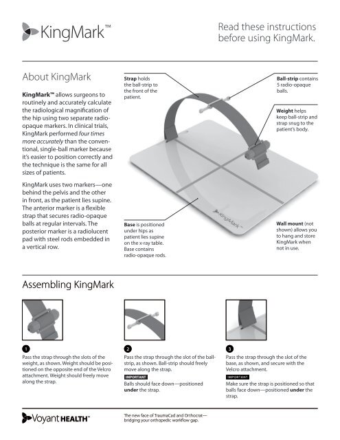
About <strong>KingMark</strong><br />
<strong>KingMark</strong> allows surgeons to<br />
routinely and accurately calculate<br />
the radiological magnification of<br />
the hip using two separate radioopaque<br />
markers. In clinical trials,<br />
<strong>KingMark</strong> performed four times<br />
more accurately than the conventional,<br />
single-ball marker because<br />
it’s easier to position correctly and<br />
the technique is the same for all<br />
sizes of patients.<br />
<strong>KingMark</strong> uses two markers—one<br />
behind the pelvis and the other<br />
in front, as the patient lies supine.<br />
The anterior marker is a flexible<br />
strap that secures radio-opaque<br />
balls at regular intervals. The<br />
posterior marker is a radiolucent<br />
pad with steel rods embedded in<br />
a vertical row.<br />
Strap holds<br />
the ball-strip to<br />
the front of the<br />
patient.<br />
Base is positioned<br />
under hips as<br />
patient lies supine<br />
on the x-ray table.<br />
Base contains<br />
radio-opaque rods.<br />
Ball-strip contains<br />
5 radio-opaque<br />
balls.<br />
Weight helps<br />
keep ball-strip and<br />
strap snug to the<br />
patient’s body.<br />
Wall mount (not<br />
shown) allows you<br />
to hang and store<br />
<strong>KingMark</strong> when<br />
not in use.<br />
Assembling <strong>KingMark</strong><br />
1 2<br />
3<br />
Pass the strap through the slots of the<br />
weight, as shown. Weight should be positioned<br />
on the opposite end of the Velcro<br />
attachment. Weight should freely move<br />
along the strap.<br />
Pass the strap through the slot of the ballstrip,<br />
as shown. Ball-strip should freely<br />
move along the strap.<br />
important<br />
Balls should face down—positioned<br />
under the strap.<br />
Pass the strap through the slot of the<br />
base, as shown, and secure with the<br />
Velcro attachment.<br />
important<br />
Make sure the strap is positioned so that<br />
balls face down—positioned under the<br />
strap.<br />
The new face of TraumaCad and Orthocrat—<br />
bridging your orthopedic workflow gap.
How to position <strong>KingMark</strong><br />
for accurate calibration.<br />
1<br />
Place the <strong>KingMark</strong> base on the x-ray<br />
table, positioned for a hip x-ray. The<br />
orange cross-marks should be facing up.<br />
2<br />
Lay patient supine on the <strong>KingMark</strong> base.<br />
3<br />
Move the base or patient so that the<br />
horizontal cross-mark is approximately in<br />
line with the tips of the greater trochanter<br />
and the vertical cross-mark is<br />
approximately midline to the patient.<br />
4<br />
Place the strap over the patient and position<br />
to approximately cover the tips of<br />
the greater trochanter.<br />
5<br />
Slide the ball-strip along the strap so that<br />
it is positioned midline over the patient’s<br />
subrapubic region.<br />
6<br />
Use the weight to hold the strap and ballstrip<br />
snuggly to the patient. Position the<br />
weight to the side of the patient, out of<br />
the x-ray image area.<br />
Tips:<br />
7<br />
You are ready to take the x-ray.<br />
The resulting radiograph should appear,<br />
as shown above. One of the 5 balls should<br />
align with the femoral head of the hip<br />
and the rods should run midline through<br />
the pelvis. If the markers are very off center<br />
and not positioned between the hips,<br />
the x-ray should be repeated.<br />
• The positioning technique remains the<br />
same for all sizes of patients. Since King-<br />
Mark is positioned midline, it will always<br />
appear within the radiograph—even on<br />
very large patients.<br />
• Flatten or remove bulky clothing that<br />
keeps the ball-strip from fitting snuggly<br />
to the patient.<br />
• Attempt to have each ball in the ballstrip<br />
touch the patient—or be as close<br />
as possible to the skin. However, do not<br />
pull the strap and ball-strip so tight that<br />
it significantly compresses the patient’s<br />
skin or adipose tissue.<br />
For more information and instruction about <strong>KingMark</strong>, visit:<br />
www.voyanthealth.com/kingmark<br />
TraumaCad will automatically detect<br />
the presence of <strong>KingMark</strong> in an x-ray,<br />
determine the magnification percentage,<br />
and size the image accurately.<br />
For information about TraumaCad, go to:<br />
www.voyanthealth.com/traumacad<br />
For surgeons who need to perform<br />
manual calibration on radiographs with<br />
<strong>KingMark</strong>, precise magnification values<br />
can be obtained by using the <strong>KingMark</strong><br />
Calculator, available at:<br />
www.voyanthealth.com/kingmark<br />
<strong>KingMark</strong> is exclusively distributed by <strong>Voyant</strong> <strong>Health</strong>, Ltd.<br />
© 2010, <strong>Voyant</strong> <strong>Health</strong>, Ltd. | All Rights Reserved | MK2S00249_A May 2010<br />
For more details, visit<br />
www.voyanthealth.com<br />
US: 866-717-0272<br />
UK: 0-800-404-8204<br />
Europe: 00-800-9290-9290<br />
International: +972-3-929-0929