Fall 2011 - Institute of Medical Science - University of Toronto
Fall 2011 - Institute of Medical Science - University of Toronto
Fall 2011 - Institute of Medical Science - University of Toronto

Create successful ePaper yourself
Turn your PDF publications into a flip-book with our unique Google optimized e-Paper software.
FEATURE<br />
Imaging and Prostate Cancer<br />
Applications <strong>of</strong> MRI<br />
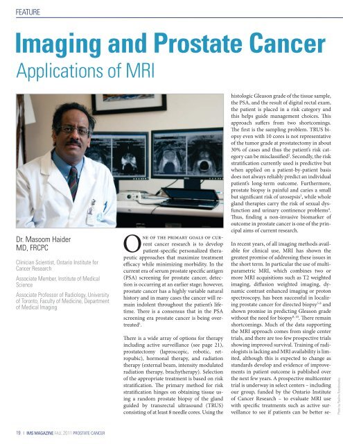
Dr. Masoom Haider<br />
MD, FRCPC<br />
Clinician Scientist, Ontario <strong>Institute</strong> for<br />
Cancer Research<br />
Associate Member, <strong>Institute</strong> <strong>of</strong> <strong>Medical</strong><br />
<strong>Science</strong><br />
Associate Pr<strong>of</strong>essor <strong>of</strong> Radiology, <strong>University</strong><br />
<strong>of</strong> <strong>Toronto</strong>, Faculty <strong>of</strong> Medicine, Department<br />
<strong>of</strong> <strong>Medical</strong> Imaging<br />
One <strong>of</strong> the primary goals <strong>of</strong> current<br />
cancer research is to develop<br />
patient-specific personalized therapeutic<br />
approaches that maximize treatment<br />
efficacy while minimizing morbidity. In the<br />
current era <strong>of</strong> serum prostate specific antigen<br />
(PSA) screening for prostate cancer, detection<br />
is occurring at an earlier stage; however,<br />
prostate cancer has a highly variable natural<br />
history and in many cases the cancer will remain<br />
indolent throughout the patient’s lifetime.<br />
There is a consensus that in the PSA<br />
screening era prostate cancer is being overtreated<br />
1 .<br />
There is a wide array <strong>of</strong> options for therapy<br />
including active surveillance (see page 21),<br />
prostatectomy (laproscopic, robotic, retropubic),<br />
hormonal therapy, and radiation<br />
therapy (external beam, intensity modulated<br />
radiation therapy, brachytherapy). Selection<br />
<strong>of</strong> the appropriate treatment is based on risk<br />
stratification. The primary method for risk<br />
stratification hinges on obtaining tissue using<br />
a random prostate biopsy <strong>of</strong> the gland<br />
guided by transrectal ultrasound (TRUS)<br />
consisting <strong>of</strong> at least 8 needle cores. Using the<br />
histologic Gleason grade <strong>of</strong> the tissue sample,<br />
the PSA, and the result <strong>of</strong> digital rectal exam,<br />
the patient is placed in a risk category and<br />
this helps guide management choices. This<br />
approach suffers from two shortcomings.<br />
The first is the sampling problem. TRUS biopsy<br />
even with 10 cores is not representative<br />
<strong>of</strong> the tumor grade at prostatectomy in about<br />
30% <strong>of</strong> cases and thus the patient’s risk category<br />
can be misclassified 2 . Secondly, the risk<br />
stratification currently used is predictive but<br />
when applied on a patient-by-patient basis<br />
does not always reliably predict an individual<br />
patient’s long-term outcome. Furthermore,<br />
prostate biopsy is painful and caries a small<br />
but significant risk <strong>of</strong> urosepsis 3 , while whole<br />
gland therapies carry the risk <strong>of</strong> sexual dysfunction<br />
and urinary continence problems 4 .<br />
Thus, finding a non-invasive biomarker <strong>of</strong><br />
outcome in prostate cancer is one <strong>of</strong> the principal<br />
aims <strong>of</strong> current research.<br />
In recent years, <strong>of</strong> all imaging methods available<br />
for clinical use, MRI has shown the<br />
greatest promise <strong>of</strong> addressing these issues in<br />
the short term. In particular the use <strong>of</strong> multiparametric<br />
MRI, which combines two or<br />
more MRI acquisitions such as T2 weighted<br />
imaging, diffusion weighted imaging, dynamic<br />
contrast enhanced imaging or proton<br />
spectroscopy, has been successful in localizing<br />
prostate cancer for directed biopsy 5-8 and<br />
shown promise in predicting Gleason grade<br />
without the need for biopsy 9, 10 . There remain<br />
shortcomings. Much <strong>of</strong> the data supporting<br />
the MRI approach comes from single center<br />
trials, and there are too few prospective trials<br />
showing improved survival. Training <strong>of</strong> radiologists<br />
is lacking and MRI availability is limited,<br />
although this is expected to change as<br />
standards develop and evidence <strong>of</strong> improvements<br />
in patient outcome is published over<br />
the next few years. A prospective multicenter<br />
trial is underway in select centers – including<br />
our group, funded by the Ontario <strong>Institute</strong><br />
<strong>of</strong> Cancer Research – to evaluate MRI use<br />
with specific treatments such as active surveillance<br />
to see if patients can be better se-<br />
Photo by Paulina Rzeczkowska<br />
19 | IMS MAGAZINE FALL <strong>2011</strong> PROSTATE CANCER