First 11 pages of thesis. - OPUS - Universität Würzburg
First 11 pages of thesis. - OPUS - Universität Würzburg
First 11 pages of thesis. - OPUS - Universität Würzburg

Create successful ePaper yourself
Turn your PDF publications into a flip-book with our unique Google optimized e-Paper software.
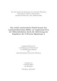
Insight into oxidative stress mediated by nitric<br />
oxide synthase (NOS) is<strong>of</strong>orms in atherosclerosis<br />
Thesis submitted towards fulfillment <strong>of</strong> the requirements for<br />
doctoral degree to the Bayerischen Julius-Maximilians-<br />
University, <strong>Würzburg</strong>, Germany,<br />
By<br />
P.Padmapriya<br />
Coimbatore, TN, INDIA<br />
<strong>Würzburg</strong>, GERMANY<br />
September, 2008<br />
i
Eingereicht am: 2 nd September 2008<br />
Mitglieder der Promotionskommission:<br />
Vorsitzender : Pr<strong>of</strong>. Dr. Martin. J. Müller<br />
Gutachter : PD. Dr. Peter. J. Kuhlencordt<br />
Gutachter : Pr<strong>of</strong>. Dr. Roland Benz<br />
Tag des Promotionskolloquiums :………………………………………....................<br />
Doktorurkunde ausgehändigt am :………………………….....................................<br />
ii
Eidesstattliche Erklärungen<br />
Hiermit erkläre ich ehrenwörtlich, dass die vorliegende Dissertation<br />
“Insight into oxidative stress mediated by nitric oxide synthase<br />
(NOS) is<strong>of</strong>orms in atherosclerosis” selbständig an der Medizinische<br />
Klinik I der <strong>Universität</strong> <strong>Würzburg</strong> unter der Anleitung von Dr.Peter<br />
Kuhlencordt angefertigt wurde und dass ich keine anderen als die<br />
angegebenen Quellen und Hilfsmittel benutzt habe.<br />
Weiterhin versichere ich, dass die vorliegende Dissertation weder in<br />
gleicher oder ähnlicher Form noch nicht in einem anderen<br />
Prüfungsverfahren vorgelegen hat und ich bisher noch keine akademischen<br />
Grade erworben oder zu erwerben versucht habe.<br />
Hiermit bewerbe ich mich erstmals um den Doktorgrad der<br />
Naturwissenschaften der Bayerischen Julius-Maximilians-<strong>Universität</strong><br />
<strong>Würzburg</strong>.<br />
iii<br />
(P.Padmapriya)<br />
September, 2008<br />
Medizinische Klinik I<br />
<strong>Universität</strong> <strong>Würzburg</strong><br />
GERMANY
I dedicate my doctoral <strong>thesis</strong>....<br />
....to all the tiny little ones, whose lives have been<br />
sacrificed for this work!<br />
iv
Acknowledgements<br />
<strong>First</strong> and foremost I would like to thank Dr. Peter Kuhlencordt for his<br />
scientific guidance and moral support. I am indebted to him for being there<br />
for me at time <strong>of</strong> utmost need. He had been very patient and understanding<br />
and had not only tolerated my failures but had also helped me overcome my<br />
frustrations, by his pep talks. He had been the source <strong>of</strong> my inspiration and<br />
motivation as a researcher and had also nurtured me into the researcher<br />
that I am today. I sincerely thank him for giving me the liberty and<br />
encouragement to make crucial decisions. I really appreciate his<br />
constructive critical comments and the brainstorming discussions/”debates”<br />
which eventually led to my progress. His amazing capability to quote<br />
citations had been highly admirable. In addition to being my project<br />
supervisor, he had also taken personal care and helped me acclimatise in<br />
Germany. In addition to research, I had also learned a lot from him to deal<br />
people diplomatically and suavely. Without his kindness, care and<br />
understanding it would have been tough to have completed this work<br />
against all the odds. I would always remember him for his smartness and<br />
sweetness, which has made my doctoral <strong>thesis</strong> work an enjoyable<br />
experience. In short, it had been a great pleasure and fun working with him.<br />
I would like to thank Pr<strong>of</strong>. Georg Ertl for giving me the opportunity to<br />
perform my <strong>thesis</strong> in his reputed institute. I really appreciate his simplicity<br />
and amicability. I also extend my sincere thanks to Pr<strong>of</strong>. Roland Benz for<br />
taking time to look into the progress <strong>of</strong> my project and his useful<br />
suggestions during our discussions.<br />
Many thanks to Dr.Fink (Noxygen Science Transfer & Diagnostics,<br />
GmbH) for his ‘online’ guidance and helping me establish the ESR and<br />
HPLC techniques in the lab. His generous help and expertise had aided me<br />
in standardising these techniques. I would also like to thank Dr. Andreas<br />
Kamlowski (Brukers Biospin GmbH) for his co-operation in establishing the<br />
ESR technique in the lab and many thanks to IZKF (Interdisziplinären<br />
Zentrum für Klinische Forschung) for the financial support.<br />
I am thankful to my colleagues Gabriele Riehl and Alla Ganscher,<br />
who had helped me in many ways and for their kindness and care. They had<br />
taken personal care <strong>of</strong> me and had always wished for my well-being. I am<br />
grateful to Gabriele for having taken immense responsibility in taking care<br />
<strong>of</strong> all my animals. I greatly appreciate Alla for her patience in helping me<br />
learn German and for having tolerated my poor German!!<br />
I sincerely thank my other lab mates, Johannes Schödle, Sebastian<br />
Rützel, Eva Ostermeier, Angelika Schröttle, Elisabeth Bendel, Nadja Miller<br />
and Carolin Knoll and institute members especially Lisa Bauer and Helga<br />
Wagner for all their timely help and for creating a good working<br />
atmosphere. I am thankful to Elisabeth and Nadja for their help in German<br />
vi
translations. Special thanks to my lab mate Eva Ostermeier for her great<br />
experimental assistance, team spirit and co-operation in completing the<br />
project.<br />
I am very thankful to all <strong>of</strong> my Indian friends who had helped me in<br />
all possible ways and for creating a home away from home ambience. My<br />
special thanks to Reena, Shruti and Tripat for their friendship and for being<br />
patient listeners <strong>of</strong> all my stories.<br />
And <strong>of</strong> course, last but not the least, I would like to thank my family<br />
for their constant motivation. I am indebted to my dad- my best friend,<br />
philosopher and guide for his encouragement through out my childhood, till<br />
date and forever and for believing more in me than what I believe in myself.<br />
I hope to live up to his dreams! Many thanks to my mom, whose prayers<br />
have always been with me and for her moral support. My mom and dad had<br />
been the best parents a kid could ever hope for! Tons <strong>of</strong> thanks to my<br />
sisters- Thara and Sasi, for their motherly care, priceless love, affection and<br />
encouragement. I also thank my nephews, Sriram and Sanath for being my<br />
future. I have no words to express my gratitude to my family. Though miles<br />
away, their warmth, love and care had always kindled my happiness. Most<br />
<strong>of</strong> all, I thank my God father for being with me always, in thoughts.<br />
vii
“Research is to see what everybody else has seen and to<br />
think what nobody else has thought”<br />
viii<br />
-Albert Szent-Gyorgyi<br />
Hungarian Biochemist,<br />
1937 Nobel Prize for Medicine
Contents<br />
Chapter 1 1<br />
1.0 Introduction 2<br />
1.1 Pathogenesis <strong>of</strong> atherosclerosis 2<br />
1.2 Risk factors 6<br />
1.2.1 Factors with a strong genetic component 6<br />
1.2.2 Environmental factors 8<br />
1.3 Oxidative stress 10<br />
1.3.1 Sources <strong>of</strong> oxidants in the vasculature <strong>11</strong><br />
1.3.2 Functional role <strong>of</strong> superoxide in atherosclerosis 12<br />
1.3.3 Functional role <strong>of</strong> nitric oxide in atherosclerosis 13<br />
1.4 The nitric oxide pathway in atherosclerosis 16<br />
1.4.1 Overview <strong>of</strong> NOS family 17<br />
1.4.2 Unique features <strong>of</strong> NOS is<strong>of</strong>orms 19<br />
1.4.2.1 Endothelial nitric oxide synthase (eNOS) 20<br />
1.4.2.2 Neuronal nitric oxide synthase (nNOS) 22<br />
1.4.2.3 Inducible nitric oxide synthase (iNOS) 25<br />
1.5 The apoE ko model <strong>of</strong> atherosclerosis 27<br />
1.6 Role <strong>of</strong> NOS is<strong>of</strong>orms in cardiovascular diseases 28<br />
1.6.1 Role <strong>of</strong> eNOS in cardiovascular diseases 28<br />
1.6.2 Role <strong>of</strong> nNOS in cardiovascular diseases 30<br />
1.6.3 Role <strong>of</strong> iNOS in cardiovascular diseases 32<br />
1.7 Aim <strong>of</strong> the study 35<br />
ix
Chapter 2 37<br />
2.0 Materials and Methods 38<br />
2.1 Materials 38<br />
2.1.1 Mice 38<br />
2.1.2 Chemicals and reagents 38<br />
2.1.3 Preparation <strong>of</strong> reagents 43<br />
2.2 Methods 44<br />
2.2.1 Detection <strong>of</strong> free radicals in the vasculature 44<br />
2.2.1.1 Electron spin resonance (ESR) 45<br />
2.2.1.2 Principle <strong>of</strong> ESR 46<br />
2.2.2 Measurement <strong>of</strong> vascular nitric oxide production by<br />
electron paramagnetic spin trapping 48<br />
2.2.3 Measurement <strong>of</strong> vascular oxygen radical production<br />
by ESR 49<br />
2.2.3.1 Sample preparation for ESR measurements 51<br />
2.2.4 Measurement <strong>of</strong> nitric oxide bioavailability in the<br />
bloodstream 51<br />
2.2.5 Measurement <strong>of</strong> intracellular superoxide production<br />
by HPLC detection <strong>of</strong> oxyethidium 52<br />
2.2.6 Tissue preparation and immunohistochemistry 53<br />
2.2.7 Histomorphometry 54<br />
2.2.8 Protein Estimation 54<br />
2.2.9 Statistical Analyses 54<br />
x
Chapter 3 55<br />
3.0 Results 56<br />
3.1 eNOS is a significant source <strong>of</strong> vascular wall nitric oxide<br />
production and circulating nitric oxide 56<br />
3.2 eNOS deletion decreases vascular production <strong>of</strong> superoxide<br />
in apoE ko vessels 58<br />
3.3 nNOS contributes little to vascular nitric oxide production 60<br />
3.4 Contribution <strong>of</strong> nNOS to vascular superoxide production in<br />
apoE ko animals 61<br />
3.5 iNOS contributes significantly to vascular nitric oxide production 62<br />
3.6 iNOS plays a major role in vascular superoxide production 63<br />
3.7 Nitric oxide and superoxide generation from iNOS results in<br />
peroxynitrite formation 65<br />
3.8 iNOS deletion influences NADPH oxidase mediated superoxide<br />
production 66<br />
Chapter 4 67<br />
4.0 Discussion 68<br />
4.1 eNOS is a major contributor <strong>of</strong> nitric oxide and superoxide<br />
generation in apoE ko vessels 68<br />
4.2 Vascular nNOS generates low amounts <strong>of</strong> nitric oxide and<br />
superoxide 70<br />
4.3 iNOS generates nitric oxide and superoxide simultaneously in<br />
apoE ko vessels 72<br />
xi
Summary and Hypo<strong>thesis</strong> 76<br />
Zusammenfassung und Hypothese 78<br />
Bibliography 80<br />
Abbreviations 95<br />
Thesaurus 96<br />
Curriculum Vitae 102<br />
xii
Chapter Chapter 1<br />
1<br />
- 1 -
1.0 Introduction<br />
Atherosclerosis, the disease <strong>of</strong> large and medium sized arteries is the<br />
most common disease in western countries and is known to be the underlying<br />
cause <strong>of</strong> 50% <strong>of</strong> all deaths. The prevalence <strong>of</strong> atherosclerosis is estimated to be<br />
17 per 1000 (NHIS95), resulting in 1 death per hour among the general<br />
population <strong>of</strong> the USA. Atherosclerosis is characterised by the chronic<br />
accumulation <strong>of</strong> lipids and fibrous elements in the wall <strong>of</strong> the blood vessel which<br />
may result in progressive narrowing <strong>of</strong> the lumen and consecutive reduction in<br />
blood flow. The reduced blood flow is the underlying cause <strong>of</strong> chronic angina<br />
pectoris and claudication. Atherosclerosis affecting other arteries causes renal<br />
impairment, hypertension, abdominal aortic aneurysms and critical limb<br />
ischemia. On the other hand, rupture <strong>of</strong> an atherosclerotic plaque may result in<br />
an acute thrombotic occlusion <strong>of</strong> a vessel, which may result in myocardial<br />
infarction, stroke or acute ischemia <strong>of</strong> the gut or an extremity.<br />
1.1 Pathogenesis <strong>of</strong> atherosclerosis<br />
Atherosclerosis is a chronic inflammatory disease which progresses with<br />
age. The development <strong>of</strong> atherosclerosis is complex, involving numerous cell<br />
types and genes. A normal artery is composed <strong>of</strong> three different layers, 1) the<br />
inner most layer called tunica intima, composed <strong>of</strong> a thin layer <strong>of</strong> collagen and<br />
proteoglycans covered by a single layer <strong>of</strong> endothelial cells which line the lumen<br />
<strong>of</strong> the artery 2) the middle layer <strong>of</strong> smooth muscle cells called the tunica media<br />
and 3) the outer most layer called tunica adventitia which consists <strong>of</strong> connective<br />
tissue, fibroblasts and smooth muscle cells (Figure 1).<br />
- 2 -
Figure 1: Structure <strong>of</strong> a normal vessel wall. The cross sectional view shows the three distinct<br />
layers <strong>of</strong> the vessel wall: the intima, media and adventitia. (Picture from Lusis AJ, Nature. 2000;<br />
407(6801):233-41)<br />
During the initial stages, lipoproteins and their aggregates accumulate in<br />
the intima <strong>of</strong> the vessel wall, at sites <strong>of</strong> lesion predilection. These predilection<br />
sites are usually the branching points or the inner curvature <strong>of</strong> the arteries,<br />
where normal blood flow dynamics are altered 1 . Subsequently, monocytes and<br />
lymphocytes adhere to the endothelium, transmigrate across the endothelial<br />
layer into the intima <strong>of</strong> the vessel where they proliferate, differentiate and take<br />
up lipoproteins to form “foam cells”. Though these early plaques or foam cells<br />
(also termed as ‘fatty streaks’) are not <strong>of</strong> clinical significance, they are the<br />
precursors <strong>of</strong> advanced lesions. As the disease progresses the foam cells die<br />
and the smooth muscle cells migrate from the medial layer into the intima,<br />
where they accumulate and proliferate. The so called advanced lesions are<br />
characterised by the accumulation <strong>of</strong> smooth muscle cells and dead foam cells,<br />
- 3 -
which contribute to the lipid rich “necrotic core”. The smooth muscle cells<br />
secrete fibrous elements which form the “fibrous cap”, enclosing the lipid rich<br />
necrotic core. Initially the lesions grow towards the adventitia until a critical point<br />
is reached after which further growth <strong>of</strong> the plaques encroaches the lumen. As<br />
leukocyte recruitment and smooth muscle cell proliferation continues plaque<br />
development progresses. Additional extracellular matrix production,<br />
accumulation <strong>of</strong> extracellular lipid and calcification leads to the development <strong>of</strong><br />
advanced atherosclerotic lesions (Figure 2).<br />
Depending on the composition, atherosclerotic lesions can be classified<br />
into two types, namely stable or vulnerable plaques (Figure 2). A “stable plaque”<br />
has a thick fibrous cap, a small lipid pool, few inflammatory cells and a dense<br />
extracellular matrix. In contrast, a “vulnerable plaque” is characterised by a thin<br />
fibrous cap, an increased number <strong>of</strong> inflammatory cells, a large lipid pool and<br />
fewer smooth muscle cells. Vulnerable plaques are unstable, which may result<br />
in plaque rupture and instantaneous occlusion <strong>of</strong> the vessel. Plaque rupture<br />
usually occurs at the shoulder <strong>of</strong> the lesion, resulting in thrombus formation.<br />
Subsequent embolization <strong>of</strong> the thrombotic material may lead to additional<br />
occlusion <strong>of</strong> distal coronary arteries or cerebral arteries which can further<br />
aggravate myocardial or cerebral ischemia.<br />
- 4 -
Figure 2: Developmental stages <strong>of</strong> atherosclerosis. (Picture from Hugh Watkins et al., Nature<br />
Reviews Genetics. 2006; 7: 163-73)<br />
- 5 -
1.2 Risk Factors<br />
Atherosclerosis has a complex aetiology influenced by a number <strong>of</strong><br />
factors. The cardiovascular risk increases with the number <strong>of</strong> risk factors <strong>of</strong> a<br />
patient. Factors which influence atherosclerosis development can be grouped<br />
into genetic and environmental. In most cases the development <strong>of</strong><br />
atherosclerosis results from complex interactions between environmental and<br />
genetic factors.<br />
1.2.1 Factors with a strong genetic component<br />
1. Elevated levels <strong>of</strong> low density lipoproteins (LDL)<br />
Low density lipoproteins play an important role in transportation <strong>of</strong> cholesterol<br />
and triglycerides from the liver to peripheral tissues and in the regulation <strong>of</strong><br />
cholesterol syn<strong>thesis</strong>. Elevated levels <strong>of</strong> LDL usually result from mutations in<br />
the LDL receptor gene, causing familial hypercholesterolemia 2 . Some <strong>of</strong> the<br />
genetic variants which cause elevated LDL levels are the apolipoprotein E 3 and<br />
the apolipoprotein (a) genes 4 .<br />
2. Reduced levels <strong>of</strong> high density lipoproteins (HDL)<br />
High density lipoproteins carry cholesterol from the systemic circulation to the<br />
liver, where they are excreted or re-utilized. Polymorphisms in the hepatic lipase<br />
encoding gene and the apolipoprotein AI-CIII-AIV gene cluster results in altered<br />
levels <strong>of</strong> HDL 5 .<br />
- 6 -
3. Elevated blood pressure<br />
Hypertension is considered one <strong>of</strong> the major cardiovascular risk factors.<br />
Consequently, treatment <strong>of</strong> hypertension reduces the risk <strong>of</strong> cardiovascular<br />
diseases by 50%, compared to patients whose blood pressure is uncontrolled 6 .<br />
4. Elevated levels <strong>of</strong> homocysteine<br />
Homocysteine, a sulphur containing amino acid is an intermediate product <strong>of</strong><br />
the metabolism <strong>of</strong> methionine and cysteine. A single mutation (677C→T) in<br />
methylenetetrahydr<strong>of</strong>olate reductase gene causes increased homocysteine<br />
levels 7 associated with premature atherosclerosis.<br />
5. Metabolic syndrome<br />
Metabolic syndrome is a cluster <strong>of</strong> metabolic disturbances that strongly<br />
predisposes to atherosclerosis development 8 . Peripheral insulin resistance<br />
seems to be the central phenomenon <strong>of</strong> the metabolic syndrome, which is<br />
characterised by impaired glucose tolerance, dyslipidemia, hypertension and<br />
obesity.<br />
6. Male gender and family history<br />
It has been reported that below 60 years <strong>of</strong> age, men develop coronary heart<br />
disease (CHD) at more than twice the rate <strong>of</strong> women 9 . Individuals with a family<br />
history, i.e first degree relatives <strong>of</strong> patients with early onset <strong>of</strong> cardiovascular<br />
disease are at a higher risk <strong>of</strong> developing atherosclerosis which may be due to<br />
a common genetic predisposition (elevated blood pressure or cholesterol levels)<br />
or non genetic effects/environmental factors (smoking or diet) 10, <strong>11</strong> .<br />
- 7 -
1.2.2 Environmental factors<br />
1. High-fat diet<br />
High fat and high cholesterol diets have been shown to increase<br />
atherosclerosis. In experimental models, high fat diets are used to induce<br />
plaque formation. In humans, regular consumption <strong>of</strong> high fat diet results in<br />
obesity and subsequent reduction <strong>of</strong> average life expectancy. Hence, reduction<br />
<strong>of</strong> body weight through diets is one <strong>of</strong> the main treatment strategies to reduce<br />
the individual cardiovascular risk. Mediterranean diets rich in olive oil or nuts<br />
have proved to reduce the cardiovascular risk more effectively than a<br />
conventional low-fat diet 12 . Additionally, omega-3 fatty acid rich diets reduce the<br />
risk <strong>of</strong> cardiovascular diseases 13 .<br />
2. Smoking<br />
It has been calculated that about 30% <strong>of</strong> cardiovascular deaths are due to<br />
smoking 14 . Cigarette smoking increases total cholesterol, triglycerides and LDL-<br />
cholesterin and decreases the cardio-protective HDL-cholesterin. Smoking is an<br />
established independent risk factor for atherosclerosis development even<br />
among young individuals 15 .<br />
3. Infectious disease/chronic inflammatory disease<br />
Recent studies have shown that inflammation plays a fundamental role in<br />
development <strong>of</strong> atherosclerosis 16, 17 . The signalling cascades that are triggered<br />
in response to inflammation have a proatherogenic role. Epidemiological and<br />
basic scientific studies have shown that pathogens like Chlamydia pneumonia 18<br />
- 8 -
and Porphyromonas gingivalis 19 are associated with atherosclerosis<br />
development. Chronic inflammatory disease secondary to infection, like<br />
acquired immunodeficiency syndrome (AIDS) due to infection <strong>of</strong> human<br />
immunodeficiency virus (HIV) 20 and auto immune diseases like systemic lupus<br />
erythematosus and rheumatoid arthritis also accelerate atherosclerosis<br />
development 21 .<br />
4. Lack <strong>of</strong> exercise<br />
Lack <strong>of</strong> physical exercise and a sedentary life style is an independent risk factor<br />
for various cardiovascular diseases. Regular exercise results in reduction <strong>of</strong><br />
body fat, LDL cholesterol, triglyceride levels and blood pressure and increases<br />
atheroprotective HDL cholesterol levels 22 . Physical exercise is <strong>of</strong> paramount<br />
importance as it positively influences many independent cardiovascular disease<br />
risk factors.<br />
5. Oxidative stress<br />
Increased levels <strong>of</strong> oxidants or decreased levels <strong>of</strong> anti-oxidants secondary to<br />
dyslipidemia, hypertension, diabetes and smoking are implicated in the<br />
pathogenesis <strong>of</strong> atherosclerosis. Oxidation <strong>of</strong> LDL is considered to be the<br />
critical step involved in ‘foam cell’ formation 23 . Oxidation <strong>of</strong> LDL results in many<br />
structural modifications and generation <strong>of</strong> numerous ‘oxidation specific epitopes’<br />
such as malondialdehyde (MDA)-lysines and 4-hydroxynonenal (4-HNE)–lysine’<br />
which are highly immunogenic. Immunization <strong>of</strong> mice with MDA and native LDL<br />
resulted in a significant reduction <strong>of</strong> atherosclerosis indicating the<br />
proatherosclerotic role <strong>of</strong> oxidized LDL (ox-LDL) 24 .<br />
- 9 -
1.3 Oxidative Stress<br />
Oxidative stress results from increased production <strong>of</strong> reactive oxygen<br />
species (ROS) in biological systems. ROS include “free radicals” and “non-free<br />
radicals” which are produced during electron transfer reactions in oxygen (O2)<br />
metabolism. Molecular O2 is essential for the survival <strong>of</strong> all aerobic organisms<br />
and acts as the electron acceptor during various metabolic reactions. The<br />
partially reduced, highly reactive metabolites formed during these reactions<br />
react more avidly compared to molecular O2. ROS are generally considered to<br />
be by-products <strong>of</strong> metabolism with a potential to cause cellular injury 25 . During<br />
evolution organisms have developed several strategies to potentially detoxify<br />
ROS. However, under physiological condition ROS are also important signalling<br />
molecules 26 and there exists a balance between production and detoxification <strong>of</strong><br />
ROS. Diseases may cause an imbalance between the production and<br />
neutralization <strong>of</strong> ROS, resulting in altered cell signalling and oxidative stress.<br />
Superoxide anion formation generates a chain <strong>of</strong> reactions which result<br />
in the formation <strong>of</strong> various highly reactive free radicals and non radicals. In<br />
atherosclerosis and other vascular diseases ROS are potent pathological<br />
mediators 27 , as they cause lipid peroxidation, smooth muscle cell proliferation,<br />
protein oxidation, inflammatory cell recruitment and vascular inflammation. For<br />
example, oxidative modification <strong>of</strong> lipoproteins initiates foam cell formation 23<br />
and vascular oxidative stress is a major cause <strong>of</strong> cardiovascular diseases 28 .<br />
ROS are implicated in the process <strong>of</strong> initiation <strong>of</strong> foam cell formation until<br />
ultimate plaque rupture. Increased oxidative stress results in reduced<br />
endothelial dysfunction 29 .<br />
- 10 -
1.3.1 Sources <strong>of</strong> oxidants in the vasculature<br />
There are numerous enzymatic sources <strong>of</strong> ROS in the vasculature,<br />
including mitochondrial electron transport chain, the arachidonic acid<br />
metabolizing enzymes lipoxygenase and cyclooxygenase, the cytochrome<br />
P450s, myeloperoxidase 30 , xanthine oxidase 31 , Nicotinamide adenine<br />
dinucleotide phosphate (NADPH) oxidase 32 , and the nitric oxide synthases<br />
(NOS).<br />
NADPH oxidase and xanthine oxidase are important contributors to<br />
oxidative stress in cardiovascular diseases 32, 33 . NADPH oxidase is expressed<br />
in endothelial cells, smooth muscle cells, fibroblasts and macrophages, while<br />
xanthine oxidase is expressed in endothelial cells and found in plasma.<br />
Myeloperoxidase (expressed in neutrophils), the enzyme that converts chloride<br />
(Cl - ) ion to hypochlorous acid, increases atherosclerosis development 30 , as<br />
expression <strong>of</strong> this enzyme was observed in human atherosclerotic lesions.<br />
Furthermore, footprints <strong>of</strong> oxidative modifications <strong>of</strong> LDL by<br />
myeloperoxidase/HOCl were observed in atherosclerotic lesions 34 .<br />
NOS enzymes produce nitric oxide (NO) by their catalytic conversion <strong>of</strong><br />
L-arginine to L-citrulline. This family <strong>of</strong> enzymes includes the endothelial NOS<br />
(eNOS or NOS III), inducible NOS (iNOS or NOS II) and neuronal NOS (nNOS<br />
or NOS I). However, NOS not only produces nitric oxide but may directly<br />
produce superoxide in special metabolic situations. Under conditions <strong>of</strong><br />
substrate L-arginine or c<strong>of</strong>actor, (tetrahydrobiopterin (BH4)) deficiency, NOS<br />
“uncouple” directing electron transfer to molecular oxygen rather than to L-<br />
arginine, resulting in generation <strong>of</strong> superoxide 35-37 . In vitro, the generation <strong>of</strong><br />
superoxide and nitric oxide results in the formation <strong>of</strong> the strong oxidant<br />
- <strong>11</strong> -
peroxynitrite 38 which by itself causes the formation <strong>of</strong> a complex array <strong>of</strong><br />
oxidants leading to lipid peroxidation and protein nitration 39, 40 . Therefore, it has<br />
been speculated that the superoxide generated by uncoupled NOS might result<br />
in peroxynitrite formation and consecutive oxidative stress.<br />
1.3.2 Functional role <strong>of</strong> superoxide in atherosclerosis<br />
Superoxide functions as a signaling molecule in cell division,<br />
differentiation and cell survival 41, 42 . Additionally, increased production <strong>of</strong><br />
superoxide is observed in hypertension, myocardial infarction, diabetes and<br />
atherosclerosis. Moreover, the severity <strong>of</strong> atherosclerosis correlates with the<br />
activation <strong>of</strong> NADPH oxidase in human carotid arteries 43 and NADPH oxidase<br />
deficient apolipoprotein E knockout (apoE ko) animals showed reduced<br />
atherosclerosis suggesting an important role <strong>of</strong> superoxide in atherosclerosis 44 .<br />
Increased expression <strong>of</strong> xanthine oxidase has also been observed in<br />
atherosclerotic plaques 45 .<br />
Superoxide production reduces nitric oxide bioavailability not only by<br />
direct inactivation <strong>of</strong> nitric oxide but also by oxidizing the NOS co-factor BH4,<br />
resulting in NOS “uncoupling”. Reactive oxygen and nitrogen species play a<br />
central role in the maintenance <strong>of</strong> vascular homeostasis. Nitric oxide dependent<br />
cell signaling, including endothelial dependent relaxation, is modulated by both<br />
superoxide and superoxide dismutase (SOD) 46, 47 . Alterations in both the rate <strong>of</strong><br />
formation and the extent <strong>of</strong> superoxide scavenging have been implicated in<br />
vascular dysfunction, hypertension, diabetes, as well as in chronic nitrate<br />
tolerance 39, 48, 49 . The evidence for the involvement <strong>of</strong> superoxide in impaired<br />
endothelium dependent relaxations is shown by the restoration <strong>of</strong> endothelium<br />
- 12 -
dependent relaxations using SOD and antioxidants 50, 51 . Further, deficiency <strong>of</strong><br />
vascular SOD results in impaired endothelial functions 47 . In addition to reducing<br />
the bioavailability <strong>of</strong> nitric oxide, superoxide generation may also promote<br />
endothelial cell apoptosis 52 .<br />
Superoxide generation causes platelet adhesion and aggregation 53 .<br />
NADPH oxidase mediated superoxide production causes increased<br />
leukocyte/endothelial cell interaction in hypercholesterolemic mice 54 . Further,<br />
SOD inhibits the expression <strong>of</strong> vascular cell adhesion molecule-1 (VCAM-1) and<br />
intercellular cell adhesion molecule-1 (ICAM-1) in endothelial cells 55 , suggesting<br />
that superoxide modulates the expression <strong>of</strong> adhesion molecules. Additionally,<br />
superoxide promotes vascular smooth muscle cell migration and proliferation 52 .<br />
Both superoxide and hydroxyl radical contribute to LDL-oxidation which induces<br />
cholesterol accumulation in macrophages and leads to foam cell formation 56, 57 .<br />
Ox-LDL acts as a chemotactic factor for monocytes and T-cells, the<br />
predominant population <strong>of</strong> blood cells found in the atherosclerotic lesions.<br />
1.3.3 Functional role <strong>of</strong> nitric oxide in atherosclerosis<br />
Nitric oxide, named the ‘molecule <strong>of</strong> the year in 1992’ is an important cell<br />
signaling, effector and vasodilator molecule with potentially strong<br />
antiatherogenic actions. Of all its functions, it’s role as endothelium dependent<br />
relaxing factor (EDFR) is the most recognized one, thought to reflect vascular<br />
homeostasis. Nitric oxide mediates vascular smooth muscle cell relaxation by a<br />
calcium-ion channel mediated activation <strong>of</strong> the cyclic guanosine<br />
monophosphate (cGMP) pathway. Further, nitric oxide inhibits smooth cell<br />
proliferation, leukocyte/endothelial cell interactions and platelet aggregation.<br />
- 13 -
Pharmacological inhibition <strong>of</strong> nitric oxide production by NOS results in increased<br />
leukocyte adhesion to microvascular endothelium 58 and expression <strong>of</strong><br />
endothelial surface adhesion molecules, including P-selectin and VCAM-1 59, 60 .<br />
Nitric oxide regulates platelet activation, platelet aggregation and<br />
platelet/endothelial cell-interactions 61-63 . It was shown in vitro that nitric oxide<br />
generated in the coronary and pulmonary vasculature inhibits platelet adhesion<br />
under constant flow conditions 64 . By it’s regulation <strong>of</strong> leukocyte and platelet<br />
adhesion to the endothelium, nitric oxide contributes to the maintenance <strong>of</strong><br />
microvascular barrier integrity and may decrease local inflammation and<br />
vascular permeability.<br />
The eNOS (endothelium) mediated nitric oxide production results in<br />
vasodilation, increased blood flow and reduced blood pressure. Impairment in<br />
the endothelial dependent relaxation, termed “endothelial dysfunction” is<br />
considered to be one <strong>of</strong> the critical steps in atherosclerosis development.<br />
Endothelial dysfunction occurs as a result <strong>of</strong> decreased nitric oxide production,<br />
decreased sensitivity to nitric oxide or decreased nitric oxide bioavailability 65 .<br />
Decreased nitric oxide production may occur secondary to reduced transcription<br />
or increased instability <strong>of</strong> eNOS mRNA 66 . Additionally, altered eNOS activity<br />
observed in hypercholesterolemia decreases nitric oxide production 67, 68 . NOS<br />
inhibitors like asymmetric dimethylarginine (ADMA) and N-monometylarginine<br />
(NMA) are involved in endothelial dysfunction 69 .<br />
The increased production <strong>of</strong> superoxide observed during condition <strong>of</strong><br />
atherosclerosis decreases the bioavailability <strong>of</strong> nitric oxide since superoxide<br />
reacts with nitric oxide at a diffusion limited rate, to form peroxynitrite. The<br />
reaction rate <strong>of</strong> superoxide with nitric oxide (6-10x10 9 M -1 sec -1 ) is faster than the<br />
- 14 -
ate at which superoxide is degraded by SOD (2x10 9 M -1 sec -1 ). Further<br />
dismutation <strong>of</strong> superoxide by SOD can occur only if the latter enzyme is present<br />
in the same compartment in which superoxide is produced. In addition to being<br />
a significant source <strong>of</strong> eNOS mediated nitric oxide production, the endothelium<br />
is also a significant source <strong>of</strong> superoxide production in atherosclerotic vessels 70 .<br />
Since under these conditioms nitric oxide and superoxide are produced in the<br />
same cellular compartment, i.e., the endothelial cell, they can immediately react<br />
to form peroxynitrite. Peroxynitrite is a strong oxidant which alters the function<br />
<strong>of</strong> biomolecules by protein nitration and lipid peroxidation 71 with secondary<br />
tissue injury 39, 40 . Subintimal lipoprotein oxidation by peroxynitrite may initiate<br />
the formation <strong>of</strong> fatty streaks and subsequent plaque development 39 .<br />
Peroxynitrite may also contribute to endothelial cell dependant vascular<br />
oxidation.<br />
Figure 3: Proposed functional role <strong>of</strong> nitric oxide and superoxide in normal and atherogenic<br />
conditions, respectively.<br />
- 15 -
1.4 The nitric oxide pathway in atherosclerosis<br />
L-arginine, a non-essential amino acid is utilized by the NOS enzyme to<br />
produce L-citrulline and nitric oxide. The nitric oxide synthesized by all NOS<br />
may enters one <strong>of</strong> the following routes: a) activation <strong>of</strong> soluble guanylate<br />
cyclase (sGC), which is responsible for most <strong>of</strong> the physiological effects <strong>of</strong> nitric<br />
oxide b) regulation <strong>of</strong> expression <strong>of</strong> adhesion molecules by inducing<br />
transcription and stabilization <strong>of</strong> IκBα 72 , an inhibitor <strong>of</strong> NF-κB through a cyclic<br />
guanylate monophosphate (cGMP) independent pathway 73 c) reaction with<br />
oxyhemoglobin to form stable metabolite nitrosylhemoglobin 74 d) formation <strong>of</strong><br />
nitrate 75 e) formation <strong>of</strong> peroxynitrite by reacting with superoxide 38 f)<br />
nitrosylation <strong>of</strong> proteins 76, 77 . Nitric oxide has no membrane receptor, but binds<br />
to the heme group <strong>of</strong> sGC producing a conformational change which increases<br />
its activity 78 . sGC converts guanylate triphosphate (GTP) into cGMP which<br />
activates protein kinase G (PKG), a cGMP dependant protein phosphorylator.<br />
PKG mediated protein phosphorylation leads to: a) inhibition <strong>of</strong> L-type calcium<br />
channels in the plasma membrane 79 ; b) activation <strong>of</strong> Ca ++ ATPase 80 and Ca ++ -<br />
Na + exchanger in the plasma membrane 81 ; c) activation <strong>of</strong> Ca ++ ATPase at the<br />
level <strong>of</strong> the sarcoplasmic reticulum 82 and d) inhibition <strong>of</strong> protein lipase C 83 .<br />
Calcium levels have differential roles in the nitric oxide pathway. Intracellular<br />
free calcium levels in endothelial cells activate NOS by binding to calmodulin 84 .<br />
Nitric oxide generated by the activated NOS in the endothelium diffuses into the<br />
smooth muscle cell layer in the media, where it activates sGC which causes<br />
feedback inhibition <strong>of</strong> calcium levels through cGMP mediated mechanisms<br />
resulting in relaxation <strong>of</strong> smooth muscle cells in the medial layers <strong>of</strong> the vessel<br />
wall. One <strong>of</strong> the many proteins which are phosphorylated in response to cGMP<br />
- 16 -
activation is the vasodilator-stimulated phosphoprotein (VASP). VASP is<br />
phosphorylated by cGMP dependent PKG which causes the nitric oxide<br />
mediated inhibition <strong>of</strong> smooth muscle cell proliferation 85 . cGMP also down<br />
regulates the function <strong>of</strong> some platelet receptors, including the fibrinogen<br />
receptor IIb/IIIa and P-selectin 86 .<br />
The activity <strong>of</strong> the enzymes involved in the nitric oxide pathway is altered<br />
during oxidative stress, as observed in atherosclerosis. As mentioned before,<br />
the expression and activity <strong>of</strong> eNOS is altered during atherosclerosis. In<br />
addition the formation <strong>of</strong> peroxynitrite is capable <strong>of</strong> impairing the activity <strong>of</strong><br />
sGC 87 . Atherosclerosis is also associated with low L-arginine availability.<br />
Subsequently, the altered functional activity <strong>of</strong> the nitric oxide pathway results in<br />
vascular smooth muscle contraction and endothelial dysfunction.<br />
1.4.1 Overview <strong>of</strong> NOS family<br />
NOS (EC 1.14.13.39) catalyses the conversion <strong>of</strong> L-arginine to L-<br />
citrulline, yielding nitric oxide as a byproduct. The NOS proteins have ~60%<br />
amino acid homology and possess similar primary structures. The is<strong>of</strong>orms are<br />
expressed in different cellular compartments and function as homodimers,<br />
composed <strong>of</strong> two identical monomers. The monomers consist <strong>of</strong> a C-terminal<br />
reductase domain and a N-terminal oxygenase domain, which differ in their<br />
structure and function between is<strong>of</strong>orms. The syn<strong>thesis</strong> <strong>of</strong> nitric oxide requires<br />
L-arginine as a substrate, calmodulin, molecular oxygen and four c<strong>of</strong>actors:<br />
flavin mononucleotide (FMN), flavin adenine dinucleotide (FAD),<br />
tetrahydrobiopterin (BH4) and nicotinamide adenine dinucleotide phosphate<br />
(NADPH). The reductase domain consists <strong>of</strong> the binding sites for one molecule<br />
- 17 -
<strong>of</strong> NADPH, FAD and FMN, whereas the oxygenase domain binds heme, BH4<br />
and the substrate L-arginine. As shown in figure 4, between these two domains<br />
lies the calmodulin binding site, which plays an important role in both structure<br />
and function <strong>of</strong> the enzyme.<br />
The biosyn<strong>thesis</strong> <strong>of</strong> nitric oxide involves a two step oxidation reaction and<br />
consumes 1.5 mol <strong>of</strong> NADPH and 2 mol <strong>of</strong> oxygen including the formation <strong>of</strong> the<br />
intermediate product, N G -hydroxy-L-arginine. The reductase domain transfers<br />
the electrons from NADPH via the flavins: FAD and FMN to the heme molecule<br />
in the oxygenase domain, where the substrate L-arginine is oxidized to L-<br />
citrulline and nitric oxide. Hence, the two domains perform catalytically distinct<br />
functions. Despite the fact that each monomer consists <strong>of</strong> both domains,<br />
dimerisation <strong>of</strong> the enzyme is essential for its catalytic activity since the<br />
electrons are transferred from the flavins in the reductase domain <strong>of</strong> one<br />
subunit to the heme centre in the oxygenase domain <strong>of</strong> the second subunit 88<br />
(Figure 4). Heme plays a key role in dimerisation <strong>of</strong> both the subunits in all three<br />
NOS is<strong>of</strong>orms and is also required for the interaction between the reductase<br />
and oxygenase domains. Calcium dependence is the key feature that<br />
distinguishes constitutive and inducible is<strong>of</strong>orms <strong>of</strong> NOS. eNOS and nNOS are<br />
activated by elevation <strong>of</strong> intracellular calcium levels, followed by subsequent<br />
binding <strong>of</strong> calcium/ calmodulin. In contrast, iNOS contains irreversibly bound<br />
calmodulin and thus its activation is independent <strong>of</strong> intracellular calcium<br />
concentration.<br />
Under conditions <strong>of</strong> either substrate L-arginine or c<strong>of</strong>actor BH4<br />
deficiency, all the is<strong>of</strong>orms <strong>of</strong> NOS can “uncouple”. The term “uncoupling”<br />
defines a situation during which the electrons flowing from the reductase<br />
- 18 -
domain to the oxygenase domain are shifted to molecular oxygen instead <strong>of</strong> L-<br />
arginine, resulting in superoxide rather than nitric oxide production. However,<br />
the conditions and mechanisms that cause uncoupling differ between the NOS<br />
is<strong>of</strong>orms. Furthermore, NOS is<strong>of</strong>orms vary in their regulation <strong>of</strong> gene<br />
expression, catalytic activity and the cellular compartment <strong>of</strong> gene expression.<br />
These features make each is<strong>of</strong>orm unique and give rise to distinct mechanistic<br />
features that are responsible for their differential function under various<br />
physiological and pathophysiological conditions.<br />
Figure 4: Structure <strong>of</strong> functional NOS dimers. Electrons in the NOS dimer flow via<br />
NADPH→FAD→FMN in the reductase domain (shaded region) <strong>of</strong> one monomer to the heme<br />
(Fe) in the oxygenase domain <strong>of</strong> the second monomer. Calmodulin (CaM) binding is essential<br />
for dimerisation <strong>of</strong> the enzyme. Dimerisation <strong>of</strong> NOS is required for conversion <strong>of</strong> L-arginine to<br />
L-citrulline and nitric oxide. (Picture adapted from Andrew PJ et al., Cardiovascular Research. 1999; 43:<br />
521-31)<br />
1.4.2 Unique features <strong>of</strong> NOS is<strong>of</strong>orms<br />
The functional relevance <strong>of</strong> nitric oxide generated by each NOS is<strong>of</strong>orm<br />
differs depending on the cellular compartment and the target proteins<br />
expressed in the compartment. Because <strong>of</strong> the versatile properties <strong>of</strong> nitric<br />
oxide, the expression, activity, spatial distribution and proximity <strong>of</strong> NOS<br />
- 19 -
is<strong>of</strong>orms to the regulatory and target proteins are tightly regulated and vary<br />
between the is<strong>of</strong>orms.<br />
Figure 5: Distinct domain structure <strong>of</strong> each NOS is<strong>of</strong>orm. Binding sites for L-arginine (Arg),<br />
heme, tetrahydrobiopterin (BH4), calmodulin (CaM), flavins (FAD and FMN) and NADPH are<br />
indicated. The oxygenase, reductase and PDZ (nNOS) domains are indicated by solid bars. The<br />
numbers indicate the amino acid residues within in each domain. Myristoylation (Myr) and<br />
palmitoylation (Palm) sites on eNOS are shown. The irreversible binding <strong>of</strong> calcium to the<br />
calmodulin in iNOS is indicated. (Picture adapted from Alderton WK et al., Biochem J. 2001; 357:<br />
593-615)<br />
1.4.2.1 Endothelial nitric oxide synthase (eNOS)<br />
eNOS is the main source <strong>of</strong> endothelial nitric oxide production in the<br />
vasculature. As mentioned in detail before, nitric oxide generated by eNOS<br />
plays an important role in the prevention <strong>of</strong> leukocyte/endothelial interactions<br />
and smooth muscle cell proliferation. In addition to the endothelium, eNOS is<br />
also expressed in cardiomyocytes and cardiac conduction tissue. eNOS<br />
belongs to the constitutively expressed, calcium dependant NOS is<strong>of</strong>orms.<br />
- 20 -
Several physiological situations like shear stress and exercise training increase<br />
eNOS expression 89 . Transforming growth factor-β (TGF-β) and tumor necrosis<br />
factor-α (TNF-α) influence eNOS mRNA levels. While TGF-β induces eNOS<br />
mRNA and protein expression as well as enzyme activity 90 , TNF-α down<br />
regulates eNOS expression 91 .<br />
The activity <strong>of</strong> eNOS is regulated by a number <strong>of</strong> mechanisms including<br />
post translational modification, mediating sub cellular localization <strong>of</strong> the<br />
enzyme 92, 93 . Hormones like estrogen, catecholamines, vasopressin and platelet<br />
derived mediators such as serotonins increase eNOS function. The activity <strong>of</strong><br />
eNOS is also determined by signaling complexes which are composed <strong>of</strong> the<br />
enzyme and a conglomerate <strong>of</strong> adaptor proteins, structural proteins, kinases,<br />
phosphatases and potentially proteins which affect association and determine<br />
intracellular localization. The kinases and phosphatases phosphorylate and<br />
dephosphorylate eNOS at various sites resulting in activation or attenuation <strong>of</strong><br />
the enzyme. For example, eNOS phosphorylation at Ser<strong>11</strong>77 activates eNOS<br />
whereas phosphorylation at Thr495 attenuates eNOS activity. It was shown that<br />
protein kinase C (PKC) promotes both the dephosphorylation <strong>of</strong> Ser<strong>11</strong>77 and<br />
the phosphorylation at Thr495, resulting in attenuated enzymatic activity 94 . In<br />
contrast cAMP dependent protein kinase (PKA) signaling leads to eNOS<br />
phosphorylation at Ser<strong>11</strong>77 and dephosphorylation at Thr495 resulting in<br />
activation <strong>of</strong> the enzyme 94 . In addition to the modulation by phosphorylation,<br />
protein-protein interactions also influence eNOS activity. Further, post<br />
translational modifications like N-terminal acylation, specifically myristoylation<br />
and palmitoylation determines the sub cellular localization <strong>of</strong> the enzyme. The<br />
modification targets eNOS to both the plasmalemmal vesicles, caveolae and the<br />
- 21 -
perinuclear/golgi region within the cell 95, 96 . Within the caveolae, eNOS is bound<br />
to caveolin-1 in its inactive form 97 . Calcium influx disrupts the caveolin-1/eNOS<br />
complex and results in eNOS activation 98 . Additionally, dynamin-2 and Hsp-90<br />
interact with eNOS and positively regulate the enzyme’s activity 99, 100 .<br />
Superoxide production by eNOS “uncoupling” is believed to result from<br />
BH4 deficiency rather than L-arginine deficiency 101 . The amount <strong>of</strong> superoxide<br />
generated by eNOS depends on calcium/calmodulin binding. In the absence <strong>of</strong><br />
calcium/calmodulin, eNOS generates low amounts <strong>of</strong> superoxide and the<br />
activation by calcium/calmodulin increases superoxide production 102 . Heme<br />
blockers like cyanide or imidazole prevent eNOS mediated superoxide<br />
generation during BH4 deficiency. This suggests that eNOS generates<br />
superoxide from the heme containing oxygenase domain 35 . One possible<br />
mechanism by which BH4 deficiency occurs is BH4 oxidation 103 .<br />
1.4.2.2 Neuronal nitric oxide synthase (nNOS)<br />
nNOS is involved in a wide variety <strong>of</strong> physiological and pathological<br />
processes, including neurotransmission, neurotoxicity, skeletal muscle<br />
contraction, body fluid homeostasis and cardiac function 104 . Though the name<br />
implies the expression <strong>of</strong> this is<strong>of</strong>orm in neuronal tissues, nNOS is expressed in<br />
epithelial cells, mesanglial cells, skeletal muscle cells and cardiomyocytes. In<br />
the vasculature, nNOS is expressed in endothelial 105 as well as smooth muscle<br />
cells <strong>of</strong> rat and human origin 106 . nNOS is the largest <strong>of</strong> the NOS is<strong>of</strong>orms<br />
containing an additional 300 amino acids at the N-terminus. This domain is<br />
called the PDZ (PSD-95 discs large/ zona occludens -1 homology domain)<br />
domain or disc-large homologous region (DHR), which is essential for nNOS<br />
- 22 -
inding to other proteins and sub cellular localization. In neurons, nNOS is<br />
associated with the rough endoplasmic reticulum and the synaptic<br />
membrane 107, 108 whereas in skeletal muscle, nNOS localizes to the<br />
sarcolemma 109 . Some studies have also shown the localization <strong>of</strong> nNOS<br />
protein in the cytosol <strong>11</strong>0-<strong>11</strong>2 . The localization <strong>of</strong> nNOS differs depending on the<br />
cellular compartment or the pathophysiological conditions.<br />
The gene structure and the expressional regulation <strong>of</strong> nNOS are highly<br />
complex. The expression <strong>of</strong> nNOS is tightly regulated by post-transcriptional<br />
and post-translational mechanisms. Several nNOS mRNA species are<br />
expressed in different tissues in a developmentally regulated manner. Post-<br />
transcriptional regulation <strong>of</strong> nNOS involves multiple promoter usage, alternate<br />
splicing through deletion and insertion <strong>of</strong> exons, varied sites for 3’ untranslated<br />
region cleavage and polyadenylation. The alternative splicing results in the<br />
generation <strong>of</strong> nNOS proteins which differ in their structural features and catalytic<br />
activity. The full length nNOS protein, nNOS-α has high catalytic activity and is<br />
coded by multiple transcripts <strong>11</strong>3 . Two additional splice variants <strong>of</strong> nNOS, the<br />
nNOS-β and nNOS-γ lack the PDZ domain. The nNOS-β and the nNOS-γ<br />
variants have about 80% and 30% <strong>of</strong> the catalytic activity <strong>of</strong> full length nNOS-α,<br />
respectively. Because <strong>of</strong> the lack <strong>of</strong> the PDZ domain, which is responsible for<br />
targeting nNOS to synaptic membranes, nNOS-β is localized to the cytosol <strong>11</strong>4 .<br />
Another splice variant, nNOS-µ possesses an in-frame insertion <strong>of</strong> 34 amino<br />
acids between the oxygenase and the reductase domains and has similar<br />
catalytic activity compared to nNOS-α <strong>11</strong>5 . Alternative splice variants <strong>of</strong> nNOS<br />
differ in their cellular compartment <strong>of</strong> expression and serve differential roles<br />
under physiological and pathological conditions.<br />
- 23 -
nNOS interacts with several proteins which determine the targeting <strong>of</strong><br />
the enzyme or the enzymatic activity. Targeting <strong>of</strong> nNOS to appropriate sites in<br />
the cell is mediated by its PDZ domain. Some <strong>of</strong> the proteins that bind to the<br />
PDZ domain and are essential for nNOS targeting are CAPON (carboxy-<br />
terminal PDZ ligand <strong>of</strong> nNOS), NIDD (nNOS interacting DHHC domain),<br />
dystrophin family <strong>of</strong> proteins and post-synaptic density proteins (PSD) 93 and<br />
95 100 . Proteins which negatively regulate nNOS activity are protein inhibitor <strong>of</strong><br />
nNOS (PIN), nitric oxide synthase-interacting protein (NOSIP) and caveolin-<br />
3 100 . nNOS is also translationally regulated by phosphorylation through<br />
calmodulin-dependent kinases. Phosphorylation <strong>of</strong> nNOS by calmodulin-<br />
dependent kinase II resulted in a decrease in the enzyme activity whereas<br />
phosphorylation by PKC caused a marked increase in enzyme activity <strong>11</strong>6 .<br />
Of all the three is<strong>of</strong>orms <strong>of</strong> NOS, nNOS was the first enzyme which was<br />
shown to “uncouple”, to produce superoxide instead <strong>of</strong> nitric oxide 36 . In the<br />
absence <strong>of</strong> its substrate L-arginine, nNOS catalyses the generation <strong>of</strong><br />
superoxide from the oxygenase domain <strong>11</strong>7 . In the presence <strong>of</strong> L-arginine nNOS<br />
can generate nitric oxide and superoxide. The ratio <strong>of</strong> the two radicals depends<br />
on the concentration <strong>of</strong> the substrate, BH4 <strong>11</strong>7, <strong>11</strong>8 . Similar to eNOS, BH4 inhibited<br />
superoxide production from nNOS in a dose dependent manner. Interestingly,<br />
L-arginine alone, independent <strong>of</strong> the dose <strong>of</strong> BH4 inhibited superoxide<br />
production, suggesting that substrate deficiency but not BH4 deficiency<br />
determines the superoxide production by nNOS <strong>11</strong>8 . Recently studies have<br />
shown that the methyl arginines, asymmetric dimethyl arginine (ADMA) and N G -<br />
monomethyl L-arginine modulate superoxide as well as nitric oxide generation<br />
from nNOS. Further this study shows that even in the presence <strong>of</strong> normal<br />
- 24 -
substrate and co-factor concentration nNOS generates superoxide <strong>11</strong>9 . In<br />
addition to superoxide nNOS is capable <strong>of</strong> generating <strong>of</strong> hydrogen peroxide<br />
(H2O2) in the absence <strong>of</strong> substrate, using molecular oxygen as the terminal<br />
electron acceptor 120 . In this reaction, BH4 plays a critical role in regulating the<br />
generation <strong>of</strong> superoxide and hydrogen peroxide 121 . In terms <strong>of</strong> enzymatic<br />
activity during uncoupling, nNOS differs from other NOS is<strong>of</strong>orms in its<br />
readiness to catalyze the uncoupled reaction i.e., nNOS oxidizes NADPH at a<br />
higher rate than the other NOS is<strong>of</strong>orms 122 . Supporting this concept, in the<br />
absence <strong>of</strong> substrate, nNOS produces higher amounts <strong>of</strong> superoxide than<br />
iNOS 123 .<br />
1.4.2.3 Inducible nitric oxide synthase (iNOS)<br />
Unlike eNOS and nNOS which are constitutively expressed, iNOS is<br />
expressed only when induced by external stimuli. Nitric oxide generated by<br />
iNOS mediates the cytotoxic actions <strong>of</strong> activated macrophages and neutrophils<br />
and plays an important role in the non specific immune response <strong>of</strong> the<br />
pathogenic defense mechanism. iNOS is expressed in many nucleated cells <strong>of</strong><br />
the cardiovascular system namely vascular smooth muscle cells, endothelial<br />
cells, cardiac myocytes, inflammatory cells found in sub endothelial space such<br />
as leukocytes, fibroblasts and mast cells during various diseased conditions. In<br />
contrast to eNOS and nNOS which are regulated by intracellular calcium levels,<br />
iNOS contains irreversibly bound calmodulin and hence is independent <strong>of</strong><br />
intracellular calcium levels. Thus, induction <strong>of</strong> iNOS results in generation <strong>of</strong><br />
tremendous levels <strong>of</strong> nitric oxide 124 .<br />
- 25 -
iNOS expression is regulated transcriptionally following cytokine (tumor<br />
necrosis factor-α, interleukin-1β, interleukin-2 or interferon gamma-γ) or<br />
bacterial lipopolysaccharide stimulation. Additionally, post transcriptional<br />
regulation is implicated. The 3’ untranslated region <strong>of</strong> iNOS possess an<br />
‘AUUUA’ motif which potentially destabilizes iNOS mRNA 125 . LPS and IFN-γ<br />
increase mRNA stability while transforming growth factor-β (TGR-β) decreases<br />
the translation <strong>of</strong> iNOS mRNA without affecting its rate <strong>of</strong> transcription and also<br />
increases iNOS protein degradation and activity 126, 127 . Alternative splice<br />
variants <strong>of</strong> iNOS have been detected in human cells, which lack the heme<br />
domain (denoted iNOS8-9-) or in the FMN binding region 128 . iNOS8-9- is<br />
functionally inactive as it is unable to form homodimers 129 . Additionally, two<br />
splice variants <strong>of</strong> iNOS were identified in normal lymphocytes and chronic<br />
lymphocytic leukemia cells 130 which regulate nitric oxide production in these<br />
cells. Further regulation <strong>of</strong> iNOS enzyme activity is achieved by<br />
phosphorylation 131 and binding <strong>of</strong> iNOS protein to caveolin-1 which results in an<br />
increased protein degradation 132 . Though iNOS does not contain specific<br />
membrane targeting sequences, it is found to be membrane associated in<br />
neutrophils and macrophages 133, 134 and localizes to both cytosol and<br />
peroxisomes in hepatocytes 135 .<br />
In contrast to eNOS and nNOS “uncoupling” <strong>of</strong> iNOS occurs in the<br />
presence <strong>of</strong> high concentration <strong>of</strong> L-arginine (5 mM) 136 . While 100 µM <strong>of</strong> L-<br />
arginine completely blocks superoxide generation from nNOS, it did not block<br />
superoxide generation by iNOS. Even in the presence <strong>of</strong> 1 mM L-arginine the<br />
superoxide production by iNOS was only partially blocked suggesting that iNOS<br />
is capable <strong>of</strong> generating superoxide even when the availability <strong>of</strong> L-arginine is<br />
- 26 -
adequate 136 . While eNOS and nNOS generate superoxide from their<br />
oxygenase domains, iNOS catalyses the production <strong>of</strong> superoxide from its<br />
reductase domain 136 . Therefore, iNOS was proposed to simultaneously<br />
generate nitric oxide from L-arginine bound to its oxygenase domain, while<br />
generating superoxide from its reductase domain (Figure 6). This simultaneous<br />
generation <strong>of</strong> superoxide and nitric oxide results in iNOS mediated peroxynitrite<br />
generation, a more potent oxidant than superoxide which enhances the anti<br />
microbial activity <strong>of</strong> iNOS 37 .<br />
Figure 6: Schematic diagram depicting superoxide generation <strong>of</strong> iNOS from its reductase<br />
domain. Solid arrows indicate electron flow. In the presence <strong>of</strong> L-arginine simultaneous<br />
generation <strong>of</strong> superoxide and nitric oxide may occur at the reductase and oxygenase domains<br />
respectively. (Picture from Xia Y et al., J Biol Chem. 1998; 273: 22635-39).<br />
1.5 The apoE ko model <strong>of</strong> atherosclerosis<br />
Experimental investigation <strong>of</strong> the mechanisms and progression <strong>of</strong><br />
atherosclerosis have been greatly facilitated by the use <strong>of</strong> mouse models. The<br />
advent <strong>of</strong> gene targeting allowed the generation <strong>of</strong> mice which lack the gene for<br />
apoE 137 . These apoE ko mice serve as a practical atherosclerosis model since<br />
they spontaneously develop complex atherosclerotic lesions closely resembling<br />
human disease. ApoE is an important component <strong>of</strong> the reverse cholesterol<br />
- 27 -
transport pathway and is an essential ligand for the uptake and clearance <strong>of</strong><br />
atherogenic lipoproteins 138 . ApoE is a constituent <strong>of</strong> chylomicrons, very low<br />
density lipoproteins (VLDL) and HDL. Genetic deletion <strong>of</strong> apoE in mice, a<br />
species normally resistant to atherosclerosis, is associated with 4-5 times<br />
increased plasma cholesterol levels. Although the pathomechanism <strong>of</strong><br />
atherosclerosis development differs from common human disease, the apoE ko<br />
model has substantially shaped our understanding <strong>of</strong> the role <strong>of</strong> apoE in lipid<br />
transport and proved to be a valid atherosclerosis model 139 .<br />
1.6 Role <strong>of</strong> NOS is<strong>of</strong>orms in cardiovascular diseases<br />
1.6.1 Role <strong>of</strong> eNOS in cardiovascular diseases<br />
Endothelium derived nitric oxide plays a major role in modulating several<br />
cardiovascular functions 140 . Nitric oxide generated by eNOS serves as an<br />
endothelium derived relaxing factor, regulates vascular tone and blood<br />
pressure. Furthermore, it exerts potential anti atherosclerotic effects as it<br />
inhibits vascular smooth muscle cell proliferation, platelet aggregation and<br />
leukocyte adhesion 140 , 141 . The importance <strong>of</strong> endothelium derived nitric oxide in<br />
maintaining normal endothelial function has been described in detail in section<br />
(1.3.3). Reduced bioavailability <strong>of</strong> nitric oxide has been associated with several<br />
cardiovascular diseases. One <strong>of</strong> the potential mechanisms leading to reduced<br />
nitric oxide bioavailability is the uncoupling <strong>of</strong> eNOS. Uncoupling <strong>of</strong> eNOS is<br />
observed in several cardiovascular diseases but may also serve as an<br />
important defense mechanism <strong>of</strong> the normal endothelium 142 . Furthermore,<br />
alteration in the sub cellular localization <strong>of</strong> eNOS resulting in decreased activity<br />
<strong>of</strong> the enzyme has been observed during various disease conditions 143 .<br />
- 28 -
Multiple lines <strong>of</strong> evidence point to an important cardioprotective effect <strong>of</strong><br />
eNOS. For example, eNOS deficiency resulted in neoinitima proliferation in a<br />
vascular injury model 144 and over expression <strong>of</strong> eNOS decreased neointimal<br />
and medial thickening, decreased leukocyte infiltration, reduced intracellular<br />
adhesion molecule (ICAM-1) and vascular cellular adhesion molecule (VCAM-1)<br />
expression, in a carotid artery ligation model <strong>of</strong> vascular remodelling 145 . eNOS<br />
protects from myocardial dysfunction. Targeted over expression <strong>of</strong> eNOS within<br />
the vascular endothelium in mice attenuates cardiac and pulmonary dysfunction<br />
and dramatically improved survival in congestive heart failure 146 . The same<br />
authors have reported that over expression <strong>of</strong> eNOS results in attenuation <strong>of</strong><br />
myocardial infarction size 147 .<br />
Atherosclerosis is associated with endothelial dysfunction, decreased<br />
eNOS activity and reduced cGMP levels 67 . Genetic deletion <strong>of</strong> eNOS resulted in<br />
increased arteriosclerosis in an aortic transplant model suggesting that eNOS<br />
protects from transplant arteriosclerosis 148 . We and others have shown that<br />
deletion <strong>of</strong> eNOS resulted in acceleration <strong>of</strong> plaque formation in apoE ko<br />
mice 149, 150 . Additionally, the apoE/eNOS dko mice developed vascular<br />
complications like abdominal aortic aneurysms, aortic dissections, distal<br />
coronary artery disease, as observed in human atherosclerosis 149 . Secondary to<br />
eNOS deletion, apoE/eNOS dko mice were hypertensive and showed impaired<br />
left ventricle function and cardiac hypertrophy, possibly a result <strong>of</strong> chronic<br />
myocardial ischemia, resulting from coronary artery disease 149 . However, eNOS<br />
may also increase atherosclerosis development as recently, over expression <strong>of</strong><br />
eNOS accelerated atherosclerosis 151 . As a potential mechanism, uncoupling <strong>of</strong><br />
eNOS with resultant superoxide production was observed in this model.<br />
- 29 -
Interestingly, BH4 supplementation resulted in decreased atherosclerosis,<br />
decreased superoxide and increased nitric oxide in this transgenic mice. Taken<br />
together, all these studies show that the presence <strong>of</strong> a functionally active eNOS<br />
is essential for the prevention <strong>of</strong> atherosclerosis.<br />
1.6.2 Role <strong>of</strong> nNOS in cardiovascular diseases<br />
Nitric oxide generated by nNOS functions as a non-adrenergic non-<br />
cholinergic neurotransmitter in the autonomous nervous system. Non-<br />
adrenergic non-cholinergic perivascular nerves (nitrergic nerves) found in the<br />
adventitia <strong>of</strong> cerebral and certain peripheral arteries (e.g. mesenteric, renal and<br />
femoral arteries) contain nNOS. In perivascular nitrergic nerves, nNOS derived<br />
nitric oxide causes relaxation <strong>of</strong> adjacent vascular smooth muscle cells,<br />
counterbalancing vasoconstriction mediated by the sympathetic nervous<br />
system 152 . nNOS expressed in cardiomyocytes plays an important role in<br />
regulating cardiac function 153 . In this respect, deletion <strong>of</strong> nNOS resulted in a<br />
higher heart rate and decreased heart rate variance compared to wildtype<br />
mice 154 . Genetic deficiency <strong>of</strong> nNOS resulted in increased myocardial infarction<br />
size and increased superoxide formation suggesting that nNOS serves a<br />
protective role in myocardial injury 155 . Following myocardial reperfusion injury,<br />
lack <strong>of</strong> nNOS resulted in a significant increase in cardiac polymorphonuclear<br />
leukocyte infiltration 156 .<br />
Recent studies have shown the expression <strong>of</strong> nNOS in normal vascular<br />
smooth muscle cells <strong>of</strong> carotid 157 , coronary 158 and pulmonary 159 arteries and the<br />
aorta 160 . In the absence <strong>of</strong> functional eNOS under pathophysiological<br />
conditions, nNOS may regulate normal vascular tone 160 . Further, studies have<br />
- 30 -
shown that nNOS inhibits leukocyte/endothelial cell interactions in the<br />
cremasteric microcirculation <strong>of</strong> mice, in the absence <strong>of</strong> eNOS 161 . In a mouse<br />
carotid artery ligation model, nNOS derived nitric oxide suppresses both<br />
neointimal formation and constrictive vascular remodelling 162 . In the same study<br />
the authors have shown that nNOS exerts an important inhibitory effect on<br />
vasoconstrictor response following balloon injury. Though there was no<br />
expression <strong>of</strong> nNOS before vascular injury, nNOS was up regulated in the<br />
neointima and medial smooth muscle cells after carotid artery ligation and<br />
balloon injury, suggesting a vasoprotective effect <strong>of</strong> nNOS in response to injury.<br />
Gene transfer <strong>of</strong> nNOS in venous bypass grafts resulted in substantial reduction<br />
<strong>of</strong> adhesion molecule expression and inflammatory cell infiltration in early<br />
venous bypass grafts (3 days after operation) 163 . In late venous bypass grafts<br />
(28 days after operation), nNOS gene transfer resulted in reduction <strong>of</strong> smooth<br />
muscle cell hyperplasia and reduced vascular superoxide production 163 .<br />
nNOS is detected in endothelial cells and macrophages in both early and<br />
advanced atherosclerotic lesions in humans, while it is absent in normal<br />
vessels 164 . nNOS is also expressed in the carotid artery <strong>of</strong> spontaneously<br />
hypertensive rats 157 and in the aorta <strong>of</strong> apoE ko 165 and apoE/iNOS double<br />
knockout (dko) mice 166 . Because nNOS is induced in various vascular<br />
pathologies like atherosclerosis, vascular injury and hypertension, nNOS should<br />
not be considered a “constitutive” enzyme. Rather, nNOS is subject to<br />
expressional regulation in the vascular system, while it is constitutively<br />
expressed in the nervous system 167 . We have recently shown that genetic<br />
deletion <strong>of</strong> nNOS resulted in accelerated atherosclerosis in apoE ko mice 165 ,<br />
suggesting that nNOS is atheroprotective. We also showed that nNOS improves<br />
- 31 -
the survival rate, as apoE/nNOS dko mice had a 30% increased mortality<br />
compared to apoE ko controls. However, the exact mechanism by which nNOS<br />
acts as an anti-atherosclerotic enzyme is still not clear. It was speculated that<br />
nNOS localized towards to the lumen <strong>of</strong> the vessel might decrease leukocyte<br />
and platelet adhesion while nNOS expressed in the adventitia might inhibit<br />
smooth muscle cell proliferation 168 . Since nNOS also uncouples under<br />
conditions <strong>of</strong> substrate deficiency, the role <strong>of</strong> nNOS derived superoxide and<br />
nitric oxide in the formation <strong>of</strong> atherosclerosis still has to be defined.<br />
1.6.3 Role <strong>of</strong> iNOS in cardiovascular diseases<br />
Under normal physiological conditions, iNOS is unlikely to have any<br />
functional role in the cardiovascular system due to its low (or absent)<br />
expression. However, a large number <strong>of</strong> reports are available which provide<br />
evidence for the expression <strong>of</strong> iNOS under pathophysiological conditions, both<br />
in humans as well as in animal models. In this respect, iNOS expression is<br />
detected in atherosclerosis, following balloon injury and restenosis, in<br />
cardiomyopathy, sepsis, transplant rejection and a variety <strong>of</strong> disorders<br />
associated with acute and chronic inflammation. Under normal physiological<br />
conditions iNOS expression has important anti microbial and anti tumor<br />
activities since it is capable <strong>of</strong> generating high cytotoxic concentration <strong>of</strong> nitric<br />
oxide. In chronic inflammation, however, the production <strong>of</strong> high cytotoxic nitric<br />
oxide and superoxide production by the enzyme may become detrimental. LPS<br />
injection was shown to increase leukocyte rolling and adhesion to post capillary<br />
venules <strong>of</strong> iNOS knockout (iNOS ko) mice suggesting that iNOS induction can<br />
act as a negative regulator <strong>of</strong> leukocyte trafficking in the microcirculation 169 .<br />
- 32 -
Transient gene transfer mediated expression <strong>of</strong> iNOS decreases smooth<br />
muscle cell proliferation and prevents neointima formation following balloon<br />
angioplasty in rats and pigs 170 . The same authors reported that iNOS gene<br />
transfer protects aortic allografts from developing allograft arteriosclerosis 171 . In<br />
mice, iNOS protects from developing transplant arteriosclerosis by inhibiting<br />
neointimal smooth muscle accumulation 172 . Another recent study showed that<br />
iNOS prevents vein graft arteriosclerosis by inhibiting vascular smooth muscle<br />
cell proliferation 173 and neointimal hyperplasia 174 .<br />
In vitro, iNOS ko mice show an improved cardiac reserve following<br />
myocardial infarction which was thought to be necessary to the reduction in<br />
oxidative stress seen in this model 175 . Genetic deletion <strong>of</strong> iNOS gene also led<br />
to partial protection against acute cardiac mechanical dysfunction mediated by<br />
pro-inflammatory cytokines 176 . The expression <strong>of</strong> iNOS is considered to be<br />
responsible for impairment <strong>of</strong> eNOS derived nitric oxide production in vessels<br />
treated with inflammatory mediators 177 . Further studies suggest that iNOS plays<br />
an important role in the impairment <strong>of</strong> endothelium dependent vascular<br />
relaxation, which may occur in part by limiting c<strong>of</strong>actor availability (BH4) and<br />
subsequent eNOS uncoupling 178 . The expression <strong>of</strong> iNOS by macrophages and<br />
smooth muscle cells in atherosclerotic lesions has been taken as evidence for<br />
its detrimental role in atherosclerosis, due to formation <strong>of</strong> peroxynitrite 179 . We<br />
and others have shown that genetic deletion <strong>of</strong> iNOS resulted in a significant<br />
reduction <strong>of</strong> lesion formation in apoE ko mice, documenting the proatherogenic<br />
potential <strong>of</strong> iNOS 180, 181 . Our results were reconfirmed by Hayashi et al. who<br />
showed that selective pharmacological inhibition <strong>of</strong> iNOS results in retardation<br />
<strong>of</strong> atherosclerosis in rabbits 182 .<br />
- 33 -
The seemingly opposing effects <strong>of</strong> iNOS under various pathological<br />
conditions may be due to differences in the cellular compartment <strong>of</strong> iNOS<br />
expression in cardiac muscle vs. vessel wall; chronic atherosclerosis vs.<br />
transplant arteriosclerosis or smooth muscle cell proliferation following balloon<br />
angioplasty. iNOS expression relevant to atherosclerosis development was<br />
detected in vascular smooth muscle cells, mononuclear cells and lymphocytes.<br />
These various cellular sources are capable <strong>of</strong> generating different amounts <strong>of</strong><br />
iNOS and subsequently target iNOS expression to various compartments <strong>of</strong> the<br />
plaque. Moreover, the cellular source determines a specific array <strong>of</strong> genes co-<br />
expressed with iNOS, which may influence their redox status 183 . For example,<br />
leukocytes can produce substantial amounts <strong>of</strong> nitric oxide and superoxide from<br />
iNOS and NADPH oxidase, resulting in the formation <strong>of</strong> peroxynitrite 184 .<br />
Peroxynitrite can oxidize LDL and cause nitrosylation <strong>of</strong> proteins which<br />
influences protein function 71 . Moreover, under conditions <strong>of</strong> substrate (L-<br />
arginine) or c<strong>of</strong>actor (BH4) deficiency NOS can “uncouple” to generate<br />
superoxide instead <strong>of</strong> nitric oxide 136 . Even more intriguing, iNOS is likely<br />
capable <strong>of</strong> producing nitric oxide and superoxide simultaneously which could<br />
directly lead to peroxynitrite formation 37 . Whether iNOS is “uncoupled” in<br />
atherosclerosis and produces substantial amounts <strong>of</strong> superoxide in addition to<br />
nitric oxide is currently unknown.<br />
- 34 -
1.7 Aim <strong>of</strong> the study<br />
As described in the previous section, NOS is<strong>of</strong>orms differ in their<br />
physiological regulation, cellular compartment <strong>of</strong> expression, sub cellular<br />
localization and catalytic activity suggesting a complex involvement in<br />
atherosclerosis development. Therefore, it is a considerable challenge to<br />
discern the role <strong>of</strong> each NOS is<strong>of</strong>orm in atherosclerosis development, as all<br />
three NOS is<strong>of</strong>orms are expressed in the vessel wall. Unfortunately, the use <strong>of</strong><br />
pharmacologic NOS inhibitors is limited by their inability to selectively and fully<br />
inhibit each NOS is<strong>of</strong>orm. In contrast, gene knockout models allow the<br />
dissection <strong>of</strong> the distinct roles <strong>of</strong> each NOS is<strong>of</strong>orm in vascular disease. In the<br />
past, we crossed eNOS ko, nNOS ko and iNOS ko mice with atherosclerotic<br />
apoE ko mice, generating apoE/eNOS dko, apoE/nNOS dko and apoE/iNOS<br />
dko mice respectively, to test the contribution <strong>of</strong> each NOS is<strong>of</strong>orm in diet<br />
induced atherosclerosis.<br />
Our previous experiments revealed that eNOS 149 and nNOS 165 are anti<br />
atherosclerotic as genetic deletion <strong>of</strong> these enzymes resulted in accelerated<br />
atherosclerosis in apoE ko mice. In contrast, genetic deletion <strong>of</strong> iNOS resulted<br />
in a significant reduction <strong>of</strong> lesion formation suggesting that iNOS is<br />
proatherogenic 180 . The contribution <strong>of</strong> the NOS is<strong>of</strong>orm with regards to local<br />
superoxide and nitric oxide in atherosclerosis is currently unknown. However,<br />
detailed information about the radical production by each NOS is<strong>of</strong>orm is <strong>of</strong> key<br />
importance to understand the pathomechanism involved in NOS mediated<br />
modulation <strong>of</strong> atherosclerosis development.<br />
- 35 -
The purpose <strong>of</strong> the study presented here was:<br />
1. Establishment and validation <strong>of</strong> Electron Spin Resonance (ESR)<br />
measurements <strong>of</strong> nitric oxide from vessel rings using a bench top Bruker,<br />
e-scan spectroscope.<br />
2. Establishment and validation <strong>of</strong> ESR measurements <strong>of</strong> superoxide from<br />
the vessel rings.<br />
3. Quantitation <strong>of</strong> the relative contribution <strong>of</strong> each NOS is<strong>of</strong>orm to the total<br />
nitric oxide levels observed in atherosclerotic vessel and bioavailable<br />
nitrosyl hemoglobin (No-Hb) in the circulation.<br />
4. Determination <strong>of</strong> superoxide production by each NOS is<strong>of</strong>orm in the<br />
atherosclerotic vessel wall.<br />
- 36 -
Chapter Chapter 2<br />
2<br />
- 37 -
2.0 Materials and Methods<br />
2.1 Materials<br />
2.1.1 Mice<br />
Mice were backcrossed for 10 generations to the C57Bl6 genetic<br />
background. eNOS ko 185 and nNOS ko 186 , provided by Paul Huang, and apoE<br />
ko (Jackson Laboratories, Bar Harbor, ME, USA) were crossed to generate<br />
double heterozygous mice. Similarly, iNOS ko mice obtained from (Jackson<br />
Laboratories, Bar Harbor, ME, USA) were crossed with apoE ko mice to<br />
generate double heterozygous mice. Offsprings were crossed and the<br />
progenies were genotyped for eNOS and nNOS by southern blotting and for<br />
apoE and iNOS by polymerase chain reaction. apoE ko, apoE/eNOS dko,<br />
apoE/iNOS dko, apoE/nNOS dko animals were weaned at 21 days and fed a<br />
western-type diet (42% <strong>of</strong> total calories from fat; 0.15% cholesterol; Harlan<br />
Teklad, USA) for 18-20 weeks. C57Bl6 mice were obtained from Charles Rivers<br />
Laboratories (Germany). The animals were maintained at 12 hours light-dark<br />
cycle.<br />
2.1.2 Chemicals and reagents<br />
Reagents Source<br />
For Krebs Hepes Buffer (KHB)<br />
Calcium chloride dihydrate Sigma<br />
Sodium chloride Sigma<br />
- 38 -
Magnesium sulphate, heptahydrate Sigma<br />
Potassium chloride Sigma<br />
Sodium bicarbonate Sigma<br />
Potassium dihydrogen phosphate Sigma<br />
D(+)Glucose Sigma<br />
Hepes Sigma<br />
Reagents Source<br />
For Electron Spin Resonance (ESR)<br />
Iron (II) sulphate heptahydrate Sigma<br />
Diethyldithiocarbamic acid. Sodium salt<br />
. trihydrate (DETC) Alexis<br />
1-hydroxy-3-methoxycarbonyl-<br />
2,2,5,5-tetramethylpyrrolidine (CMH) Noxygen<br />
3-carboxyl-2,2,5,5-tetramethyl-<br />
1-pyrrolidinyloxy (CP) Noxygen<br />
Deferoxamine mesylate salt Sigma<br />
For High Performance Liquid Chromatography (HPLC)<br />
HPLC water AppliChem<br />
Acetonitrile Sigma<br />
Trifluoroacetic acid Sigma<br />
Dihydroethidium Molecular Probes<br />
Methanol J.T.Baker<br />
- 39 -
Reagents Source<br />
For Immunohistochemistry<br />
Acetone J.T.Baker<br />
Hydrogen peroxide Sigma<br />
Blocking solution Dako<br />
ABC reagent Vector Laboratorties<br />
DAB reagent Vector Laboratorties<br />
For Mayer’s Haemalaun stain<br />
Haematoxylin Roth<br />
Sodium iodate Merck<br />
Aluminium calcium sulphate Merck<br />
Chloral hydrate Merck<br />
Citric acid Merck<br />
Antibodies<br />
Anti nitrotyrosine antibody Upstate<br />
Anti rabbit IgG Vector Laboratories<br />
Buffers and Solutions<br />
Krebs Hepes Buffer (KHB)<br />
KHB with the following composition, prepared in Milli Q water, was used for the<br />
experiments<br />
Sodium chloride 99 mM<br />
Potassium chloride 4.69 mM<br />
- 40 -
Calcium chloride 2.5 mM<br />
Magnesium sulphate 1.20 mM<br />
Sodium bicarbonate 25 mM<br />
Potassium dihydrogen phosphate 1.03 mM<br />
D (+) Glucose 5.6 mM<br />
Sodium Hepes 20 mM<br />
The above reagents were dissolved in 500 ml <strong>of</strong> MilliQ water. The pH<br />
was adjusted to 7.4. The pH was checked every day before use and the buffer<br />
was filtered using a 0.22 µm filter (Schleicher&Schuell).<br />
Phosphate Buffered Saline (PBS)<br />
9.55 g <strong>of</strong> PBS were dissolved in 1 litre <strong>of</strong> distilled water to obtain a<br />
working solution <strong>of</strong> PBS.<br />
Mayer’s Haemalaun stain<br />
Haematoxylin 6 g<br />
Sodium iodate 1 g<br />
Aluminium calcium sulphate 250 g<br />
Chloral hydrate 250 g<br />
Citric acid 5 g<br />
All these reagents were dissolved in 5 liters <strong>of</strong> distilled water.<br />
- 41 -
Others<br />
Reagents Source<br />
Pegulated Superoxide Dismutase<br />
(PEG-SOD) Sigma<br />
Apocynin Sigma<br />
Phosphate buffered saline Biochrom AG<br />
Sodium Hydroxide Merck<br />
BCA protein assay kit Pierce<br />
Nitric oxide synthase inhibitors<br />
L-arginine methyl ester hydrochloride<br />
(L-NAME) Sigma<br />
L-NIO Alexis<br />
N-(3-aminomethyl) benzyl-acetamidine<br />
(1400W) Alexis<br />
N-[(4S)-4-amino-5-[(2-aminoethyl)<br />
amino] pentyl]-N'-nitroguanidine tris<br />
(trifluoroacetate) salt (N-AANG) Sigma<br />
Instruments and Accessories<br />
Stereosome microscope Leica<br />
e-scan ESR spectroscope Bruker BioSpin GmbH<br />
Akta HPLC Amersham Biosciences<br />
Fluorescence Detector Jasko<br />
Image Pro-Plus s<strong>of</strong>tware Media Cybernetics<br />
Elisa reader S<strong>of</strong>tmax (Molecular Devices)<br />
- 42 -
Spectrophotometer Pharmacia Biotech<br />
Cold plate Noxygen<br />
12 well and 96 well plates Falcon<br />
Incubator WTC Binder<br />
pH meter InoLab<br />
Centrifuge Eppendorf<br />
Water Bath Thermomix<br />
Homogenisers Roth<br />
0.22 µm Filters Schleicher&Schuell<br />
0.22 µm 13mm-PTFE Filters Millipore<br />
Tissue Tek Sakura Finetek<br />
2.1.3 Preparation <strong>of</strong> reagents<br />
KHB was filtered using a 0.22 µm filter and the pH was checked daily.<br />
KHB solution containing 25 µM Deferoxamine and 5 µM DETC was used to<br />
prepare the spin probe, 1-hydroxy-3-methoxycarbonyl-2,2,5,5-<br />
tetramethylpyrrolidine (CMH). The solutions were prepared fresh just before<br />
use. Filtered 0.9% NaCl was used to prepare 1.6 mM FeSO4 and 3.2 mM DETC<br />
solutions for nitric oxide measurements. PBS was obtained from Biochrom AG,<br />
Germany.<br />
- 43 -
2.2 Methods<br />
2.2.1 Detection <strong>of</strong> free radicals in the vasculature<br />
The measurement <strong>of</strong> vascular free radical production is difficult for<br />
several reasons. Since radicals are very short lived, they usually do not occur at<br />
high concentrations in the biological environment. Low intracellular steady-state<br />
concentrations <strong>of</strong> superoxide result from the balance between endogenous<br />
partial reduction <strong>of</strong> oxygen to superoxide and the scavenging <strong>of</strong> superoxide by<br />
highly efficient cytoplasmic and mitochondrial SOD, resulting in intracellular<br />
superoxide concentrations which rarely exceed 1 nmol/L. Extra cellular release<br />
<strong>of</strong> small proportions <strong>of</strong> intracellularly formed superoxide may occur via anion<br />
channels. In addition, superoxide levels formed from plasma membrane bound<br />
oxidases are maintained at low local concentrations due to extra cellular fluid<br />
components, including low molecular weight oxidant scavengers and the<br />
heparin binding extracellular (EC)-SOD. Similarly, the intracellular<br />
concentrations <strong>of</strong> nitric oxide depend on the balance between the rate <strong>of</strong><br />
formation from L-arginine and the reaction <strong>of</strong> nitric oxide with superoxide,<br />
resulting in peroxynitrite formation. Thus, the relatively short half-life (seconds)<br />
<strong>of</strong> these radicals and the efficient systems which evolved to scavenge radicals<br />
require that any detection technique must be sensitive enough to effectively<br />
compete with these intracellular and extra cellular antioxidant components for<br />
reaction with the substance in question.<br />
Some <strong>of</strong> the currently available methods include chemiluminescence<br />
techniques, fluorescent based assays, enzymatic assays and electron spin<br />
resonance. Reduction <strong>of</strong> ferricytochrome c has been used to measure rates <strong>of</strong><br />
formation <strong>of</strong> superoxide by numerous enzymes, tissue extracts, and whole cells.<br />
- 44 -
The generation <strong>of</strong> other reactive oxygen intermediates, such as hydrogen<br />
peroxide or hydroxyl radical can also cause oxidation <strong>of</strong> ferricytochrome C and<br />
consequently result in the underestimation <strong>of</strong> superoxide production 187 .<br />
Furthermore, because <strong>of</strong> its inability to penetrate cells it can be used only to<br />
measure extra cellular superoxide. Chemiluminescent methods <strong>of</strong> superoxide<br />
and nitric oxide detection in vascular tissues have been widely used because <strong>of</strong><br />
the ability <strong>of</strong> the method to measure intracellular radical production, the alleged<br />
specificity <strong>of</strong> the reaction, the minimal cellular toxicity and the purported<br />
increased sensitivity compared with chemical measurements. However, there is<br />
uncertainity concerning the precise mechanism <strong>of</strong> enhanced luminescence-<br />
dependent radical formation, particularly in a cell or tissue based experimental<br />
system. Therefore, chemiluminescent techniques do not reflect actual cellular<br />
radical production.<br />
2.2.1.1 Electron spin resonance (ESR)<br />
The most commonly used methods for evaluation <strong>of</strong> superoxide and nitric<br />
production in biological systems are based on reduction <strong>of</strong> cytochrome C and<br />
chemiluminescence, respectively. But these techniques have limitations for the<br />
quantitation <strong>of</strong> superoxide and nitric oxide due to the fact that cytochrome c and<br />
luminescent probes can readily react with other products from activated cells 188 .<br />
The only analytical approach that permits a highly specific and sensitive direct<br />
detection <strong>of</strong> free radicals is ESR, also termed electron paramagnetic resonance<br />
spectroscopy.<br />
- 45 -
2.2.1.2 Principle <strong>of</strong> ESR<br />
ESR spectroscopy reports on the magnetic properties <strong>of</strong> unpaired<br />
electrons and their molecular environment. Electrons possess a property called<br />
spin. The unpaired electrons exist in two orientations, either parallel or<br />
antiparallel with respect to an applied magnetic field. Electrons in the<br />
antiparallel state possess higher energy than electrons in the parallel state.<br />
Resonance is the term used to describe the total energy between the two spin<br />
states. Paired electrons possess no net spin and hence produce no ESR signal,<br />
while free radicals which contain one or more unpaired electrons, produce an<br />
ESR signal. In ESR spectroscopy a fixed frequency <strong>of</strong> microwave irradiation is<br />
used to excite the electrons at the lower energy level to the higher energy level.<br />
An external magnetic field <strong>of</strong> a specific strength is applied for the transition to<br />
occur such that the difference in energy levels is matched by the microwave<br />
frequency. A unique spectrum is obtained when a spin-trapped free radical is<br />
exposed to an applied magnetic field (Figure 7). The unpaired electrons which<br />
produce the ESR spectrum are very sensitive to their surrounding.<br />
- 46 -
Figure 7: Principle <strong>of</strong> ESR spectroscopy. (Picture from Bruker Biospin GmbH, Karlsruhe,<br />
Germany).<br />
Free radicals like nitric oxide, superoxide and peroxynitrite are too low in<br />
concentration and too short lived to be directly detectable by ESR in biological<br />
systems. This problem can be overcome by the addition <strong>of</strong> exogenous spin<br />
traps that react with free radicals to form secondary ESR detectable radicals<br />
with a higher stability. These spin traps, frequently nitroxide and nitrone<br />
derivatives, can also be used to label biomolecules and probe basal and<br />
oxidation induced events in protein and lipid microenvironments. With a<br />
sensitivity limit <strong>of</strong> ~10 -9 mol/L, ESR spectroscopy is also capable <strong>of</strong> detecting<br />
the more stable free radical species produced in the vascular compartment<br />
during oxidative stress and inflammation, including ascorbyl radical,<br />
tocopheroxyl radical, and heme-nitrosyl complexes directly 189 .<br />
Reaction <strong>of</strong> nitric oxide with endogenous heme and non-heme iron<br />
proteins leads to the formation <strong>of</strong> iron-nitrosyl complexes with characteristic<br />
- 47 -
spectra 190 . Addition <strong>of</strong> exogenous iron-dithiocarbamates and nitronyl nitroxides<br />
has also been used to detect nitric oxide formation 191 . Endogenous ascorbate is<br />
oxidized to ascorbate radical which can be directly detected at room<br />
temperature 192 . An emerging approach to the use <strong>of</strong> ESR in vascular biology<br />
has been the use <strong>of</strong> cyclic hydroxylamines. These molecules are not spin traps,<br />
in that they do not “trap” radicals, but they are oxidized to form very stable<br />
radicals with half-lives <strong>of</strong> several hours, permitting ESR detection. The spin<br />
probe 1-hydroxy-3-methoxycarbonyl-2,2,5,5-tetramethylpyrrolidine.HCl (CMH)<br />
is oxidized by superoxide and peroxynitrite to form CM . radical. This molecule<br />
has successfully been used with intact cells and in vivo to detect reactive<br />
oxygen species released into the circulation. Thus, the cyclic hydroxylamines<br />
and similar compounds are very useful for the detection <strong>of</strong> superoxide and other<br />
reactive oxygen species in vascular tissues. Overall, ESR has proven to be<br />
useful as a free radical detection strategy. In this respect superoxide detection<br />
by ESR was proven to be 20 times more sensitive than the cytochrome C<br />
reduction assay.<br />
2.2.2 Measurement <strong>of</strong> vascular nitric oxide production by<br />
electron paramagnetic spin trapping<br />
Mice were injected with 100 U <strong>of</strong> heparin, anaesthetized with Avertin (10<br />
mg/kg, i.p.) after 5 minutes and dissected on a styr<strong>of</strong>oam board. The aorta was<br />
removed rapidly and was cleaned from the perivascular fatty tissues while being<br />
maintained at 4°C in chilled KHB on a cold plate. The aorta was cut into 2mm<br />
rings and the rings were placed in each well <strong>of</strong> a 12 well plate containing 1.5 ml<br />
KHB. For the preparation <strong>of</strong> the Fe-(DETC)2 spin trap, 4.5 mg <strong>of</strong> FeSO4 (1.6<br />
- 48 -
mM) and 7.2 mg <strong>of</strong> DETC (3.2 mM) were prepared separately in 10 ml <strong>of</strong> 0.9%<br />
filtered NaCl. The solutions were deoxygenated by bubbling N2 gas for 30<br />
minutes. Just before use, equal amounts (500 µl) <strong>of</strong> FeSO4 and DETC solutions<br />
were mixed quickly in an eppendorf tube pre-filled with N2 gas, to obtain a<br />
0.8mM <strong>of</strong> Fe-(DETC)2 colloidal, brown solution. The spin trap (500 µl) was then<br />
added to the vessels and incubated for 1 hour at 37°C. Subsequently, the<br />
vessel rings were frozen at the end <strong>of</strong> the column <strong>of</strong> KHB buffer in a 1 ml<br />
syringe. Nitric oxide production was quantitated using an e-scan bench top<br />
spectroscope with the following instrumental settings: Centre field: 3308 G.<br />
Sweep width: 80 G. Microwave frequency: 9.495 GHz. Microwave power: 50<br />
mW. Modulation Amplitude: 4.6 G. Modulation frequency: 86 kHz. Time<br />
constant: 81.92 ms. Conversion Time: 20.48 ms. Number <strong>of</strong> scans: 100. The<br />
method was adapted from a previously published protocol 190 . The intensity <strong>of</strong><br />
the ESR signal was normalized to the protein content <strong>of</strong> the sample.<br />
2.2.3 Measurement <strong>of</strong> vascular oxygen radical production by<br />
ESR<br />
ESR measurements <strong>of</strong> superoxide formation were obtained as the ESR<br />
detectable nitroxide radical CM . formed through oxidation <strong>of</strong> the spin probe<br />
CMH by superoxide and peroxynitrite. The concentrations <strong>of</strong> reactive oxygen<br />
species in each sample were calculated from the ESR amplitude using a<br />
calibration curve <strong>of</strong> a standard solution <strong>of</strong> 3-carboxy-proxyl (CP . ) radical. In<br />
order to obtain the maximum signal amplitude, the position <strong>of</strong> the finger dewar<br />
was optimized in the cavity using a standard CP . -radical solution. The vessel<br />
- 49 -
ings were prepared as mentioned above. The aortas were cut into 2 mm rings<br />
and five rings were placed in 100 µl KHB containing 25 µM deferoxamine and 5<br />
µM DETC in each well <strong>of</strong> a 98 well plate. Deferoxamine and DETC stock<br />
solutions were prepared by dissolving the compounds in KHB. The reagents<br />
were freshly prepared everyday. Samples were incubated for 1hour at 37°C<br />
with the spin probe, CMH (250 µM). Superoxide production was assessed by<br />
pre incubating aortic rings with PEG-SOD (100 U/ml) parallel to CMH for 1 hour.<br />
The experimental procedure was carried out according to a previously<br />
published protocol 193 . The instrumental settings for superoxide measurements<br />
are as follows: Centre field: 3388 G. Sweep width: 132 G. Microwave frequency:<br />
9.497 GHz. Microwave power: 1.25 mW. Modulation Amplitude: 1.63 G.<br />
Modulation frequency: 86 kHz. Time constant: 40.96 ms. Conversion Time:<br />
10.24 ms. Number <strong>of</strong> scans: 50. The intensity <strong>of</strong> the ESR signal was normalized<br />
to the protein content <strong>of</strong> the sample.<br />
We chose to use the cyclic hydroxylamine CMH because <strong>of</strong> its higher<br />
stability when compared to the widely used nitrone spin traps DMPO and<br />
DEPMO 194 . Furthermore, CMH is more sensitive and reliable than the nitrone<br />
spin traps. In particular CMH has a higher scavenging efficacy for superoxide,<br />
compared to CP-H, which is another cyclic hydroxylamine.The higher stability <strong>of</strong><br />
CM . even in the presence <strong>of</strong> reducing agents like ascorbate or glutathione 195<br />
and the ability <strong>of</strong> CMH to penetrate cells are additionally advantageous.<br />
- 50 -
2.2.3.1 Sample preparation for ESR measurements<br />
After incubation the vessel rings were frozen at the end <strong>of</strong> a column <strong>of</strong><br />
KHB solution. In order to do this, the needle-end <strong>of</strong> a one ml syringe was cut,<br />
300 µl <strong>of</strong> KHB were taken up and frozen in liquid nitrogen. Subsequently the<br />
plunger was retracted an additional 5 mm. A plastic forceps (metal forceps was<br />
avoided to prevent oxidation caused by metals) were used to collect the rings<br />
from the 12 well-plate for nitric oxide measurements, without the buffer. Using a<br />
200 µl Eppendorf tip aortic rings were collected from the 98 well-plate along with<br />
the buffer containing the spin probe, for superoxide measurements. The rings<br />
were then layered over the frozen column <strong>of</strong> KHB in a syringe and were frozen<br />
in liquid nitrogen again. The syringe plunger was used to push the frozen<br />
column directly into a liquid nitrogen containing finger dewar vacuum flask. A<br />
constant supply <strong>of</strong> dry air was provided to the cavity <strong>of</strong> the spectrometer to<br />
avoid condensation. After measurement, the samples were stored in -80°C and<br />
used for the estimation <strong>of</strong> the total protein concentration <strong>of</strong> the vessel rings.<br />
2.2.4 Measurement <strong>of</strong> nitric oxide bioavailability in the<br />
bloodstream<br />
Nitrosyl hemoglobin, a reaction product <strong>of</strong> deoxygenated hemoglobin<br />
(Hb) with nitric oxide can be used as a marker for nitric oxide bioavailability 196 .<br />
Nitrosyl hemoglobin can be detected as a characteristic triplet peak by ESR<br />
spectroscopy. Blood samples were prepared according to a published<br />
method 197 . Briefly, venous blood was drawn from the right ventricle. Following<br />
centrifugation at 2000 g the red cell cast was frozen in syringes and transferred<br />
into a liquid nitrogen containing finger dewar. Spectra were acquired using an<br />
- 51 -
X-band EMX spectroscope (Bruker Biospin GmbH, Germany) with the following<br />
instrument settings: Centre field: 3340 G. Sweep width: 230 G. Microwave<br />
frequency: 9.452 GHz. Microwave power: 47.6 mW. Modulation Amplitude: 4.76<br />
G. Modulation frequency: 86 kHz. Time constant: 40.96 ms. Conversion Time:<br />
10.24 ms. Number <strong>of</strong> scans: 24. The amount <strong>of</strong> detected nitric oxide was<br />
determined from a calibration curve generated by incubating blood samples with<br />
known concentrations <strong>of</strong> nitrite and sodium dithionite (Na2S2O4).<br />
2.2.5 Measurement <strong>of</strong> intracellular superoxide production by<br />
HPLC detection <strong>of</strong> oxyethidium<br />
HPLC measurements served as a second, independent method for<br />
superoxide detection. Superoxide was measured in aortic rings by detection <strong>of</strong><br />
oxyethidium, the fluorescent reaction product <strong>of</strong> superoxide and<br />
dihydroethidium 198 . Vessel rings were prepared as mentioned before and<br />
incubated in a 12 well plate, containing KHB and 50 µM dihydroethidium, at<br />
37°C for 15 minutes. Inhibitors (L-NIO: 100 µM, 1400W: 10 µM, N-AANG: 10<br />
µM, L-NAME: 100 µM, apocynin: 100 µM) were added and incubated at 37°C<br />
for 30 minutes, prior to the addition <strong>of</strong> DHE. Subsequently, extracellular<br />
dihydroethidium was washed <strong>of</strong>f and the rings were incubated for 1 h for<br />
intracellular accumulation <strong>of</strong> oxyethidium. The plate was covered with<br />
aluminium foil to prevent the exposure <strong>of</strong> dihydroethidium to light. Aortic rings<br />
were then homogenized in 350 µl <strong>of</strong> ice cold methanol. A 50 µl aliquot <strong>of</strong> the<br />
homogenate was stored for protein measurements. The homogenate was<br />
filtered using a syringe top filter (0.2 µm pore size) and separated by reverse<br />
phase HPLC using a C-18 column (Nucleosil 250, 4.5 mm; Sigma-Aldrich)<br />
- 52 -
along with an ÄKTA HPLC system (Amersham Biosciences, GE Healthcare).<br />
The mobile phase was composed <strong>of</strong> a gradient containing 2.6 column volumes<br />
<strong>of</strong> 37- 47% acetonitrile containing 0.1% trifluoroacetic acid. Oxyethidium was<br />
quantified with a fluorescence detector (Jasco, UK) at 580 nm (emission) and<br />
480 nm (excitation). The detected oxyethidium was normalised to the sample’s<br />
protein content 199 .<br />
2.2.6 Tissue preparation and immunohistochemistry<br />
Aortic arches were cleaned <strong>of</strong> adherent tissue, embedded in Tissue-Tek ®<br />
(Sakura Finetek) and snap-frozen in liquid nitrogen. Serial sections (5 µm) were<br />
cut at -20°C, mounted on silane treated slides (SuperFrost ® Plus, Menzel GmbH<br />
& Co KG, Braunschweig, Germany), thoroughly air-dried and fixed in acetone<br />
for 10 minutes at room temperature prior to staining. The slides were allowed to<br />
dry at room temperature for 1 hour followed by washing thrice with PBS. The<br />
sections were incubated with 0.3% hydrogen peroxide in methanol for 30<br />
minutes and washed with PBS. A commercially available blocking reagent<br />
(Dako REAL, antibody dilutent) was used for 30 minutes and subsequently<br />
incubated along with the primary anti nitrotyrosine antibody (1:80 dilution,<br />
Upstate Biotechnology, Inc.) for 1 hour. After washing <strong>of</strong>f unbound antibody, the<br />
sections were incubated for 30 minutes with the secondary biotinylated anti<br />
rabbit IgG antibody (1:200 dilution, Vector Laboratories, Inc.) at room<br />
temperature. Following three PBS washing steps sections were stained for 30<br />
minutes with ABC reagent (Vectastain ® ABC kit, Vector Laboratories, Inc.).<br />
Sections were then washed with PBS and incubated with DAB reagent for 5-20<br />
- 53 -
minutes. At last sections were washed with distilled water and stained with<br />
Mayer’s Haemalaun stain for 1 minute.<br />
2.2.7 Histomorphometry<br />
Photomicrographs <strong>of</strong> the vessel sections were obtained with an Olympus<br />
camera mounted on a light microscope (Zeiss, Axiophot, Germany). Pictures<br />
were digitalized with the Cell B Imaging s<strong>of</strong>tware and transferred to a PC for<br />
planimetry using Image Pro Plus (Media Cybernetics). All images were<br />
analyzed at 400 fold magnification. Results were expressed as the positive<br />
staining area per total area <strong>of</strong> the plaque.<br />
2.2.8 Protein Estimation<br />
For ESR measurements vessel rings were incubated in 1N NaOH at<br />
50°C for 2- 3 hours. The protein concentration <strong>of</strong> the samples was determined<br />
at 562 nM using a BCA Protein Assay Kit (Pierce,USA). In order to quantify total<br />
sample protein content, a protein standard curve was generated using bovine<br />
serum albumin. Samples from vessel homogenates <strong>of</strong> HPLC samples were<br />
estimated for their protein content using the BCA Protein Assay Kit in an elisa<br />
reader (S<strong>of</strong>tmax, Molecular devices).<br />
2.2.9 Statistical Analyses<br />
The data is represented as mean±SE. Statistical significance was<br />
determined by Student’s t test for unpaired data. Two groups <strong>of</strong> data were<br />
considered to be significantly different at a p value <strong>of</strong> < 0.05.<br />
- 54 -
Chapter Chapter 33<br />
3 3<br />
- 55 -
3.0 Results<br />
3.1 eNOS is a significant source <strong>of</strong> vascular wall nitric oxide<br />
production and circulating nitric oxide<br />
Quantitation <strong>of</strong> baseline nitric oxide production in the vasculature is a<br />
challenging task, due to the radical’s short half-life and very low concentrations<br />
<strong>of</strong> bioavailable nitric oxide. We used ESR, a method <strong>of</strong> highest sensitivity and<br />
specificity, to measure vascular nitric oxide production and circulating nitric<br />
oxide levels in blood samples. Spin trapping <strong>of</strong> nitric oxide with colloidal Fe-<br />
(DETC)2 was used to measure baseline nitric oxide production <strong>of</strong> the vessel<br />
wall. This is considered a very specific method for detection <strong>of</strong> nitric oxide, since<br />
the Fe-(DETC)2 bound to nitric oxide gives rise to a specific triplet hyperfine<br />
nitric oxide-Fe-(DETC)2 peak, whereas Fe-(DETC)2 alone does not give any<br />
signal (Figure 8a). Our experiments show that the baseline nitric oxide<br />
production <strong>of</strong> the vessel wall is significantly lower in apoE/eNOS dko (920±287<br />
AU/µg protein, n=12) than in apoE ko mice (2554±195 AU/µg protein, n=14,<br />
p
Figure 8. Contribution <strong>of</strong> vascular and circulating nitric oxide by eNOS: a) Representative<br />
ESR spectrum <strong>of</strong> NO-Fe-(DETC)2 in aortic segments <strong>of</strong> apoE ko and apoE/eNOS dko mice.<br />
Bold lines indicate apoE ko, stripped lines indicate apoE/eNOS dko, patterned lines indicate<br />
buffer with spin trap. The arrows show the typical 3 peak signal observed for NO-Fe-(DETC) 2,<br />
which is absent in the buffer only sample. The vertical axis represents signal intensity in<br />
arbitrary units (AU). b) Aortic nitric oxide production <strong>of</strong> apoE /eNOS dko mice was reduced by<br />
60%, compared to apoE ko (*p
3.2 eNOS deletion decreases vascular production <strong>of</strong> superoxide<br />
in apoE ko vessels<br />
To determine vascular superoxide production the formation <strong>of</strong><br />
oxyethidium from dihydroethidium was measured by HPLC. The measurements<br />
revealed higher levels <strong>of</strong> superoxide in vessel rings <strong>of</strong> apoE ko and apoE/eNOS<br />
dko mice, compared to C57Bl6 (9.4±0.5 AU/µg protein, n=14, p=0.000001 and<br />
6.9±0.9 AU/µg protein, n=13, p=0.04 respectively, vs. 4.4±0.32 AU/µg protein,<br />
n=9, Figure 9a). Total superoxide formation in apoE/eNOS dko mice was<br />
decreased compared to apoE ko (6.9±0.9 AU/µg protein vs. 9.4±0.5 AU/µg<br />
protein, p=0.02, Figure 9a). In addition to the experiments using chronic<br />
deletion <strong>of</strong> eNOS, acute pharmacological inhibition <strong>of</strong> NOS by L-NAME resulted<br />
in a significant reduction <strong>of</strong> superoxide formation in apoE ko vessels (9.4±0.5<br />
AU/µg protein, n=14 vs. 4.8±0.6 AU/µg protein, n=14, p=0.00001, Figure 9b),<br />
suggesting that NOS is uncoupled and contributes to superoxide formation in<br />
apoE ko mice. Moreover, L-NIO, an eNOS specific inhibitor significantly<br />
decreased vascular superoxide production in apoE ko mice fed with the western<br />
diet for 24 weeks, supporting partial uncoupling <strong>of</strong> eNOS (9.3±0.6 AU/µg<br />
protein, n=22 vs. <strong>11</strong>.6±0.7 AU/µg protein, n=22, p=0.02, Figure 9c). Superoxide<br />
levels in these older apoE ko mice were significantly higher than in apoE ko<br />
mice fed with the western diet for 18 weeks (<strong>11</strong>.6±0.7 AU/µg protein, n=22,<br />
p=0.05, Figure 9c) documenting increasing superoxide production with<br />
progressive atherosclerosis.<br />
- 58 -
Figure 9. Determination <strong>of</strong> eNOS derived superoxide production: a) HPLC measurements<br />
<strong>of</strong> superoxide formation showed significantly lower levels <strong>of</strong> superoxide production in<br />
apoE/eNOS dko, compared to apoE ko mice following 18 weeks <strong>of</strong> western diet. Hyperlipidemic<br />
apoE ko and apoE/eNOS dko mice had significantly higher superoxide levels, compared to<br />
normocholesterolemic C57Bl6 animals (*p=0.000001; § p=0.02; # p=0.04). b) Unselective NOS<br />
inhibition by L-NAME resulted in inhibition <strong>of</strong> superoxide production in apoE ko mice<br />
(*p=0.00001). c) Vascular superoxide production from 24 weeks western diet fed apoE ko mice<br />
were significantly elevated, compared to the 18 weeks time point (*p=0.05). Additionally,<br />
treatment <strong>of</strong> apoE ko vessel rings with the eNOS specific inhibitor L-NIO decreased vascular<br />
superoxide formation in 24 weeks western diet fed apoE ko animals ( § p=0.02).<br />
- 59 -
3.3 nNOS contributes little to vascular nitric oxide production<br />
Determination <strong>of</strong> vascular nitric oxide production using Fe-(DETC)2<br />
showed no significant difference between apoE ko and apoE/nNOS dko animals<br />
(2471±189 AU/µg protein, n=16 vs. 2293±124 AU/µg protein, n=17, p=0.43,<br />
Figure 10). However, a trend towards reduction <strong>of</strong> vascular nitric oxide<br />
production (7%) was observed in apoE/nNOS dko animals suggesting that<br />
nNOS may have a minor contribution.<br />
Figure 10. Contribution <strong>of</strong> vascular nitric oxide by nNOS: ESR measurements <strong>of</strong> vascular<br />
nitric oxide levels using Fe-(DETC)2 showed no significant difference between apoE ko and<br />
apoE/nNOS dko animals (NS).<br />
- 60 -
3.4 Contribution <strong>of</strong> nNOS to vascular superoxide production in<br />
apoE ko animals<br />
HPLC measurements <strong>of</strong> intracellular superoxide levels, measured by<br />
DHE showed no significant difference between apoE ko (19±1.0 AU/µg protein,<br />
n=12) and apoE/nNOS dko animals (17±1.6 AU/µg protein, n=10, p=0.28,<br />
Figure <strong>11</strong>a), suggesting that chronic nNOS deletion does not alter superoxide<br />
production in apoE ko animals. However, acute inhibition <strong>of</strong> nNOS using N-<br />
[(4S)-4-amino-5-[(2-aminoethyl)amino]pentyl]-N'-nitroguanidine tris<br />
(trifluoroacetate) salt (N-AANG) showed a significant reduction <strong>of</strong> superoxide<br />
formation in apoE ko vessels compared to basal levels (19±1.0 AU/µg protein,<br />
n=12 vs. 16±1.0 AU/µg protein, n=14, p
3.5 iNOS contributes significantly to vascular nitric oxide<br />
production<br />
Detection <strong>of</strong> vascular wall nitric oxide production using Fe-(DETC)2 spin<br />
trap showed significantly lower nitric oxide levels in apoE/iNOS dko, compared<br />
to apoE ko mice (1726±201 AU/µg protein, n=26 vs. 2554±195 AU/µg protein,<br />
n=14, p≤0.01, Figure 12). Interestingly, nitric oxide production in apoE ko mice<br />
was increased, compared to C57Bl6 animals (2554±195 AU/µg protein, n=14<br />
vs. 1453±263 AU/µg protein, n=12, p
3.6 iNOS plays a major role in vascular superoxide production<br />
ESR measurements <strong>of</strong> SOD inhibitable superoxide levels showed that<br />
apoE ko aortas produce significantly higher superoxide levels than apoE/iNOS<br />
dko vessels (10.8±0.8 nM/µg protein, n=18 vs. 7.5±0.8 nM/µg protein, n=20,<br />
p≤0.01, Figure 13a). Additionally, superoxide was measured by HPLC<br />
quantification <strong>of</strong> oxyethidium, the specific reaction product <strong>of</strong> dihydroethidium<br />
and superoxide. In support <strong>of</strong> the ESR data, the HPLC assay showed that apoE<br />
ko vessels produce higher superoxide levels compared to apoE/iNOS dko mice<br />
(8.9±0.6 AU/µg protein, n=15 vs. 5.7±0.8 AU/µg protein, n=16, p≤0.01, Figure<br />
13b). Superoxide levels <strong>of</strong> C57Bl6 animals (4.5±0.3 AU/µg protein, n=9,<br />
p
Figure 13. Determination <strong>of</strong> iNOS derived superoxide production: a) Superoxide levels<br />
were significantly lower in apoE/iNOS dko animals, compared to apoE ko controls (ESR<br />
measurements <strong>of</strong> SOD inhibitable CM . signal (**p≤0.01)). b) Additionally, HPLC measurements<br />
revealed that iNOS deletion reduced vascular superoxide production in apoE ko animals. ApoE<br />
ko had significantly higher vascular superoxide levels than C57Bl6 animals (*p≤0.01,<br />
**p≤0.0001). c) Acute inhibition <strong>of</strong> iNOS in vessels <strong>of</strong> apoE ko mice, using iNOS specific inhibitor<br />
1400W, significantly reduced superoxide production ( § p≤0.0001).<br />
- 64 -
3.7 Nitric oxide and superoxide generation from iNOS results in<br />
peroxynitrite formation<br />
Peroxynitrite, a reaction product <strong>of</strong> nitric oxide and superoxide was<br />
quantified by measuring 3-nitrotyrosine positive atherosclerotic plaque areas<br />
(Figure 14a). Peroxynitrite staining was significantly reduced in apoE/iNOS dko,<br />
compared to apoE ko animals (0.4±0.1 positive staining area/total area, n=8 vs.<br />
1.5±0.3 positive staining area/total area, n=8, p
3.8 iNOS deletion influences NADPH oxidase mediated<br />
superoxide production<br />
Since iNOS expression may regulate NADPH oxidase activity and<br />
expression we determined the contribution <strong>of</strong> NADPH oxidase to superoxide<br />
formation in apoE ko vessels. Our results show that apocynin, a NADPH<br />
oxidase inhibitor significantly reduced superoxide levels compared to basal<br />
superoxide production <strong>of</strong> apoE ko vessels (13±0.6 AU/µg protein, (n=13) vs.<br />
19±1.0 AU/µg protein, (n=12), p
Chapter Chapter 4<br />
4<br />
- 67 -
4.0 Discussion<br />
4.1 eNOS is a major contributor <strong>of</strong> nitric oxide and superoxide<br />
generation in apoE ko vessels<br />
We have previously shown that eNOS protects from atherosclerosis, as<br />
genetic deletion <strong>of</strong> eNOS resulted in increased plaque formation in apoE ko<br />
mice. Further, we also observed increased leukocyte/endothelial cell<br />
interactions, increased macrophage infiltration and increased expression <strong>of</strong><br />
vascular cell adhesion molecule (VCAM-1) in apoE/eNOS dko animals<br />
compared to apoE ko controls (data not shown/ unpublished data). Considering<br />
the expression <strong>of</strong> all three NOS is<strong>of</strong>orms in the atherosclerotic plaques, nitric<br />
oxide production by the vessel wall could be decreased secondary to eNOS<br />
deletion or increased since the increase in leukocyte/endothelial interactions,<br />
macrophage infiltration and plaque area observed in apoE/eNOS dko could<br />
increase cytotoxic nitric oxide formation by iNOS. Moreover, eNOS deletion<br />
could have changed the bioavailability <strong>of</strong> substrate and co-factor, thereby<br />
influencing the chemistry <strong>of</strong> iNOS and nNOS. Considering the alternative<br />
chemistry <strong>of</strong> eNOS, deletion <strong>of</strong> the enzyme could also reduce superoxide<br />
production and thereby indirectly increase nitric oxide bioavailability by nNOS<br />
and iNOS. Because <strong>of</strong> this alternative chemistry <strong>of</strong> eNOS, we measured both<br />
vascular nitric oxide and superoxide formation in aortic segments <strong>of</strong> apoE ko<br />
and apoE/eNOS dko mice using ESR and HPLC methods. Spin trapping <strong>of</strong><br />
nitric oxide using Fe-(DETC)2 is considered the utmost specific and sensitive<br />
method for detection <strong>of</strong> nitric oxide in biological systems. Unlike<br />
chemiluminescence, a frequently used method which indirectly measures nitric<br />
- 68 -
oxide’s metabolite nitrite, ESR directly detects nitric oxide as a specific hyperfine<br />
triplet peak 190 . Our experiments show a significant reduction <strong>of</strong> baseline nitric<br />
oxide formation in the vessel wall <strong>of</strong> apoE/eNOS dko, compared to apoE ko<br />
mice. In addition, the in vivo concentration <strong>of</strong> circulating nitrosyl hemoglobin, a<br />
measure for nitric oxide bioavailability in the circulation, was markedly reduced<br />
in apoE/eNOS dko mice. Our data suggest that eNOS is the major contributor <strong>of</strong><br />
vascular and circulating nitric oxide despite all these possibilities. Since eNOS<br />
is expressed in the endothelium lining the lumen <strong>of</strong> the vessel, the nitric oxide<br />
generated by eNOS can easily diffuse into the circulation, explaining the<br />
significant contribution <strong>of</strong> eNOS to circulating levels <strong>of</strong> nitric oxide.<br />
To assess vascular superoxide formation we used a HPLC assay which<br />
measures the formation <strong>of</strong> oxyethidium, a specific reaction product <strong>of</strong><br />
dihydroethidium with superoxide 199 . These measurements revealed a significant<br />
reduction in superoxide production following genetic deletion <strong>of</strong> the enzyme<br />
(chronic) in apoE/eNOS dko, and acute pharmacological inhibition <strong>of</strong> eNOS in<br />
apoE ko. The reduction <strong>of</strong> superoxide generation observed in apoE/eNOS dko<br />
reflects the specific contribution <strong>of</strong> eNOS derived superoxide production in<br />
endothelial cells. Since both, significant nitric oxide and superoxide production<br />
by eNOS is detectable in apoE ko mice, we conclude that eNOS is partially<br />
uncoupled during atherosclerosis development in this model. We speculate that<br />
eNOS produces nitric oxide in the healthy regions <strong>of</strong> the vessel, whereas in the<br />
diseased regions, it is uncoupled and contributes to superoxide production.<br />
As recently shown in eNOS transgenic apoE ko mice, reduced nitric<br />
oxide and increased superoxide generation by uncoupled eNOS can accelerate<br />
atherosclerosis 151 . However, despite decreased vascular superoxide formation<br />
- 69 -
in our apoE/eNOS dko model, these animals develop increased atherosclerosis,<br />
suggesting that the atheroprotective effect <strong>of</strong> decreased superoxide production<br />
does not outweigh the proatherogenic effects <strong>of</strong> nitric oxide deficiency. Ample<br />
evidence implicates increased superoxide production in atherosclerosis 43, 70 .<br />
Our data is consistent with this hypo<strong>thesis</strong> since we observed increased<br />
superoxide production in apoE ko and apoE/eNOS dko compared to C57Bl6<br />
animals. In fact our results suggests that uncoupled eNOS is a significant<br />
source <strong>of</strong> superoxide production in apoE ko. It appears that the decrease in<br />
superoxide production from eNOS is not able to counterbalance the<br />
proatherogenic effects that resulted from the inhibition <strong>of</strong> eNOS dependant nitric<br />
oxide production.<br />
4.2 Vascular nNOS generates low amounts <strong>of</strong> nitric oxide and<br />
superoxide<br />
We had previously shown that nNOS has an anti atherogenic role, as<br />
genetic deletion <strong>of</strong> nNOS resulted in accelerated atherosclerosis in apoE ko<br />
mice 165 . Since nNOS is expressed in endothelial cell, smooth muscle cells and<br />
macrophages 164 we determined to estimate the contribution by nNOS to total<br />
vascular nitric oxide production in atherosclerosis. Our ESR experiments show<br />
that nNOS only contributes little to total vascular nitric oxide production (7%<br />
reduction <strong>of</strong> total nitric oxide production, p>0.05). However, atheroprotection by<br />
nNOS may be mediated through several direct or indirect mechanisms. nNOS is<br />
expressed outside the cardiovascular system, for example in nerves innervating<br />
endocrine organs such as pituitary and adrenal medulla 104 and hence may<br />
regulate hormones which potentially modulate atherosclerosis development.<br />
- 70 -
Further, since nNOS is expressed in smooth muscle cells and macrophages <strong>of</strong><br />
the atherosclerotic plaques 164, 165 nitric oxide generated by nNOS may directly<br />
inhibit smooth muscle cell proliferation. And indeed, we observed increased<br />
smooth muscle cell proliferation in apoE/nNOS dko animals. Within the smooth<br />
muscle cells and macrophages nNOS can potentially co-localize with other PDZ<br />
domain containing proteins which in turn might target nNOS towards<br />
intracellular components which regulate migration and proliferation.<br />
Furthermore, it is plausible that nNOS might decrease leukocyte and platelet<br />
adhesion to the endothelium when localized toward the vessel lumen, whereas<br />
nNOS localized toward the adventitia might inhibit smooth muscle cell<br />
proliferation. In the small intestine <strong>of</strong> rats nNOS negatively regulates the<br />
expression <strong>of</strong> iNOS 200 . However, this regulation does not seem to occur in<br />
apoE ko mice, since we observed equal amounts <strong>of</strong> iNOS expression in apoE<br />
ko and apoE/nNOS dko animals 165 .<br />
Studies in nNOS ko mice have shown that deficiency <strong>of</strong> nNOS resulted in<br />
increased xanthine oxidase mediated superoxide formation 201 suggesting that<br />
nNOS mediated nitric oxide decreases superoxide production. In order to check<br />
the influence <strong>of</strong> nNOS on superoxide production in apoE ko vessels, we<br />
performed HPLC measurements <strong>of</strong> superoxide. Our results show that genetic<br />
deletion <strong>of</strong> nNOS did not alter vascular superoxide levels as the enzyme<br />
produces only low levels <strong>of</strong> nitric oxide. However, acute pharmacological<br />
inhibition <strong>of</strong> nNOS resulted in a significant reduction in superoxide formation,<br />
suggesting that nNOS might be uncoupled in atherosclerosis. Our data raises<br />
the possibility, that chronic nNOS deletion initiates compensatory mechanisms,<br />
like increased SOD or glutathione activity. In vitro studies have shown that<br />
- 71 -
under conditions <strong>of</strong> substrate or co-factor deficiency, nNOS uncouples to<br />
generate superoxide <strong>11</strong>7, 121 . It was recently shown that induced expression <strong>of</strong><br />
nNOS in neuroepithelial cells 202 and over expression <strong>of</strong> nNOS in neuroblastoma<br />
cells resulted in increased nNOS mediated superoxide formation 203 . These<br />
results are consistent with our observation in atherosclerotic vessel where the<br />
expression <strong>of</strong> nNOS is induced during atherosclerosis 164 .<br />
4.3 iNOS generates nitric oxide and superoxide simultaneously<br />
in apoE ko vessels<br />
Our ESR measurements show that iNOS significantly contributes to nitric<br />
oxide production in the vasculature <strong>of</strong> atherosclerotic apoE ko mice.<br />
Interestingly, total aortic nitric oxide formation is increased in apoE ko,<br />
compared to healthy wild type C57Bl6 mice. Vascular nitric oxide production in<br />
apoE/iNOS dko mice was reduced to the levels <strong>of</strong> C57Bl6 mice, suggesting that<br />
the source <strong>of</strong> increased nitric oxide production in apoE ko mice is iNOS. Since<br />
iNOS is expressed in smooth muscle cells and leukocytes in the plaque, these<br />
cell types mediate the additional aortic nitric oxide production 164 . iNOS is<br />
regulated transcriptionally and catalyses the production <strong>of</strong> high concentrations<br />
<strong>of</strong> nitric oxide, once the enzyme is expressed. In contrast, eNOS and nNOS<br />
produce low amounts <strong>of</strong> nitric oxide in a highly regulated way.<br />
Indirect measurements <strong>of</strong> nitric oxide like the L-arginine to L-citrulline<br />
conversion assay and cGMP levels suggest that there is reduced eNOS<br />
activity in atherosclerotic vessels 67 . The impaired endothelial nitric oxide<br />
production results in impaired endothelial vasodilator function also termed<br />
endothelial dysfunction. Therefore, atherosclerosis is generally considered a<br />
- 72 -
state <strong>of</strong> decreased nitric oxide bioavailability. In contrast, our data shows that<br />
iNOS increases total nitric oxide production <strong>of</strong> the vessel wall in the apoE ko<br />
model. Since iNOS activity is not regulated by calcium influx, the iNOS<br />
mediated nitric oxide production does not locally mediate endothelium derived<br />
vasorelaxation. Instead, the iNOS mediated increased nitric oxide production<br />
observed during bacterial sepsis can result in life threatening hypotension due<br />
to systemic vasorelaxation 204 . Our study is consistent with the previously<br />
published report by Minor et al., who showed increased nitrogen oxides in the<br />
aorta <strong>of</strong> atherosclerotic rabbits 205 . This study employed an indirect method for<br />
nitric oxide detection and didn’t specify the source <strong>of</strong> nitric oxide production.<br />
Thus, the induction <strong>of</strong> iNOS expression in atherosclerosis results in increased<br />
vascular nitric oxide production, compared to wild type mice.<br />
Increased superoxide production can decrease nitric oxide bioavailability<br />
and result in proatherogenic oxidative stress. Aside from other enzymatic<br />
sources <strong>of</strong> superoxide, iNOS itself may contribute directly to superoxide<br />
production. Therefore, we evaluated superoxide production in apoE ko and<br />
apoE/iNOS dko mice. Employing two independent direct methods, we found a<br />
significant decrease in vascular wall superoxide production in apoE/iNOS dko,<br />
compared to apoE ko. In addition, total vascular reactive oxygen species (ROS)<br />
production, assessed by measuring the conversion <strong>of</strong> CMH to nitroxide CM .<br />
was significantly reduced in apoE/iNOS dko, compared to apoE ko animals<br />
(data not shown). Since decreased oxidative stress in chronically iNOS deficient<br />
mice could also result from secondary changes, i.e. iNOS mediated regulation<br />
<strong>of</strong> gene expression; we tested whether acute pharmacological iNOS inhibition<br />
reduces vascular superoxide production. And indeed, pretreatment <strong>of</strong> the<br />
- 73 -
vessels with the iNOS specific inhibitor 1400W significantly reduced superoxide<br />
levels, indicating that iNOS itself contributes to superoxide production in<br />
atherosclerosis. To the best <strong>of</strong> our knowledge this is the first evidence<br />
supporting superoxide generation by iNOS in atherosclerosis. In contrast, ample<br />
evidence supports eNOS “uncoupling” and subsequent superoxide, instead <strong>of</strong><br />
nitric oxide production 151, 206 . The mechanism <strong>of</strong> superoxide generation by iNOS<br />
differs from eNOS and nNOS. The latter two enzymes produce superoxide from<br />
the heme centers <strong>of</strong> their oxygenase domains 35, <strong>11</strong>7 , while iNOS produces<br />
superoxide from the reductase domain 136 . While 100 µM <strong>of</strong> L-arginine was<br />
sufficient to block superoxide generation from nNOS <strong>11</strong>7 , there was no effect <strong>of</strong><br />
100 µM L-arginine on iNOS mediated superoxide generation 136 . Higher<br />
concentrations <strong>of</strong> L-arginine (1mM–5mM) only reduced, but did not abolish<br />
superoxide production, indicating that superoxide generation by iNOS may even<br />
occur when substrate is not limited 136 . Xia et al. reported that induction <strong>of</strong> iNOS<br />
in macrophages reduced L-arginine concentrations, indicating high substrate<br />
demand 37 . These authors hypothesized that iNOS may catalyze nitric oxide<br />
from the oxygenase- and superoxide from the reductase domain<br />
simultaneously, which could directly lead to peroxynitrite formation.<br />
Quantification <strong>of</strong> tissue 3-nitrotyrosine, considered a footprint <strong>of</strong><br />
peroxynitrite mediated protein damage showed reduced levels <strong>of</strong> 3-nitrotyrosine<br />
in apoE/iNOS dko vessels, compared to apoE ko vessels. Peroxynitrite oxidizes<br />
BH4, a c<strong>of</strong>actor <strong>of</strong> NOS 207 which in its oxidized form causes uncoupling <strong>of</strong> the<br />
constitutive NOS enzymes 103 . Further, peroxynitrite can directly cause<br />
irreversible uncoupling and inactivation <strong>of</strong> nNOS 208 . iNOS mediated<br />
peroxynitrite formation and its high substrate demand may thus cause<br />
- 74 -
uncoupling <strong>of</strong> constitutive NOS is<strong>of</strong>orms, resulting in further superoxide<br />
production. The residual peroxynitrite detected in apoE/iNOS dko might be due<br />
to the reaction <strong>of</strong> nitric oxide from constitutive NOS with superoxide generated<br />
from myeloperoxidase, NADPH oxidase and xanthine oxidase.<br />
iNOS and NADPH oxidase are pro inflammatory enzymes and known to<br />
be expressed in atherosclerotic vessels 43, 164 and the expression <strong>of</strong> both<br />
enzymes coincides with localized oxidative stress in the lesions. NADPH<br />
oxidase is proposed to negatively regulate the expression <strong>of</strong> iNOS in smooth<br />
muscle cells 209 . While increased nitric oxide generated by iNOS is suppresses<br />
the expression and activity <strong>of</strong> NADPH oxidase under certain inflammatory<br />
conditions 210 interleukin-1 induced nitric oxide (i.e iNOS) increases NADPH<br />
oxidase activity in smooth muscle cells <strong>of</strong> coronary arteries 2<strong>11</strong> . Since complex<br />
interactions exists between iNOS and NADPH oxidase we determined the<br />
contribution <strong>of</strong> NADPH oxidase to superoxide production in apoE ko and<br />
apoE/iNOS dko atherosclerosis. Consistent with the previous observation that<br />
NADPH oxidase is one <strong>of</strong> the major sources <strong>of</strong> superoxide generation in<br />
atherosclerosis 44 , we found that NADPH oxidase significantly contributes to<br />
superoxide production in atherosclerosis. The reduction <strong>of</strong> superoxide<br />
production mediated by the NADPH oxidase inhibitor apocynin was similar to<br />
that observed in apoE/iNOS dko animals and inhibition <strong>of</strong> NADPH oxidase by<br />
apocynin did not further reduce the superoxide levels in apoE/iNOS dko<br />
vessels. These results suggest that the decrease <strong>of</strong> superoxide production<br />
observed in apoE/iNOS dko aorta is largely due to decreased NADPH oxidase<br />
activity in this genotype.<br />
- 75 -
Summary and Hypo<strong>thesis</strong><br />
The principle product <strong>of</strong> each NOS is nitric oxide. However, under<br />
conditions <strong>of</strong> substrate and c<strong>of</strong>actor deficiency the enzymes directly catalyze<br />
superoxide formation. Considering this alternative chemistry <strong>of</strong> each NOS, the<br />
effects <strong>of</strong> each single enzyme on key events <strong>of</strong> atherosclerosis are difficult to<br />
predict. Here, we evaluate nitric oxide and superoxide production by all three<br />
NOS is<strong>of</strong>orms in atherosclerosis.<br />
ESR measurements <strong>of</strong> circulating and vascular wall nitric oxide<br />
production showed significantly reduced nitric oxide levels in apoE/eNOS<br />
double knockout (dko) and apoE/iNOS dko animals but not in apoE/nNOS dko<br />
animals suggesting that eNOS and iNOS majorly contribute to vascular nitric<br />
oxide production in atherosclerosis. Pharmacological inhibition and genetic<br />
deletion <strong>of</strong> eNOS and iNOS reduced vascular superoxide production suggesting<br />
that eNOS and iNOS are uncoupled in atherosclerotic vessels. Though genetic<br />
deletion <strong>of</strong> nNOS did not alter superoxide production, acute inhibition <strong>of</strong> nNOS<br />
showed that nNOS contributes significantly to superoxide production.<br />
In conclusion, uncoupling <strong>of</strong> eNOS occurs in apoE ko atherosclerosis but<br />
eNOS mediated superoxide production does not outweigh the protective effects<br />
<strong>of</strong> eNOS mediated nitric oxide production. We show that although nNOS is not<br />
a major contributor <strong>of</strong> the vascular nitric oxide formation, it prevents<br />
atherosclerosis development. Acute inhibition <strong>of</strong> nNOS showed a significant<br />
reduction <strong>of</strong> superoxide formation suggesting that nNOS is uncoupled. The<br />
exact mechanism <strong>of</strong> action <strong>of</strong> nNOS in atheroprotection is yet to be elucidated.<br />
Genetic deletion <strong>of</strong> iNOS reduced NADPH oxidase activity. Thus, iNOS has<br />
both direct and indirect proatherosclerotic effects, as it directly generates both<br />
- 76 -
nitric oxide and superoxide simultaneously resulting in peroxynitrite formation<br />
and indirectly modulates NADPH oxidase activity.<br />
We hypothesize that eNOS is coupled in the disease free regions <strong>of</strong> the<br />
vessel and contributes to nitric oxide generation whereas in the diseased region<br />
<strong>of</strong> the vessel it is uncoupled to produce superoxide (Figure 16). nNOS<br />
expressed in the smooth muscle cells <strong>of</strong> the plaque contributes to the local<br />
superoxide generation. iNOS expressed in smooth muscle cells and leukocytes<br />
<strong>of</strong> the plaque generates superoxide and nitric oxide simultaneously to produce<br />
the strong oxidant peroxynitrite.<br />
Figure 16. Proposed hypo<strong>thesis</strong>.<br />
- 77 -
Zusammenfassung und Hypothese<br />
Stickst<strong>of</strong>fmonoxid (NO) ist das prinzipielle Produkt aller<br />
Stickst<strong>of</strong>fmonoxid-Synthasen (NOS). Im Falle eines Mangels an Substrat (L-<br />
arginin) und K<strong>of</strong>aktoren (Tetrahydrobiopterin, BH4) katalysieren die NOS-<br />
Enzyme direkt Superoxid (O2 - ). Diese Veränderung in der Radikalproduktion<br />
wird auch als Entkopplung der NOS bezeichnet. Die alternative Produktion von<br />
NO oder O2 - durch die NOS bedingen, dass eine Voraussage über die<br />
Schlüsselfunktion der einzelnen Enzyme in der Entstehung der Atherosklerose<br />
schwierig ist. In unserer Studie evaluieren wir die Produktion von NO sowie O2 -<br />
in atherosklerotischen Läsionen von apoE ko Mäusen und apoE/NOS doppel<br />
knockout (dko) Mäusen denen jeweils eine NOS-Is<strong>of</strong>orm fehlt.<br />
Elektronen Spin Resonanz (ESR) Messungen konnten eine signifikante<br />
Reduktion sowohl des zirkulierenden, als auch der Gefäßwand eigenen<br />
Produktion von NO in apoE/eNOS dko und apoE/iNOS dko Mäusen zeigen,<br />
nicht jedoch in apoE/nNOS dko Mäusen. Dies lässt darauf schließen, dass<br />
eNOS und iNOS den hauptsächlichen Anteil der vaskulären NO-Produktion in<br />
atherosklerotischen Läsionen bewerkstelligen.<br />
Die pharmakologische Inhibierung wie auch die genetische Deletion von<br />
eNOS und iNOS führten ebenfalls zu einer reduzierten vaskulären O2 -<br />
produktion, was die partielle Entkopplung beider Enzyme in atherosklerotisch<br />
veränderten Gefäßen nahe legt. Obwohl die chronische genetische Deletion<br />
von nNOS in apoE/nNOS dko die O2 - Produktion nicht verändert, zeigte sich bei<br />
der akuten pharmakologischen Inhibierung von nNOS (durch L-NAANG) eine<br />
maßgebliche Beteiligung von nNOS an der O2 - produktion in apoE ko Mäusen.<br />
- 78 -
Schlussfolgernd lässt sich sagen, dass in atherosklerotischen Gefäßen<br />
von apoE ko Tieren eine Entkopplung von eNOS statt findet, diese jedoch zu<br />
keinem Ausgleich der protektiven Effekte der eNOS vermittelten NO-Produktion<br />
führt. Unsere Ergebnisse in apoE/nNOS dko Mäusen zeigen eine<br />
atheroprotektive Rolle der nNOS, die sich nicht allein durch eine lokale,<br />
vaskuläre NO-Produktion durch das Enzym erklären lässt. Wir postulieren<br />
weitere systemisch atheroprotektive Eigenschaften der nNOS. Die signifikante<br />
Reduktion der Superoxidproduktion durch eine akute Inhibierung der nNOS<br />
weist auf eine Entkopplung der nNOS hin. Der exakte Wirkungsmechansimus<br />
von nNOS in der Atheroskleroseprävention ist weiterhin noch zu eruieren. Die<br />
genetische Deletion von iNOS führt zu einer reduzierten Aktivität der NADPH-<br />
Oxidase. Demnach sind für iNOS direkte sowie indirekte<br />
atherosklerosefördernde Effekte anzunehmen, da sie auf direktem Wege<br />
gleichzeitig NO und O2 - produziert, was in einer Peroxynitritbildung resultiert.<br />
Wir stellen die Hypothese auf, dass eNOS in den läsionsfreien<br />
Gefäßregionen gekoppelt ist und dort seine atheroprotektiven Effekte durch die<br />
NO-Produktion vermittelt, während die eNOS in atherosklerotischen Läsionen<br />
entkoppelt vorliegt und hier O2 - produziert (Fig. 16). iNOS, welches vor allem in<br />
den Plaques, in glatten Muskelzellen und Leukozyten zu finden ist, produziert<br />
gleichzeitig hohe Konzentrationen von O2 - und NO, die als gemeinsames<br />
Endprodukt das stark oxidierende Peroxynitrit ergeben und die von uns<br />
dokumentierte proatherosklerotische Wirkung der iNOS vermittelt.<br />
- 79 -
Bibliography<br />
1. Gimbrone MA, Jr. Vascular endothelium, hemodynamic forces, and<br />
atherogenesis. Am J Pathol. Jul 1999;155(1):1-5.<br />
2. Goldstein JL, Brown MS. The LDL receptor locus and the genetics <strong>of</strong><br />
familial hypercholesterolemia. Annual review <strong>of</strong> genetics. 1979;13:259-289.<br />
3. Dzau VJ, Gibbons GH, Cooke JP, Omoigui N. Vascular biology and<br />
medicine in the 1990s: scope, concepts, potentials, and perspectives.<br />
Circulation. Mar 1993;87(3):705-719.<br />
4. van den Ende A, van der Hoek YY, Kastelein JJ, Koschinsky ML, Labeur<br />
C, Rosseneu M. Lipoprotein [a]. Advances in clinical chemistry. 1996;32:73-<br />
134.<br />
5. Cohen JC, Wang Z, Grundy SM, Stoesz MR, Guerra R. Variation at the<br />
hepatic lipase and apolipoprotein AI/CIII/AIV loci is a major cause <strong>of</strong><br />
genetically determined variation in plasma HDL cholesterol levels. J Clin<br />
Invest. Dec 1994;94(6):2377-2384.<br />
6. Kohlman-Trigob<strong>of</strong>f D. Hypertension management in patients with vascular<br />
disease. J Vasc Nurs. Jun 2004;22(2):53-56.<br />
7. Frosst P, Blom HJ, Milos R, Goyette P, Sheppard CA, Matthews RG, Boers<br />
GJ, den Heijer M, Kluijtmans LA, van den Heuvel LP, et al. A candidate<br />
genetic risk factor for vascular disease: a common mutation in<br />
methylenetetrahydr<strong>of</strong>olate reductase. Nature genetics. May 1995;10(1):<strong>11</strong>1-<br />
<strong>11</strong>3.<br />
8. Peterfy M, Davis RC, Lusis AJ. Metabolic syndrome as a modifier <strong>of</strong><br />
atherosclerosis in murine models. Current drug targets. Nov<br />
2007;8(<strong>11</strong>):1215-1220.<br />
9. Nathan L, Chaudhuri G. Estrogens and atherosclerosis. Annual review <strong>of</strong><br />
pharmacology and toxicology. 1997;37:477-515.<br />
10. Shea S, Ottman R, Gabrieli C, Stein Z, Nichols A. Family history as an<br />
independent risk factor for coronary artery disease. Journal <strong>of</strong> the<br />
American College <strong>of</strong> Cardiology. Oct 1984;4(4):793-801.<br />
<strong>11</strong>. Barrett-Connor E, Khaw K. Family history <strong>of</strong> heart attack as an<br />
independent predictor <strong>of</strong> death due to cardiovascular disease. Circulation.<br />
Jun 1984;69(6):1065-1069.<br />
12. Estruch R, Martinez-Gonzalez MA, Corella D, Salas-Salvado J, Ruiz-<br />
Gutierrez V, Covas MI, Fiol M, Gomez-Gracia E, Lopez-Sabater MC,<br />
Vinyoles E, Aros F, Conde M, Lahoz C, Lapetra J, Saez G, Ros E. Effects <strong>of</strong><br />
a Mediterranean-style diet on cardiovascular risk factors: a randomized<br />
trial. Annals <strong>of</strong> internal medicine. Jul 4 2006;145(1):1-<strong>11</strong>.<br />
13. Kris-Etherton PM, Harris WS, Appel LJ. Fish consumption, fish oil,<br />
omega-3 fatty acids, and cardiovascular disease. Arterioscler Thromb Vasc<br />
Biol. Feb 1 2003;23(2):e20-30.<br />
14. Peto R, Lopez AD, Boreham J, Thun M, Heath C, Jr. Mortality from<br />
tobacco in developed countries: indirect estimation from national vital<br />
statistics. Lancet. May 23 1992;339(8804):1268-1278.<br />
15. Naya T, Hosomi N, Ohyama H, Ichihara S, Ban CR, Takahashi T,<br />
Taminato T, Feng A, Kohno M, Koziol JA. Smoking, fasting serum insulin,<br />
and obesity are the predictors <strong>of</strong> carotid atherosclerosis in relatively young<br />
subjects. Angiology. Dec-2008 Jan 2007;58(6):677-684.<br />
- 80 -
16. Becker AE, de Boer OJ, van Der Wal AC. The role <strong>of</strong> inflammation and<br />
infection in coronary artery disease. Annual review <strong>of</strong> medicine.<br />
2001;52:289-297.<br />
17. Leinonen M, Saikku P. Evidence for infectious agents in cardiovascular<br />
disease and atherosclerosis. The Lancet infectious diseases. Jan<br />
2002;2(1):<strong>11</strong>-17.<br />
18. Hu H, Pierce GN, Zhong G. The atherogenic effects <strong>of</strong> chlamydia are<br />
dependent on serum cholesterol and specific to Chlamydia pneumoniae. J<br />
Clin Invest. Mar 1999;103(5):747-753.<br />
19. Gibson FC, 3rd, Yumoto H, Takahashi Y, Chou HH, Genco CA. Innate<br />
immune signaling and Porphyromonas gingivalis-accelerated<br />
atherosclerosis. Journal <strong>of</strong> dental research. Feb 2006;85(2):106-121.<br />
20. de Saint Martin L, Vandhuick O, Guillo P, Bellein V, Bressollette L,<br />
Roudaut N, Amaral A, Pasquier E. Premature atherosclerosis in HIV<br />
positive patients and cumulated time <strong>of</strong> exposure to antiretroviral therapy<br />
(SHIVA study). Atherosclerosis. Apr 2006;185(2):361-367.<br />
21. Farzaneh-Far A, Roman MJ. Accelerated atherosclerosis in rheumatoid<br />
arthritis and systemic lupus erythematosus. International journal <strong>of</strong> clinical<br />
practice. Jul 2005;59(7):823-824.<br />
22. Berg A, Frey I, Baumstark MW, Halle M, Keul J. Physical activity and<br />
lipoprotein lipid disorders. Sports medicine (Auckland, N.Z. Jan<br />
1994;17(1):6-21.<br />
23. Steinberg D, Parthasarathy S, Carew TE, Khoo JC, Witztum JL. Beyond<br />
cholesterol. Modifications <strong>of</strong> low-density lipoprotein that increase its<br />
atherogenicity. N Engl J Med. Apr 6 1989;320(14):915-924.<br />
24. Freigang S, Horkko S, Miller E, Witztum JL, Palinski W. Immunization <strong>of</strong><br />
LDL receptor-deficient mice with homologous malondialdehyde-modified<br />
and native LDL reduces progression <strong>of</strong> atherosclerosis by mechanisms<br />
other than induction <strong>of</strong> high titers <strong>of</strong> antibodies to oxidative neoepitopes.<br />
Arterioscler Thromb Vasc Biol. Dec 1998;18(12):1972-1982.<br />
25. Freeman BA, Crapo JD. Biology <strong>of</strong> disease: free radicals and tissue injury.<br />
Laboratory investigation; a journal <strong>of</strong> technical methods and pathology. Nov<br />
1982;47(5):412-426.<br />
26. Thannickal VJ, Fanburg BL. Reactive oxygen species in cell signaling. Am J<br />
Physiol Lung Cell Mol Physiol. Dec 2000;279(6):L1005-1028.<br />
27. Rubanyi GM. Vascular effects <strong>of</strong> oxygen-derived free radicals. Free Radic<br />
Biol Med. 1988;4(2):107-120.<br />
28. Landmesser U, Harrison DG. Oxidant stress as a marker for<br />
cardiovascular events: Ox marks the spot. Circulation. Nov 27<br />
2001;104(22):2638-2640.<br />
29. Landmesser U, Spiekermann S, Dikalov S, Tatge H, Wilke R, Kohler C,<br />
Harrison DG, Hornig B, Drexler H. Vascular oxidative stress and<br />
endothelial dysfunction in patients with chronic heart failure: role <strong>of</strong><br />
xanthine-oxidase and extracellular superoxide dismutase. Circulation. Dec<br />
10 2002;106(24):3073-3078.<br />
30. Daugherty A, Dunn JL, Rateri DL, Heinecke JW. Myeloperoxidase, a<br />
catalyst for lipoprotein oxidation, is expressed in human atherosclerotic<br />
lesions. J Clin Invest. Jul 1994;94(1):437-444.<br />
31. White CR, Darley-Usmar V, Berrington WR, McAdams M, Gore JZ,<br />
Thompson JA, Parks DA, Tarpey MM, Freeman BA. Circulating plasma<br />
- 81 -
xanthine oxidase contributes to vascular dysfunction in<br />
hypercholesterolemic rabbits. Proc Natl Acad Sci U S A. Aug 6<br />
1996;93(16):8745-8749.<br />
32. Spiekermann S, Landmesser U, Dikalov S, Bredt M, Gamez G, Tatge H,<br />
Reepschlager N, Hornig B, Drexler H, Harrison DG. Electron spin<br />
resonance characterization <strong>of</strong> vascular xanthine and NAD(P)H oxidase<br />
activity in patients with coronary artery disease: relation to endotheliumdependent<br />
vasodilation. Circulation. Mar 18 2003;107(10):1383-1389.<br />
33. Harrison D, Griendling KK, Landmesser U, Hornig B, Drexler H. Role <strong>of</strong><br />
oxidative stress in atherosclerosis. Am J Cardiol. Feb 6 2003;91(3A):7A-<br />
<strong>11</strong>A.<br />
34. Hazen SL, Heinecke JW. 3-Chlorotyrosine, a specific marker <strong>of</strong><br />
myeloperoxidase-catalyzed oxidation, is markedly elevated in low density<br />
lipoprotein isolated from human atherosclerotic intima. J Clin Invest. May<br />
1 1997;99(9):2075-2081.<br />
35. Xia Y, Tsai AL, Berka V, Zweier JL. Superoxide generation from<br />
endothelial nitric-oxide synthase. A Ca2+/calmodulin-dependent and<br />
tetrahydrobiopterin regulatory process. J Biol Chem. Oct 2<br />
1998;273(40):25804-25808.<br />
36. Pou S, Pou WS, Bredt DS, Snyder SH, Rosen GM. Generation <strong>of</strong><br />
superoxide by purified brain nitric oxide synthase. J Biol Chem. Dec 5<br />
1992;267(34):24173-24176.<br />
37. Xia Y, Zweier JL. Superoxide and peroxynitrite generation from inducible<br />
nitric oxide synthase in macrophages. Proceedings <strong>of</strong> the National Academy<br />
<strong>of</strong> Sciences <strong>of</strong> the United States <strong>of</strong> America. Jun 24 1997;94(13):6954-6958.<br />
38. Huie RE, Padmaja S. The reaction <strong>of</strong> no with superoxide. Free Radic Res<br />
Commun. 1993;18(4):195-199.<br />
39. White CR, Brock TA, Chang LY, Crapo J, Briscoe P, Ku D, Bradley WA,<br />
Gianturco SH, Gore J, Freeman BA, et al. Superoxide and peroxynitrite in<br />
atherosclerosis. Proc Natl Acad Sci U S A. Feb 1 1994;91(3):1044-1048.<br />
40. Radi R, Beckman JS, Bush KM, Freeman BA. Peroxynitrite oxidation <strong>of</strong><br />
sulfhydryls. The cytotoxic potential <strong>of</strong> superoxide and nitric oxide. J Biol<br />
Chem. Mar 5 1991;266(7):4244-4250.<br />
41. Brandes RP. Role <strong>of</strong> NADPH oxidases in the control <strong>of</strong> vascular gene<br />
expression. Antioxidants & redox signaling. Dec 2003;5(6):803-8<strong>11</strong>.<br />
42. Buetler TM, Krauskopf A, Ruegg UT. Role <strong>of</strong> superoxide as a signaling<br />
molecule. News Physiol Sci. Jun 2004;19:120-123.<br />
43. Sorescu D, Weiss D, Lassegue B, Clempus RE, Szocs K, Sorescu GP,<br />
Valppu L, Quinn MT, Lambeth JD, Vega JD, Taylor WR, Griendling KK.<br />
Superoxide production and expression <strong>of</strong> nox family proteins in human<br />
atherosclerosis. Circulation. Mar 26 2002;105(12):1429-1435.<br />
44. Barry-Lane PA, Patterson C, van der Merwe M, Hu Z, Holland SM, Yeh<br />
ET, Runge MS. p47phox is required for atherosclerotic lesion progression<br />
in ApoE(-/-) mice. J Clin Invest. Nov 2001;108(10):1513-1522.<br />
45. Patetsios P, Song M, Shutze WP, Pappas C, Rodino W, Ramirez JA,<br />
Panetta TF. Identification <strong>of</strong> uric acid and xanthine oxidase in<br />
atherosclerotic plaque. Am J Cardiol. Jul 15 2001;88(2):188-191, A186.<br />
46. Rubanyi GM, Vanhoutte PM. Superoxide anions and hyperoxia inactivate<br />
endothelium-derived relaxing factor. Am J Physiol. May 1986;250(5 Pt<br />
2):H822-827.<br />
- 82 -
47. Lynch SM, Frei B, Morrow JD, Roberts LJ, 2nd, Xu A, Jackson T, Reyna<br />
R, Klevay LM, Vita JA, Keaney JF, Jr. Vascular superoxide dismutase<br />
deficiency impairs endothelial vasodilator function through direct<br />
inactivation <strong>of</strong> nitric oxide and increased lipid peroxidation. Arterioscler<br />
Thromb Vasc Biol. Nov 1997;17(<strong>11</strong>):2975-2981.<br />
48. Munzel T, Sayegh H, Freeman BA, Tarpey MM, Harrison DG. Evidence<br />
for enhanced vascular superoxide anion production in nitrate tolerance. A<br />
novel mechanism underlying tolerance and cross-tolerance. J Clin Invest.<br />
Jan 1995;95(1):187-194.<br />
49. Rajagopalan S, Kurz S, Munzel T, Tarpey M, Freeman BA, Griendling KK,<br />
Harrison DG. Angiotensin II-mediated hypertension in the rat increases<br />
vascular superoxide production via membrane NADH/NADPH oxidase<br />
activation. Contribution to alterations <strong>of</strong> vasomotor tone. J Clin Invest. Apr<br />
15 1996;97(8):1916-1923.<br />
50. Mugge A, Elwell JH, Peterson TE, H<strong>of</strong>meyer TG, Heistad DD, Harrison<br />
DG. Chronic treatment with polyethylene-glycolated superoxide dismutase<br />
partially restores endothelium-dependent vascular relaxations in<br />
cholesterol-fed rabbits. Circ Res. Nov 1991;69(5):1293-1300.<br />
51. Beckman JA, Goldfine AB, Gordon MB, Creager MA. Ascorbate restores<br />
endothelium-dependent vasodilation impaired by acute hyperglycemia in<br />
humans. Circulation. Mar 27 2001;103(12):1618-1623.<br />
52. Taniyama Y, Griendling KK. Reactive oxygen species in the vasculature:<br />
molecular and cellular mechanisms. Hypertension. Dec 2003;42(6):1075-<br />
1081.<br />
53. Salvemini D, de Nucci G, Sneddon JM, Vane JR. Superoxide anions<br />
enhance platelet adhesion and aggregation. Br J Pharmacol. Aug<br />
1989;97(4):<strong>11</strong>45-<strong>11</strong>50.<br />
54. Stokes KY, Clanton EC, Russell JM, Ross CR, Granger DN. NAD(P)H<br />
oxidase-derived superoxide mediates hypercholesterolemia-induced<br />
leukocyte-endothelial cell adhesion. Circ Res. Mar 16 2001;88(5):499-505.<br />
55. Lin SJ, Shyue SK, Hung YY, Chen YH, Ku HH, Chen JW, Tam KB, Chen<br />
YL. Superoxide dismutase inhibits the expression <strong>of</strong> vascular cell adhesion<br />
molecule-1 and intracellular cell adhesion molecule-1 induced by tumor<br />
necrosis factor-alpha in human endothelial cells through the JNK/p38<br />
pathways. Arterioscler Thromb Vasc Biol. Feb 2005;25(2):334-340.<br />
56. Boullier A, Bird DA, Chang MK, Dennis EA, Friedman P, Gillotre-Taylor<br />
K, Horkko S, Palinski W, Quehenberger O, Shaw P, Steinberg D, Terpstra<br />
V, Witztum JL. Scavenger receptors, oxidized LDL, and atherosclerosis.<br />
Ann N Y Acad Sci. Dec 2001;947:214-222; discussion 222-213.<br />
57. Heinecke JW. Oxidants and antioxidants in the pathogenesis <strong>of</strong><br />
atherosclerosis: implications for the oxidized low density lipoprotein<br />
hypo<strong>thesis</strong>. Atherosclerosis. Nov 1998;141(1):1-15.<br />
58. Kubes P, Suzuki M, Granger DN. Nitric oxide: an endogenous modulator <strong>of</strong><br />
leukocyte adhesion. Proc Natl Acad Sci U S A. Jun 1 1991;88(<strong>11</strong>):4651-4655.<br />
59. Gauthier TW, Davenpeck KL, Lefer AM. Nitric oxide attenuates leukocyteendothelial<br />
interaction via P-selectin in splanchnic ischemia-reperfusion.<br />
Am J Physiol. Oct 1994;267(4 Pt 1):G562-568.<br />
60. De Caterina R, Libby P, Peng HB, Thannickal VJ, Rajavashisth TB,<br />
Gimbrone MA, Jr., Shin WS, Liao JK. Nitric oxide decreases cytokineinduced<br />
endothelial activation. Nitric oxide selectively reduces endothelial<br />
- 83 -
expression <strong>of</strong> adhesion molecules and proinflammatory cytokines. J Clin<br />
Invest. Jul 1995;96(1):60-68.<br />
61. Michelson AD, Benoit SE, Furman MI, Breckwoldt WL, Rohrer MJ,<br />
Barnard MR, Loscalzo J. Effects <strong>of</strong> nitric oxide/EDRF on platelet surface<br />
glycoproteins. Am J Physiol. May 1996;270(5 Pt 2):H1640-1648.<br />
62. Stamler J, Mendelsohn ME, Amarante P, Smick D, Andon N, Davies PF,<br />
Cooke JP, Loscalzo J. N-acetylcysteine potentiates platelet inhibition by<br />
endothelium-derived relaxing factor. Circ Res. Sep 1989;65(3):789-795.<br />
63. Cerwinka WH, Cooper D, Krieglstein CF, Feelisch M, Granger DN. Nitric<br />
oxide modulates endotoxin-induced platelet-endothelial cell adhesion in<br />
intestinal venules. Am J Physiol Heart Circ Physiol. Mar 2002;282(3):H<strong>11</strong><strong>11</strong>-<br />
<strong>11</strong>17.<br />
64. Venturini CM, Del Vecchio PJ, Kaplan JE. Thrombin induced platelet<br />
adhesion to endothelium is modified by endothelial derived relaxing factor<br />
(EDRF). Biochem Biophys Res Commun. Feb 28 1989;159(1):349-354.<br />
65. Harrison DG. Cellular and molecular mechanisms <strong>of</strong> endothelial cell<br />
dysfunction. J Clin Invest. Nov 1 1997;100(9):2153-2157.<br />
66. Oemar BS, Tschudi MR, Godoy N, Brovkovich V, Malinski T, Luscher TF.<br />
Reduced endothelial nitric oxide synthase expression and production in<br />
human atherosclerosis. Circulation. Jun 30 1998;97(25):2494-2498.<br />
67. d'Uscio LV, Baker TA, Mantilla CB, Smith L, Weiler D, Sieck GC, Katusic<br />
ZS. Mechanism <strong>of</strong> endothelial dysfunction in apolipoprotein E-deficient<br />
mice. Arterioscler Thromb Vasc Biol. Jun 2001;21(6):1017-1022.<br />
68. Feron O, Dessy C, Moniotte S, Desager JP, Balligand JL.<br />
Hypercholesterolemia decreases nitric oxide production by promoting the<br />
interaction <strong>of</strong> caveolin and endothelial nitric oxide synthase. J Clin Invest.<br />
Mar 1999;103(6):897-905.<br />
69. Miyazaki H, Matsuoka H, Cooke JP, Usui M, Ueda S, Okuda S, Imaizumi<br />
T. Endogenous nitric oxide synthase inhibitor: a novel marker <strong>of</strong><br />
atherosclerosis. Circulation. Mar 9 1999;99(9):<strong>11</strong>41-<strong>11</strong>46.<br />
70. Ohara Y, Peterson TE, Harrison DG. Hypercholesterolemia increases<br />
endothelial superoxide anion production. J Clin Invest. Jun<br />
1993;91(6):2546-2551.<br />
71. Beckmann JS, Ye YZ, Anderson PG, Chen J, Accavitti MA, Tarpey MM,<br />
White CR. Extensive nitration <strong>of</strong> protein tyrosines in human atherosclerosis<br />
detected by immunohistochemistry. Biological chemistry Hoppe-Seyler. Feb<br />
1994;375(2):81-88.<br />
72. Peng HB, Libby P, Liao JK. Induction and stabilization <strong>of</strong> I kappa B alpha<br />
by nitric oxide mediates inhibition <strong>of</strong> NF-kappa B. J Biol Chem. Jun 9<br />
1995;270(23):14214-14219.<br />
73. Lee SK, Kim JH, Yang WS, Kim SB, Park SK, Park JS. Exogenous nitric<br />
oxide inhibits VCAM-1 expression in human peritoneal mesothelial cells.<br />
Role <strong>of</strong> cyclic GMP and NF-kappaB. Nephron. Apr 2002;90(4):447-454.<br />
74. Gow AJ, Luchsinger BP, Pawloski JR, Singel DJ, Stamler JS. The<br />
oxyhemoglobin reaction <strong>of</strong> nitric oxide. Proc Natl Acad Sci U S A. Aug 3<br />
1999;96(16):9027-9032.<br />
75. Hausladen A, Gow AJ, Stamler JS. Nitrosative stress: metabolic pathway<br />
involving the flavohemoglobin. Proceedings <strong>of</strong> the National Academy <strong>of</strong><br />
Sciences <strong>of</strong> the United States <strong>of</strong> America. Nov 24 1998;95(24):14100-14105.<br />
- 84 -
76. Ahern GP, Klyachko VA, Jackson MB. cGMP and S-nitrosylation: two<br />
routes for modulation <strong>of</strong> neuronal excitability by NO. Trends in<br />
neurosciences. Oct 2002;25(10):510-517.<br />
77. Davis KL, Martin E, Turko IV, Murad F. Novel effects <strong>of</strong> nitric oxide.<br />
Annual review <strong>of</strong> pharmacology and toxicology. 2001;41:203-236.<br />
78. Sharma VS, Magde D. Activation <strong>of</strong> soluble guanylate cyclase by carbon<br />
monoxide and nitric oxide: a mechanistic model. Methods (San Diego, Calif.<br />
Dec 1999;19(4):494-505.<br />
79. Blatter LA, Wier WG. Nitric oxide decreases [Ca2+]i in vascular smooth<br />
muscle by inhibition <strong>of</strong> the calcium current. Cell calcium. Feb<br />
1994;15(2):122-131.<br />
80. Vrolix M, Raeymaekers L, Wuytack F, H<strong>of</strong>mann F, Casteels R. Cyclic<br />
GMP-dependent protein kinase stimulates the plasmalemmal Ca2+ pump<br />
<strong>of</strong> smooth muscle via phosphorylation <strong>of</strong> phosphatidylinositol. The<br />
Biochemical journal. Nov 1 1988;255(3):855-863.<br />
81. Furukawa K, Ohshima N, Tawada-Iwata Y, Shigekawa M. Cyclic GMP<br />
stimulates Na+/Ca2+ exchange in vascular smooth muscle cells in primary<br />
culture. J Biol Chem. Jul 5 1991;266(19):12337-12341.<br />
82. Cohen RA, Weisbrod RM, Gericke M, Yaghoubi M, Bierl C, Bolotina VM.<br />
Mechanism <strong>of</strong> nitric oxide-induced vasodilatation: refilling <strong>of</strong> intracellular<br />
stores by sarcoplasmic reticulum Ca2+ ATPase and inhibition <strong>of</strong> storeoperated<br />
Ca2+ influx. Circ Res. Feb 5 1999;84(2):210-219.<br />
83. Lang D, Lewis MJ. Inhibition <strong>of</strong> inositol 1,4,5-trisphosphate formation by<br />
cyclic GMP in cultured aortic endothelial cells <strong>of</strong> the pig. Br J Pharmacol.<br />
Jan 1991;102(1):277-281.<br />
84. Marletta MA. Nitric oxide synthase: aspects concerning structure and<br />
catalysis. Cell. Sep 23 1994;78(6):927-930.<br />
85. Chen L, Daum G, Chitaley K, Coats SA, Bowen-Pope DF, Eigenthaler M,<br />
Thumati NR, Walter U, Clowes AW. Vasodilator-stimulated<br />
phosphoprotein regulates proliferation and growth inhibition by nitric<br />
oxide in vascular smooth muscle cells. Arterioscler Thromb Vasc Biol. Aug<br />
2004;24(8):1403-1408.<br />
86. Mendelsohn ME, O'Neill S, George D, Loscalzo J. Inhibition <strong>of</strong> fibrinogen<br />
binding to human platelets by S-nitroso-N-acetylcysteine. J Biol Chem. Nov<br />
5 1990;265(31):19028-19034.<br />
87. Weber M, Lauer N, Mulsch A, Kojda G. The effect <strong>of</strong> peroxynitrite on the<br />
catalytic activity <strong>of</strong> soluble guanylyl cyclase. Free radical biology &<br />
medicine. Dec 1 2001;31(<strong>11</strong>):1360-1367.<br />
88. Siddhanta U, Presta A, Fan B, Wolan D, Rousseau DL, Stuehr DJ. Domain<br />
swapping in inducible nitric-oxide synthase. Electron transfer occurs<br />
between flavin and heme groups located on adjacent subunits in the dimer.<br />
J Biol Chem. Jul 24 1998;273(30):18950-18958.<br />
89. Indolfi C, Torella D, Coppola C, Curcio A, Rodriguez F, Bilancio A, Leccia<br />
A, Arcucci O, Falco M, Leosco D, Chiariello M. Physical training increases<br />
eNOS vascular expression and activity and reduces restenosis after balloon<br />
angioplasty or arterial stenting in rats. Circ Res. Dec 13 2002;91(12):<strong>11</strong>90-<br />
<strong>11</strong>97.<br />
90. Inoue N, Venema RC, Sayegh HS, Ohara Y, Murphy TJ, Harrison DG.<br />
Molecular regulation <strong>of</strong> the bovine endothelial cell nitric oxide synthase by<br />
- 85 -
transforming growth factor-beta 1. Arterioscler Thromb Vasc Biol. Aug<br />
1995;15(8):1255-1261.<br />
91. Yoshizumi M, Perrella MA, Burnett JC, Jr., Lee ME. Tumor necrosis<br />
factor downregulates an endothelial nitric oxide synthase mRNA by<br />
shortening its half-life. Circ Res. Jul 1993;73(1):205-209.<br />
92. Dimmeler S, Fleming I, Fisslthaler B, Hermann C, Busse R, Zeiher AM.<br />
Activation <strong>of</strong> nitric oxide synthase in endothelial cells by Akt-dependent<br />
phosphorylation. Nature. Jun 10 1999;399(6736):601-605.<br />
93. Montagnani M, Chen H, Barr VA, Quon MJ. Insulin-stimulated activation<br />
<strong>of</strong> eNOS is independent <strong>of</strong> Ca2+ but requires phosphorylation by Akt at<br />
Ser(<strong>11</strong>79). J Biol Chem. Aug 10 2001;276(32):30392-30398.<br />
94. Michell BJ, Chen Z, Tiganis T, Stapleton D, Katsis F, Power DA, Sim AT,<br />
Kemp BE. Coordinated control <strong>of</strong> endothelial nitric-oxide synthase<br />
phosphorylation by protein kinase C and the cAMP-dependent protein<br />
kinase. J Biol Chem. May 25 2001;276(21):17625-17628.<br />
95. Liu J, Hughes TE, Sessa WC. The first 35 amino acids and fatty acylation<br />
sites determine the molecular targeting <strong>of</strong> endothelial nitric oxide synthase<br />
into the Golgi region <strong>of</strong> cells: a green fluorescent protein study. The Journal<br />
<strong>of</strong> cell biology. Jun 30 1997;137(7):1525-1535.<br />
96. Garcia-Cardena G, Oh P, Liu J, Schnitzer JE, Sessa WC. Targeting <strong>of</strong><br />
nitric oxide synthase to endothelial cell caveolae via palmitoylation:<br />
implications for nitric oxide signaling. Proceedings <strong>of</strong> the National Academy<br />
<strong>of</strong> Sciences <strong>of</strong> the United States <strong>of</strong> America. Jun 25 1996;93(13):6448-6453.<br />
97. Ju H, Zou R, Venema VJ, Venema RC. Direct interaction <strong>of</strong> endothelial<br />
nitric-oxide synthase and caveolin-1 inhibits synthase activity. J Biol Chem.<br />
Jul 25 1997;272(30):18522-18525.<br />
98. Michel JB, Feron O, Sacks D, Michel T. Reciprocal regulation <strong>of</strong><br />
endothelial nitric-oxide synthase by Ca2+-calmodulin and caveolin. J Biol<br />
Chem. Jun 20 1997;272(25):15583-15586.<br />
99. Cao S, Yao J, McCabe TJ, Yao Q, Katusic ZS, Sessa WC, Shah V. Direct<br />
interaction between endothelial nitric-oxide synthase and dynamin-2.<br />
Implications for nitric-oxide synthase function. J Biol Chem. Apr 27<br />
2001;276(17):14249-14256.<br />
100. Kone BC, Kuncewicz T, Zhang W, Yu ZY. Protein interactions with nitric<br />
oxide synthases: controlling the right time, the right place, and the right<br />
amount <strong>of</strong> nitric oxide. American journal <strong>of</strong> physiology. Aug<br />
2003;285(2):F178-190.<br />
101. Bevers LM, Braam B, Post JA, van Zonneveld AJ, Rabelink TJ, Koomans<br />
HA, Verhaar MC, Joles JA. Tetrahydrobiopterin, but not L-arginine,<br />
decreases NO synthase uncoupling in cells expressing high levels <strong>of</strong><br />
endothelial NO synthase. Hypertension. Jan 2006;47(1):87-94.<br />
102. Vasquez-Vivar J, Kalyanaraman B, Martasek P, Hogg N, Masters BS,<br />
Karoui H, Tordo P, Pritchard KA, Jr. Superoxide generation by endothelial<br />
nitric oxide synthase: the influence <strong>of</strong> c<strong>of</strong>actors. Proc Natl Acad Sci U S A.<br />
Aug 4 1998;95(16):9220-9225.<br />
103. Landmesser U, Dikalov S, Price SR, McCann L, Fukai T, Holland SM,<br />
Mitch WE, Harrison DG. Oxidation <strong>of</strong> tetrahydrobiopterin leads to<br />
uncoupling <strong>of</strong> endothelial cell nitric oxide synthase in hypertension. J Clin<br />
Invest. Apr 2003;<strong>11</strong>1(8):1201-1209.<br />
- 86 -
104. Bredt DS, Hwang PM, Snyder SH. Localization <strong>of</strong> nitric oxide synthase<br />
indicating a neural role for nitric oxide. Nature. Oct 25 1990;347(6295):768-<br />
770.<br />
105. Papapetropoulos A, Desai KM, Rudic RD, Mayer B, Zhang R, Ruiz-Torres<br />
MP, Garcia-Cardena G, Madri JA, Sessa WC. Nitric oxide synthase<br />
inhibitors attenuate transforming-growth-factor-beta 1-stimulated capillary<br />
organization in vitro. Am J Pathol. May 1997;150(5):1835-1844.<br />
106. Papadaki M, Tilton RG, Eskin SG, McIntire LV. Nitric oxide production by<br />
cultured human aortic smooth muscle cells: stimulation by fluid flow. Am J<br />
Physiol. Feb 1998;274(2 Pt 2):H616-626.<br />
107. Rodrigo J, Riveros-Moreno V, Bentura ML, Uttenthal LO, Higgs EA,<br />
Fernandez AP, Polak JM, Moncada S, Martinez-Murillo R. Subcellular<br />
localization <strong>of</strong> nitric oxide synthase in the cerebral ventricular system,<br />
subfornical organ, area postrema, and blood vessels <strong>of</strong> the rat brain. The<br />
Journal <strong>of</strong> comparative neurology. Feb 24 1997;378(4):522-534.<br />
108. Saitoh F, Tian QB, Okano A, Sakagami H, Kondo H, Suzuki T. NIDD, a<br />
novel DHHC-containing protein, targets neuronal nitric-oxide synthase<br />
(nNOS) to the synaptic membrane through a PDZ-dependent interaction<br />
and regulates nNOS activity. J Biol Chem. Jul 9 2004;279(28):29461-29468.<br />
109. Kobzik L, Reid MB, Bredt DS, Stamler JS. Nitric oxide in skeletal muscle.<br />
Nature. Dec 8 1994;372(6506):546-548.<br />
<strong>11</strong>0. Franco MC, Arciuch VG, Peralta JG, Galli S, Levisman D, Lopez LM,<br />
Romorini L, Poderoso JJ, Carreras MC. Hypothyroid phenotype is<br />
contributed by mitochondrial complex I inactivation due to translocated<br />
neuronal nitric-oxide synthase. The Journal <strong>of</strong> biological chemistry. Feb 24<br />
2006;281(8):4779-4786.<br />
<strong>11</strong>1. Blottner D, Luck G. Nitric oxide synthase (NOS) in mouse skeletal muscle<br />
development and differentiated myoblasts. Cell and tissue research. May<br />
1998;292(2):293-302.<br />
<strong>11</strong>2. Frandsen U, Lopez-Figueroa M, Hellsten Y. Localization <strong>of</strong> nitric oxide<br />
synthase in human skeletal muscle. Biochemical and biophysical research<br />
communications. Oct 3 1996;227(1):88-93.<br />
<strong>11</strong>3. Wang Y, Newton DC, Marsden PA. Neuronal NOS: gene structure, mRNA<br />
diversity, and functional relevance. Critical reviews in neurobiology.<br />
1999;13(1):21-43.<br />
<strong>11</strong>4. Huber A, Saur D, Kurjak M, Schusdziarra V, Allescher HD.<br />
Characterization and splice variants <strong>of</strong> neuronal nitric oxide synthase in rat<br />
small intestine. Am J Physiol. Nov 1998;275(5 Pt 1):G<strong>11</strong>46-<strong>11</strong>56.<br />
<strong>11</strong>5. Larsson B, Phillips SC. Isolation and characterization <strong>of</strong> a novel, human<br />
neuronal nitric oxide synthase cDNA. Biochem Biophys Res Commun. Oct<br />
29 1998;251(3):898-902.<br />
<strong>11</strong>6. Nakane M, Mitchell J, Forstermann U, Murad F. Phosphorylation by<br />
calcium calmodulin-dependent protein kinase II and protein kinase C<br />
modulates the activity <strong>of</strong> nitric oxide synthase. Biochem Biophys Res<br />
Commun. Nov 14 1991;180(3):1396-1402.<br />
<strong>11</strong>7. Pou S, Keaton L, Surichamorn W, Rosen GM. Mechanism <strong>of</strong> superoxide<br />
generation by neuronal nitric-oxide synthase. J Biol Chem. Apr 2<br />
1999;274(14):9573-9580.<br />
<strong>11</strong>8. Vasquez-Vivar J, Hogg N, Martasek P, Karoui H, Pritchard KA, Jr.,<br />
Kalyanaraman B. Tetrahydrobiopterin-dependent inhibition <strong>of</strong> superoxide<br />
- 87 -
generation from neuronal nitric oxide synthase. J Biol Chem. Sep 17<br />
1999;274(38):26736-26742.<br />
<strong>11</strong>9. Cardounel AJ, Xia Y, Zweier JL. Endogenous methylarginines modulate<br />
superoxide as well as nitric oxide generation from neuronal nitric-oxide<br />
synthase: differences in the effects <strong>of</strong> monomethyl- and dimethylarginines<br />
in the presence and absence <strong>of</strong> tetrahydrobiopterin. J Biol Chem. Mar 4<br />
2005;280(9):7540-7549.<br />
120. Heinzel B, John M, Klatt P, Bohme E, Mayer B. Ca2+/calmodulindependent<br />
formation <strong>of</strong> hydrogen peroxide by brain nitric oxide synthase.<br />
The Biochemical journal. Feb 1 1992;281 ( Pt 3):627-630.<br />
121. Rosen GM, Tsai P, Weaver J, Porasuphatana S, Roman LJ, Starkov AA,<br />
Fiskum G, Pou S. The role <strong>of</strong> tetrahydrobiopterin in the regulation <strong>of</strong><br />
neuronal nitric-oxide synthase-generated superoxide. J Biol Chem. Oct 25<br />
2002;277(43):40275-40280.<br />
122. Andrew PJ, Mayer B. Enzymatic function <strong>of</strong> nitric oxide synthases.<br />
Cardiovasc Res. Aug 15 1999;43(3):521-531.<br />
123. Weaver J, Porasuphatana S, Tsai P, Pou S, Roman LJ, Rosen GM. A<br />
comparative study <strong>of</strong> neuronal and inducible nitric oxide synthases:<br />
generation <strong>of</strong> nitric oxide, superoxide, and hydrogen peroxide. Biochimica<br />
et biophysica acta. Nov 30 2005;1726(3):302-308.<br />
124. Nathan C, Xie QW. Nitric oxide synthases: roles, tolls, and controls. Cell.<br />
Sep 23 1994;78(6):915-918.<br />
125. Galea E, Reis DJ, Feinstein DL. Cloning and expression <strong>of</strong> inducible nitric<br />
oxide synthase from rat astrocytes. Journal <strong>of</strong> neuroscience research. Feb 15<br />
1994;37(3):406-414.<br />
126. Vodovotz Y, Bogdan C, Paik J, Xie QW, Nathan C. Mechanisms <strong>of</strong><br />
suppression <strong>of</strong> macrophage nitric oxide release by transforming growth<br />
factor beta. J Exp Med. Aug 1 1993;178(2):605-613.<br />
127. Musial A, Eissa NT. Inducible nitric-oxide synthase is regulated by the<br />
proteasome degradation pathway. J Biol Chem. Jun 29 2001;276(26):24268-<br />
24273.<br />
128. Eissa NT, Strauss AJ, Haggerty CM, Choo EK, Chu SC, Moss J.<br />
Alternative splicing <strong>of</strong> human inducible nitric-oxide synthase mRNA.<br />
tissue-specific regulation and induction by cytokines. J Biol Chem. Oct 25<br />
1996;271(43):27184-27187.<br />
129. Eissa NT, Yuan JW, Haggerty CM, Choo EK, Palmer CD, Moss J. Cloning<br />
and characterization <strong>of</strong> human inducible nitric oxide synthase splice<br />
variants: a domain, encoded by exons 8 and 9, is critical for dimerization.<br />
Proc Natl Acad Sci U S A. Jun 23 1998;95(13):7625-7630.<br />
130. Tiscornia AC, Cayota A, Landoni AI, Brito C, Oppezzo P, Vuillier F,<br />
Robello C, Dighiero G, Gabus R, Pritsch O. Post-transcriptional regulation<br />
<strong>of</strong> inducible nitric oxide synthase in chronic lymphocytic leukemia B cells in<br />
pro- and antiapoptotic culture conditions. Leukemia. Jan 2004;18(1):48-56.<br />
131. Pan J, Burgher KL, Szczepanik AM, Ringheim GE. Tyrosine<br />
phosphorylation <strong>of</strong> inducible nitric oxide synthase: implications for<br />
potential post-translational regulation. The Biochemical journal. Mar 15<br />
1996;314 ( Pt 3):889-894.<br />
132. Felley-Bosco E, Bender FC, Courjault-Gautier F, Bron C, Quest AF.<br />
Caveolin-1 down-regulates inducible nitric oxide synthase via the<br />
- 88 -
proteasome pathway in human colon carcinoma cells. Proc Natl Acad Sci U<br />
S A. Dec 19 2000;97(26):14334-14339.<br />
133. Wheeler MA, Smith SD, Garcia-Cardena G, Nathan CF, Weiss RM, Sessa<br />
WC. Bacterial infection induces nitric oxide synthase in human neutrophils.<br />
The Journal <strong>of</strong> clinical investigation. Jan 1 1997;99(1):<strong>11</strong>0-<strong>11</strong>6.<br />
134. Vodovotz Y, Russell D, Xie QW, Bogdan C, Nathan C. Vesicle membrane<br />
association <strong>of</strong> nitric oxide synthase in primary mouse macrophages. J<br />
Immunol. Mar 15 1995;154(6):2914-2925.<br />
135. Loughran PA, Stolz DB, Vodovotz Y, Watkins SC, Simmons RL, Billiar<br />
TR. Monomeric inducible nitric oxide synthase localizes to peroxisomes in<br />
hepatocytes. Proc Natl Acad Sci U S A. Sep 27 2005;102(39):13837-13842.<br />
136. Xia Y, Roman LJ, Masters BS, Zweier JL. Inducible nitric-oxide synthase<br />
generates superoxide from the reductase domain. The Journal <strong>of</strong> biological<br />
chemistry. Aug 28 1998;273(35):22635-22639.<br />
137. Zhang SH, Reddick RL, Burkey B, Maeda N. Diet-induced atherosclerosis<br />
in mice heterozygous and homozygous for apolipoprotein E gene<br />
disruption. J Clin Invest. Sep 1994;94(3):937-945.<br />
138. Mahley RW, Huang Y, Weisgraber KH. Putting cholesterol in its place:<br />
apoE and reverse cholesterol transport. J Clin Invest. May<br />
2006;<strong>11</strong>6(5):1226-1229.<br />
139. Breslow JL. Mouse models <strong>of</strong> atherosclerosis. Science. May 3<br />
1996;272(5262):685-688.<br />
140. Ignarro LJ. Nitric oxide as a unique signaling molecule in the vascular<br />
system: a historical overview. J Physiol Pharmacol. Dec 2002;53(4 Pt 1):503-<br />
514.<br />
141. Furchgott RF, Zawadzki JV. The obligatory role <strong>of</strong> endothelial cells in the<br />
relaxation <strong>of</strong> arterial smooth muscle by acetylcholine. Nature. Nov 27<br />
1980;288(5789):373-376.<br />
142. Rabelink TJ, Luscher TF. Endothelial nitric oxide synthase: host defense<br />
enzyme <strong>of</strong> the endothelium? Arterioscler Thromb Vasc Biol. Feb<br />
2006;26(2):267-271.<br />
143. Sullivan JC, Pollock JS. NOS 3 subcellular localization in the regulation <strong>of</strong><br />
nitric oxide production. Acta physiologica Scandinavica. Oct<br />
2003;179(2):<strong>11</strong>5-122.<br />
144. Moroi M, Zhang L, Yasuda T, Virmani R, Gold HK, Fishman MC, Huang<br />
PL. Interaction <strong>of</strong> genetic deficiency <strong>of</strong> endothelial nitric oxide, gender, and<br />
pregnancy in vascular response to injury in mice. J Clin Invest. Mar 15<br />
1998;101(6):1225-1232.<br />
145. Kawashima S, Yamashita T, Ozaki M, Ohashi Y, Azumi H, Inoue N, Hirata<br />
K, Hayashi Y, Itoh H, Yokoyama M. Endothelial NO synthase<br />
overexpression inhibits lesion formation in mouse model <strong>of</strong> vascular<br />
remodeling. Arterioscler Thromb Vasc Biol. Feb 2001;21(2):201-207.<br />
146. Jones SP, Greer JJ, van Haperen R, Duncker DJ, de Crom R, Lefer DJ.<br />
Endothelial nitric oxide synthase overexpression attenuates congestive heart<br />
failure in mice. Proc Natl Acad Sci U S A. Apr 15 2003;100(8):4891-4896.<br />
147. Jones SP, Greer JJ, Kakkar AK, Ware PD, Turnage RH, Hicks M, van<br />
Haperen R, de Crom R, Kawashima S, Yokoyama M, Lefer DJ. Endothelial<br />
nitric oxide synthase overexpression attenuates myocardial reperfusion<br />
injury. Am J Physiol Heart Circ Physiol. Jan 2004;286(1):H276-282.<br />
- 89 -
148. Lee PC, Wang ZL, Qian S, Watkins SC, Lizonova A, Kovesdi I, Tzeng E,<br />
Simmons RL, Billiar TR, Shears LL, 2nd. Endothelial nitric oxide synthase<br />
protects aortic allografts from the development <strong>of</strong> transplant<br />
arteriosclerosis. Transplantation. Mar 27 2000;69(6):<strong>11</strong>86-<strong>11</strong>92.<br />
149. Kuhlencordt PJ, Gyurko R, Han F, Scherrer-Crosbie M, Aretz TH, Hajjar<br />
R, Picard MH, Huang PL. Accelerated atherosclerosis, aortic aneurysm<br />
formation, and ischemic heart disease in apolipoprotein E/endothelial nitric<br />
oxide synthase double-knockout mice. Circulation. Jul 24 2001;104(4):448-<br />
454.<br />
150. Knowles JW, Reddick RL, Jennette JC, Shesely EG, Smithies O, Maeda N.<br />
Enhanced atherosclerosis and kidney dysfunction in eNOS(-/-)Apoe(-/-)<br />
mice are ameliorated by enalapril treatment. J Clin Invest. Feb<br />
2000;105(4):451-458.<br />
151. Ozaki M, Kawashima S, Yamashita T, Hirase T, Namiki M, Inoue N,<br />
Hirata K, Yasui H, Sakurai H, Yoshida Y, Masada M, Yokoyama M.<br />
Overexpression <strong>of</strong> endothelial nitric oxide synthase accelerates<br />
atherosclerotic lesion formation in apoE-deficient mice. J Clin Invest. Aug<br />
2002;<strong>11</strong>0(3):331-340.<br />
152. Toda N, Okamura T. Nitroxidergic nerve: regulation <strong>of</strong> vascular tone and<br />
blood flow in the brain. Journal <strong>of</strong> hypertension. Apr 1996;14(4):423-434.<br />
153. Barouch LA, Harrison RW, Skaf MW, Rosas GO, Cappola TP, Kobeissi<br />
ZA, Hobai IA, Lemmon CA, Burnett AL, O'Rourke B, Rodriguez ER,<br />
Huang PL, Lima JA, Berkowitz DE, Hare JM. Nitric oxide regulates the<br />
heart by spatial confinement <strong>of</strong> nitric oxide synthase is<strong>of</strong>orms. Nature. Mar<br />
21 2002;416(6878):337-339.<br />
154. Jumrussirikul P, Dinerman J, Dawson TM, Dawson VL, Ekelund U,<br />
Georgakopoulos D, Schramm LP, Calkins H, Snyder SH, Hare JM, Berger<br />
RD. Interaction between neuronal nitric oxide synthase and inhibitory G<br />
protein activity in heart rate regulation in conscious mice. J Clin Invest. Oct<br />
1 1998;102(7):1279-1285.<br />
155. Saraiva RM, Minhas KM, Raju SV, Barouch LA, Pitz E, Schuleri KH,<br />
Vandegaer K, Li D, Hare JM. Deficiency <strong>of</strong> neuronal nitric oxide synthase<br />
increases mortality and cardiac remodeling after myocardial infarction:<br />
role <strong>of</strong> nitroso-redox equilibrium. Circulation. Nov 29 2005;<strong>11</strong>2(22):3415-<br />
3422.<br />
156. Jones SP, Girod WG, Huang PL, Lefer DJ. Myocardial reperfusion injury<br />
in neuronal nitric oxide synthase deficient mice. Coronary artery disease.<br />
Dec 2000;<strong>11</strong>(8):593-597.<br />
157. Boulanger CM, Heymes C, Benessiano J, Geske RS, Levy BI, Vanhoutte<br />
PM. Neuronal nitric oxide synthase is expressed in rat vascular smooth<br />
muscle cells: activation by angiotensin II in hypertension. Circ Res. Dec 14-<br />
28 1998;83(12):1271-1278.<br />
158. Tambascia RC, Fonseca PM, Corat PD, Moreno H, Jr., Saad MJ, Franchini<br />
KG. Expression and distribution <strong>of</strong> NOS1 and NOS3 in the myocardium <strong>of</strong><br />
angiotensin II-infused rats. Hypertension. Jun 2001;37(6):1423-1428.<br />
159. Loesch A, Burnstock G. Ultrastructural localization <strong>of</strong> nitric oxide synthase<br />
and endothelin in rat pulmonary artery and vein during postnatal<br />
development and ageing. Cell and tissue research. Mar 1996;283(3):355-365.<br />
160. Schwarz PM, Kleinert H, Forstermann U. Potential functional significance<br />
<strong>of</strong> brain-type and muscle-type nitric oxide synthase I expressed in<br />
- 90 -
adventitia and media <strong>of</strong> rat aorta. Arterioscler Thromb Vasc Biol. Nov<br />
1999;19(<strong>11</strong>):2584-2590.<br />
161. Sanz MJ, Hickey MJ, Johnston B, McCafferty DM, Raharjo E, Huang PL,<br />
Kubes P. Neuronal nitric oxide synthase (NOS) regulates leukocyteendothelial<br />
cell interactions in endothelial NOS deficient mice. Br J<br />
Pharmacol. Sep 2001;134(2):305-312.<br />
162. Morishita T, Tsutsui M, Shimokawa H, Horiuchi M, Tanimoto A, Suda O,<br />
Tasaki H, Huang PL, Sasaguri Y, Yanagihara N, Nakashima Y.<br />
Vasculoprotective roles <strong>of</strong> neuronal nitric oxide synthase. Faseb J. Dec<br />
2002;16(14):1994-1996.<br />
163. West NE, Qian H, Guzik TJ, Black E, Cai S, George SE, Channon KM.<br />
Nitric oxide synthase (nNOS) gene transfer modifies venous bypass graft<br />
remodeling: effects on vascular smooth muscle cell differentiation and<br />
superoxide production. Circulation. Sep 25 2001;104(13):1526-1532.<br />
164. Wilcox JN, Subramanian RR, Sundell CL, Tracey WR, Pollock JS,<br />
Harrison DG, Marsden PA. Expression <strong>of</strong> multiple is<strong>of</strong>orms <strong>of</strong> nitric oxide<br />
synthase in normal and atherosclerotic vessels. Arteriosclerosis, thrombosis,<br />
and vascular biology. Nov 1997;17(<strong>11</strong>):2479-2488.<br />
165. Kuhlencordt PJ, Hotten S, Schodel J, Rutzel S, Hu K, Widder J, Marx A,<br />
Huang PL, Ertl G. Atheroprotective effects <strong>of</strong> neuronal nitric oxide<br />
synthase in apolipoprotein e knockout mice. Arterioscler Thromb Vasc Biol.<br />
Jul 2006;26(7):1539-1544.<br />
166. Chen J, Kuhlencordt P, Urano F, Ichinose H, Astern J, Huang PL. Effects<br />
<strong>of</strong> chronic treatment with L-arginine on atherosclerosis in apoE knockout<br />
and apoE/inducible NO synthase double-knockout mice. Arterioscler<br />
Thromb Vasc Biol. Jan 1 2003;23(1):97-103.<br />
167. Forstermann U, Boissel JP, Kleinert H. Expressional control <strong>of</strong> the<br />
'constitutive' is<strong>of</strong>orms <strong>of</strong> nitric oxide synthase (NOS I and NOS III). Faseb<br />
J. Jul 1998;12(10):773-790.<br />
168. Lowenstein CJ. Beneficial effects <strong>of</strong> neuronal nitric oxide synthase in<br />
atherosclerosis. Arterioscler Thromb Vasc Biol. Jul 2006;26(7):1417-1418.<br />
169. Hickey MJ, Sharkey KA, Sihota EG, Reinhardt PH, Macmicking JD,<br />
Nathan C, Kubes P. Inducible nitric oxide synthase-deficient mice have<br />
enhanced leukocyte-endothelium interactions in endotoxemia. Faseb J. Oct<br />
1997;<strong>11</strong>(12):955-964.<br />
170. Shears LL, 2nd, Kibbe MR, Murdock AD, Billiar TR, Lizonova A, Kovesdi<br />
I, Watkins SC, Tzeng E. Efficient inhibition <strong>of</strong> intimal hyperplasia by<br />
adenovirus-mediated inducible nitric oxide synthase gene transfer to rats<br />
and pigs in vivo. J Am Coll Surg. Sep 1998;187(3):295-306.<br />
171. Shears LL, Kawaharada N, Tzeng E, Billiar TR, Watkins SC, Kovesdi I,<br />
Lizonova A, Pham SM. Inducible nitric oxide synthase suppresses the<br />
development <strong>of</strong> allograft arteriosclerosis. J Clin Invest. Oct 15<br />
1997;100(8):2035-2042.<br />
172. Koglin J, Glysing-Jensen T, Mudgett JS, Russell ME. Exacerbated<br />
transplant arteriosclerosis in inducible nitric oxide-deficient mice.<br />
Circulation. May 26 1998;97(20):2059-2065.<br />
173. Garg UC, Hassid A. Nitric oxide-generating vasodilators and 8-bromocyclic<br />
guanosine monophosphate inhibit mitogenesis and proliferation <strong>of</strong><br />
cultured rat vascular smooth muscle cells. The Journal <strong>of</strong> clinical<br />
investigation. May 1989;83(5):1774-1777.<br />
- 91 -
174. Mayr U, Zou Y, Zhang Z, Dietrich H, Hu Y, Xu Q. Accelerated<br />
arteriosclerosis <strong>of</strong> vein grafts in inducible NO synthase(-/-) mice is related to<br />
decreased endothelial progenitor cell repair. Circulation research. Feb 17<br />
2006;98(3):412-420.<br />
175. Liu YH, Carretero OA, Cingolani OH, Liao TD, Sun Y, Xu J, Li LY,<br />
Pagano PJ, Yang JJ, Yang XP. Role <strong>of</strong> inducible nitric oxide synthase in<br />
cardiac function and remodeling in mice with heart failure due to<br />
myocardial infarction. Am J Physiol Heart Circ Physiol. Dec<br />
2005;289(6):H2616-2623.<br />
176. Csont T, Viappiani S, Sawicka J, Slee S, Altarejos JY, Batinic-Haberle I,<br />
Schulz R. The involvement <strong>of</strong> superoxide and iNOS-derived NO in cardiac<br />
dysfunction induced by pro-inflammatory cytokines. Journal <strong>of</strong> molecular<br />
and cellular cardiology. Nov 2005;39(5):833-840.<br />
177. Kessler P, Bauersachs J, Busse R, Schini-Kerth VB. Inhibition <strong>of</strong> inducible<br />
nitric oxide synthase restores endothelium-dependent relaxations in<br />
proinflammatory mediator-induced blood vessels. Arterioscler Thromb Vasc<br />
Biol. Sep 1997;17(9):1746-1755.<br />
178. Gunnett CA, Lund DD, McDowell AK, Faraci FM, Heistad DD.<br />
Mechanisms <strong>of</strong> inducible nitric oxide synthase-mediated vascular<br />
dysfunction. Arterioscler Thromb Vasc Biol. Aug 2005;25(8):1617-1622.<br />
179. Buttery LD, Springall DR, Chester AH, Evans TJ, Standfield EN, Parums<br />
DV, Yacoub MH, Polak JM. Inducible nitric oxide synthase is present<br />
within human atherosclerotic lesions and promotes the formation and<br />
activity <strong>of</strong> peroxynitrite. Laboratory investigation; a journal <strong>of</strong> technical<br />
methods and pathology. Jul 1996;75(1):77-85.<br />
180. Kuhlencordt PJ, Chen J, Han F, Astern J, Huang PL. Genetic deficiency <strong>of</strong><br />
inducible nitric oxide synthase reduces atherosclerosis and lowers plasma<br />
lipid peroxides in apolipoprotein E-knockout mice. Circulation. Jun 26<br />
2001;103(25):3099-3104.<br />
181. Detmers PA, Hernandez M, Mudgett J, Hassing H, Burton C, Mundt S,<br />
Chun S, Fletcher D, Card DJ, Lisnock J, Weikel R, Bergstrom JD, Shevell<br />
DE, Hermanowski-Vosatka A, Sparrow CP, Chao YS, Rader DJ, Wright<br />
SD, Pure E. Deficiency in inducible nitric oxide synthase results in reduced<br />
atherosclerosis in apolipoprotein E-deficient mice. J Immunol. Sep 15<br />
2000;165(6):3430-3435.<br />
182. Hayashi T, Matsui-Hirai H, Fukatsu A, Sumi D, Kano-Hayashi H, Rani PJ,<br />
Iguchi A. Selective iNOS inhibitor, ONO1714 successfully retards the<br />
development <strong>of</strong> high-cholesterol diet induced atherosclerosis by novel<br />
mechanism. Atherosclerosis. Aug 2006;187(2):316-324.<br />
183. Kubes P. Inducible nitric oxide synthase: a little bit <strong>of</strong> good in all <strong>of</strong> us. Gut.<br />
Jul 2000;47(1):6-9.<br />
184. Beckman JS, Beckman TW, Chen J, Marshall PA, Freeman BA. Apparent<br />
hydroxyl radical production by peroxynitrite: implications for endothelial<br />
injury from nitric oxide and superoxide. Proceedings <strong>of</strong> the National<br />
Academy <strong>of</strong> Sciences <strong>of</strong> the United States <strong>of</strong> America. Feb 1990;87(4):1620-<br />
1624.<br />
185. Huang PL, Huang Z, Mashimo H, Bloch KD, Moskowitz MA, Bevan JA,<br />
Fishman MC. Hypertension in mice lacking the gene for endothelial nitric<br />
oxide synthase. Nature. Sep 21 1995;377(6546):239-242.<br />
- 92 -
186. Huang PL, Dawson TM, Bredt DS, Snyder SH, Fishman MC. Targeted<br />
disruption <strong>of</strong> the neuronal nitric oxide synthase gene. Cell. Dec 31<br />
1993;75(7):1273-1286.<br />
187. Radi R, Cosgrove TP, Beckman JS, Freeman BA. Peroxynitrite-induced<br />
luminol chemiluminescence. Biochem J. Feb 15 1993;290 ( Pt 1):51-57.<br />
188. Dahlgren C, Stendahl O. Role <strong>of</strong> myeloperoxidase in luminol-dependent<br />
chemiluminescence <strong>of</strong> polymorphonuclear leukocytes. Infect Immun. Feb<br />
1983;39(2):736-741.<br />
189. Quaresima V, Ferrari M. Current status <strong>of</strong> electron spin resonance (ESR)<br />
for in vivo detection <strong>of</strong> free radicals. Phys Med Biol. Jul 1998;43(7):1937-<br />
1947.<br />
190. Kleschyov AL, Mollnau H, Oelze M, Meinertz T, Huang Y, Harrison DG,<br />
Munzel T. Spin trapping <strong>of</strong> vascular nitric oxide using colloid Fe(II)diethyldithiocarbamate.<br />
Biochem Biophys Res Commun. Aug 28<br />
2000;275(2):672-677.<br />
191. Fujii H, Berliner LJ. Nitric oxide: prospects and perspectives <strong>of</strong> in vivo<br />
detection by L-band EPR spectroscopy. Phys Med Biol. Jul 1998;43(7):1949-<br />
1956.<br />
192. Hubel CA, Kagan VE, Kisin ER, McLaughlin MK, Roberts JM. Increased<br />
ascorbate radical formation and ascorbate depletion in plasma from women<br />
with preeclampsia: implications for oxidative stress. Free Radic Biol Med.<br />
1997;23(4):597-609.<br />
193. Dikalova A, Clempus R, Lassegue B, Cheng G, McCoy J, Dikalov S, San<br />
Martin A, Lyle A, Weber DS, Weiss D, Taylor WR, Schmidt HH, Owens<br />
GK, Lambeth JD, Griendling KK. Nox1 overexpression potentiates<br />
angiotensin II-induced hypertension and vascular smooth muscle<br />
hypertrophy in transgenic mice. Circulation. Oct 25 2005;<strong>11</strong>2(17):2668-<br />
2676.<br />
194. Dikalov SI, Dikalova AE, Mason RP. Noninvasive diagnostic tool for<br />
inflammation-induced oxidative stress using electron spin resonance<br />
spectroscopy and an extracellular cyclic hydroxylamine. Arch Biochem<br />
Biophys. Jun 15 2002;402(2):218-226.<br />
195. Dikalov S, Skatchkov M, Bassenge E. Spin trapping <strong>of</strong> superoxide radicals<br />
and peroxynitrite by 1-hydroxy-3-carboxy-pyrrolidine and 1-hydroxy-2,2,6,<br />
6-tetramethyl-4-oxo-piperidine and the stability <strong>of</strong> corresponding nitroxyl<br />
radicals towards biological reductants. Biochem Biophys Res Commun. Feb<br />
24 1997;231(3):701-704.<br />
196. Hall DM, Buettner GR. In vivo spin trapping <strong>of</strong> nitric oxide by heme:<br />
electron paramagnetic resonance detection ex vivo. Methods Enzymol.<br />
1996;268:188-192.<br />
197. Dikalov S, Fink B. ESR techniques for the detection <strong>of</strong> nitric oxide in vivo<br />
and in tissues. Methods Enzymol. 2005;396:597-610.<br />
198. Fink B, Laude K, McCann L, Doughan A, Harrison DG, Dikalov S.<br />
Detection <strong>of</strong> intracellular superoxide formation in endothelial cells and<br />
intact tissues using dihydroethidium and an HPLC-based assay. Am J<br />
Physiol Cell Physiol. Oct 2004;287(4):C895-902.<br />
199. Zhao H, Kalivendi S, Zhang H, Joseph J, Nithipatikom K, Vasquez-Vivar J,<br />
Kalyanaraman B. Superoxide reacts with hydroethidine but forms a<br />
fluorescent product that is distinctly different from ethidium: potential<br />
- 93 -
implications in intracellular fluorescence detection <strong>of</strong> superoxide. Free<br />
Radic Biol Med. Jun 1 2003;34(<strong>11</strong>):1359-1368.<br />
200. Qu XW, Wang H, De Plaen IG, Rozenfeld RA, Hsueh W. Neuronal nitric<br />
oxide synthase (NOS) regulates the expression <strong>of</strong> inducible NOS in rat small<br />
intestine via modulation <strong>of</strong> nuclear factor kappa B. Faseb J. Feb<br />
2001;15(2):439-446.<br />
201. Kinugawa S, Huang H, Wang Z, Kaminski PM, Wolin MS, Hintze TH. A<br />
defect <strong>of</strong> neuronal nitric oxide synthase increases xanthine oxidase-derived<br />
superoxide anion and attenuates the control <strong>of</strong> myocardial oxygen<br />
consumption by nitric oxide derived from endothelial nitric oxide synthase.<br />
Circ Res. Feb 18 2005;96(3):355-362.<br />
202. Boissel JP, Bros M, Schrock A, Godtel-Armbrust U, Forstermann U. Cyclic<br />
AMP-mediated upregulation <strong>of</strong> the expression <strong>of</strong> neuronal NO synthase in<br />
human A673 neuroepithelioma cells results in a decrease in the level <strong>of</strong><br />
bioactive NO production: analysis <strong>of</strong> the signaling mechanisms that are<br />
involved. Biochemistry. Jun 8 2004;43(22):7197-7206.<br />
203. Shang T, Kotamraju S, Zhao H, Kalivendi SV, Hillard CJ, Kalyanaraman<br />
B. Sepiapterin attenuates 1-methyl-4-phenylpyridinium-induced apoptosis<br />
in neuroblastoma cells transfected with neuronal NOS: role <strong>of</strong><br />
tetrahydrobiopterin, nitric oxide, and proteasome activation. Free radical<br />
biology & medicine. Oct 15 2005;39(8):1059-1074.<br />
204. Hallemeesch MM, Janssen BJ, de Jonge WJ, Soeters PB, Lamers WH,<br />
Deutz NE. NO production by cNOS and iNOS reflects blood pressure<br />
changes in LPS-challenged mice. Am J Physiol Endocrinol Metab. Oct<br />
2003;285(4):E871-875.<br />
205. Minor RL, Jr., Myers PR, Guerra R, Jr., Bates JN, Harrison DG. Dietinduced<br />
atherosclerosis increases the release <strong>of</strong> nitrogen oxides from rabbit<br />
aorta. J Clin Invest. Dec 1990;86(6):2109-2<strong>11</strong>6.<br />
206. Hattori Y, Hattori S, Wang X, Satoh H, Nakanishi N, Kasai K. Oral<br />
administration <strong>of</strong> tetrahydrobiopterin slows the progression <strong>of</strong><br />
atherosclerosis in apolipoprotein E-knockout mice. Arteriosclerosis,<br />
thrombosis, and vascular biology. Apr 2007;27(4):865-870.<br />
207. Laursen JB, Somers M, Kurz S, McCann L, Warnholtz A, Freeman BA,<br />
Tarpey M, Fukai T, Harrison DG. Endothelial regulation <strong>of</strong> vasomotion in<br />
apoE-deficient mice: implications for interactions between peroxynitrite<br />
and tetrahydrobiopterin. Circulation. Mar 6 2001;103(9):1282-1288.<br />
208. Sun J, Druhan LJ, Zweier JL. Dose dependent effects <strong>of</strong> reactive oxygen<br />
and nitrogen species on the function <strong>of</strong> neuronal nitric oxide synthase. Arch<br />
Biochem Biophys. Mar 15 2008;471(2):126-133.<br />
209. Ginnan R, Guikema BJ, Halligan KE, Singer HA, Jourd'heuil D.<br />
Regulation <strong>of</strong> smooth muscle by inducible nitric oxide synthase and<br />
NADPH oxidase in vascular proliferative diseases. Free radical biology &<br />
medicine. Apr 1 2008;44(7):1232-1245.<br />
210. Pleskova M, Beck KF, Behrens MH, Huwiler A, Fichtlscherer B, Wingerter<br />
O, Brandes RP, Mulsch A, Pfeilschifter J. Nitric oxide down-regulates the<br />
expression <strong>of</strong> the catalytic NADPH oxidase subunit Nox1 in rat renal<br />
mesangial cells. Faseb J. Jan 2006;20(1):139-141.<br />
2<strong>11</strong>. Kaur J, Dhaunsi GS, Turner RB. Interleukin-1 and nitric oxide increase<br />
NADPH oxidase activity in human coronary artery smooth muscle cells.<br />
Med Princ Pract. Jan-Feb 2004;13(1):26-29.<br />
- 94 -
Apo E : Apolipoprotein E<br />
BH4 : Tetrahydrobiopterin<br />
CAD : Coronary artery disease<br />
Abbreviations<br />
DETC : Diethyldithiocarbamic acid<br />
ESR : Electron spin resonance<br />
KHB : Krebs Hepes Buffer<br />
L-NAME : L-arginine methyl ester<br />
LDL : Low density lipoprotein<br />
HDL : High density lipoprotein<br />
HPLC : High Performance Liquid Chromatography<br />
N-AANG :N-[(4S)-4-amino-5-[(2-aminoethyl) amino] pentyl]-N'-<br />
nitroguanidine tris (trifluoroacetate)<br />
NADPH : Nicotinamide adenine dinucleotide phosphate<br />
NHIS 95 : National Health Interview Survey 1995<br />
NO : Nitric oxide<br />
NOS : Nitric oxide synthase<br />
Ox-LDL : Oxidized LDL<br />
TNF-α : Tumor necrosis factor- α<br />
1400W : N-(3-aminomethyl) benzyl-acetamidine<br />
- 95 -
Definitions<br />
Thesaurus<br />
• Allograft / vein graft arteriosclerosis refers to arteriosclerosis developed<br />
following tissue or vein graft transplantation. An allograft or homograft is a<br />
transplant in which transplanted cells, tissues, or organs are sourced from a<br />
genetically non-identical member <strong>of</strong> the same species. Most human tissue and<br />
organ transplants are allografts.<br />
• Angina pectoris, commonly known as angina, is chest pain due to ischemia <strong>of</strong><br />
the heart muscle, generally due to obstruction or spasm <strong>of</strong> the coronary arteries<br />
(the heart's blood vessels). Coronary artery disease, the main cause <strong>of</strong> angina, is<br />
due to atherosclerosis <strong>of</strong> the cardiac arteries.<br />
• Aortic aneurysm is a general term for any swelling <strong>of</strong> the aorta, usually<br />
representing an underlying weakness in the wall <strong>of</strong> the aorta. While the stretched<br />
vessel may occasionally cause discomfort, a greater concern is the risk <strong>of</strong><br />
rupture which causes severe pain; massive internal bleeding and without prompt<br />
treatment results in quick death.<br />
• Aortic dissection is a tear in the wall <strong>of</strong> the aorta that causes blood to flow<br />
between the layers <strong>of</strong> the wall <strong>of</strong> the aorta and force the layers apart. Aortic<br />
dissection is a medical emergency and can quickly lead to death, even with<br />
optimal treatment.<br />
- 96 -
• Balloon angioplasty is a widely used catheter-based technique for opening<br />
blocked arteries and treating coronary artery disease (CAD) and is one <strong>of</strong> the<br />
standard treatments for CAD. To restore normal blood flow, a balloon-tipped<br />
catheter is guided into one <strong>of</strong> the clogged arteries, and the balloon is rapidly<br />
inflated. This action helps blood flow more freely through the vessel by pushing<br />
the plaque back against the artery wall, creating small cracks or fissures called<br />
plaque fractures within the brittle, fatty deposit and by stretching the artery.<br />
• Cardiac hypertrophy is a thickening <strong>of</strong> the heart muscle (myocardium) which<br />
results in a decrease in size <strong>of</strong> the chamber <strong>of</strong> the heart, including the left and<br />
right ventricles.<br />
• Cardiomyopathy is the deterioration <strong>of</strong> the function <strong>of</strong> the myocardium (i.e.,<br />
the heart muscle) for any reason. People with cardiomyopathy are <strong>of</strong>ten at risk <strong>of</strong><br />
sudden cardiac death.<br />
• Carotid artery ligation model is an animal model that is used to study arterial<br />
injuries. The carotid artery is ligated using a wire, to cause denudation <strong>of</strong> the<br />
endothelium, representing vascular injury during angioplasty.<br />
• Caveolae (Latin for little caves) are small (50–100 nanometer) invaginations <strong>of</strong><br />
the plasma membrane in many vertebrate cell types, especially in endothelial<br />
cells and adipocytes.<br />
- 97 -
• Claudication refers to leg pain brought on by exertion and relieved with rest.<br />
Claudication is also known as intermittent claudication, peripheral vascular<br />
disease, or "poor circulation." The primary cause <strong>of</strong> claudication is<br />
atherosclerosis, or narrowing <strong>of</strong> the arteries that feed the leg muscles.<br />
• Congestive heart failure or heart failure is a condition in which the heart can't<br />
pump enough blood to the body's other organs.<br />
• Coronary artery disease (CAD)- a disease in which the blood flow to the heart<br />
is restricted due to hardened arteries (atherosclerosis) that are clogged with<br />
plaque deposits.<br />
• Endothelial dysfunction is the physiological dysfunction <strong>of</strong> normal<br />
biochemical processes carried out by the endothelium.<br />
• Hypercholesterolemia is the presence <strong>of</strong> high levels <strong>of</strong> cholesterol in the blood.<br />
It is not a disease but a metabolic derangement that can be secondary to many<br />
diseases and can contribute to many forms <strong>of</strong> cardiovascular disease.<br />
• Hyperplasia is a general term referring to the proliferation <strong>of</strong> cells within an<br />
organ or tissue beyond that which is ordinarily seen in constantly dividing cells.<br />
Hyperplasia may result in the gross enlargement <strong>of</strong> an organ, the formation <strong>of</strong> a<br />
benign tumor, or may be visible only under a microscope.<br />
- 98 -
• Ischemia is a restriction in blood supply, generally due to factors in the blood<br />
vessels, with resultant damage or dysfunction <strong>of</strong> tissue.<br />
• L type calcium channels are a type <strong>of</strong> voltage-dependent calcium channel.<br />
Voltage-dependent calcium channels are a group <strong>of</strong> voltage-gated ion channels<br />
found in excitable cells (e.g., muscle, glial cells, neurons, etc.) with a<br />
permeability to the ion Ca 2+ . At physiologic or resting membrane potential,<br />
voltage-dependent calcium channels are normally closed. They are activated<br />
(i.e., opened) at depolarized membrane potentials and this is the source <strong>of</strong> the<br />
"voltage-dependent" epithet. Activation <strong>of</strong> particular voltage-dependent calcium<br />
channels allows Ca 2+ entry into the cell, which depending on the cell type,<br />
results in muscular contraction, excitation <strong>of</strong> neurons, up-regulation <strong>of</strong> gene<br />
expression, or release <strong>of</strong> hormones or neurotransmitters.<br />
• Myocardial infarction more commonly known as a heart attack is a medical<br />
condition that occurs when the blood supply to a part <strong>of</strong> the heart is interrupted,<br />
most commonly due to rupture <strong>of</strong> a vulnerable plaque.<br />
• Myocardial ischemia is a condition in which oxygen deprivation to the heart<br />
muscle is accompanied by inadequate removal <strong>of</strong> metabolites because <strong>of</strong><br />
reduced blood flow or perfusion.<br />
• Neuroblastoma is the most common extracranial solid cancer in childhood and<br />
the most common cancer in infancy, with an annual incidence <strong>of</strong> about 650 new<br />
- 99 -
cases per year in the US. It is a neuroendocrine tumor, arising from any neural<br />
crest element <strong>of</strong> the sympathetic nervous system.<br />
• Nitrergic nerves: Nerve where transmission is mediated by nitric oxide.<br />
• Non-adrenergic non-cholinergic neuron usually refers to autonomic efferent<br />
neuron whose transmission is not blocked by blocking adrenergic<br />
(catecholamines) and cholinergic (acetylcholine) transmission. Nitric oxide may<br />
be the transmitter in some cases.<br />
• Protein kinase G (PKG) is a serine/threonine-specific protein kinase that is<br />
activated by cGMP. It phosphorylates a number <strong>of</strong> biologically important targets<br />
and is implicated in the regulation <strong>of</strong> smooth muscle relaxation, platelet<br />
function, sperm metabolism, cell division and nucleic acid syn<strong>thesis</strong>.<br />
• Reperfusion injury refers to damage to tissue caused when blood supply<br />
returns to the tissue after a period <strong>of</strong> ischemia. The absence <strong>of</strong> oxygen and<br />
nutrients from blood creates a condition in which the restoration <strong>of</strong> circulation<br />
results in inflammation and oxidative damage through the induction <strong>of</strong> oxidative<br />
stress rather than restoration <strong>of</strong> normal function.<br />
• Restenosis literally means the reoccurrence <strong>of</strong> stenosis. This is usually<br />
restenosis <strong>of</strong> an artery, or other blood vessel that has been "unblocked" by<br />
mechanically widening a narrowed or totally obstructed blood vessel (as a result<br />
<strong>of</strong> atherosclerosis) using interventional techniques like angioplasty.<br />
- 100 -
• Sepsis is a serious medical condition characterized by a whole-body<br />
inflammatory state caused by infection. Sepsis is broadly defined as the presence<br />
<strong>of</strong> various pus-forming and other pathogenic organisms, or their toxins, in the<br />
blood or tissues.<br />
• Soluble guanylate cyclase (sGC, (EC 4.6.1.2), also known as guanylyl cyclase<br />
or GC) is a lyase enzyme. It catalyzes the conversion <strong>of</strong> guanosine triphosphate<br />
(GTP) to 3',5'-cyclic guanosine monophosphate (cGMP) and pyrophosphate.<br />
sGC is a receptor for NO (thus also called NO receptor). It is soluble i.e.<br />
completely intracellular. It is most notably involved in vasodilation.<br />
• Transplant arteriosclerosis develops following organ transplantation.<br />
• Vasodilator is a drug or chemical that relaxes the smooth muscle in blood<br />
vessels, which causes them to dilate.<br />
- 101 -
Curriculum Curriculum vitae<br />
vitae<br />
Name : P. Padmapriya<br />
Sex : Female<br />
Date <strong>of</strong> Birth : 03-03-1980<br />
Nationality : Indian<br />
Present address : <strong>11</strong>4, Schwestern Wohnheim-2<br />
Straubmülweg No. Nr<br />
<strong>Würzburg</strong>, D97078<br />
Germany<br />
Tel : 0049 1766<strong>11</strong>18717<br />
Fax : 0049 931 201 36664<br />
Email : pppris@yahoo.com<br />
Permanent address : 61/23, Sowthamalar layout<br />
Educational Qualification<br />
Ramaswamy nagar<br />
Udumalpet-642126<br />
Coimbatore District, Tamilnadu<br />
INDIA<br />
1. Bachelor <strong>of</strong> Science : B.Sc Biotechnology (2000)<br />
Bharathiyar University, Coimbatore<br />
Tamilnadu, India<br />
Division: <strong>First</strong> class with distinction<br />
2. Master <strong>of</strong> Science : M.Sc Biotechnology (2002)<br />
Bharathidasan University, Tiruchirapalli<br />
Tamilnadu, India<br />
Division: <strong>First</strong> class<br />
- 102 -
Awards<br />
Awards<br />
• Pr<strong>of</strong>iciency Prize holder in graduation.<br />
• Secured Gold Medal and University 3 rd rank in graduation.<br />
• Won prizes in inter-department quiz competitions in college.<br />
• Won prize in state level essay writing competition.<br />
Oral Oral and and poster poster presentations<br />
presentations<br />
presentations<br />
• Padmapriya Ponnuswamy, E. Ostermeier, P. Kuhlencordt . Inducible nitric<br />
oxide synthase (iNOS) modulates oxidative stress in vessels <strong>of</strong> atherosclerotic<br />
mice; Oral presentation at the 72 nd Annual conference <strong>of</strong> the German society for<br />
cardiology (DGK), 20-22 nd April, 2006.<br />
• Padmapriya Ponnuswamy, E. Ostermeier, A.Schröttle, G. Ertl and P.<br />
Kuhlencordt. Alternative chemistry <strong>of</strong> inducible nitric oxide synthase (iNOS) in<br />
atherosclerosis; Oral presentation at the 36 th Annual conference <strong>of</strong> the German<br />
society for angiology (DGA) and the 14 th Conference <strong>of</strong> the German, Austrian<br />
and Swiss societies for angiology, 9-12 th September, 2007.<br />
• Padmapriya Ponnuswamy, Angelika Schröttle, Eva Ostermeier, Sabine<br />
Grüner, David Varga-Szabo, Paul Huang, Georg Ertl, Bernhard Nieswandt,<br />
Peter Kuhlencordt. Endothelial nitric oxide production inhibits leukocyte<br />
endothelial cell- but not platelet endothelial cell interactions; Poster<br />
presentation at the Scientific sessions meeting <strong>of</strong> American Heart Association,<br />
4-7 th November, 2007.<br />
• Padmapriya Ponnuswamy, Angelika Schröttle, Sabine Grüner, Andreas<br />
Schäfer, Paul Huang , Johann Bauersachs, Bernhard Nieswandt, Georg Ertl and<br />
Peter Kuhlencordt. Role <strong>of</strong> nNOS in endothelial relaxation and<br />
leukocyte/endothelial cell (L/E-) interactions in atherosclerosis; Oral<br />
- 103 -
presentation at the 37 th Annual conference <strong>of</strong> the German society for angiology<br />
(DGA), 24 th -27 th September, 2008.<br />
List List <strong>of</strong> <strong>of</strong> publications<br />
publications<br />
• Schwedler SB, Kuhlencordt PJ, Ponnuswamy PP, Hatiboglu G, Quaschning T,<br />
Widder J, Wanner C, Potempa LA, Galle J. Native C-reactive protein induces<br />
endothelial dysfunction in ApoE-/- mice: implications for iNOS and reactive<br />
oxygen species. Atherosclerosis, 2007; 195, Issue 2: e76-e84.<br />
• Peter J. Kuhlencordt*, P. Padmapriya*, S. Rützel, J. Schödel, K. Hu, A.<br />
Schäfer, P.L. Huang, G. Ertl, J. Bauersachs. Ezetimibe potently reduces vascular<br />
inflammation and arteriosclerosis in eNOS-deficient ApoE ko mice.<br />
Atherosclerosis 2008 Apr 6 [Epub ahead <strong>of</strong> print].<br />
• P. Padmapriya, A. Schröttle, E. Ostermeier, S. Grüner, D. Varga-Szabo, P.L.<br />
Huang, G. Ertl, B. Nieswandt and P.J. Kuhlencordt. Endothelial nitric oxide<br />
inhibits leukocyte endothelial cell- but not platelet endothelial cell-interactions<br />
in atherosclerosis. (In revision Arter. Thromb. Vas. Biol.)<br />
• P. Padmapriya, Eva Ostermeier, Angelika Schröttle, Jiqiu Chen, Paul L.<br />
Huang, Georg Ertl, Bernhard Nieswandt, Peter J. Kuhlencordt. Oxidative stress<br />
and compartment <strong>of</strong> gene expression determine proatherosclerotic effects <strong>of</strong><br />
inducible nitric oxide synthase. (In revision Am J <strong>of</strong> Pathology).<br />
• Johannes Schödel, P. Padmapriya, Alexander Marx, Paul L. Huang, Georg Ertl,<br />
Peter J. Kuhlencordt. Expression <strong>of</strong> neuronal nitric oxide synthase splice<br />
variants in atherosclerotic plaques <strong>of</strong> apoE knockout mice. (In revision<br />
Atherosclerosis)<br />
- 104 -
Declaration<br />
Declaration<br />
I hereby declare that all the above information provided is true to the best <strong>of</strong> my<br />
knowledge.<br />
Date :<br />
Place : (P.Padmapriya<br />
- 105 -<br />
P.Padmapriya)<br />
P.Padmapriya P.Padmapriya