First 11 pages of thesis. - OPUS - Universität Würzburg
First 11 pages of thesis. - OPUS - Universität Würzburg
First 11 pages of thesis. - OPUS - Universität Würzburg

You also want an ePaper? Increase the reach of your titles
YUMPU automatically turns print PDFs into web optimized ePapers that Google loves.
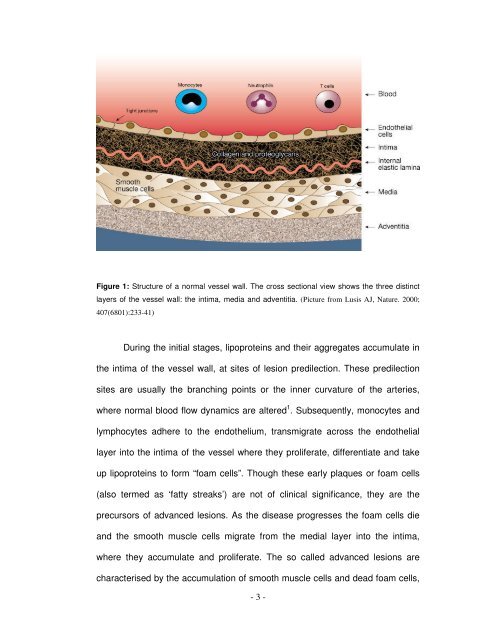
Figure 1: Structure <strong>of</strong> a normal vessel wall. The cross sectional view shows the three distinct<br />
layers <strong>of</strong> the vessel wall: the intima, media and adventitia. (Picture from Lusis AJ, Nature. 2000;<br />
407(6801):233-41)<br />
During the initial stages, lipoproteins and their aggregates accumulate in<br />
the intima <strong>of</strong> the vessel wall, at sites <strong>of</strong> lesion predilection. These predilection<br />
sites are usually the branching points or the inner curvature <strong>of</strong> the arteries,<br />
where normal blood flow dynamics are altered 1 . Subsequently, monocytes and<br />
lymphocytes adhere to the endothelium, transmigrate across the endothelial<br />
layer into the intima <strong>of</strong> the vessel where they proliferate, differentiate and take<br />
up lipoproteins to form “foam cells”. Though these early plaques or foam cells<br />
(also termed as ‘fatty streaks’) are not <strong>of</strong> clinical significance, they are the<br />
precursors <strong>of</strong> advanced lesions. As the disease progresses the foam cells die<br />
and the smooth muscle cells migrate from the medial layer into the intima,<br />
where they accumulate and proliferate. The so called advanced lesions are<br />
characterised by the accumulation <strong>of</strong> smooth muscle cells and dead foam cells,<br />
- 3 -