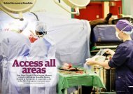
EQUINE CLINICAL PATHOLOGY - Rossdale & Partners
EQUINE CLINICAL PATHOLOGY - Rossdale & Partners
EQUINE CLINICAL PATHOLOGY - Rossdale & Partners

Create successful ePaper yourself
Turn your PDF publications into a flip-book with our unique Google optimized e-Paper software.
G u i d e t o e q u i n e c l i n i c a l p a t h o l o g y<br />
diagnostic aid for lungworm (Dictyocaulus<br />
arnfieldi) infestation, in horses and ponies<br />
coughing at pasture. Bacterial culture can<br />
be useful in identifying specific pathogens<br />
and guiding antimicrobial therapy.<br />
Cerebrospinal (CSF) samples<br />
CSF samples are most commonly collected<br />
in horses showing neurological signs<br />
for the diagnosis and differentiation of<br />
meningitis or traumatic injury. In horses<br />
imported from countries where equine<br />
protozoal myeloencephalitis occurs and<br />
who develop neurological signs, CSF<br />
analysis is indicated.<br />
For a CSF tap, samples are collected from<br />
the atlanto-occipital space with the horse<br />
restrained in lateral recumbency with the<br />
poll flexed. In foals it may be possible to<br />
perform this under heavy sedation whereas<br />
in adults, general anaesthesia is mandatory.<br />
The skin is clipped and prepared as if for<br />
surgical intervention and a bleb of local<br />
anaesthetic is placed in the midline of the<br />
dorsal neck at a level defined by a line<br />
joining the cranial borders or the atlas,<br />
which may be clearly palpated. A sterile<br />
18 gauge 2 inch needle is then inserted<br />
through the skin at 90° and advanced<br />
until it ‘pops’ through the meninges and<br />
CSF drips or pours from the needle into a<br />
sterile container or is aspirated into a sterile<br />
syringe.<br />
Alternatively, in adult horses, CSF may<br />
be collected via the lumbosacral (LS)<br />
space, by lumbosacral tap. The horse is<br />
restrained, sedated, in stocks. The skin<br />
between the tuber coxae is clipped and<br />
prepared as if for surgical intervention. A<br />
sizeable (3-4 ml) bleb of local anaesthetic<br />
is placed in and under the skin in the<br />
midline at a line bisecting the caudal<br />
borders of the tuber coxae. With the<br />
horse standing ’square’ with weight evenly<br />
distributed on both hind legs, a sterile 18<br />
gauge 6 inch spinal needle with stylette in<br />
place is then inserted though the skin in<br />
the midline at the line bisecting the tuber<br />
coxae and down through the palpable<br />
depression just caudal to the sixth lumbar<br />
spinous process. The horse will often flinch<br />
when the subarachnoid space is penetrated<br />
and this is an indication to start aspiration<br />
into a sterile syringe for a fluid sample.<br />
Gross examination of CSF reveals a clear<br />
almost colourless fluid in normality and<br />
a turbid and/or bloodstained fluid with<br />
meningitis or following traumatic injury.<br />
Laboratory examinations of CSF need to be<br />
able to measure very low total nucleated<br />
cell counts (