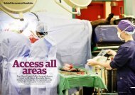
EQUINE CLINICAL PATHOLOGY - Rossdale & Partners
EQUINE CLINICAL PATHOLOGY - Rossdale & Partners
EQUINE CLINICAL PATHOLOGY - Rossdale & Partners

Create successful ePaper yourself
Turn your PDF publications into a flip-book with our unique Google optimized e-Paper software.
G u i d e t o e q u i n e c l i n i c a l p a t h o l o g y<br />
endometrial epithelial cells is used as a<br />
test of smear quality and the presence or<br />
absence of polymorphonuclear leucocytes<br />
is used as the diagnostic test for acute<br />
endometritis.<br />
Smears may be collected during oestrus<br />
by a variety of methods, but we favour<br />
a simple non-guarded technique. An<br />
extended, sterile, large tipped swab is<br />
passed via a sterile speculum, through<br />
the relaxed cervix and rotated onto the<br />
endometrial lining. The swab is withdrawn<br />
and should be rolled onto gelatine-coated<br />
slides immediately and fixed with 'Smearfix',<br />
carbowax or even hair spray prior to<br />
transport to the laboratory for staining.<br />
Excellent results have been obtained by<br />
rolling smears onto 'Testsimplets' (Boehringer<br />
Mannheim UK Ltd.) pre-stained slides,<br />
then after two to three minutes at room<br />
temperature, washing off the background<br />
blue colour, drying and cover slipping. This<br />
technique provides an excellent permanent<br />
smear sample for immediate reading and/or<br />
for sending for a second opinion.<br />
Histology<br />
Using our state-of-the-art microwave<br />
processing system, same day results can be<br />
provided for most fixed tissue specimens.<br />
In general terms, equine lesions are best<br />
sampled, if possible and appropriate,<br />
by total removal and submission<br />
for histopathological processing and<br />
examination. If total removal is not possible,<br />
samples of representative size and location<br />
should be collected by wedge resection.<br />
Fine needle aspirates should only be<br />
collected if it is not possible to collect<br />
larger samples. Experience suggests that<br />
they often result in a traumatised lesion<br />
without a conclusive diagnosis and with<br />
a recommendation for the collection of a<br />
larger and more representative sample,<br />
delaying resolution. Total lesion removal is<br />
always preferable, if possible.<br />
Removed lesions or biopsies should be<br />
placed immediately into an adequate<br />
volume (10 x sample volume) of 10%<br />
formol saline. Reproductive tissues are<br />
best fixed in Bouin’s fluid because of their<br />
higher water content. Large lesions should<br />
be sectioned before submersion to allow<br />
adequate penetration of fixative. Sectioning<br />
should be performed with a thought to how<br />
the sample will need to be trimmed and<br />
orientated for histopathological processing<br />
to allow the pathologist to make a complete<br />
and meaningful appraisal. When sampling<br />
organs and tissues, we recommend that<br />
samples are taken from adjacent, grossly<br />
normal sites as well as from lesional<br />
sites. Notes of your own postmortem<br />
examination findings should be sent to aid<br />
histopathological interpretations. Samples<br />
should be carefully packed for transport to<br />
avoid leakages and breakages.<br />
47