

Buccal fat pad flap - Vula - University of Cape Town
Buccal fat pad flap - Vula - University of Cape Town
Buccal fat pad flap - Vula - University of Cape Town
- No tags were found...

You also want an ePaper? Increase the reach of your titles
YUMPU automatically turns print PDFs into web optimized ePapers that Google loves.
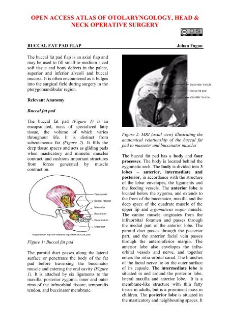
extends up to the inferior orbital fissureand surrounds the temporalis muscle, andextends down to the superior rim <strong>of</strong> themandibular body, and back to the anteriorrim <strong>of</strong> the temporalis tendon and ramus. Indoing so it forms the buccal, pterygopalatineand temporal processes.Four processes (buccal, pterygoid, superficialand deep temporal) extend from thebody into surrounding spaces such as thepterygomandibular and infratemporal fossae.IndicationsReconstruction <strong>of</strong> small to medium(
Surgical StepsSurgery may be done under local orgeneral anaesthesiaThree approaches (Figure 6)o Incise buccal mucosal membrane1cm below the opening <strong>of</strong> parotidduct (Matarasso’s method)o Incise behind the opening <strong>of</strong> parotidduct (Stuzin’s method)o Incise superior gingivobuccal sulcusFigure 7: Careful delivery <strong>of</strong> <strong>fat</strong> <strong>pad</strong> afterincising the capsulePosition the buccal <strong>fat</strong> <strong>pad</strong> <strong>flap</strong> indefect and secure it with absorbablesutures (Figures 8, 9)Cover the <strong>flap</strong> with mucosa if feasible(Figure 9)Figure 6: Position <strong>of</strong> <strong>fat</strong> <strong>pad</strong> relative toparotid ductCut through the buccinator muscle withdiathermy and dissect bluntly until thebuccal <strong>fat</strong> <strong>pad</strong> is foundIncise the thin capsule <strong>of</strong> the buccal <strong>fat</strong><strong>pad</strong>Gently deliver the required volume <strong>of</strong>buccal <strong>fat</strong> tissue into oral cavity bygentle to-and-fro traction on the buccal<strong>fat</strong>, so as not to disrupt the bloodsupply and hence devascularise the <strong>flap</strong>(Figure 7)Take care not to injure the inferiorbuccinator branches <strong>of</strong> facial artery soas to avoid causing a haematomaFreshen the edges <strong>of</strong> the recipient siteFigure 8: Flap placed over an oronasaldefectFigure 9: Flap sutured to defect, andpedicle covered with mucosa3
Await epithelialisation <strong>of</strong> the <strong>flap</strong>which usually occurs within 1 month(Figure 10)Figure 11: Defect filled with buccal fad<strong>pad</strong>SummaryFigure 10: Mucosalised <strong>flap</strong> approximatelya month postoperativelyComplicationsComplications rarely occur and mayinclude partial necrosis and excessivescarring. With large <strong>flap</strong>s used for buccaldefects there is a risk <strong>of</strong> fibrosis andtrismus.Clinical exampleThe buccal <strong>fat</strong> <strong>pad</strong> is a simple, reliable <strong>flap</strong>for repair <strong>of</strong> small-to-medium sized oraldefects. It has an excellent blood supplyand causes minimal donor site morbidity.Author & EditorJohan Fagan MBChB, FCORL, MMedPr<strong>of</strong>essor and ChairmanDivision <strong>of</strong> Otolaryngology<strong>University</strong> <strong>of</strong> <strong>Cape</strong> <strong>Town</strong><strong>Cape</strong> <strong>Town</strong>South Africajohannes.fagan@uct.ac.zaTHE OPEN ACCESS ATLAS OFOTOLARYNGOLOGY, HEAD &NECK OPERATIVE SURGERYwww.entdev.uct.ac.zaFigure 10: <strong>Buccal</strong> <strong>fat</strong> <strong>pad</strong> adjacent tointeralveolar defectThe Open Access Atlas <strong>of</strong> Otolaryngology, Head &Neck Operative Surgery by Johan Fagan (Editor)johannes.fagan@uct.ac.za is licensed under a CreativeCommons Attribution - Non-Commercial 3.0 UnportedLicense4


