Paediatric adenoidectomy surgery technique - Vula - University of ...
Paediatric adenoidectomy surgery technique - Vula - University of ...
Paediatric adenoidectomy surgery technique - Vula - University of ...
Create successful ePaper yourself
Turn your PDF publications into a flip-book with our unique Google optimized e-Paper software.
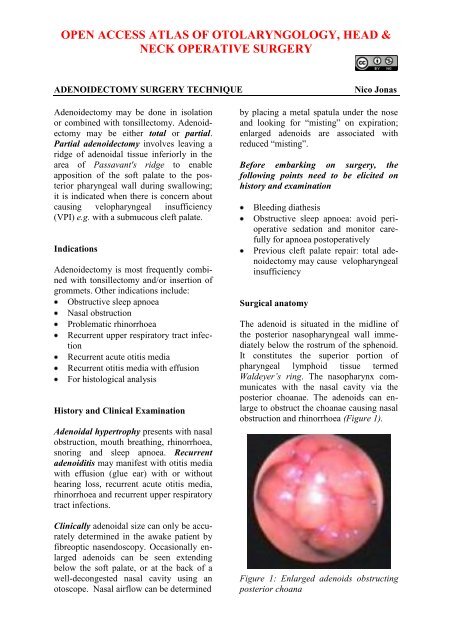
The space lateral to the adenoid and posteromedialto the orifice <strong>of</strong> the Eustachiantube is termed the fossa <strong>of</strong> Rosenmüller.Gerlach’s tonsil refers to a collection <strong>of</strong>lymphoid tissue located within the lip <strong>of</strong>the fossa <strong>of</strong> Rosenmüller and can extendinto the Eustachian tube. Inferiorly, theadenoids abut the upper margin <strong>of</strong> thesuperior constrictor or Passavant’s ridge(Figure 2).(Table 1). This is best achieved by flexiblenasendoscopy in an awake patient (ifpossible) or with a dental mirror placed inthe postnasal space in an anaesthetisedpatient.Grade Choanal obstruction1 < 1/32 1/3 - 2/33 2/3 - 3/3Table 1: Grading system for adenoid sizeSurgical equipmentFigure 2: View from below <strong>of</strong> adenoidsextending superiorly from Passavant’sridge (broken yellow line)The arterial blood supply to the adenoidarises from branches <strong>of</strong> the external carotidartery i.e. ascending pharyngeal, ascendingpalatine, sphenopalatine, pharyngealbranch <strong>of</strong> maxillary artery, and artery <strong>of</strong>the pterygoid canal. Venous drainage is tothe facial and internal jugular systems.Sensory innervation is provided by theglossopharyngeal (IX) and vagus (X)nerves; this explains the referred pain thatpatients experience to the ear and throatwith adenoid infection and following<strong>adenoidectomy</strong>.Grading <strong>of</strong> adenoidal sizeThe percentage <strong>of</strong> choanal obstruction is<strong>of</strong>ten used to grade the size <strong>of</strong> adenoidsAn adenoid curette and/or suctiondiathermy are commonly used to perform<strong>adenoidectomy</strong>. Advantages <strong>of</strong> suction diathermyinclude targeted, directed removal<strong>of</strong> the adenoids avoiding injury to adjacentstructures, clearing choanal adenoidaltissue and haemostasis. Figures 3-5 illustratethe equipment required to perform<strong>adenoidectomy</strong> with a curette and withsuction diathermy.Figure 3: Instruments used to performcurette <strong>adenoidectomy</strong>: curettes, Boyle-Davis gag and tonsil swabs2
clip to retract the s<strong>of</strong>t palate anteriorly(Figure 7)Figure 4: Instruments used to perform<strong>adenoidectomy</strong> with suction diathermy:monopolar suction diathermy, Boyle-Davisgag, dental mirror, Burkitt straight forcepsand suction catheterFigure 6: Boyle Davis gag in placesuspended and held in place withDrafton rodsFigure 5: Drafton rods are used tosuspend the Boyle Davis gag and stabilizethe headPreliminary stepsGeneral anaesthesia with endotrachealintubation using an angled tube or witha laryngeal maskPatient is positioned supine with ashoulder roll to achieve extension <strong>of</strong>the neckA Boyle Davis mouth gag is inserted;ensure that the ventilation tube and tongueare in the midline (Figures 3, 4, 6)Open the gag to expose the oropharynxStabilise the patient’s head in thedesired position by inserting Draftonsuspension rods (Figure 6)To improve the view <strong>of</strong> the nasopharynx,insert a nasal catheter in one/bothnostrils and bring it out through themouth; secure both ends with an arteryFigure 7: Note the nasal catheterretracting s<strong>of</strong>t palate anteriorlyAssess the size <strong>of</strong> the adenoids andexclude an aberrant or dehiscent internalcarotid artery by examining thenasopharynx with a dental mirror and /or by digital palpationPalpate the palate to exclude an occultsubmucous cleft palate; proceedingwith <strong>adenoidectomy</strong> in such cases cancause rhinolalia aperta3
Curette AdenoidectomyUse the largest <strong>adenoidectomy</strong> curettethat will engage the adenoidsUsing a dental mirror aids the surgeonto place the curette under direct visionStabilise the head with the nondominanthandRemove the adenoids with a singlefirm scraping motion from superiorlyto inferiorlyInspect the adenoid bed to assesswhether removal is complete; if not,repeat the process until completeremoval has been achievedPlace swabs/sponges in the postnasalspace while continuing with the tonsillectomyif indicatedRemove the swabs after a few minutesConfirm haemostasis by inspecting thenasopharynx with a mirrorHaemostasis can be achieved by usingmonopolar suction diathermy; tt isabsolutely essential that there iscomplete haemostasisRemove all clots from the nose andnasopharynx with a suction catheterpassed through the noseDocument in the operation notes thathaemostasis has been achieved andclots clearedUsing a combination <strong>of</strong> sweepingmotions and localised "spot welding",remove or ablate the adenoids underdirect vision until a clear view <strong>of</strong> theposterior choanae is obtained (Figure9)Suction Diathermy AdenoidectomyBend the tip <strong>of</strong> the suction diathermyto 90 degrees with the introducer stillin place so as to prevent kinking andocclusion <strong>of</strong> the lumen (Figure 8)Remove the introducer and connectcontinuous suctionSet the monopolar diathermy at 38WattsWith a mirror held in the non-dominanthand, pass the suction diathermybehind the palate4
Postoperative CareAdenoidectomy is commonly performed asa day case procedure. Paracetamol isusually sufficient to control postoperativepain. If suction diathermy was used, broadspectrum antibiotics (co-amoxiclav) isadministered for a week post-operativelyto treat the resulting nasal discharge.Patients are advised to miss school for 5days and generally recover within a week.Complications following <strong>adenoidectomy</strong>EarlyLateBleeding (more commonly with curette<strong>adenoidectomy</strong>)Aspiration <strong>of</strong> retained blood clotcausing acute airway obstruction(Coronor’s clot)Nasal discharge (suction diathermy)Grisel syndrome (atlanto-axial instability)Scarring <strong>of</strong> Eustachian tube openingcausing middle ear dysfunctionNasopharyngeal stenosisVelopharyngeal insufficiencyRegrowth <strong>of</strong> adenoidsAuthor and <strong>Paediatric</strong> Section EditorNico Jonas MBChB, FCORL, MMed<strong>Paediatric</strong> OtolaryngologistAddenbrooke’s HospitalCambridgeUnited Kingdomnico.jonas@gmail.comEditorJohan Fagan MBChB, FCORL, MMedPr<strong>of</strong>essor and ChairmanDivision <strong>of</strong> Otolaryngology<strong>University</strong> <strong>of</strong> Cape TownCape Town, South Africajohannes.fagan@uct.ac.zaTHE OPEN ACCESS ATLAS OFOTOLARYNGOLOGY, HEAD &NECK OPERATIVE SURGERYwww.entdev.uct.ac.zaThe Open Access Atlas <strong>of</strong> Otolaryngology, Head &Neck Operative Surgery by Johan Fagan (Editor)johannes.fagan@uct.ac.za is licensed under a CreativeCommons Attribution - Non-Commercial 3.0 UnportedLicense5


