

Passalora perplexa, an important pleoanamorphic leaf blight ... - CBS
Passalora perplexa, an important pleoanamorphic leaf blight ... - CBS
Passalora perplexa, an important pleoanamorphic leaf blight ... - CBS

Create successful ePaper yourself
Turn your PDF publications into a flip-book with our unique Google optimized e-Paper software.
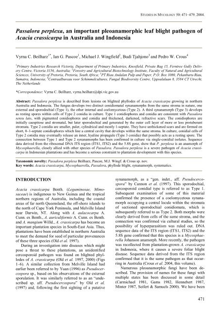
STUDIES IN MYCOLOGY 50: 471–479. 2004.<strong>Passalora</strong> <strong>perplexa</strong>, <strong>an</strong> import<strong>an</strong>t pleo<strong>an</strong>amorphic <strong>leaf</strong> <strong>blight</strong> pathogen ofAcacia crassicarpa in Australia <strong>an</strong>d IndonesiaVyrna C. Beilharz 1* , I<strong>an</strong> G. Pascoe 1 , Michael J. Wingfield 2 , Budi Tjahjono 3 <strong>an</strong>d Pedro W. Crous 41 Primary Industries Research Victoria, Department of Primary Industries, Knoxfield, Private Bag 15, Ferntree Gully DeliveryCentre, Victoria 3156, Australia; 2 Forestry <strong>an</strong>d Agricultural Biotechnology Institute, Faculty of Natural <strong>an</strong>d AgriculturalSciences, University of Pretoria, Pretoria, South Africa; 3 PT Riau Andal<strong>an</strong> Pulp <strong>an</strong>d Paper, P.O. Box 1080, Pek<strong>an</strong>baru-Riau,Sumatra, Indonesia; 4 Centraalbureau voor Schimmelcultures, Fungal Biodiversity Centre, Uppsalala<strong>an</strong> 8, 3584 CT Utrecht,The Netherl<strong>an</strong>ds*Correspondence: Vyrna C. Beilharz, vyrna.beilharz@dpi.vic.gov.auAbstract: <strong>Passalora</strong> <strong>perplexa</strong> is described from lesions on <strong>blight</strong>ed phyllodes of Acacia crassicarpa growing in northernAustralia <strong>an</strong>d Indonesia. The fungus develops two distinct conidiomatal syn<strong>an</strong>amorphs from the same stroma in nature, oneexternal <strong>an</strong>d sporodochial (Type 1), the other internal <strong>an</strong>d coelomycetous (Type 2). A third syn<strong>an</strong>amorph (Type 3) developsas resting spores within cells of Type 2 conidia in culture. Type 1 conidiophores <strong>an</strong>d conidia are consistent with <strong>Passalora</strong>sensu lato, with pigmented conidiophores <strong>an</strong>d conidia <strong>an</strong>d thickened, darkened, refractive scars. The conidiophores areinitially caespitose <strong>an</strong>d stromatal, but later sporodochial <strong>an</strong>d generated by the outer cell layer of more or less protuber<strong>an</strong>tstromata. Type 2 conidia are smaller, paler, cylindrical <strong>an</strong>d mostly 1-septate. They have unthickened scars <strong>an</strong>d are formed onshort, 0–1-septate conidiophores which line a central cavity that develops within the same stroma. In culture, conidial cells ofType 2 conidia may eventually release <strong>an</strong> inner, hyaline propagule (Type 3 conidia) that possibly acts as a resting spore. Theconnection between Type 1 <strong>an</strong>d Type 2 syn<strong>an</strong>amorphs has been confirmed in culture via single-conidial isolates. Sequencedata derived from the ribosomal DNA ITS region (ITS1, ITS2) <strong>an</strong>d the 5.8S gene, show that P. <strong>perplexa</strong> is <strong>an</strong> <strong>an</strong>amorph ofMycosphaerella, closely allied with other species of <strong>Passalora</strong>. <strong>Passalora</strong> <strong>perplexa</strong> is a severe pathogen of Acacia crassicarpain Indonesi<strong>an</strong> pl<strong>an</strong>tations <strong>an</strong>d has become a serious constraint to pl<strong>an</strong>tation development with this species.Taxonomic novelty: <strong>Passalora</strong> <strong>perplexa</strong> Beilharz, Pascoe, M.J. Wingf. & Crous sp. nov.Key words: Acacia crassicarpa, Mycosphaerella, <strong>Passalora</strong>, phyllode <strong>blight</strong>, syn<strong>an</strong>amorph, systematics.INTRODUCTIONAcacia crassicarpa Benth. (Leguminosae, Mimosaceae)is indigenous to New Guinea <strong>an</strong>d the tropicalnorthern regions of Australia, including the coastalareas of far north Queensl<strong>an</strong>d, the off-shore isl<strong>an</strong>ds tothe north of Cape York Peninsula, <strong>an</strong>d Melville Isl<strong>an</strong>dnear Darwin, NT. Along with A. aulacocarpa A.Cunn. ex Benth., A. auriculiformis A. Cunn. ex Benth.<strong>an</strong>d A. m<strong>an</strong>gium Willd., A. crassicarpa has become <strong>an</strong>import<strong>an</strong>t pl<strong>an</strong>tation species in South-East Asia. Thus,pl<strong>an</strong>tations have been established in northern Australiato meet the dem<strong>an</strong>d for seed of particular proven<strong>an</strong>cesof these three species (Old et al. 1997).During <strong>an</strong> investigation into diseases which mightpose a threat to these pl<strong>an</strong>tations, <strong>an</strong> unidentifiedcercosporoid pathogen was found on <strong>blight</strong>ed phyllodesof A. crassicarpa (Old et al. 1997, 2000) (Figs1–6). A similar collection from Melville Isl<strong>an</strong>d hadearlier been referred to by Yu<strong>an</strong> (1996) as Pseudocercosporasp., based on his observations of the externalsporulation. It was similarly referred to as <strong>an</strong> “undescribedsp. aff. Pseudocercospora” by Old et al.(1997) <strong>an</strong>d, following the first sighting of a putativesyn<strong>an</strong>amorph, as a “gen. indet., aff. Pseudocercospora”by C<strong>an</strong>non et al. (1997). This sporodochial,cercosporoid conidial type is referred to as Type 1.Subsequent examination of some of this materialconfirmed the presence of a coelomycetous syn<strong>an</strong>amorphoccupying a central locule within the stromataof sectioned sporodochial conidiomata, which issubsequently referred to as Type 2. Both morphs wereclearly derived from cells of the same stroma, <strong>an</strong>d theconnection was confirmed via cultural studies, so thepossibility of hyperparasitism was ruled out. DNAsequence data of the ITS region (ITS1, ITS2) <strong>an</strong>d the5.8S gene confirmed that this species is a MycosphaerellaJoh<strong>an</strong>son <strong>an</strong>amorph. More recently, the pathogenwas recollected from pl<strong>an</strong>tation-grown A. crassicarpain Indonesia, where it causes a severe foliar <strong>blight</strong>disease. Sequence data derived from the ITS regionconfirmed that it is the same pathogen as that occurringin Australia (Crous et al. 2004, this volume).Numerous pleo<strong>an</strong>amorphic fungi have been described.The provision of names for these fungi withmultiple states has been discussed in some detail(Carmichael 1981, Gams 1982, Hennebert 1987,Minter 1987, Seifert & Samuels 2000). We have been471
BEILHARZ ET AL.unable to find <strong>an</strong>y combination of syn<strong>an</strong>amorphs thatwould equate with the cercosporoid fungus infectingphyllodes of A. crassicarpa <strong>an</strong>d described here. Thecorrect or most logical me<strong>an</strong>s of treating the nomenclatureof pleo<strong>an</strong>amorphic fungi remains somewhatsubjective, <strong>an</strong>d has not been defined in the Code ofBot<strong>an</strong>ical Nomenclature. The well-known genericname <strong>Passalora</strong> Fr. sensu Crous & Braun (2003) iscorrect for the hyphomycetous syn<strong>an</strong>amorph, <strong>an</strong>d it isparticularly useful in this case because most coloniesof the fungus show liberal <strong>an</strong>d conspicuous sporulationof this morph, whereas the cryptic Type 2 syn<strong>an</strong>amorphis less likely to be observed, <strong>an</strong>d isunlikely to be found in isolation. Gams (1982) suggestedthat the <strong>an</strong>amorph form with the greatest differentiationshould have priority (unless it is rare), aview which further supports the application of thename <strong>Passalora</strong> to the fungus on A. crassicarpa.The conidia of the Type 1 <strong>an</strong>d Type 2 syn<strong>an</strong>amorphsof the cercosporoid fungus from A. crassicarpa,although easily distinguished, show somesimilarity in morphology. The Type 2 conidia aresomewhat cercosporoid in type <strong>an</strong>d reminiscent ofsome species of Colletogloeum Petr., but the conidiomatado not fit with the acervular conidiomata of thatgenus. Critical differences between the two types ofconidia, including pigmentation, c<strong>an</strong> be linked to theirrelative positions in relation to the host tissue. Forexample, the thickened hila of Type 1 conidia areprobably associated with their readiness to secede(Beilharz 1994). In contrast, the broader, unthickenedhila of Type 2 conidia are more appropriate to passiverelease following breakdown of overlying fungal <strong>an</strong>dhost tissues.Acacia crassicarpa is a species that has becomeincreasingly import<strong>an</strong>t in pl<strong>an</strong>tations in various partsof South-East Asia, where it is grown specifically forthe production of pulp. The relatively recent outbreakof a serious <strong>leaf</strong> <strong>blight</strong> disease caused by a cercosporoidfungus has dem<strong>an</strong>ded <strong>an</strong> appropriate taxonomictreatment of this org<strong>an</strong>ism. This study represents acollaborative effort by a number of research groupswho have <strong>an</strong> interest in this fungus <strong>an</strong>d the diseasewith which it is associated. Here, we describe a potentiallydevastating, newly recognised disease of A.crassicarpa, <strong>an</strong>d describe the causal org<strong>an</strong>ism, a novelpleo<strong>an</strong>amorphic species of <strong>Passalora</strong>.MATERIALS AND METHODSIsolatesAt VPRI, cultures were derived from the Australi<strong>an</strong>specimen VPRI 21125 by the following me<strong>an</strong>s. Naturally-producedType 1 conidia were lifted from lesionsen masse with a fine needle <strong>an</strong>d jab-inoculated on to 2% potato-dextrose agar (PDA; Difco) plates emendedwith achromycin (0.05 mg/mL) (PDA+A). Similarly472harvested Type 1 conidia were also suspended in adrop of water containing a trace of Tween 80, streakedout on to 2 % tap water agar <strong>an</strong>d tr<strong>an</strong>sferred individuallyto PDA+A the following day, after germinating.Type 2 conidia formed in vitro in PDA cultures derivedfrom individual Type 1 conidia were used toprovide single-conidial isolates as described above. Inaddition, whole, pale, smooth protuber<strong>an</strong>t stromatalacking external conidiophores <strong>an</strong>d putatively containingType 2 conidia, were lifted from the phyllodesurface with a fine, sterile needle <strong>an</strong>d placed directlyonto PDA+A. All PDA+A cultures were tr<strong>an</strong>sferred toPDA after 3–7 d <strong>an</strong>d grown on for up to 2 mo in thedark at 22 °C. The choice of the various forms ofinoculum was determined by the ease with which theycould be harvested from infected phyllodes or cultures.For example, Type 1 conidia were abund<strong>an</strong>t onthe natural subtrate, but occurred in comparativelysmall numbers in culture, where they were liable to becontaminated with Type 2 conidia. Type 2 conidiawere abund<strong>an</strong>t in wet masses in culture; in nature,however, they tended to remain aggregated <strong>an</strong>d oftencould not be completely freed from excised conidiomata,despite the application of pressure on coverslipsor attempts to tease the elements apart in a drop ofwater on a microscope slide. Type 3 conidia were notseen in 2-mo-old cultures on PDA, <strong>an</strong>d Type 2 conidiafrom these cultures germinated normally on fresh agarplates. The Australi<strong>an</strong> specimens <strong>an</strong>d a dried cultureof VPRI 21125 have been deposited in herb. VPRI,Knoxfield, Victoria, Australia.At <strong>CBS</strong>, single Type 1 conidial isolates werederived from Indonesi<strong>an</strong> specimens <strong>an</strong>d cultivated on2 % malt extract agar (MEA; Difco) as described byCrous (1998). Colonies sporulated on MEA after 1–2mo incubation on the laboratory bench in daylight atroom temperature, forming conidiomata containingType 1 <strong>an</strong>d Type 2 conidia. After 3 mo incubation onMEA, conidiomata with Type 2 conidia were observedto also give rise to Type 3 conidia, a formobserved only in culture. Specimens <strong>an</strong>d cultures havebeen deposited in the herbarium <strong>an</strong>d culture collectionof <strong>CBS</strong> in Utrecht, the Netherl<strong>an</strong>ds.A phylogeny of the cercosporoid fungi occurringon Acacia, including <strong>Passalora</strong> <strong>perplexa</strong>, is presentedelsewhere in this volume (Crous et al. 2004).MorphologySlide preparations were made in lactic acid <strong>an</strong>d 50examples of each structure were measured under a×100 oil immersion lens using Olympus BH–2 (VPRI)or Zeiss Axioskop (<strong>CBS</strong>) light microscopes. The 95 %confidence intervals were also determined for conidialdimensions, with the extremes in conidium length <strong>an</strong>dwidth given in parentheses. Colony colour was determinedon 2 % MEA after 3 mo at 25 °C in the darkusing the colour designations of Rayner (1970).
PASSALORA PERPLEXA AND ITS SYNANAMORPHSFigs 1–6. <strong>Passalora</strong> <strong>perplexa</strong>. 1. Trees showing defoliation. 2–6. Symptoms associated with Crassicarpa <strong>leaf</strong> <strong>blight</strong>.RESULTSDisease symptomsLesions occur primarily on the phyllodes of A. crassicarpabut they c<strong>an</strong> also form on the petioles <strong>an</strong>dyoung shoots. Phyllode lesions are initially small <strong>an</strong>dtypically elliptical, <strong>an</strong>d are surrounded by a distinctchlorotic halo (Figs 1–6). On freshly formed lesions,fascicles of grey-brown conidiophores <strong>an</strong>d denseolivaceous spore masses c<strong>an</strong> easily be seen. Lesionsformed at the edges of phyllodes or abutting primaryveins c<strong>an</strong> cause severe malformation <strong>an</strong>d curling ofthe phyllodes (Figs 2–6). Infections are often severe,causing the dramatic malformation of the apicalportions of young (1–2-yr-old) trees (Fig. 1).TaxonomySequences obtained for the ITS region in the laboratoriesof both VPRI <strong>an</strong>d <strong>CBS</strong>, confirmed that the Australi<strong>an</strong><strong>an</strong>d Indonesi<strong>an</strong> specimens represented the sametaxon. The DNA sequence <strong>an</strong>alyses also showed thatthe fungus is <strong>an</strong> <strong>an</strong>amorph of Mycosphaerella, clusteringwith Cercospora lor<strong>an</strong>thi McAlpine (Crous et al.2004, fig. 1), which is a species of <strong>Passalora</strong>. Theserelationships have been discussed elsewhere (V.C.Beilharz, in press). Sequences of P. <strong>perplexa</strong> havebeen deposited in GenB<strong>an</strong>k, <strong>an</strong>d the alignment ofsequence data in TreeBASE (Crous et al. 2004).The habit, morphology, pigmentation <strong>an</strong>d scarcharacteristics of Type 1 conidiophores <strong>an</strong>d conidiaare characteristic of the genus <strong>Passalora</strong>. This observationis consistent with the results of the DNA-basedcomparisons. Currently there are no species of <strong>Passalora</strong>known from Acacia (Crous & Braun 2003), <strong>an</strong>dhence this species with its pigmented Type 1 conidia<strong>an</strong>d thickened, darkened, refractive conidial hila c<strong>an</strong>be described as new. Prior to the discovery of additionalcoelomycetes resembling the Type 2 syn<strong>an</strong>amorph<strong>an</strong>d their affiliations being established, it wouldbe inappropriate to provide a separate generic namefor the Type 2 syn<strong>an</strong>amorph or to name this syn<strong>an</strong>amorph.<strong>Passalora</strong> <strong>perplexa</strong> Beilharz, Pascoe, M.J. Wingf.& Crous, sp. nov. MycoB<strong>an</strong>k MB500123. Figs7–27.Etymology: Named because of the unusual combinationof conidial syn<strong>an</strong>amorphs.Fungus pleo<strong>an</strong>amorphicus conidia generis <strong>Passalora</strong>e etcoelomycitica form<strong>an</strong>s. Conidiophora solitaria vel laxeaggregata, pallide vel medio-brunnea, levia vel verruculosa,subcylindrica, ramosa vel simplicia, pluriseptata, sympodialiterproliferentia, 15–80(–116) µm longa, 3–5 µm lata.Cellulae conidiogenae terminales, verruculosae vel rugosae,simplices, subcylindricae, apicem rotundatum versus<strong>an</strong>gustatae, 15–20 × 3–4 µm; cicatrices modice inspissataeet fuscatae, refringentes, 1–2 µm diam. Conidia solitaria,pallide olivacea vel medio-brunnea, levia vel eximie verruculosa,recta vel curvata, <strong>an</strong>guste obclavata vel subcylindri-473
BEILHARZ ET AL.ca, sursum obtusa, ad basim longe obconice subtruncata,(1–)3–9(–13)-septata, (16–)50–100(–153) µm longa, 2.5–5.5 µm lata, hilo modice inspissato et fuscato, refringente,1–2 µm diam.Holotype: Indonesia, South Sumatra, Kerinci, Herb. <strong>CBS</strong>9907 holotype, on phyllodes of Acacia crassicarpa, Feb.2004, M.J. Wingfield, ex-type cultures <strong>CBS</strong> 116363 = STE-U 11147–11149.Pleo<strong>an</strong>amorphic, producing stromatic conidiomata onphyllodes of Acacia crassicarpa. Leaf spots hologenous,initially pale brown, orbicular <strong>an</strong>d non-necrotic,becoming medium brown, necrotic, elongated, narrowlyellipsoidal to sub-circular, with <strong>an</strong> inconspicuousborder, often distorted <strong>an</strong>d wrinkled <strong>an</strong>d causingdistortion of the phyllode, limited by main secondaryveins, to at least 15 mm long, 2–5 mm wide. Myceliuminternal, consisting of smooth, br<strong>an</strong>ched, septate,brown hyphae, 3–4 µm wide. Stromata mediumbrown throughout or r<strong>an</strong>ging from brown in the exposedapical cells to hyaline or sub-hyaline in thedeeper tissues, initiated in the substomatal cavity,usually becoming erumpent, protuber<strong>an</strong>t <strong>an</strong>d pulvinate,composed of textura <strong>an</strong>gularis, 50–80µm wide,50–90 µm high. Conidiomata amphigenous, eustromatic,comprising either (a) pale-yellowish, fleshy,protuber<strong>an</strong>t stromata containing Type 2 conidia but, atleast initially, bearing no Type 1 conidiophores; (b)brown immersed or erumpent stromata bearing Type 1conidiophores but, at least initially, containing noType 2 conidia, or (c) mature brown conidiomata, upto 80 µm diam, 60 µm high, bearing numerous Type 1conidiophores <strong>an</strong>d containing numerous Type 2 conidia.Type 1 syn<strong>an</strong>amorph: Conidiophores occasionallysolitary, usually aggregated in loose fascicles arisingfrom the upper cells of a brown stroma, up to at least62 in number, pale to medium brown, smooth towardsthe base <strong>an</strong>d often becoming rugose towards the apexwith age, subcylindrical, br<strong>an</strong>ched or unbr<strong>an</strong>ched,walls slightly thickened, straight to variously curvedor geniculate-sinuous, having a basal septum <strong>an</strong>d 0–11additional septa; proliferation sympodial, with endohyphalregeneration or proliferation also commonlyexhibited, 15–80(–116) µm long, 3–5 µm wide. Conidiogenouscells terminal, verruculose or rugose,unbr<strong>an</strong>ched, subcylindrical, tapering to roundedapices proliferating sympodially, 15–20 × 3–4 µm.Conidiogenous scars slightly thickened <strong>an</strong>d darkened,refractive, flat against the side of the conidiophore, onshort pegs or on sloping shoulders following proliferationof the conidiogenous cell, sometimes protruber<strong>an</strong>t,often somewhat disguised by the dark, rugosewall of mature conidiophores but clearly seen onpaler, more newly generated conidiogenous cells, 1–2µm diam. Conidia solitary, pale olivaceous to mediumbrown, dry, smooth, rarely finely verruculose, straightor curved, narrowly obclavate to sub-cylindrical,474tapering gradually to <strong>an</strong> obtuse apex <strong>an</strong>d to a roundedor long-obconically-subtruncate base, often constrictedat one or more septa or with <strong>an</strong> otherwiseuneven edge-line, (1–)3–9(–13)-septate, (16–)50–100(–153) µm long, <strong>an</strong>d 4–4.5(–5.5) µm wide in vivo(Australi<strong>an</strong> specimens), or (2.5–)3–4 µm wide (Indonesi<strong>an</strong>specimens). Secondary conidiation was occasionallyseen. Hila slightly but distinctly thickened<strong>an</strong>d darkened, refractive, 1–2 µm diam.Fig. 7. Conidia of <strong>Passalora</strong> <strong>perplexa</strong> in vivo. Scale bar =10 µm.Fig. 8. Type 2 conidia <strong>an</strong>d conidiophores of <strong>Passalora</strong><strong>perplexa</strong> in vivo. Scale bar = 10 µm.
PASSALORA PERPLEXA AND ITS SYNANAMORPHSType 2 syn<strong>an</strong>amorph: Conidiophores reduced, hyalineto sub-hyaline, 0–1-septate, lining a single, initiallyill-defined cavity, which develops within either asubstomatal or protuber<strong>an</strong>t stroma exactly as describedabove. Conidia initially hyaline <strong>an</strong>d inconspicuous,later pale olivaceous, ± cylindrical, barely ifat all tapering to the apex or the base, sometimesswollen at the apex or broadening to the base, occasionallyconstricted, smooth, (0–)1(–3) septate,(12–)15–21(–25) µm long, 2.5–4 µm wide; hila broad,truncate to slightly convex, not darkened, unthickened,non-refractive, 2–2.5 µm diam. No pore or slithas been detected that would allow ready release ofType 2 conidia. On the other h<strong>an</strong>d, the contents ofcertain old conidiomata have become exposed by theapparent breakdown <strong>an</strong>d peeling back of both fungal<strong>an</strong>d host tissues. These conidiomata eventually resembleacervuli, although they are no longer activelysporulating. It is possible that Type 2 conidia dependon tissue breakdown for their dissemination, <strong>an</strong>d thatinsects or other <strong>an</strong>imals may aid their dispersal.Type 3 syn<strong>an</strong>amorph: After 1 mo, Type 2 conidiafrom 3-mo-old MEA cultures exposed to daylightdevelop thick-walled hyphal swellings (reminiscent ofchlamydospores that develop in conidial cells ofcertain Fusarium spp.); these inner propagule cellseventually burst free from the cells of the Type 2conidia, frequently still having pigmented remn<strong>an</strong>ts ofthe conidial wall attached to their hyaline walls. Type3 conidia are 6–20 x 4–6 µm, 0(–1)-septate, ellipsoid<strong>an</strong>d hyaline. Type 3 conidia did not develop in 2-mooldPDA cultures grown mostly in the dark.Cultural characteristics: Colonies slow-growing,reaching up to 20 mm diam after 3 mo on 2 % MEA at25 °C under near-UV light; colonies erumpent, marginsfeathery, irregular; outer region (surface) sepia(15”k) due to submerged, radiating mycelium; innerregion whitish to cream, with slimy sporodochialspore masses, fuscous black (7””k); central regionwith moderate hazel (17”’i) aerial mycelium; reversebrown-vinaceous (5”’m).(ex DFR 162), VPRI 20901; Melville Isl<strong>an</strong>d, pl<strong>an</strong>tationproven<strong>an</strong>ce, 17 Sept. 1992, K.M. Old (ex DFR 138), VPRI20903.Fig. 9. Type 1 conidiophores of <strong>Passalora</strong> <strong>perplexa</strong> in vivo.A. Conidiophores emerging from stomata. B. Sporodochialconidiophores. C. Conidiophores arising from a subcuticularhypha. Scale bar = 10 µm.Substrate <strong>an</strong>d distribution: Pathogenic to phyllodes ofAcacia crassicarpa; Australia, Indonesia.Additional specimens examined: All on phyllodes of Acaciacrassicarpa, Indonesia, Southern Sumatra, Kerinci, Feb.2004, M.J. Wingfield, herb. <strong>CBS</strong> 9908, 9909, 9911, culturesderived from <strong>CBS</strong> 9911, <strong>CBS</strong> 116364 = STE-U11150–11151. Australia, Queensl<strong>an</strong>d, Cooktown–CapeTribulation Hwy, 4 km W of Bloomfield, 8 Apr. 1995,K.M. Old (ex DFR 257) (CSIRO Forestry <strong>an</strong>d ForestProducts herbarium, C<strong>an</strong>berra Australia), VPRI 20902; 5km W of Bloomfield, 8 Apr. 1995, K.M. Old (ex DFR 252),VPRI 20904; Ingham, Shell trial site, L<strong>an</strong>nercost StateForest, 3 Apr. 1995, K.M. Old (ex DFR 255), VPRI 20905;Edmund Kennedy National Park, K.M. Old (ex DFR 305),4 Apr. 1995, VPRI 20906; Ingham, Shell trial site, L<strong>an</strong>nercostState Forest, K.M. Old (#6), 9 Apr. 1996, VPRI 21125; N.T., Yapilika, Melville Isl<strong>an</strong>d, 17 May 1994, Z.Q. Yu<strong>an</strong>,Fig. 10. Conidia of <strong>Passalora</strong> <strong>perplexa</strong> in vitro on PDA.Type 1 conidia (top), <strong>an</strong>d Type 2 conidia (bottom). Scalebar = 10 µm.475
BEILHARZ ET AL.Notes: In culture, colonies sporulate on MEA after 1–2 mo, forming sporodochia situated on globose conidiomatathat are pycnidioid but lacking a clearostiole. Conidia formed on sporodochia are of Type 1.A conidial type intermediate between Type 1 <strong>an</strong>dType 2 was also observed. These conidia were initiallyhyaline, becoming medium brown, smooth,cylindrical to subcylindrical, apex obtuse, base obconicallysubtruncate with a darkened (not thickened<strong>an</strong>d refractive as in Type 1) scar, <strong>an</strong>d a minute marginalfrill, 20–35 × 3–6 µm, (1–)3(–4)-septate. Insidethe conidiomata Type 2 conidia were formed. After 3mo, these conidia were observed to give rise to Type 3conidia which were 0(–1)-septate, ellipsoid <strong>an</strong>d hyaline.Both Type 1 <strong>an</strong>d Type 2 conidia were also generatedin cultures on PDA held in the dark for 2 mo.When the superficial mycelium was lifted away from2.5-mo-old colonies, wet, dark masses of Type 2conidia were found in small, well-defined cavities inthe mycelium. The conidia resembled naturally producedType 2 conidia in shape, pigmentation <strong>an</strong>d scarcharacteristics, but were longer, broader, <strong>an</strong>d up to 5-septate. Type 2 conidia were sparsely present inmounts of both superficial hyphae <strong>an</strong>d the fine, featherymycelium at the more or less flat colony margins.They had been produced terminally on lateral conidiophoresor hyphae of indeterminate length. Type 1conidia produced in vitro were smooth to verruculose(the latter especially towards the base), occasionallyverrucose, shorter <strong>an</strong>d fewer-septate th<strong>an</strong> those producedin vivo, but similar in shape <strong>an</strong>d width <strong>an</strong>dexhibiting the narrow, darkened, slightly thickenedhila of conidia produced in vivo. The outer wall layerof these Type 1 conidia was often slightly retractedfrom the hilum. Cottony mycelium from the colonysurfaces often contained a few small, dense hyphalaggregates composed largely of clusters of shortconidiogenous cells producing Type 2 conidia. Theseconidia tended to be straighter th<strong>an</strong> Type 2 conidiaproduced en masse, whether in vivo or in vitro.DISCUSSIONIn this study, we have provided a name for the import<strong>an</strong>tfungal pathogen that causes <strong>leaf</strong> <strong>blight</strong> specificallyon Acacia crassicarpa. <strong>Passalora</strong> <strong>perplexa</strong> ispresent both in Australia, where it is apparently native,<strong>an</strong>d in the extensive pl<strong>an</strong>tations in Indonesia to whichit has spread. In pl<strong>an</strong>tations, the disease associatedwith this fungus c<strong>an</strong> be very severe <strong>an</strong>d it is likely toprovide signific<strong>an</strong>t challenges for forestry comp<strong>an</strong>iesthat pl<strong>an</strong>t A. crassicarpa. Having a name for thepathogen that causes Crassicarpa <strong>leaf</strong> <strong>blight</strong> is <strong>an</strong>import<strong>an</strong>t step towards the recognition of the disease<strong>an</strong>d the development of m<strong>an</strong>agement strategies to dealwith it.Crassicarpa <strong>leaf</strong> <strong>blight</strong> was first noted on MelvilleIsl<strong>an</strong>d, Northern Territory, Australia in 1996 (Yu<strong>an</strong>1996). There has been some confusion regarding thetaxonomy of this fungus, particularly because verylittle work has been done on the taxonomy of <strong>leaf</strong>pathogens of tropical Acacia spp. Thus two fungi, aspecies of Pseudocercospora <strong>an</strong>d a species of Cercospora,were recorded on the leaves of A. crassicarpa<strong>an</strong>d A. m<strong>an</strong>gium respectively. Although the fungi arereadily distinguished, there has been confusion in thefield regarding the causes of the two diseases. <strong>Passalora</strong><strong>perplexa</strong>, described in this study, may be specificto A. crassicarpa, <strong>an</strong>d certainly appears unable toinfect A. m<strong>an</strong>gium, which shows no signs of thedisease even when pl<strong>an</strong>ted in close proximity toheavily infected A. crassicarpa trees.The pleo<strong>an</strong>amorphy displayed by P. <strong>perplexa</strong> isunusual in that one form could be characterised as ahyphomycete, a second form represents a coelomycete,<strong>an</strong>d a third morph appears to represent a restingspore form. Because of this, care was taken to demonstrateunequivocally that Type 1 <strong>an</strong>d Type 2 conidiadid indeed belong to the same fungus. Careful characterisationof the relationship between morphs includedsingle-spore culturing <strong>an</strong>d connection of the variousforms based on DNA sequence data. The conidiophoresof the Type 1 <strong>an</strong>d Type 2 morphs are generated,often concurrently, by cells of the same stroma.This appears to be a unique feature. Type 3 conidiawere only observed in certain cultures, <strong>an</strong>d we suspectthat this conidial form is associated with the growthmedium or conditions of incubation.Different <strong>an</strong>amorphs in single fungal taxa oftendevelop on conidiogenous cells having distinctlydifferent morphological forms. These are often associatedwith different functions such as, for example, raindispersal (conidia in wet slimy masses), winddispersal(conidia thin-walled, dry) <strong>an</strong>d survival(conidia less numerous, larger, pigmented <strong>an</strong>d thickwalled)(Carmichael 1981, Seifert & Samuels 2000).The conidia of the syn<strong>an</strong>amorphs of P. <strong>perplexa</strong> differin pigmentation <strong>an</strong>d wall thickness, as well as in theirmode of liberation. The hyphomycetous (Type 1)conidia are ideally suited to wind dispersal, <strong>an</strong>d aretypical of cercosporoid fungi. The coelomycetous(Type 2) conidia probably require moisture for dispersal,<strong>an</strong>d the Type 3 conidia, which c<strong>an</strong> form inside theconidial cells of Type 2 conidia in culture, are probablyassociated with longer term survival.476
PASSALORA PERPLEXA AND ITS SYNANAMORPHSFigs 11–24. <strong>Passalora</strong> <strong>perplexa</strong>. 11. Vertical section through <strong>an</strong> array of conidiomata. 12. Particularly long Type 1 conidiophoreson excised sporodochial conidioma. 13. Type 1 conidiophores on sporodochial conidioma. 14, 15. Vertical sectionthrough substomatal conidiomata showing Type 1 conidiophores <strong>an</strong>d Type 2 conidia. 16–17. Vertical sections through conidiomatacontaining Type 2 conidia but lacking Type 1 conidiophores. 18. Vertical section through conidioma. 19. Type 1conidium in vivo. 20–22. Type 1 conidia in vitro. 23, 24. Type 2 conidia in vitro. Scale bars = 10 µm.477
BEILHARZ ET AL.Figs 25–27. Type 2 conidia of <strong>Passalora</strong> <strong>perplexa</strong> forming Type 3 conidia (arrows) in vitro. Scale bars = 5 µm.There appears to be only one previously describedpleo<strong>an</strong>amorphic cercosporoid fungus. Alcorn (1992)described Parapithomyces clitoriae Alcorn, whichproduced a Pseudocercospora Speg. syn<strong>an</strong>amorphsporulating from epigenous, erumpent stromata onleaves of Clitoria sp. In contrast, the Parapithomycesmorph sporulated from conidiophores borne on hypogenous,superficial hyphae. Both spore types of thisfungus were produced in culture from single Pseudocercosporasp. conidia. Intermediate spore types werealso found, as indeed occurred in P. <strong>perplexa</strong>, in bothinst<strong>an</strong>ces emphasising the validity of the link betweenthe respective syn<strong>an</strong>amorphs. In contrast to <strong>Passalora</strong><strong>perplexa</strong>, both <strong>an</strong>amorphs of Parapithomyces clitoriaewere hyphomycetes <strong>an</strong>d their conidiophores wereindistinguishable from each other.Acacia crassicarpa has become one of the mostwidely pl<strong>an</strong>ted pl<strong>an</strong>tation tree species in the tropics<strong>an</strong>d various forestry comp<strong>an</strong>ies depend on it for theproduction of pulp. Early pl<strong>an</strong>tings of this tree werevirtually free of disease. Thus the wide-scale appear<strong>an</strong>ceof <strong>leaf</strong> <strong>blight</strong> caused by P. <strong>perplexa</strong>, particularlyin Sumatra, is of considerable concern. Earlier recordsof Australi<strong>an</strong> collections were referred to Pseudocercospora(Yu<strong>an</strong> 1996, Old et al. 1997), as the Type 2syn<strong>an</strong>amorph had not been observed. The presentstudy represents the first detailed taxonomic evaluationof the causal agent of Crassicarpa <strong>leaf</strong> <strong>blight</strong>.The distribution of P. <strong>perplexa</strong> on native A. crassicarpain Australia suggests that it may be indigenousacross the humid tropical north of Australia (Old et al.1997) <strong>an</strong>d that it has been accidentally introduced intoIndonesia. Although there is no direct proof that this isthe case, we believe that the pathogen has been movedwith seed. This appears to be typical of Mycosphaerellaspp. such as those on Eucalyptus leaves that havebeen widely distributed throughout the world, largelyin the absence of <strong>an</strong>y movement of pl<strong>an</strong>ts. The fungi478might not specifically occur on seeds, but seed consignmentsoften include fragments of leaves <strong>an</strong>d fruitsthat bear fruiting structures of the pathogens. Greatcare should thus be taken in the future to prevent themovement of additional new <strong>an</strong>d devastating pathogensof forest trees (Wingfield et al. 2001).Virtually nothing is known regarding the biologyof P. <strong>perplexa</strong>, <strong>an</strong>d this alone represents <strong>an</strong> import<strong>an</strong>tconstraint to efforts to control the <strong>leaf</strong> <strong>blight</strong> diseasethat it causes. The epidemiology of Crassicarpa <strong>leaf</strong><strong>blight</strong> will need to be elucidated in order that m<strong>an</strong>agementstrategies to reduce its impact may be implemented.The presence of healthy trees alongsideseverely <strong>blight</strong>ed individuals suggests that subst<strong>an</strong>tialopportunity exists to breed <strong>an</strong>d select for toler<strong>an</strong>ce tothis disease.ACKNOWLEDGEMENTSJames Cunnington, Rebecca Mills (VPRI) <strong>an</strong>d EwaldGroenewald (<strong>CBS</strong>) are th<strong>an</strong>ked for valuable assist<strong>an</strong>ce inproviding the sequence data for specimens from Australia<strong>an</strong>d Indonesia, respectively. Mark Dudzinski, Division ofForestry <strong>an</strong>d Forest Products, CSIRO, C<strong>an</strong>berra, Australia,is th<strong>an</strong>ked for kindly providing VPRI with the Australi<strong>an</strong>specimens. Walter Gams is also th<strong>an</strong>ked for providing theLatin diagnosis.REFERENCESAlcorn JL (1992). Parapithomyces clitoriae sp. nov.(Fungi: Hyphomycetes) <strong>an</strong>d its Pseudocercospora <strong>an</strong>amorph.Australi<strong>an</strong> Systematic Bot<strong>an</strong>y 5: 711–715.Beilharz V (1994). Cercosporoid fungi on Australi<strong>an</strong> nativepl<strong>an</strong>ts. PhD Dissertation, University of Melbourne.C<strong>an</strong>non P, Pascoe I, Beilharz V, Yu<strong>an</strong> Z-Q (1997). Reporton fungi from diseased acacia samples examined at In-
PASSALORA PERPLEXA AND ITS SYNANAMORPHSstitute of Horticultural Development, Knoxfield Victoria.In: Diseases of tropical acacias (Old KM, Lee SS,Sharma JK, eds). Proceedings of <strong>an</strong> international workshop,Sub<strong>an</strong>jeriji (South Sumatra), 28 April – 3 May1996. Center for International Forestry Research specialpublication, Jakarta: 108–113.Carmichael JW (1981). Pleomorphism. In: Biology ofconidial fungi (Cole CT & Kendrick WB, eds). AcademicPress: London, U.K. 1: 135–143.Crous PW (1998). Mycosphaerella spp. <strong>an</strong>d their <strong>an</strong>amorphsassociated with <strong>leaf</strong> spot diseases of Eucalyptus.Mycologia Memoir 21: 1–170.Crous PW, Braun U (2003). Mycosphaerella <strong>an</strong>d its <strong>an</strong>amorphs.1. Names published in Cercospora <strong>an</strong>d <strong>Passalora</strong>.<strong>CBS</strong> Biodiversity Series 1: 1–571.Crous PW, Groenewald JZ, Pongp<strong>an</strong>ich K, Himam<strong>an</strong> W,Arz<strong>an</strong>lou M, Wingfield MJ (2004). Cryptic speciation<strong>an</strong>d host specificity among Mycosphaerella spp. occurringon Australi<strong>an</strong> Acacia species grown as exotics inthe tropics. Studies in Mycology 50: 457–469.Gams, W. (1982). Generic names for syn<strong>an</strong>amorphs?Mycotaxon 15: 459–464.Hennebert GL (1987). Pleo<strong>an</strong>amorphy <strong>an</strong>d its nomenclaturalproblem. In: Pleomorphic fungi: the diversity <strong>an</strong>d itstaxonomic implications (Sugiyama J ed.). Kod<strong>an</strong>shaLtd., Tokyo & Elsevier: Amsterdam: 263–290.Minter DW (1987). The signific<strong>an</strong>ce of conidiogenesis inpleo<strong>an</strong>amorphy. In: Pleomorphic fungi: the diversity<strong>an</strong>d its taxonomic implications (Sugiyama J ed.). Kod<strong>an</strong>shaLtd., Tokyo & Elsevier: Amsterdam: 241–262.Old KM, Hood IA, Yu<strong>an</strong> Z-Q (1997). Diseases of tropicalacacias in northern Queensl<strong>an</strong>d. In: Diseases of tropicalacacias (Old KM, Lee SS, Sharma JK, eds). Proceedingsof <strong>an</strong> international workshop, Sub<strong>an</strong>jeriji (SouthSumatra), 28 April – 3 May 1996. Center for InternationalForestry Research special publication, Jakarta: 1–22.Old KM, Lee SS, Sharma JK, Yu<strong>an</strong> Z-Q (2000). A m<strong>an</strong>ualof diseases of tropical acacias in Australia, South-EastAsia <strong>an</strong>d India. Centre for International Forestry Research,Jakarta, Indonesia.Rayner RW (1970). A mycological colour chart. CommonwealthMycological Institute, Kew.Seifert KA, Samuels GJ (2000). How should we look at<strong>an</strong>amorphs? Studies in Mycology 45: 5–18.Wingfield MJ, Slippers B, Roux J, Wingfield BD (2001).Worldwide movement of exotic forest fungi, especiallyin the tropics <strong>an</strong>d the Southern Hemisphere. BioScience51: 134–140.Yu<strong>an</strong> Z-Q (1996). Fungi <strong>an</strong>d associated tree diseases inMelville Isl<strong>an</strong>d, Northern Territory, Australia. Australi<strong>an</strong>Systematic Bot<strong>an</strong>y 9: 337–360.479


