


You also want an ePaper? Increase the reach of your titles
YUMPU automatically turns print PDFs into web optimized ePapers that Google loves.
Studies in Mycology 61 (2008)<strong>Black</strong> <strong>fungal</strong> <strong>extremes</strong>Edited by G.S. de Hoog and M. Grube<strong>CBS</strong> Fungal Biodiversity Centre,Utrecht, The NetherlandsAn institute of the Royal Netherlands Academy of Arts and Sciences
<strong>Black</strong> <strong>fungal</strong> <strong>extremes</strong>St u d i es in My c o l o g y 61, 2008
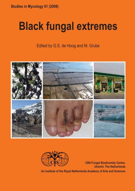
Studies in MycologyThe Studies in Mycology is an international journal which publishes systematic monographs of filamentous fungi and yeasts, and in rareoccasions the proceedings of special meetings related to all fields of mycology, biotechnology, ecology, molecular biology, pathology andsystematics. For instructions for authors see www.cbs.knaw.nl.Ex e c u t i v e Ed i t o rProf. dr Robert A. Samson, <strong>CBS</strong> Fungal Biodiversity Centre, P.O. Box 85167, 3508 AD Utrecht, The Netherlands.E-mail: r.samson@cbs.knaw.nlLay o u t Ed i t o rsManon van den Hoeven-Verweij, <strong>CBS</strong> Fungal Biodiversity Centre, P.O. Box 85167, 3508 AD Utrecht, The Netherlands.E-mail: m.verweij@cbs.knaw.nlKasper Luijsterburg, <strong>CBS</strong> Fungal Biodiversity Centre, P.O. Box 85167, 3508 AD Utrecht, The Netherlands.E-mail: k.luijsterburg@cbs.knaw.nlScientific Ed i t o rsProf. dr Uwe Braun, Martin-Luther-Universität, Institut für Geobotanik und Botanischer Garten, Herbarium, Neuwerk 21, D-06099 Halle, Germany.E-mail: uwe.braun@botanik.uni-halle.deProf. dr Pedro W. Crous, <strong>CBS</strong> Fungal Biodiversity Centre, P.O. Box 85167, 3508 AD Utrecht, The Netherlands.E-mail: p.crous@cbs.knaw.nlProf. dr David M. Geiser, Department of Plant Pathology, 121 Buckhout Laboratory, Pennsylvania State University, University Park, PA, U.S.A. 16802.E-mail: dgeiser@psu.eduDr Lorelei L. Norvell, Pacific Northwest Mycology Service, 6720 NW Skyline Blvd, Portland, OR, U.S.A. 97229-1309.E-mail: llnorvell@pnw-ms.comDr Erast Parmasto, Institute of Zoology & Botany, 181 Riia Street, Tartu, Estonia EE-51014.E-mail: e.parmasto@zbi.eeProf. dr Alan J.L. Phillips, Faculdade de Ciências e Tecnologia, Universidade Nova de Lisboa, Quinta de Torre, 2829-516 Caparica, Portugal.E-mail: alp@mail.fct.unl.ptDr Amy Y. Rossman, Rm 304, Bldg 011A, Systematic Mycology & Microbiology Laboratory, Beltsville, MD, U.S.A. 20705.E-mail: amy@nt.ars-grin.govDr Keith A. Seifert, Research Scientist / Biodiversity (Mycology and Botany), Agriculture & Agri-Food Canada, KW Neatby Bldg, 960 Carling Avenue,Ottawa, ON, Canada K1A OC6.E-mail: seifertk@agr.gc.caProf. dr Jeffrey K. Stone, Department of Botany & Plant Pathology, Cordley 2082, Oregon State University, Corvallis, OR, U.S.A. 97331-2902.E-mail: stonej@bcc.orst.eduDr Richard C. Summerbell, 27 Hillcrest Park, Toronto, Ont. M4X 1E8, Canada.E-mail: summerbell@aol.comCopyright 2008 <strong>CBS</strong> Fungal Biodiversity Centre, P.O. Box 85167, 3508 AD Utrecht, The Netherlands.You are free to share — to copy, distribute and transmit the work, under the following conditions:Attribution:You must attribute the work in the manner specified by the author or licensor (but not in any way that suggests that they endorse youor your use of the work).Non-commercial: You may not use this work for commercial purposes.No derivative works: You may not alter, transform, or build upon this work.For any reuse or distribution, you must make clear to others the license terms of this work, which can be found at http://creativecommons.org/licenses/bync-nd/3.0/legalcode.Any of the above conditions can be waived if you get permission from the copyright holder. Nothing in this license impairs or restrictsthe author"s moral rights.Publication date: 18 December 2008Published and distributed by <strong>CBS</strong> Fungal Biodiversity Centre, P.O. Box 85167, 3508 AD Utrecht, The Netherlands. Internet: www.cbs.knaw.nl.E-mail: info@cbs.knaw.nl.ISBN/EAN : 978-90-70351-73-1Online ISSN : 1872-9797Print ISSN : 0166-0616Cover: Images of various habitats on black yeast-like fungi. Top from left to right: flying foxes at temple of Chachoengsao province, Thailand, the hypothesizednatural niche of Exophiala dermatitidis. Saltpans at Sečovlje, Slovenia, the natural habitat of Hortaea werneckii. Gas station, The Netherlands, humanmadehabitat of Cladophialophora immunda. Bottom from left to right: rock formations in the McMurdo Dry Valley, Antarctica, one of the natural habitatsof Recurvomyces mirabilis. Human foot with lesions of Coniosporium epidermidis. Sea ice from Kongsvegen glacier in Spitsbergen, natural habitat ofAureobasidium pullulans var. subglaciale.
<strong>Black</strong> <strong>fungal</strong> <strong>extremes</strong>edited byG. S. de Hoog<strong>CBS</strong> Fungal Biodiversity Centre, Uppsalalaan 8, 3584 CT Utrecht, The NetherlandsM. GrubeInstitute of Plant Sciences, Karl-Franzens-University, Holteigasse 6, 010 Graz, Austria<strong>CBS</strong> Fungal Biodiversity Centre,Utrecht, The NetherlandsAn institute of the Royal Netherlands Academy of Arts and Sciences
PREFACEThe terms "black fungi" or "dematiaceous fungi" are practical tags that recall the early days of mycological classification. At the beginning ofthe 19 th century the grouping of anamorphic fungi relied on characters that were easy to observe with minimal optical equipment, such ascolour mycelial. Since these times, and till today, hyphomycetes producing olive-grey, brown or black pigment in their cell wall or conidia areclassified as "dematiaceous fungi" (referring to the meanwhile obsolete genus Dematium, originally introduced for black, clumpy fungi). Thedark pigments are presumed to be DHN-like (dihydroxynaphthalene) melanins, although biochemical and structural characterisation has notalways been achieved and may involve various precursor molecules.It has now become clear that black fungi do not comprise a single phylogenetic lineage, but stem from divergent branches of the <strong>fungal</strong>tree of life. The lineages share production of melanin-like pigments, which has had profound evolutionary consequences for these groups.In human- and phytopathogenic fungi melanins are linked to increased virulence. Melanins also provide protection from a broad range ofenvironmental stress conditions.With their adaptive potential to uncommon habitats, black fungi have raised increasing interest of mycologists in medical sciences as wellas in environmental ecology. It has become clear that a comprehensive understanding of black <strong>fungal</strong> evolution, ecology and functionalityrequires a synergic interdisciplinary approach, supported by a cooperative effort among specialists. A first step to bundle the interests inblack fungi and to create a common forum was the foundation of a Working Group "<strong>Black</strong> Yeasts" under auspices of the International Societyfor Human and Animal Mycology (ISHAM). A small-scale meeting was held in Graz, Austria (May 30–June 1, 2006), and focused on theextremophilic ecology of black fungi. The growing interest in the topic was reflected in the second workshop held in Utrecht, The Netherlands(April 26–28, 2007), jointly with the ISHAM-affiliated Working Group on "Chromoblastomycosis", with 55 participants from 19 countries.The workshops helped to further establish the scientific network among fundamental scientists, clinicians and workers in applied fields.Oral contributions at the Utrecht workshop covered such diverse topics as human infections, diseases on cold-blooded animals, fungi growingin lichens or on rock under extreme climatic conditions, <strong>fungal</strong> use in bioremediation of polluted environments, and black yeasts in drinkingwater, but also susceptibility testing, and molecular evolution. Nearly all contributions included new material and quickly the idea was bornthat this information would be valuable to be published in a coherent fashion. For the majority of the medical papers we refer to the journal"Medical Mycology", issue 46(1), 2009. The present issue of Studies in Mycology entitled "<strong>Black</strong> Fungal Extremes" reveals unexpectedtypes of ecology, such as growth in Arctic glaciers, Mediterranean rock, in lichens, in pure acid, and in nearly saturated salt solutions. Otherpapers investigate the evolutionary origins of black fungi, expression of relevant genes, medical aspects, and technical advances in culturingtechniques.This interdisciplinary blend of approaches gives an insight in current research on black fungi. We hope that the present issue will attractthe interest of more mycologists, who will join in our initiative to shed more light on the fascinating biology of extremophilic and pathogenicblack fungi.The Editors November 2008The papers in this issue of Studies in Mycology emerged from a workshop organised by the WorkingGroups on <strong>Black</strong> Yeasts and on Chromoblastomycosis, under auspices of the non-profit organisation“International Society for Human and Animal Mycology” (ISHAM). All authors report no conflicts ofinterest. The authors alone are responsible for the content and writing of the papers.
CONTENTSDrought meets acid: three new genera in a dothidealean clade of extremotolerant fungiL. Selbmann, G.S. de Hoog, L. Zucconi, D. Isola, S. Ruisi, A.H.G. Gerrits van den Ende, C. Ruibal, F. De Leo, C. Urzì and S. Onofri ... 1Redefinition of Aureobasidium pullulans and its varietiesP. Zalar, Gostinčar, G.S. de Hoog, V. Uršič, M. Sudhadham and N. Gunde-Cimerman ........................................................................... 21The influence of ortho- and par)a diphenoloxidase substrates on pigment formation in black yeast-like fungiN.A. Yurlova, G.S. de Hoog and L.G. Fedorova ...................................................................................................................................... 39Expression of fatty-acid-modifying enzymes in the halotolerant black yeast Aureobasidium pullulans (de Bary) G. Arnaudunder salt stressC. Gostinčar, M. Turk, T. Trbuha, T. Vaupotič, A. Plemenitaš and N. Gunde-Cimerman .......................................................................... 51HMG-CoA reductase is regulated by environmental salinity and its activity is essential for halotolerance in halophilic fungiT. Vaupotič, P. Veranic, U. Petrovič, N. Gunde-Cimerman and A. Plemenitaš ......................................................................................... 61Adaptation of extremely halotolerant black yeast Hortaea werneckii to increased osmolarity: a molecular perspective at a glanceA. Plemenitaš, T. Vaupotič, M. Lenassi, T. Kogej and N. Gunde-Cimerman ............................................................................................ 67Tinea nigra by Hortaea werneckii, a report of 22 cases from MexicoA. Bonifaz, H. Badali, G.S. de Hoog, M. Cruz, J. Araiza, M.A. Cruz, L. Fierro and R.M. Ponce .............................................................. 77<strong>Black</strong> fungi in lichens from seasonally arid habitatsS. Harutyunyan, L. Muggia and M. Grube ............................................................................................................................................... 83Cellular responses of microcolonial rock fungi to long-term desiccation and subsequent rehydrationA.A. Gorbushina, E.R. Kotlova and O.A. Sherstneva .............................................................................................................................. 91Resistance of Antarctic black fungi and cryptoendolithic communities to simulated space and Martian conditionsS. Onofri, D. Barreca, L. Selbmann, D. Isola, E. Rabbow, G. Horneck, J.P.P. de Vera, J. Hatton and L. Zucconi .................................. 99A rock-inhabiting ancestor for mutualistic and pathogen-rich <strong>fungal</strong> lineagesC. Gueidan, C. Ruibal Villaseñor, G.S. de Hoog, A.A. Gorbushina, W.A. Untereiner and F. Lutzoni ..................................................... 111Evolution of CDC42-1, a putative virulence factor triggering meristematic growth in black yeastsS. Deng, A.H.G. Gerrits van den Ende, A. Ram, M. Arendhorst, H. Hu and G.S. de Hoog ................................................................... 121Coniosporium epidermidis sp. nov., a new species from human skinD.M. Li, G.S. de Hoog, D.M. Lindhardt Saunte, A.H.G. Gerrits van den Ende and X.R. Chen .............................................................. 131Environmental isolation of black yeast-like fungi involved in human infectionV.A. Vicente, D. Attili-Angelis, M.R. Pie, F. Queiroz-Telles, L.M. Cruz, M.J. Najafzadeh, G.S. de Hoog J. Zhaoand A. Pizzirani-Kleiner .......................................................................................................................................................................... 137The neurotropic black yeast Exophiala dermatitidis has a possible origin in the tropical rain forestM. Sudhadham, P. Sihanonth, S. Sivichai, R. Chaiwat, S.B.J. Menken, G.M. Dorrestein and G.S. de Hoog ....................................... 145Selective factors involved in oil flotation isolation of black yeasts from the environmentM.M. Satow, D. Attili-Angelis, G.S. de Hoog, D.F. Angelis and V.A. Vicente .......................................................................................... 157Characterisation of the substrate specificity of the nitrile hydrolyzing system of the acidotolerant black yeast Exophialaoligosperma R1S. Rustler, A. Chmura, R.A. Sheldon and A. Stolz ................................................................................................................................. 165Biodiversity of the genus CladophialophoraH. Badali, C. Gueidan, M.J. Najafzadeh, A. Bonifaz, A.H.G. Gerrits van den Ende and G.S. de Hoog ................................................. 175
available online at www.studiesinmycology.orgdoi:10.3114/sim.2008.61.01St u d i es in My c o l o g y 61: 1–20. 2008.Drought meets acid: three new genera in a dothidealean clade of extremotolerantfungiL. Selbmann 1 *, G.S. de Hoog 2,3 , L. Zucconi 1 , D. Isola 1 , S. Ruisi 1 , A.H.G. Gerrits van den Ende 2 , C. Ruibal 2 , F. De Leo 4 , C. Urzì 4 andS. Onofri 11DECOS, Università degli Studi della Tuscia, Largo dell’Università, Viterbo, Italy; 2 <strong>CBS</strong> Fungal Biodiversity Centre, P.O. Box 85167, NL-3508 AD Utrecht, The Netherlands;3Institute for Biodiversity and Ecosystem Dynamics, University of Amsterdam, Kruislaan 315, NL-1098 SM Amsterdam, The Netherlands; 4 Dipartimento di Scienze Microbiologiche,Genetiche e Molecolari, Università di Messina, Salita Sperone 31, I-98166 Messina, Italy*Correspondence: Laura Selbmann, selbmann@unitus.itAbstract: Fungal strains isolated from rocks and lichens collected in the Antarctic ice-free area of the Victoria Land, one of the coldest and driest habitats on earth, were foundin two phylogenetically isolated positions within the subclass Dothideomycetidae. They are here reported as new genera and species, Recurvomyces mirabilis gen. nov., sp.nov. and Elasticomyces elasticus gen. nov., sp. nov. The nearest neighbours within the clades were other rock-inhabiting fungi from dry environments, either cold or hot. PlantassociatedMycosphaerella-like species, known as invaders of leathery leaves in semi-arid climates, are also phylogenetically related with the new taxa. The clusters are alsorelated to the halophilic species Hortaea werneckii, as well as to acidophilic fungi. One of the latter, able to grow at pH 0, is Scytalidium acidophilum, which is ascribed here tothe newly validated genus Acidomyces. The ecological implications of this finding are discussed.Key words: Acidophilic fungi, Antarctica, black fungi, extremotolerance, halophilic fungi, ITS, lichens, phylogeny, rock-inhabiting fungi, SSU, taxonomy.Taxonomic novelties: Recurvomyces Selbmann & de Hoog, gen. nov.; Recurvomyces mirabilis Selbmann & de Hoog, sp. nov.; Elasticomyces Zucconi & Selbmann, gen. nov.;Elasticomyces elasticus Zucconi & Selbmann, sp. nov.; Acidomyces Selbmann, de Hoog & De Leo, gen. nov.; Acidomyces acidophilus (Sigler & J.W. Carmich.) Selbmann, deHoog & De Leo, comb. nov.INTRODUCTIONContrary to expectations, bare rocks in arid and semi-arid climatesmay harbour a bewildering biodiversity of black fungi. Many specieshave been reported from the Mediterranean basin (Sterflinger et al.1997, Wollenzien et al. 1997, Bogomolova & Minter 2003, De Leoet al. 1999, 2003, Bills et al. 2004, Ruibal et al. 2005, Ruibal et al.2008). These extremotolerant fungi live and even thrive on surfacesthat are too harsh to support growth of competing microorganisms;they shelter in small depressions in the marble surface, calledmicropits (Sterflinger 1998). Similar extremotolerant fungi werediscovered in the extremely cold and ice-free McMurdo Dry Valleys,a desert area in the Antarctic (Nienow & Friedmann 1993), wheretemperatures are only occasionally above zero, dropping to about–50 °C in winter. Onofri et al. (1999) and Selbmann et al. (2005)even reported on the existence of possibly endemic genera,Friedmanniomyces Onofri and Cryomyces Selbmann, de Hoog,Mazzaglia, Friedmann & Onofri in these habitats, which apparentlyshow active evolution under conditions of near-permanent frost andextreme dryness (Friedmann et al. 1987). These fungi may escapeprohibitive environmental conditions by colonising air spaces inrocks, living in association with lichens and algae in cryptoendolithiccommunities (Friedmann & Ocampo 1976, Friedmann 1982).In the present paper we describe three new <strong>fungal</strong> genera andspecies; their novelty is supported by molecular phylogeny, taking aclearly separate position within the Dothideomycetidae. One genusincludes two strains isolated from rocks in the Antarctic desert, onestrain from rocks collected in Monte Rosa in the Alps, Italy, and anunidentified rock fungus from Puebla de la Sierra, Spain; the othergenus includes three strains isolated from different thalli of Antarcticlichens, one from cryptoendolithic Antarctic communities and onefrom rocks collected in Aconcagua in the Argentinian Andes. Incontrast to most rock-inhabiting black fungi, which are generallyscarcely differentiated, they show peculiar and distinguishedmorphological traits.Fungi may also be encountered in extremely acidicenvironments. Some are able to grow at pH values down to pH 0(Starkey & Waksman 1943, Harrison et al. 1966, Gould et al. 1974,Ivarsson & Morita 1982, Gimmler et al. 2001). Sigler & Carmichael(1974) compared four strains from an acidic soil (pH 1.4–3.5) withthe ones previously isolated by Starkey & Waksman (1943) andIvarsson & Morita (1982), referring them to the genus ScytalidiumPesante on the basis of scarcely differentiated brown arthroconidia.Our SSU and ITS comparison proved these fungi also to bemembers of a clade within the Dothideomycetidae, amidst rockinhabitingfungi from cold and semi-arid climates.MATERIALS AND METHODSStrains<strong>Black</strong> fungi were isolated from rock samples harbouring acryptoendolithic lichen-dominated community and from epilithiclichens collected in different locations of Northern and SouthernVictoria Land, Antarctica, in the framework of the Italian expeditionCopyright 2008 <strong>CBS</strong> Fungal Biodiversity Centre, P.O. Box 85167, 3508 AD Utrecht, The Netherlands.You are free to share - to copy, distribute and transmit the work, under the following conditions:Attribution:You must attribute the work in the manner specified by the author or licensor (but not in any way that suggests that they endorse you or your use of the work).Non-commercial: You may not use this work for commercial purposes.No derivative works: You may not alter, transform, or build upon this work.For any reuse or distribution, you must make clear to others the license terms of this work, which can be found at http://creativecommons.org/licenses/by-nc-nd/3.0/legalcode. Any of the above conditions can be waived if you getpermission from the copyright holder. Nothing in this license impairs or restricts the author’s moral rights.1
Se l b m a n n e t a l.Table 1. List of strains studied.Species Strain no. Source Geography ITS ReferenceAcidomyces acidophilum <strong>CBS</strong> 335.97 Acidophilic algae Dunaliella acidophila pH 1.0 Germany AJ244237 Gimmler et al. 2001Soil near acidic elemental sulphur pile pH 1.1 Canada – Sigler & Carmichael 1974Acidomyces acidophilum (depositedas Scytalidium acidophilum)Acidomyces acidophilum (depositedas Botryomyces caespitosus)<strong>CBS</strong> 270.74 T (ATCC 26772; UAMH 3460; IMI183518)<strong>CBS</strong> 899.87 Pyrite ore acidic drainage pH 2.0 Germany – –dH 13081 = det 106/2003 2N Sulphuric acid pH 1 Danmark (supplied by GC Frisvad) – Starkey & Waksman 1943dH 11526 = det 237-1999 Volcanic soil Iceland (supplied by S Gross, Berlin) – –dH 12881 = det 142-AF1 – –Acidomyces sp. dH 13119 Acidic industrial process water pH 1.5 Emmen, The Netherlands – –Batcheloromyces proteae <strong>CBS</strong> 110696; CPC 1518 Protea cynaroides South Africa – Crous et al. 2007Capnobotryella renispora <strong>CBS</strong> 214.90 T (<strong>CBS</strong> 176.88; IAM 13014; JCM 6932) Capnobotrys neessii Japan – –Catenulostroma abietis <strong>CBS</strong> 290.90 Man, skin lesion The Netherland AY128698 Crous et al. 2007<strong>CBS</strong> 145.97 (dH 15396) sandstone of cathedral Zeitz, Germany AY128699 Butin et al. 1996<strong>CBS</strong> 300.81 Juniperus communis (Cupressaceae), needle Graubünden, Grüsc, Switzerland AJ244264 –<strong>CBS</strong> 279.86 Kiel, Germany – Butin et al. 1996; Sterflinger et al.1999dH 12687 = det 396/2001 Painted wall Sweeden – –TRN 128 Limestone Mallorca AY559363 –dH 12697 = det 373/2001 RMF N113 Desert soil Namibia – –dH 13593 See snail Italy – –<strong>CBS</strong> 618.84 Ilex sp. leaf Germany AY128696 –<strong>CBS</strong> 118765 (TRN 127; dH 14531) Limestone Cala San Vincenc, Mallorca AY559362 –Catenulostroma elginense <strong>CBS</strong> 111030 (CPC 1958) Protea grandiceps South Africa AY260093 Crous et al. 2007Catenulostroma germanicum <strong>CBS</strong> 539.88 Stone Germany, former West-Germany EU019253 Crous et al. 2007Catenulostroma macowanii <strong>CBS</strong> 110756 (CPC 1872) Protea nitida South Africa – Crous et al. 2007CPC 1488 – –Elasticomyces elasticus <strong>CBS</strong> 122538 (CCFEE 5313) Lecanora fuscobrunnea Kay Island, Northern Victoria Land, Antarctica FJ415474 –<strong>CBS</strong> 122539 (CCFEE 5319) Lecanora sp. Inexpressible Island, Northern Victoria Land,AntarcticaFJ415475 –<strong>CBS</strong> 122540 (CCFEE 5320) Usnea antarctica Edmondson Point, Northern Victoria Land, Antarctica FJ415476 –Da-004-06 Rock Mount Aconcagua, Andes, Argentina – –CCFEE 5474 (D007-06) Sandstone Tarn Flat, Northern Victoria Land, Antarctica – –Friedmanniomyces endolithicus <strong>CBS</strong> 119423 (CCFEE 5208) Sandstone Northern Victoria Land, Antarctica – Selbmann et al. 2005<strong>CBS</strong> 119424 (CCFEE 5195) Rock Northern Victoria Land, Antarctica – Selbmann et al. 2005<strong>CBS</strong> 119429 (CCFEE 5193) Sandstone Timber Peak, Northern Victoria Land, Antarctica – Selbmann et al. 20052
Dr o u g h t m e e t s a c i dTable 1. (Continued).Species Strain no. Source Geography ITS ReferenceFriedmanniomyces endolithicus <strong>CBS</strong> 119428 (CCFEE 5001) Sandstone Timber Peak, Northern Victoria Land, Antarctica – Selbmann et al. 2005Friedmanniomyces simplex <strong>CBS</strong> 116775 T (CCFEE 5184) Sandstone Battleship Promontory, Southern Victoria Land,DQ028271 Selbmann et al. 2005AntarcticaHobsonia santessonii Peltigera scabrosa Sweden – Sikaroodi et al. 2001Hortaea acidophila <strong>CBS</strong> 113389 (dH 11932) Lignite pH 1.0 Germany – Hölker et al. 2004Hortaea werneckii (preserved as <strong>CBS</strong> 110352 (dH 12843 = VPCI 176) Hollow tree Sudan – –Pseudotaeniolina globosa)Hortaea werneckii dH 12322; Poonwan 13-44-08648 – – – –<strong>CBS</strong> 373.92 (dH 15813) Ceach soil La Palma, Spain AJ238474 –<strong>CBS</strong> 359.66 (dH 15803) Can, tinea nigra palmaris Suriname, Paramaribo AJ244249 –<strong>CBS</strong> 122.32 (dH 15340) Can, tinea nigra palmaris – AJ238473 –<strong>CBS</strong> 117.90 (UAMH 4978; dH 15327) Salted fish, Osteoglossum bicirrhosum Brazil AJ238472 Mok et al. 1981<strong>CBS</strong> 116.90 (ATCC 52681; UAMH 5389; dH 15311) Cantharus cantharus, eye infection, black sea Italy AJ238471 Todaro et al. 1983bream in aquarium<strong>CBS</strong> 115.90 (UAMH 4985; dH 15303) Bufo granulosus, kidney Brazil AJ238470 –<strong>CBS</strong> 111.31 (dH 15284) Man, keratomycosis nigricans palmaris Brazil AJ238679 –<strong>CBS</strong> 100455 (MZKI B-675) Coral, sea water Croatia AY128704 Zalar et al. 1999<strong>CBS</strong> 107.67 (dH 15206) Man, tinea nigra Lisboa, Portugal AJ238468 McGinnis 1979MZKI B-987 Ipersaline water Spain – –dH 13416 Angelfish – – –<strong>CBS</strong> 117931 (TRN 122, dH 14528) Limestone Cala San Vincenc, Mallorca AY559357 –BMU00057 Patient’s foot China – –Mycocalicium victoriae <strong>CBS</strong> 109863 Soil, garden museum Messina, Italy AJ312123 –Mycosphaerella tasmaniensis <strong>CBS</strong> 114556 (CMW 14663; STE-U 1556) E. nitens Australia DQ267592 –<strong>CBS</strong> 111687 (CMW 14780; STE-U 1555) E. nitens Australia AY667578 –Pseudotaeniolina globosa <strong>CBS</strong> 110353 (dH 12840; det M108/2002) 58-yr-old man, aorta, at autopsy Würzburg, Germany – Kurzai et al. 2003<strong>CBS</strong> 109889 T (MC 769) Church roof Sicily, Italy AY128700 De Leo et al. 2003<strong>CBS</strong> 113249 (dH 13060) – – – –<strong>CBS</strong> 303.84 Wood Germany AJ244268 de Hoog et al. 1999<strong>CBS</strong> 119923 (dH 16905) Window Japan – –Recurvomyces mirabilis <strong>CBS</strong> 119434 (CCFEE 5264; dH 14759) Sandstone Battleship Promontory, Southern Victoria Land,AntarcticaFJ415477 –Recurvomyces mirabilis CCFEE 5480 (D016-02) Sandstone Battleship Promontory, Southern Victoria Land,Antarctica– –CCFEE 5391 Rock Mount Rosa, P.ta Indren, Alps, Italy – –Recurvomyces sp. <strong>CBS</strong> 117957 (TRN 491; dH 14566) Quarzite Puebla de la Sierra, Madrid, Spain AY1843175 –Teratosphaeria cryptica TC 0.56 E. globulus Australia DQ665661 –www.studiesinmycology.org3
Se l b m a n n e t a l.Table 1. (Continued).Species Strain no. Source Geography ITS ReferenceTeratosphaeria mexicana <strong>CBS</strong> 110502 (CMW 14461) E. globulus Manjimup, Darling View, Plantation, Western Australia AY725558 Crous et al. 2007Teratosphaeria microspora <strong>CBS</strong> 110890 (CPC 1832) Protea leaf South Africa AY260097 Taylor et al. 2003Teratosphaeria molleriana <strong>CBS</strong> 111164 (CMW 4940; STE-U 1214) E. globulus Abrantes, Portugal AF309620 Crous et al. 2007CPC11842 Eucalyptus sp. Portugal DQ302989 Crous et al. 2006CPC11845 Eucalyptus sp. Portugal DQ302990 Crous et al. 2006<strong>CBS</strong> 116370 (CPC 10397) E. globulus Spain AY725561CPC 12056 DQ302991 Crous et al. 2006Teratosphaeria nubilosa <strong>CBS</strong> 116005 E (CMW 3282; CPC 937) E. globulus Austria AY725572 Crous et al. 2007CPC 4661 E. globulus Spain AY725570 Crous et al. 2004<strong>CBS</strong> 111445 (CPC 4660) E. globulus Spain AY725569 Crous et al. 2004CPC 3722 E. globulus Spain AY725568 Crous et al. 2004CPC 1099 E. globulus Tanzania AY725567 Crous et al. 2004<strong>CBS</strong> 111969 (CPC 1078) E. globulus Kenia AY725563 Crous et al. 2004CPC 4663 E. globulus Spain AY725571 Crous et al. 2004CPC 11882 E. globulus Portugal DQ302999 Crous et al. 2006CPC 11723 –CPC 11487 E. globulus Spain DQ302994 Crous et al. 2006CPC 11249 E. globulus Spain DQ302993 Crous et al. 2006CPC 11246 E. globulus Spain DQ302992 Crous et al. 2006<strong>CBS</strong> 114419 (CPC 10497) Eucalyptus globulus (Myrtaceae) New Zealand AY725574 Crous et al. 2007TC 0.42 E. globulus Australia DQ665659 –TC 0.40 E. globulus Australia DQ665657 –TC 0.47 E. globulus Australia DQ665658 –<strong>CBS</strong> 116283 (CPC 10495) E. globulus New Zeland AY725573 Crous et al. 2004Teratosphaeria nubilosa ( CPC 11885 E. globulus Portugal DQ303000 Crous et al. 2006Teratosphaeria ohnowa CPC 1005 – – AF309605 Crous et al. 2001<strong>CBS</strong> 112896* (CMW 4937; CPC 1004 ) E. globulus South Africa AF309604 Crous et al. 2001– – AF173299 –CMW 9103 – – AF468881 –<strong>CBS</strong> 110949 (STE-U 1006) E. grandis (Myrtaceae), leaves Hazyview, South Africa AY725575 –Teratosphaeria toledana <strong>CBS</strong> 113313 H (CMW 14457) Eucalyptus sp., leaves Toledo, Spain AY725580 –CPC 10840 Eucalyptus sp. Spain AY725581 Crous et al. 2004Abbreviations used: ATCC – American Type Culture Collection, Manassas, VA, U.S.A.; <strong>CBS</strong> – Centraalbureau voor Shimmelcultures, Utrecht, The Netherlands; dH – GS de Hoog private collection, <strong>CBS</strong>, Utrecht, The Netherlands; CCFEE – CultureCollection of Fungi From Extreme Environments, Università degli Studi della Tuscia, Viterbo, Italy; CMW – Culture collection of the Forestry and Agricultural Biotechnology Institute (FABI), University of Pretoria, Pretoria, South Africa; CPC – Culturecollection of P Crous, housed at the <strong>CBS</strong>; IMI – International Mycological Institute, U.K.; MC – Collection of Istituto di Microbiologia di Messina, Italy; MZKI – Microbiological Culture Collection, National Institute of Chemistry, Ljubljana, Slovenia; STE-U –University of Stellenbosch <strong>fungal</strong> culture collection, Stellenbosch, South Africa; TRN – T Ruibal private collection; UAMH – The University of Alberta Microfungus Collection and Herbarium, Edmonton, AB, Canada.*Ex-type cultures.4
Dr o u g h t m e e t s a c i d2003/2004, and from rock samples collected in Mount Aconcagua,Andes, Argentina, and Monte Rosa in the Alps, Italy, as reportedin Table 1. The samples were collected using a sterile chisel,sterlilised in the field before each sampling, and preserved in sterilebags at –20 °C. To remove potential contaminants, rocks collectedin other environments than Antarctica were washed 15 min insterile physiological solution added with 0.1 % Tween 20 (Sigma- Aldrich, Munich, Germany) and rinsed 4 times with physiologicalsolution to remove any trace of the detergent. Isolations from rockswere performed by powdering the samples and seeding fragmentsin triplicate Petri dishes filled with 2 % malt extract agar (MEA,AppliChem, GmbH) and dichloran-rose bengal agar (DRBC, OxoidLtd., Basingstoke, Hampshire, U.K.). Media were supplementedwith chloramphenicol 100 ppm to prevent bacterial growth. Fungifrom lichens were isolated as follows: small fragments of thalli weresterilised by washing with H 2O 2(8 %) for 5 min, washed with steriledeionised water to remove any trace of H 2O 2and finally seededon MEA and DRBC. Plates were incubated at 5 and 15 °C andinspected every 15 d until no new colonies appeared. Colonieswere transferred to MEA slants and incubated at 15 °C. Strainsanalysed for comparison are listed in Table 1; they were taken fromreference collections of the Culture Collection of Fungi from ExtremeEnvironments (CCFEE, Viterbo, Italy) and the Centraalbureau voorSchimmelcultures (<strong>CBS</strong>, Utrecht, The Netherlands), including alarge set of strains sampled by P.W. Crous from plants with leatheryleaves in a semi-arid climate.MorphologyHyphal maturation and conidiogenesis were studied using bothlight and scanning electron microscope (SEM). Slide cultureswere seeded onto MEA, incubated for 10 wk and mounted in lacticacid. Samples for SEM observations were prepared according tomethods described by Onofri et al. (1980).DNA extraction and sequencingDNA was extracted from mycelial fragments taken from 6-mos-oldMEA slants grown at 10 °C, using Nucleospin Plant kit (Macherey-Nagel, Düren, Germany) following the protocol optimised forfungi. PCR reactions were performed using BioMix (BioLineGmbH, Luckenwalde, Germany). In each 25 µL reaction tube 5pmol of each primer and 40 ng on template DNA were added. Theamplification was carried out using MiniCycler (MJ Research,Waltham, Massachusetts, U.S.A.) equipped with a heated lid.The first denaturation step at 95 °C for 3 min was followed by:denaturation at 95 °C for 2 s, annealing at 55 °C for 30 s, extensionat 72 °C for 30 s. The last three steps were repeated 35 times,with a last extension 72 °C for 5 min. The products were purifiedusing Nucleospin Extract kit (Macherey-Nagel, Düren, Germany).Primers NS1, NS2, NS3, NS4, NS5, NS8, ITS1, ITS4 (White etal. 1990), SR10R (Bruns et al. 1992), ITS5, and ITS4a (Larenaet al. 1999) were employed to amplify SSU and ITS rDNAportions. Sequencing reactions were performed according to thedideoxynucleotide method (Sanger et al. 1977) using the TF BigDye Terminator 1,1 RR kit (Applied Biosystems). Fragments wereanalysed using an ABI 310 Genetic Analyser (Applied Biosystems).Sequence assembly was done using the software Chromas (v. 1.451996–1998, Conor McCarthy School of Health Science, GriffithUniversity, Southport, Queensland, Australia).www.studiesinmycology.orgAlignment and tree reconstructionSSU sequences were aligned with ARB beta-package (v. 22-08-2003, Ludwig et al. 2004; www.mikro.biologie.tu-muenchen.de/pub/ARB). The SSU alignment spanned positions 141–2512,which corresponds to 1515 bp with reference to Saccharomycescerevisiae. Trees based on SSU sequences were reconstructedwith neighbour-joining in ARB.ITS sequences were aligned iteratively with Ward’s averaging(Van Ooyen 2002) in a research data base of black yeasts presentat <strong>CBS</strong> using the BioNumerics package (Applied Maths, Kortrijk,Belgium). Due to gaps necessary for alignment, the ITS1 domainspanned 187 positions (real lengths 147–154 bp), the 5.8S gene156 positions and the ITS2 domain 184 positions (real lengths143–155 bp). The alignments were based on the positions 28–481,the initial and the final parts were cut off to compare fragmentswith the same length. Alignments were exported and the best-fitsubstitution model was determined using Modeltest MrAic.pl 1.4.3(Nylander 2004, program distributed by the author) estimatedusing Ph y m l (Guindon & Gascuel 2003) through hierarchicallikelihood ratio tests. MrAic calculates the Akaike InformationCriterion (AIC), corrected Akaike Information Criterion (AICc)and Bayesian Information Criterion (BIC); Akaike weights fornucleotide substitution model and model uncertainty. All 56 modelsimplemented in Modeltest were evaluated. Phylogenetic trees werereconstructed by Maximum Likelihood, using Tr e e f i n d e r (Jobbet al. 2004) and the resulting tree was displayed using Tr e e v i e wv. 1.6.6 (Page 1996). The robustness of the phylogenetic inferencewas estimated using the bootstrap method (Felsenstein 1985) with100 pseudoreplicates generated and analysed with Tr e e f i n d e r.As alignment over the entire complex was highly ambiguous, analgorithm for tree reconstruction without alignment, the DNA-walkDivergence method (DNAWD, Licinio & Caligiorne 2004; Caligiorneet al. 2005) was used, involving the entire spacer region. DNAwalksare defined by incrementing walk steps for each nucleotide inthe sequence (for example a positive step for purines, and negativefor pyrimidines). It makes simultaneous comparisons of the threedimensionalwalks (representing three composition skews): AG-TC, AC-TG, and AT-CG for each pair of sequences. One sequenceslides against the other until the minimum squared walk difference isfound, corresponding to a global alignment. This is then taken as ameasure of their distance since statistically independent mutationsand indels increase the mean square walk differences linearly. Theresulting distance matrices are then fed into the Kitsch program ofthe Phylip package (v. 3.572c, Felsenstein 1996).Cultural preferencesCultural characteristics and growth rates were recorded onPotato-Dextrose Agar (PDA), MEA, Czapek Dox Agar (CzA) andOatmeal Agar (OA). Strain <strong>CBS</strong> 119434 was incubated at 10 °C,strains <strong>CBS</strong> 122538, 122539 and 122540 at 20 °C and strains<strong>CBS</strong> 899.87, <strong>CBS</strong> 335.97, dH 12881, dH 11526 and dH 13081 at25 °C. The diameter of the colonies was recorded monthly. Testswere performed in triplicate.Temperature preferencesTemperature preferences for the strains <strong>CBS</strong> 119434, 122538,122539 and 122540 were tested by incubating them on MEA,5
Se l b m a n n e t a l.Table 2. Physiological profiles of Antarctic strains.Species Strain no. Cultural preferences Thermal preferences (° C)PDA MEA CzA OA 0 5 10 15 20 25 30 35Recurvomyces mirabilis <strong>CBS</strong> 119434 1.5±0.14 1.8±0.14 0.5±0.14 1.65±0.07 0.45±0.07 0.8±0.2 1.5±0.26 1.9±0.26 0.78±0.2 – – –Elasticomyces elasticus <strong>CBS</strong> 122538 1.7±0.2 1.5±0.14 0.5±0.14 1.5±0.14 0.82±0.064 0.64±0.079 1.2 ±0.2 1.6 ±0.26 1.54 ±0.09 0.95 ±0.15 – –<strong>CBS</strong> 122539 1.5±0.1 1.43±0.15 0.3±0.02 1.23±0.2 0.77±0.1 0.6±0.2 1.1±0 1.5±0.14 1.2±0.2 0.6±0.2 – –<strong>CBS</strong> 122540 1.0±0.2 1.1±0.1 0.2±0.02 1.0±0.2 0.72±0.1 0.5±0.14 1.13±0.2 1.4±0.17 1.2±0.2 0.8±0.08 – –Cultural and thermal preferences reported as diameter of the colonies (cm), after two mos of incubation. The values represent the average of three different tests. Plates for cultural preferences were incubated at 10 °C for strain <strong>CBS</strong> 119434 and at 20 °Cfor strains <strong>CBS</strong> 122538, 122539 and 122540; plates for temperature preferences were seeded on MEA; – = no growth.Table 3. Cultural preferences and salt tolerance of acidophilic strains.Species Strain no. Cultural preferences NaCl %PDA MEA CzA OA 1.2 1.5 3 5 7 10 12Acidomyces acidophilum <strong>CBS</strong> 899.87 1.17±0.1 1.33±0.15 0.3±0.02 1.17±0.1 1.3±0.02 0.7±0.02 0.6±0.03 0.6±0 0.5±0 0.2±0.02 –<strong>CBS</strong> 335.97 1.08±0.1 1.9±0.14 0.6±0.2 1.0±0.2 1.5±0.02 0.5±0.02 0.4±0.04 0.3±0.02 0.2±0.02 – –dH 12881 2.0±0.14 1.5±0.14 1.2±0.2 1.5±0.1 1.5±0.02 1.5±0.02 1.5±0.02 0.7±0.03 0.5±0.04 – –dH 11526 2.5±0.2 3.0±0.2 1.5±0.14 1.2±0.2 2.5±0.02 2.0±0.04 2±0.04 0.8±0.03 0.6±0.008 – –dH 13081 1.8±0.14 2.2±0.2 1.1±0.1 1.0±0.2 1.0±0.04 0.8±0.02 0.8±0.02 0.6±0.02 0.3±0.02 – –Growth on different media and salt concentration expressed as diameters of the colonies (cm); – = no growth.Table 4. Thermal and pH preferences of acidophilic strains.Species Strain no. Thermal preferences (° C) pH4 10 18 25 30 37 1 3 5 7 9Acidomyces acidophilum <strong>CBS</strong> 899.87 – – + ++ + – + + ++ + ±<strong>CBS</strong> 335.97 + + ++ + + – + ++ ++ + –dH 12881 + + ++ ++ + – + ++ + ± –dH 11526 + + + + ++ – + ++ ++ + –dH 13081 + + + ++ + – ± ++ ++ + –++ = maximum growth recorded; + = growth; ± = weak growth; – = no growth.6
Dr o u g h t m e e t s a c i din Petri dishes at 0–35 °C (in 5° intervals) ± 1 °C. The diameter of thecolonies was recorded monthly. Tests were performed in triplicate.Optimum temperatures for growth and development of the strains<strong>CBS</strong> 899.87, <strong>CBS</strong> 335.97, dH12881, dH11526 and dH13081,were determined by seeding 25 mL flasks containing 2 % MaltExtract Broth (MEB) with 0.25 mL of a 10 5 cells/mL suspension andincubating in shaken culture at 70 r.p.m. After 30 d of incubation attemperatures of 4, 10, 18, 25, 30, 37 °C, cultures were filtered andthe biomass dry-weighed. The test was performed in duplicate.Growth at different salt concentrationsThe ability to grow at different salinities was tested in duplicate onplates of MEA amended with 1.2, 1.5, 3, 5, 7, 10 or 12 % NaCl.Strains were inoculated in three spots on each plate and incubatedat 25 °C for one mo, when the colony diameter was recorded.Colonies with a diameter >2 mm were considered positive (Kane& Summerbell 1987).Growth at different pHThe ability to grow at different pH values for the strains <strong>CBS</strong>899.87, <strong>CBS</strong> 335.97, dH12881, dH11526 and dH13081 was testedin duplicate using MEB2 % medium at pH 1, 3, 5, 7 and 9. Valuesof pH 5 were obtained by the addition of 1N HCl; remaining pHvalues were obtained according to Küster & Thiel (1990) as follows:McIlvaine solution for pH 2–7, Clark & Lubs solution for pH 8–9;buffer HCl/KCl for pH 1. Strains were incubated at 25 °C in shakenculture at 70 r.p.m. for one mo, cultures were filtered and thebiomass dry-weighed.RESULTSPhysiologyTemperature relations and cultural features of the strains <strong>CBS</strong>119434, 122538, 122539 and 122540 are shown in Table 2.Physiological data for the strains <strong>CBS</strong> 899.87, <strong>CBS</strong> 335.97,dH12881, dH11526 and dH13081 are reported in Tables 3 and 4.All fungi tested were able to grow on natural media showing clearlyvisible growth on MEA as well as on PDA and OA, whilst there wasa marked reduction of ultimate colony diameter when seeded onCzA. <strong>CBS</strong> 119434 was able to grow in the range 0–20 °C, withan optimum at 15 °C, whereas strains <strong>CBS</strong> 122538, 122539 and122540 grew in a wider range of temperatures between 0 and25 °C, with an optimum at 15–20 °C. Strains <strong>CBS</strong> 899.87, <strong>CBS</strong>335.97, dH12881, dH11526 and dH13081 grew best in the rangeof 18–30 °C, with the optimum temperature at 18 °C for the strains<strong>CBS</strong> 335.97 and dH 12881, 25 °C for the strains <strong>CBS</strong> 899.87 anddH 13081, and 30 °C for the strain dH 11526. Since almost allstrains (except <strong>CBS</strong> 899.87) were still able to reproduce at 4 °C,they can be referred as mesophilic-psycrotolerant (Zucconi et al.1996). None of them grew at 37 °C. Furthermore, they were able togrow in very acidic conditions (pH 1) and most of them grew best atpH 5 or below. In particular the optimum was pH 5 for the strain <strong>CBS</strong>899.87, around pH 3–5 for the strains <strong>CBS</strong> 335.97, dH11526 anddH 13081 and pH 3 for the strain dH 12881. All the strains studiedshowed a decreasing growth at pH 7 and stopped to grow at pH 9,with the exception of strain <strong>CBS</strong> 899.87 which was able to grow ina very wide range of pH values showing a weak growth even at pHwww.studiesinmycology.org9. The strains demonstrated also to be moderately halophilic beingable to grow in up to 7 % NaCl (strains <strong>CBS</strong> 335.97, dH11526, dH13081, dH 12881) and in up to 10 % for the strain <strong>CBS</strong> 899.87.PhylogenySSU sequences were analysed for 71 strains of ascomycetousblack yeasts and relatives belonging to the orders Capnodiales,Dothideales, Myriangiales as well as the recently proposed neworder Botryosphaeriales (Schoch et al. 2006). The recentlydescribed taxon Baudoinia compniacensis (Richon) J.A. Scott& Unter. (Scott et al. 2007) with an SSU similarity around 97 %with some capnodialean/dothidealean strains, was not includedin this comparison. Fig. 1 shows a Neighbour Joining tree basedon the SSU comparison where the outgroup is represented byAliquandostipite khaoyaiensis (Jahnulales).Three Antarctic rock strains (<strong>CBS</strong> 122538, 122539 and122540), with nearly identical sequences, were included in a groupparaphyletic to Friedmanniomyces endolithicus Onofri. The strainsshared a remarkable morphology, with straight fertile hyphae ofwhich fragments detached, sometimes by basauxically expandingconnectives.Strain <strong>CBS</strong> 119434 clustered in a clade composed of mainlymeristematic species, which included the Antarctic rock-inhabitinggenus Friedmanniomyces. This strain also had characteristicmorphology, showing a unique pattern of recurved hyphal branchingat conidiation. Another strain with nearly identical sequence, <strong>CBS</strong>117957 from Mediterranean rocks, showed a simple, undiagnosticmeristematic micromorphology. The lichenicolous fungus Hobsoniasantessonii Lowen & D. Hawksw. belonged to the same group,together with the black fungus Mycocalicium victoriae (C. Knightex F. Wilson) Tibell.The acidophilic species Hortaea acidophila Hölker et al.and Acidomyces acidophilum as “Acidomyces richmondensis”B.J. Baker et al. composed a sister clade to the Friedmanniomycescomplex. The halophilic species Hortaea werneckii (Horta) Nishimura& Miyaji was found at a larger distance, in a heterogeneous cladewith Pseudotaeniolina globosa De Leo et al., Catenulostromaabietis (Butin & Pehl) Crous & Braun and Coccodinium bartschii A.Massal. As the backbone of the tree shows low bootstrap values formost clades, the exact phylogenetic positions of the newly addedAntarctic and acidophilic species are difficult to determine.The ITS tree shown in Fig. 2 was generated on the basis ofthe manually optimised alignment containing 95 sequences ofhalophilic, acidophilic, rock and plant-pathogenic fungi; it is basedon a length of 452 characters, including alignment gaps. The AICcselected HKY+G (Hasegawa et al. 1985) as the best model. Thebase frequencies was as follows: T = 0.2293, C = 0.3120, A =0.2009, G = 0.2574, TC = 0.5413, AG = 0.4583. The entire ITSregion showed too many polymorphisms to allow alignment witha sufficient degree of confidence. For this reason, DNAWD wasapplied, which is insensitive to alignment. Topology of this tree wasidentical (data not shown).In general the ITS trees showed excellent resolution of entities.Inter-group variability was constant and invariably significantlylarger than intra-group variability. The tree contained anamorphtaxa from extreme environments, as well as a number of plantassociatedspecies of Teratosphaeria Syd. & P. Syd. Three mainclades were discernible: an upper clade around Teratosphaeriamicrospora J.E. Taylor & Crous, a clade with T. nubilosa (Cook)Crous & U. Braun as central species, and an outgroup with strainsisolated at very low pH (Fig. 2).A single cluster is composed of strains listed as Teratosphaeria7
Se l b m a n n e t a l.0.01857697959599100Friedmanniomyces endolithicus CCFEE 52274 Friedmanniomyces endolithicus CCFEE 6709790Friedmanniomyces endolithicus CCFEE 524Friedmanniomyces endolithicus CCFEE 5180Friedmanniomyces endolithicus CCFEE 519999 Friedmanniomyces endolithicus <strong>CBS</strong> 11942497Friedmanniomyces endolithicus <strong>CBS</strong> 11942380 Friedmanniomyces endolithicus <strong>CBS</strong> 119428Friedmanniomyces endolithicus <strong>CBS</strong> 119429Friedmanniomyces simplex <strong>CBS</strong> 116775Elasticomyces elasticus <strong>CBS</strong> 12253910097Elasticomyces elasticus <strong>CBS</strong> 122540Elasticomyces elasticus <strong>CBS</strong> 122538Hobsonia santessonii AF289658100Mycocalicium victoriae <strong>CBS</strong> 109863Recurvomyces sp. <strong>CBS</strong> 117957Recurvomyces mirabilis <strong>CBS</strong> 119434100Acidomyces acidophilum AY374300Acidomyces acidophilum AY374299Acidomyces acidophilum AY374298Hortaea acidophila <strong>CBS</strong> 113389Hortaea werneckii Y18700Hortaea werneckii <strong>CBS</strong> 107.67Pseudotaeniolina globosa <strong>CBS</strong> 109889Catenulostroma abietis <strong>CBS</strong> 618.84Unidentified Antarctic rock black fungus CCFEE 451Capnobotryella renispora <strong>CBS</strong> 215.90Capnobotryella renispora <strong>CBS</strong> 214.90Capnobotryella renispora <strong>CBS</strong> 572.89Capnobotryella renispora UAMH 987099Trimmatostroma salinum <strong>CBS</strong> 100461100 Cladosporium cladosporioides AF548070Cladosporium cladosporioides AF548071Ramichloridium apiculatum <strong>CBS</strong> 156.59 T100 Myriangium duriaei AF242266Myriangium duriaei <strong>CBS</strong> 260.36Comminutispora agavaciensis <strong>CBS</strong> 619.95Lasallia rossica AF088238Umbilicaria subglabra AF088253100 Cryomyces antarcticus CCFEE 514Cryomyces antarcticus CCFEE 535Cryomyces antarcticus <strong>CBS</strong> 116301Cryomyces antarcticus <strong>CBS</strong> 119418Cryomyces antarcticus CCFEE 453Cryomyces antarcticus CCFEE 536Cryomyces antarcticus CCFEE 515Cryomyces minteri <strong>CBS</strong> 116902Phaeotheca fissurella <strong>CBS</strong> 520.89Aureobasidium pullulans AY030322Discosphaerina fagi AY016342Aureobasidium pullulans AY137507Aureobasidium pullulans AY13750887Aureobasidium pullulans AY137509Aureobasidium pullulans M55639100Aureobasidium pullulans AY137505Aureobasidium pullulans AY137506100 Delphinella strobiligena <strong>CBS</strong> 735.71Hormonema dematioides AY150054Scleroconidioma sphagnicola UAMH 9731100Dothidea hippophaeos U42475Dothidea insculpta U42474Neoscytalidium dimidiatum AF258604100 Neoscytalidium dimidiatum IP 127881Neoscytalidium dimidiatum AF258605Lasiodiplodia theobromae U42476Neofusicoccom ribis <strong>CBS</strong> 121.26Guignardia mangiferae <strong>CBS</strong> 398.80100 Neoplaconema gloeosporioides AJ534443100 Phaeotrichum benjaminii AY016348Phaeosclera dematioides Y11716Tubeufia helicoma AF201455Aliquandostipite khaoyaiensis AF201453CapnodialesMyriangialesDothidealesBotryosphaerialesFig. 1. Molecular phylogeny based on SSU sequences indicating the positions of the clades in Dothideomycetidae; the described new genera were highlighted with colouredrectangles. The tree has been built with neighbour-joining algorithm in ARB package with 100 replications. Branches of the clades supported by a bootstrap value above95 % are in bold.8
Dr o u g h t m e e t s a c i d991007063Acidomyces acidophilum dH 11526Acidomyces acidophilum <strong>CBS</strong> 335.97Acidomyces acidophilum <strong>CBS</strong> 270.74 TAcidomyces acidophilum dH 13081100Acidomyces acidophilum dH 12881Acidomyces acidophilum <strong>CBS</strong> 899.87Acidomyces sp. dH 131195877621006035567974100100100100557910079Hortaea werneckii <strong>CBS</strong> 122.32Hortaea werneckii <strong>CBS</strong> 111.31Hortaea werneckii dH 12322Hortaea werneckii dH 13416Hortaea werneckii <strong>CBS</strong> 115.90Hortaea werneckii <strong>CBS</strong> 100455Hortaea werneckii <strong>CBS</strong> 117931Hortaea werneckii MZKI B-987Hortaea wernekii <strong>CBS</strong> 359.66Hortaea werneckii <strong>CBS</strong> 117.90Hortaea werneckii BMU 00057Hortaea werneckii <strong>CBS</strong> 110352Hortaea werneckii <strong>CBS</strong> 107.67Hortaea wernekii <strong>CBS</strong> 116.90Hortaea werneckii <strong>CBS</strong> 373.92Pseudotaeniolina globosa <strong>CBS</strong> 109889 TPseudotaeniolina globosa <strong>CBS</strong> 303.84Pseudotaeniolina globosa <strong>CBS</strong> 119923Pseudotaeniolina globosa <strong>CBS</strong> 113249Pseudotaeniolina globosa <strong>CBS</strong> 110353Recurvomyces mirabilis CCFEE 5480Recurvomyces mirabilis <strong>CBS</strong> 119434Recurvomyces mirabilis CCFEE 5391Recurvomyces sp. <strong>CBS</strong> 117957Catenulostroma abietis dH 13593Catenulostroma abietis TRN 128Teratosphaeria microspora <strong>CBS</strong> 110890Catenulostroma abietis dH 12687Catenulostroma germanicum <strong>CBS</strong> 53988Catenulostroma abietis <strong>CBS</strong> 118765Catenulostroma abietis <strong>CBS</strong> 290.90Catenulostroma abietis <strong>CBS</strong> 618.84Catenulostroma abietis dH 12697Catenulostroma abietis <strong>CBS</strong> 300.81Catenulostroma abietis <strong>CBS</strong> 145.97Catenulostroma abietis <strong>CBS</strong> 279.86Teratosphaeria ohnowa <strong>CBS</strong> 110949Teratosphaeria ohnowa AF173299100 Teratosphaeria ohnowa CPC 1005Teratosphaeria ohnowa <strong>CBS</strong> 112896Teratosphaeria ohnowa CMW 9103Batcheloromyces proteae <strong>CBS</strong> 110696Elasticomyces elasticus <strong>CBS</strong> 122539Elasticomyces elasticus <strong>CBS</strong> 122538100 Elasticomyces elasticus <strong>CBS</strong> 122540Elasticomyces elasticus Da-00406Elasticomyces elasticus CCFEE 5474Hobsonia santessoniiMycocalicium victoriae <strong>CBS</strong> 109863Catenulostroma elginense <strong>CBS</strong> 111030Friedmanniomyces endolithicus <strong>CBS</strong> 119423100 Friedmanniomyces endolithicus <strong>CBS</strong> 11942992 Friedmanniomyces endolithicus <strong>CBS</strong> 119424Friedmanniomyces endolithicus <strong>CBS</strong> 119428Friedmanniomyces simplex <strong>CBS</strong> 116775 TTeratosphaeria nubilosa CPC 4663Teratosphaeria nubilosa <strong>CBS</strong> 116005 ETeratosphaeria nubilosa CPC 4661Teratosphaeria nubilosa <strong>CBS</strong> 111445Teratosphaeria nubilosa CPC 3722Teratosphaeria nubilosa CPC 1099Teratosphaeria nubilosa <strong>CBS</strong> 111969Teratosphaeria nubilosa CPC 11246Teratosphaeria nubilosa CPC 11249Teratosphaeria nubilosa CPC 1148797 Teratosphaeria nubilosa CPC 11723Teratosphaeria nubilosa CPC 11882100 Teratosphaeria nubilosa CPC 11885Teratosphaeria nubilosa TC 0.47Teratosphaeria nubilosa TC 0.4097 Teratosphaeria nubilosa <strong>CBS</strong> 114419Teratosphaeria nubilosa <strong>CBS</strong> 11628386Teratosphaeria nubilosa TC 0.4297Teratosphaeria molleriana CPC <strong>CBS</strong> 116370Teratosphaeria molleriana CPC 11842Teratosphaeria molleriana <strong>CBS</strong> 111164100 99 Teratosphaeria molleriana CPC 11845Teratosphaeria molleriana CPC 12056100Teratosphaeria toledana <strong>CBS</strong> 113313 HTeratosphaeria toledana CPC 10840Teratosphaeria cryptica T0.56Catenulostroma macowanii <strong>CBS</strong> 110756Catenulostroma macowanii CPC 1488Teratosphaeria mexicana <strong>CBS</strong> 110502Capnobotryella renispora <strong>CBS</strong> 214.90 THortaea acidophila <strong>CBS</strong> 113389Mycosphaerella tasmaniensis <strong>CBS</strong> 114556Mycosphaerella tasmaniensis <strong>CBS</strong> 1116870.1Fig. 2. Molecular phylogeny of the analysed ITS rDNAs showing the relationship of the new genera described, highlighted with coloured rectangles, in the Capnodiales. TheFIG. neighbour-joining 2. Molecular tree, based phylogeny on 95 sequences of the and 452 analized nucleotide positions, ITS rDNAs has been fungi. generated The using tree, HKY+G based model; the on model 95 was sequences calculated using and ML in MrAIC 452 software. nucleotidepositions, Bootstrap values was from obtained 100 resampled by data a sets maximum-likelihood are shown. analysis using the program Treefinder with a HKY+Gmodel.www.studiesinmycology.org9
Se l b m a n n e t a l.Fig. 3. Recurvomyces mirabilis. A. 1- celled conidia and ramoconidia. B–D. Septate and branched conidiophores; branches forming right angle and bent down. E. Conidiophorewith recurved hyphal branching. F. Conidiogenous cells producing conidia by enteroblastic proliferation. G. Schyzolytic conidial secession. H. Swelling cells. Scale bar = 10µm.microspora, Catenulostroma abietis (Butin & Pehl) Crous & U.Braun and C. germanicum Crous & U. Braun. The strain TRN128 from Mediterranean rock is found in a paraphyletic position.Pseudotaeniolina globosa is a member of the same clade, whilethe endemic Antarctic Friedmanniomyces species are membersof a neighbouring clade which includes Mycocalicium victoriae<strong>CBS</strong> 109663 and the lichenicolous fungus Hobsonia santessonii,without any known teleomorph. Five further undescribed Antarcticstrains, among which was <strong>CBS</strong> 122539, constituted a sister clade(Fig. 2), and phenetically all showed conidial dehiscence withbasauxically expanding connectives; we here describe a newgenus, Elasticomyces Zucconi & Selbmann for this group. Another,separate cluster of four strains from Antarctic and Mediterraneanrocks was found around <strong>CBS</strong> 119434, strains microscopically eithershowing recurved conidial branches or a reduced, meristematicmorphology; for which we describe a new genus, RecurvomycesSelbmann & de Hoog. The halo- respectively acidophilic Hortaeaspecies, H. werneckii and H. acidophila, were located at clearlyseparate positions. The same clade included also the plantpathogenicteleomorph species Teratosphaeria ohnowa (Crous &M.J. Wingf.) Crous & U. Braun occurring on leaves of Myrtaceae.A basal cluster comprised some strains, among which Scytalidiumacidophilum <strong>CBS</strong> 270.74 was selected as outgroup. The cladecontained the plant-pathogenic Mycosphaerella nubilosa (Cooke)Hansf. and, as sister clade, a group of acidophilic fungi whichincluded the ex-type strain of Scytalidium acidophilum Sigler & J.W.Carmich. <strong>CBS</strong> 270.74, isolated from acidic soil, was found to beidentical to that species. No teleomorph relationships are knownfor this group. S. acidophilum was found to be identical to strainsof the the invalidly described genus “Acidomyces” B.J. Baker et al.That genus is validated below as Acidomyces B.J. Baker et al. exSelbmann et al.DESCRIPTIONSRecurvomyces Selbmann & de Hoog, gen. nov. – MycoBankMB511293.Ad fungos anamorphos, hyphomycetes pertinens. Coloniae in agaro maltoso lentecrescentes, compactae, velutinae, nigrae vel olivaceo-nigrae. Mycelium ex hyphislongis, levibus, pallide brunneis et crassitunicatis compositum. Hyphae torulosaeinterdum productae, brunneae, crassitunicatae, enteroblastice proliferantes.Conidiophora macronematosa vel semi-macronematosa, saepe ramosa, cum ramislateralibus saepe angulo recto dispositis deorsumque inflexis. Conidia enteroblastica,schizolytice secedentia. Ramoconidia interdum producta. Teleomorphosis ignota.Species typica: Recurvomyces mirabilis Selbmann & de Hoog, sp. nov.10
Dr o u g h t m e e t s a c i dAnamorphic fungi, hyphomycetes. Colonies very slowly growing,compact, heaped, black or olive-black. Mycelium composed of long,smooth, yellowish brown and thick-walled hyphae. Torulose hypaesometimes present, brown, thick-walled, smooth to verrucose,enteroblastically proliferating. Conidiophores macronematousor semi-macronematous, often branched with lateral branchesmostly at roughly right angle and bent down. Conidia producedby enteroblastic proliferation released by schizolytic secession.Ramoconidia sometimes present.Teleomorph: Unknown; phylogenetic affinity to the ascomyceteorder Capnodiales.Type species: Recurvomyces mirabilis Selbmann & de Hoog, sp.nov.Recurvomyces mirabilis Selbmann & de Hoog, sp. nov. –MycoBank MB511294, Figs 3–5.Coloniae in agaro maltoso acervatae, compactae, margine regulari atque plano,velutinae, nigrae vel olivaceo-nigrae, reversus nigrum, lente crescentes, posttres menses usque ad 18 mm diam. Mycelium ex hyphis longis, levibus, pallidebrunneis et crassitunicatis, apicem versus gradatim pallidioribus et subtiliustunicatis, subhyalinis, interdum anastomoses ferentibus, septatis, ramosiscompositum. Torulosae hypae interdum productae, brunneae, crassitunicatae,leves vel verrucosae, partibus inflationis 4.5–7.5 µm crassis, proliferatione apicalienteroblastica ortae. Conidiophora ex mycelio superficiali orientia, macronematosavel semi-macronematosa, mononematosa, erecta, brevia, septata, levia,crassitunicata, brunnea, apicem versus gradatim pallidiora et tenuitunicata, exproliferatione enteroblastica percurrente orientia, saepe ramosa, cum ramislateralibus saepe angulo recto dispositis, deorsum inflexis. Cellulae conidiogenaepolyblasticae, integratae, terminales vel intercalares, tenuitunicatae, brunneae,enteroblasticae, post schizolyticam secessionem conidiorum tenui cicatricepraeditae. Conidia enteroblastica, sicca, solitaria, 0–1-septata, subhyalina veldilute brunnea, tenuitunicata, levia, ellipsoidea vel obovoidea, raro modice adseptum constricta, ad apicem rotundata, plano hilo basilari incospicuo praedita postschizolyticam secessionem, 7.2–11.2 × 2.5–4.7 µm. Propagula liberata e partibusterminalibus conidiophororum vel cellulis conidiogenis orientia; ramoconidia etiamproducta, saepe 1-septata, brunnea, crassitunicata, forma irregulare, quae a latereenteroblastica conidia gignere possunt. Teleomorphosis ignota.Holotypus: <strong>CBS</strong> H-20178, cultura ex-typo <strong>CBS</strong> 119434 = CCFEE 5264, in PromontorioPugnae Navalis, Terra Victoriae Meridionalis, Antarctica, isolatus ex arenari saxo.L. Zucconi legit, 24.I.2004.Cultural characteristics: Description based on strain <strong>CBS</strong> 119434at 10 °C.Colonies on MEA and PDA heaped, with flat and regular margin, onOA remaining almost completely flat; growth very slow: diameterafter two mos 18 mm on MEA2, 16.5 mm on OA, 15 mm on PDA,and 5 mm on CzA. Colony obverse olive-black, compact, felty;reverse black. Mycelium composed of long, smooth, yellowishbrown and thick-walled hyphae, paler and thinner-walled towardsthe subhyaline apices, septate, branched, with occasionalanastomoses. Torulose hyphae sometimes present, brown,thick-walled, smooth to verrucose, with swellings 4.5–7.5 µmwide, formed by enteroblastic apical proliferation. Conidiophoreserect, semi-macronematous, mononematous, short, septate,smooth, thick-walled, brown, paler and thinner towards the apex,developing from percurrent enteroblastic proliferations of shortsimple conidiophores, repeatedly branched, with lateral branchesmostly at roughly right angle and bent downward. Conidiogenouscells polyblastic, integrated, terminal or intercalary, thin-walled,brown, producing conidia by enteroblastic proliferation, with slightabscission scars remaining after schizolytic conidial secession.www.studiesinmycology.orgConidia enteroblastic, dry, solitary, 0–1-septate, subhyaline toyellowish brown, thin-walled, smooth, ellipsoidal to obovoidal,sometimes slightly constricted at the median septum, rounded atthe apex and with a flat scar at the base, 7.2–11.2 × 2.5–4.7 µm;conidial secession schizolytic. Terminal parts of conidiophoresor apical conidiogenous cells generally breaking off producingpropagules; ramoconidia also present, formed by schizoliticsecession, mostly 1-septate, brown, thick-walled, irregular in shape,laterally producing conidia by enteroblastic proliferation.Teleomorph: Unknown.Holotype: <strong>CBS</strong> H-20178, culture ex-type <strong>CBS</strong> 119434 = CCFEE5264, Battleship Promontory, McMurdo Dry Valleys, SouthernVictoria Land, Antarctica, isolated from sandstone. Leg. L. Zucconi,24 Jan. 2004.Strains examined: <strong>CBS</strong> 119434; CCFEE 5480; CCFEE 5391; <strong>CBS</strong>117957.Elasticomyces Zucconi & Selbmann, gen. nov. – MycoBankMB511296.Ad fungos anamorphos, hyphomycetes pertinens. Coloniae compactae, coactae,cumulatae, nigrae, lente crescentes. Mycelium ex hyphis longis, septatis, ramosis,tenuitunicatis, hyalinis vel pallide pigmentatis compositum. Hyphae fertilesobscuriores et magis crassitunicatae, repetite ramosae, septatae, fissione ad septain conidia disarticulantes. Arthroconidia catenata, uni- vel pluricellularia, cylindrica,utrimque secessione schizolytica truncata. Teleomorphosis ignota.Species typica: Elasticomyces elasticus Zucconi & Selbmann, sp. nov.Anamorphic fungi, hyphomycetes. Colonies compact, felted,clumped, black, slow growing. Mycelium composed of long,branched, septate, thin-walled, colourless or yellowish to palebrown hyphae. Fertile hyphae more pigmented and thicker-walled,repeatedly branched at roughly right angle, septate, forming conidiaby fragmentation. Arthroconidia catenate, one or pluricellular,cylindrical, with truncated ends due to schizolytic secession.Teleomorph: Unknown.Phylogenetic affinity to the ascomycete order Capnodiales.Type species: Elasticomyces elasticus Zucconi & Selbmann, sp.nov.Elasticomyces elasticus Zucconi & Selbmann, sp. nov. –MycoBank MB511297, Figs 6, 7.Coloniae in agaro maltoso compactae, coactae, partim immersae, margineplerumque irregulari, cumulatae, obversum reversumque nigra, lente crescentes,post duos menses usque ad 15 mm diam. Mycelium ex hyphis longis, septatis,levibus, tenuitunicatis, luteolis vel pallide brunneis, ramosis, anastomosantibus,3.5–5.3 µm latis compositum. Hyphae fertiles brunneae, crassitunicatae, ad angulorecto repetite ramosae, septatae, primum leves, demum crenulatae, fissionead septa in numerosa brevia segmenta, ex uno vel pluribus conidiis composita,disarticulantes, interdum connectivo coniuncta. Arthroconidia catenata, plerumquebicellularia, rarius unicellularia, levia vel crenulata, cylindrica, truncata et magiscrassitunicata ad apices secessione schizolytica, modice ad septum constricta,12.5–16 × 3.5–5.3 µm.Cellulae intercalares chlamydosporarum similes interdumorientes, parietibus crassis et brunneis. Teleomorphosis ignota.Holotypus: <strong>CBS</strong> H-20177, cultura ex-typo <strong>CBS</strong> 122538 = CCFEE 5313, Insula Kay,Terra Victoriae Settentrionalis, Antarctica, isolatus ex thallo Usneae antarcticae. L.Zucconi legit, 30.I.2004.11
Se l b m a n n e t a l.Fig. 4. A. Battleship Promontory, Southern Victoria Land, Antarctica, view from the helicopter. B. Battleship Promontory, Southern Victoria Land, Antarctica, landscape. C.Sandstone sample from which Recurvomyces mirabilis <strong>CBS</strong> 119434 has been isolated. The surface is weathered by the activity of cryptoendolithic microorganisms. Scale bar= 5 cm. D. Magnification at the dissecting microscope of the black fungi endolithic colonization inside the sample of sandstone used for the isolation. Scale bar = 2 mm. E, F.Rock colonised by the fungus observed at the Scanning Electron Microscope.12
Dr o u g h t m e e t s a c i dFig. 5. Recurvomyces mirabilis, <strong>CBS</strong> 119434 (= CCFEE 5264). A. Strain grown on different media after two mo of incubation at 15 °C. B–D. Hyphae with branched andunbranched conidiophores producing 0–1 septated conidia. E. Curved branched conidiophores schyzolytically seceding (black arrow) showing enteroblastic elongation (whitearrow) at the apex. F, G. high magnification of branched conidiophores producing 1-celled conidia. H, I. 1- and 2-celled conidia and ramoconidia. L. Terminal swelling cell. M, N.swelling hyphae showing enteroblastic elongation. O, P. Unbranched conidiophores producing 1-celled conidia, scar is visible after schizolytic secession (P, arrow). Q. Branchedconidiophore. R, S. swelling cell at intercalary (R) and terminal position (S). B–N. Light microscopy; Scale bars = 10 µm. O–S. SEM.www.studiesinmycology.org13
Se l b m a n n e t a l.Fig. 6. Elasticomyces elasticus. A. Vegetative hyphae. B, C. Branched fertile hyphae producing conidia by fragmentation. D. Disarticulating fertile hyphae where cells remainjoint by connectives. E. Swelling hyphae. Scale bar = 20 µm.Cultural characteristics: Description based on strain <strong>CBS</strong> 122538at 20 °C.Colonies compact, felted, partially immersed in the agar, marginrather irregular, obverse and reverse black, clumped on MEA2 %and PDA, almost completely flat on OA; slow growth: diameterafter two mos 15 mm on MEA2 % and OA, 17 mm on PDA, and5 mm on CzA. Mycelium composed of long, septate, branched,smooth, thin-walled, yellowish to pale brown hyphae, 3.5–5.3 µmwide, with anastomoses. Fertile hyphae more pigmented, thickerwalled,at roughly right angle repeatedly branched, septate, at firstsmooth, then crenulate, forming by fragmentation numerous shortsegments, composed of one or more conidia, sometimes joinedby connectives. Arthroconidia catenate, mostly bicellular, rarelyaseptate, smooth or crenulate, cylindrical, with thickened andtruncated ends due to schizolytic secession, slightly constricted atthe septum, 12.5–16 × 3.5–5.3 µm. Intercalary chlamydospore-likecells, with thickened and brown wall, sometimes present.Teleomorph: Unknown.Holotype: <strong>CBS</strong> H-20177, culture ex-type <strong>CBS</strong> 122538 = CCFEE5313, Kay Island (75°04’13.7’’S, 165°19’0.2.0’’E), Northern VictoriaLand, Antarctica, isolated from lichen thallus (Usnea antarctica DuRietz).Leg. L. Zucconi, 30 Jan. 2004.Strains examined: <strong>CBS</strong> 122538; <strong>CBS</strong> 122539; <strong>CBS</strong> 122540;Da-004-06; CCFEE 5474.Notes: The conidium ontogeny is holoarthric, involving an irregularbasipetal maturation of cells and fragmentation of fertile hyphae.Often short portions of fertile hyphae are released by fragmentation14
Dr o u g h t m e e t s a c i dFig. 7. Elasticomyces elasticus, CCFEE 5313. A. Strain grown on different media after 1.5 mo of incubation at 15 °C. B–D. Vegetative and fertile hyphae. E–H. High magnificationof fertile hyphae; anastomoses in the vegetative hyphae (black arrows in E). I–O. Uncompleted disarticulation of artic conidia and hyphal fragments remaining joint by connectives(white arrows). P. High magnification of conidia remaining joint after disarticulation. Q, R. 1- and 2-celled conidia produced after schyzolithic secession (white arrow). S–U.Enteroblastic elongation at the apexes (white arrows). Scale bars = 20 µm.www.studiesinmycology.org15
Se l b m a n n e t a l.Fig. 8. Acidomyces acidophilus. A. Strain <strong>CBS</strong> 899.87 grown on different media after 1 mo of incubation at 25 °C. B–D, G. Toruloid unbranched hyphae with melanised andthick-walled cells. E. Meristematic development of the hyphae. F. Fungus grown at pH 1 in liquid culture. H. Chain of 1- 2- and 3- celled conidia. I. Strain <strong>CBS</strong> 335.97 grownon different media after 1 mo of incubation at 25 °C. L, M. Filamentous hyphae with intercalary and terminal swelling cells (black arrows). N. Fungus grown at pH 1 in liquidculture. Scale bars = 20 µm.of longer hyphae, functioning as propagules. Apical growth of fertilehyphae occurs concomitantly with holoarthric development. Acircumscissile scar remains at both ends, after schizolytic secessionof adjacent conidia; complete disarticulation is retarded by thepresence of thin strands of wall material at the central convexityof septa. Sometimes conidial secession is not completed; new wallmaterial is laid down in the existing septum, and adjacent cellsremain connected by narrow and pale connectives. Connectivescan eventually elongate to form new pale hyphae, sometimesevolving in new fertile hyphae.Acidomyces B.J. Baker, M.A. Lutz, S.C. Dawson, P.L. Bond & J.F.Banfield ex Selbmann, de Hoog & De Leo, gen. nov. – MycoBankMB511298.Ad fungos anamorphos, hyphomycetes pertinens. Coloniae lente crescentes,celerius in acido agaro, compactae, nigrae. Mycelium ex septatis, interdum ramosis,brunneis crassitunicatisque hyphis compositum, demum meristematice increscens.Conidia arthrice secedentia. Teleomorphosis ignota.Anamorphic fungi, hyphomycetes. Colonies growing slowly, fasterin acidic medium, compact, dark. Mycelium composed of septate,scarcely branched, brown and thick-walled hyphae, eventually16
Dr o u g h t m e e t s a c i dconverting into a meristematic mycelium. Conidia produced byarthric disarticulation of hyphae.Teleomorph: Unknown; phylogenetic affinity to the ascomyceteorder Capnodiales.Type species: Acidomyces acidophilus (Sigler & J.W. Carmich.) deHoog & De LeoAcidomyces acidophilus (Sigler & J.W. Carmich.) Selbmann,de Hoog & De Leo, comb. nov. – MycoBank MB511856, Fig. 8.Basionym: Scytalidium acidophilum Sigler & J.W. Carmich., Canad.J. Microbiol. 20: 267, 1974.≡ ‘Fungus D’, Starkey & Waksman, J. Bacteriol. 45: 512, 1943 (withoutdescription, without type).≡ ‘Acidomyces richmondensis’ B.J. Baker, M.A. Lutz, S.C. Dawson, P.L.Bond & J.F. Banfield, Appl. Environm. Microbiol. 70: 6270, 2004 (nom.inval., Arts 36.1, 37.1 ICBN).Type: UAMH 3460, from field soil, adjacent to elementary sulphurstockpile from natural gas purification plant, Bowden, Alberta,Canada [as Scytalidium acidophilum].Ex-type strain: <strong>CBS</strong> 270.74 (= ATCC 26772 = IMI 183518 = UAMH3460).Additional strains examined: See Table 2.DISCUSSIONMelanised rock-inhabiting fungi are phylogenetically quiteheterogeneous and have been grouped at least in four different<strong>fungal</strong> orders: Capnodiales, Dothideales, Chaetothyriales andPleosporales (Sterflinger et al. 1997, Selbmann et al. 2005, Ruibalet al. 2008). Now that suitable isolation and identification techniqueshave been developed for these fungi (Wollenzien et al. 1995, Ruibalet al. 2005), they appear to be very common in arid climates, fromPolar Regions to the subtropics. Stringent environmental conditionsinclude high solar radiation (Urzì et al. 1995), strongly fluctuatingtemperatures (Nienow & Friedmann 1993), repeated freezing andthawing cycles (Selbmann et al. 2002), low water activity (Sterflinger1998, Zalar et al. 1999), acidity (Price 2000), and nutrient deficiency(Sterflinger et al. 1999).The physiological studies indicated that all the strainsstudied were well adapted to the particular stressing conditionscharacterising their natural environments. For instance, the strainsbelonging to the newly validated genus Acidomyces, all isolatedfrom very acidic environments (see Table 1), showed an acidophilicprofile being able to grow very well at pH 1 and with optimum wellbelow the neutral value (pH 3–5). This means that most probablythey are scarcely competitive in other environments and remaintrapped in the extremely acidic ones where they have been isolated.Furthermore, they showed only a moderate tolerance to salinity.Similar data have been already reported for other acidophilic fungiphylogenetically distant from Acidomyces strains; for instance,Hortaea acidophila, was reported to be very sensitive to osmoticstresses being unable to grow at NaCl concentration above 2 %(Hölker et al. 2004). These data suggest that the adaptation toacidic environments does not require a concomitant toleranceto osmotic stresses. The situation seems to be different for fungicolonising rocks; Sterflinger (1998) observed a certain degree ofwww.studiesinmycology.orghalotolerance for some <strong>fungal</strong> strains in a selected, phylogeneticallyheterogeneous, group of rock fungi. Halotolerance has beenproven to be particularly pronounced in some fungi isolated fromthe exposed rocks of the Antarctic desert (Onofri et al. 2007),among the driest terrestrial ice-free areas on Earth; there, the highevaporation leads also to salt accumulation on rock surfaces andfungi adapted to that environment have to cope with both waterdeficiency and salinity (Ruisi et al. 2007).All Antarctic strains studied can also be referred to aspsychrophilic according to criteria outlined by van Uden (1984) andVishniac (1987), having an optimum at 15 °C and being unable togrow at temperatures above 20–25 °C. This result confirms whathas been recently published for other strains isolated from Antarcticrocks (Selbmann et al. 2005). Remarkable is the finding that strainsbelonging to the genus Elasticomyces here described, isolated fromAntarctic lichens, showed a wider temperature range for growth withrespect to the Antarctic cryptoendolithic strain of Recurvomycesmirabilis Selbmann & de Hoog. Their ability to grow at 0 as wellas at 25 °C can be an adaptive strategy to withstand not only thevery low temperatures characterising their natural environment butalso the very wide thermal fluctuations, much more marked in theepilithic rather than in the endolithic environment.Common features enhancing survival are high degrees ofmelanisation and thick cell walls (Figueras et al. 1996), slow,isodiametric growth, isodiametric expansion ensuring an optimalsurface/volume ratio (Wollenzien et al. 1995), ability to changecellular polarity (Yoshida et al. 1996), and hence little differentiationalthough with great polymorphism. These characters seem tofavour a marked degree of convergent evolution.In the SSU phylogeny (Fig. 1) a clade is recognisable, containinga large number of melanised fungi that can be isolated from barerocks, among which there is Pseudotaeniolina globosa isolatedfrom rock surfaces in Sicily (De Leo et al. 2003). The clade alsocontains some Teratosphaeria species inhabiting leathery plantleaves, mostly in semi-arid climates (Crous et al. 2004); this genuswas re-established as separate from Mycosphaerella because ofthis ecology and because of its phylogenetic position (Crous et al.2007). The phylogenetic relationship to numerous extremotolerantspecies is remarkable. Most plant-associated fungi are knownwith their teleomorph, while for the epi- and endolithic species noteleomorphs are known. Teratosphaeria microspora can be foundboth on rock and plant leaves, suggesting that adaptation to a life atthe extreme starts with dispersal under semi-arid conditions.In general the ITS tree shows excellent resolution of species.Recognised entities were ecologically consistent (Fig. 2), suchas the halophilic species Hortaea werneckii and the acidophilicAcidomyces acidophilus.Trimmatostroma Corda, with the generic type species T.salicis Corda, was recently treated as a genus of Leotiales, whilethe capnodialean species were reclassified in CatenulostromaCrous & U. Braun (Crous et al. 2007). Teleomorph connectionsof Catenulostroma are in Teratosphaeria. Crous et al. (2007)mentioned differences in ecology and geography, as Teratosphaeriamicrospora would be an endemic species of Protea in South Africa,while other taxa within the T. microspora complex were observed onconifer needles and rocks in the Northern Hemisphere. Members ofthe complex are commonly observed on rocks and other relativelyinert surfaces in temperate climates, but on conifer needles theyproduce well-defined acervuli (Butin et al. 1996). This suggests thatsuperficial rock-colonising strains of this group may have originatedfrom leathery plant leaves; we did not find any match betweenobserved ITS polymorphisms and geography or ecology (Table17
Se l b m a n n e t a l.1). Crous et al. (2007) distinguished two additional anamorphspecies in the complex of T. microspora, viz. Catenulostromaabietis and C. germanicum. ITS sequences of these species arenearly identical (Fig. 2). At reduced water activity, C. abietis adaptswith a meristematic form (Figueras et al. 1996). Catenulostromagermanicum was claimed to be different from C. abietis in havingoccasional oblique conidial septa, in contrast to the remainingspecies. In addition, in C. abietis the transformation to meristematicmorphology reported above can be reproduced in vitro whenreaching the stationary phase (Figueras et al. 1996, Yoshida et al.1996), leading to the formation of septa in all directions, as in C.germanicum.The genus Friedmanniomyces consists merely of speciesoccurring cryptendolithically in rocks in the Antarctic, suggestinga further degree of extremotolerant specialisation (Selbmann etal. 2005). The strains belonging to the new genus and specieshere described, Elasticomyces elasticus, are located as a sistergroup of Friedmanniomyces. They have been firstly isolated fromAntarctic lichens, but later on also from Antarctic lichen-dominatedcryptoendolithic communities, i.e. microorganisms living insiderocks in the airspaces between crystals (Friedmann & Ocampo1976; Friedmann 1982), as well as from Andean rocks (4885 a.s.l.)colonised by epilithic lichens. In this respect lichens seem to be arecurrent element in the environments where these strains havebeen found. Therefore, Elasticomyces seems to be particularlysensitive to the oligotrophic conditions of rocks and the epi- orendolithic lichens could play a pivotal role as nutrient suppliers. Thepeculiar ability to produce connectives seems a distinctive featureof the genus, being observed both in strains from the Antarctic andthe Andes.The Antarctic strain <strong>CBS</strong> 119434 is at some distance fromFriedmanniomyces. The group to which the strain belongs isdescribed as Recurvomyces mirabilis, a further cryptoendolithicmember of the Capnodiales. The ex-type strain and the strainCCFEE 5480 were both isolated from inside sandstone asa member of an Antarctic lichen-dominated cryptoendolithiccommunity. Additional, sterile strains with a nearly identical ITSsequence, <strong>CBS</strong> 117957 (TRN 491) and CCFEE 5391, were isolatedfrom Spanish rocks (Ruibal 2004) and the Alps (unpublished data)respectively. Recurvomyces mirabilis thus is an example of a rockinhabitingfungus with a distribution spanning both hemispheres.Antarctic and Mediterranean environments have very differenttemperature regimens, but share high solar radiation and waterdeficiency at least during part of the yr, suggesting that toleranceto such stresses is promoted by the same set of morphologicaland physiological factors (Ruisi et al. 2007). No cryptoendolithicbehaviour is known for Mediterranean rock-colonisers, but theirprevalent mode of action is superficial biopitting (Sterflinger et al.1997). Their phylogenetic affinity could be related to the productionof easily air-dispersed propagules. Particularly the catenate conidiaof R. mirabilis resemble Cladosporium, which is abundantly presentin Antarctic air (Marshall 1997). Otherwise air-dispersed conidia areuncommon among black rock-inhabiting fungi.Hortaea werneckii was found in derived position in the tree(Fig. 2). The species is one of the most pronounced halophilicfungi known to date (Sterflinger 1998, Zalar et al. 1999). It has agrowth optimum at 17 % salt and still shows good growth near thesaturation point of NaCl (Plemenitaš & Gunde-Cimerman 2005). Itsnatural niche is in waters of solar salterns worldwide, reaching itsoptimum distribution during the hot summer period.Hortaea acidophila is a further related species with a verypeculiar ecology. It was isolated from a lignite extract at pH 0.6,using humic and fulvic acids as carbon sources. It was placed inthe monotypic genus Hortaea which only included the halophilicspecies H. werneckii, in the order Dothideales, based on SSUrDNA sequences (Hölker et al. 2004).The outgroup of the tree is composed of the genus Acidomyceswhich includes a single acidophilic species. The name Acidomyceswas invalidly introduced (Baker et al. 2004) for a fungus, named“Acidomyces richmondensis”, isolated from warm (35 to 57 °C)pyrite ore mine drainage at pH between 0.5 and 0.9. A funguswith identical properties is Scytalidium acidophilum (Sigler &Carmichael 1974), which was invariably isolated from extremelyacidic environments (Table 1). Sequencing reference and additionalstrains of this species, we noticed that “Acidomyces richmondensis”is indeed identical to S. acidophilum. The phylogenetic position ofthe latter fungus is far away from that of the generic type speciesof Scytalidium, S. lignicola Pesante. The ex-type strain of thatfungus, <strong>CBS</strong> 233.57 = UAMH 1502, was recently proven to belongto the subclass Leotiomycetidae (Hambleton & Sigler 2005), whilethe present study highlighted that S. acidophilum phylogeneticallybelongs to the Dothideomycetidae, order Capnodiales. This resultjustifies the validation of the genus Acidomyces and the synonymyof A. richmondensis with S. acidophilum.Acidomyces acidophilus has a remarkable ecology. Starkey& Waksman (1943) first found it in extremely acidic, sulphatecontainingindustrial water. Gould et al. (1974) reported the speciesas the only organism isolated from a sulphur-containing soil at apH of 1.1, where it occurred at high CFU counts. Harrison et al.(1966) found it in uranium mine drain water and Gimmler et al.(2001) on an acidophilic moss species. Ivarsson & Morita (1982)showed that acidity is a crucial factor in the ecology of this fungus,obtaining good growth when adjusting the pH to 0.5 with HCl. Inpre-molecular times strains of this <strong>fungal</strong> species – defined byslow-growing cultures producing arthric conidia – frequently werenot recognised, because strains tend to convert to meristematicgrowth, reluctantly disarticulating clumps of cells being produced, orremain entirely hyphal, without conidiation. ITS-sequencing provedthe strict identity of all these strains. All positively identified strainsoriginated from environments with pH of 2.0 or below (Table 1).All the fungi described above are highly melanised. Melaninis frequently viewed as a virulence factor playing a role in <strong>fungal</strong>pathogenicity to humans (Wheeler & Stipanovic 1985, Schnitzleret al. 1999, Paolo et al. 2006). Increasing amounts of melaninmade Madurella mycetomatis (Laveran) Brumpt more virulent,apparently scavenging oxygen radicals (van de Sande et al.2006). Meristematic growth also is a known virulence factor(Matsumoto et al. 1984). Nevertheless, members of the subclassDothideomycetidae are only exceptionally encountered as agents ofinfection (Clark et al. 1995, Caporale et al. 1996, Kurzai et al. 2003).In contrast, a large number of agents of severe mycoses is knownin black yeasts belonging to the subclass Chaetothyriomycetidae,order Chaetothyriales (de Hoog et al. 2001). All factors discussedabove of melanisation, meristematic morphology, growth at lowwater activity and at high/low temperature, and acid tolerance,are encountered in Dothideales as well as in Chaetothyriales, invarying combinations, but only in Chaetothyriales they play a rolein infection. For example, meristematic morphology, particularlyexpressed at low pH (Mendoza et al. 1993), determines theinvasive form in humans with the black yeast-specific skin diseasechromoblastomycosis. The natural habitat of one of the agents ofthis disease, Cladophialophora carrionii (Trejos) de Hoog et al.,was found to be in cactus debris in semi-arid climates (de Hoog et18
Dr o u g h t m e e t s a c i dal. 2007), where the same morphology is expressed as prevalent inhuman skin. This suggests that in C. carrionii the extremotolerantmorphology directly enhances human invasion. Nevertheless, deHoog et al. (2005) noticed that human pathogenicity is associatedwith a stress-factor like osmotolerance at the order level, but thetwo factors are nearly mutually exclusive at the species level. Thismeans that extremotolerance may facilitate pathogenic evolution,but this has to be additive to other factors, such as, in the caseof Chaetothyriales, oligotrophy with the ability to assimilatemonoaromates (Prenafeta-Boldύ et al. 2006). We thereforeconsider the characters listed above as primarily suited for growthon exposed surfaces under harsh climatic conditions, rather thanfor the capacity to evade immune cells.In summary, we determined a group with pronouncedextremotolerance among semi-arid plant-associated fungi. It isprobable that all these fungi share elaborate complexes of factors,as an adaptive response to these extreme conditions (Plemenitaš& Gunde-Cimerman 2005). Having acquired a basic set of vitalityfactors, a shift to a different habitat with a comparable degreeof stress seems to be allowed. Phylogeny thus is predictive forecology in that overall tendencies within a single clade are similar;the shifts to other extreme conditions may be possible providedthat they fit the same framework of extremotolerance. In this groupof fungi, the winning strategy consists in escaping competitors bycolonising selective niches.ACKNOWLEDGEMENTSThis research received support from the SYNTHESYS Project http://www.synthesys.info/ which is financed by European Community Research InfrastructureAction under the FP6 "Structuring the European Research Area" Programme. Theauthors would like to thank PNRA (Italian National Program for Antarctic Research)for supporting samples collecting, Italian National Antarctic Museum “Felice Ippolito”for supporting CCFEE (Culture Collection of Fungi From Extreme Environments).The Alpine guides A Serafini and M Heltai are kindly acknowledged for collectingrock samples in the Alps and Aconcagua respectively.REFERENCESBaker BJ, Lutz MA, Dawson SC, Bond PL, Banfield JF (2004). Metabolicallyactive eukaryotic communities in extremely acidic mine drainage. Applied andEnvironmental Microbiology 70: 6264–6271.Bills GF, Collado J, Ruibal C, Peláez F, Platas G (2004). Hormonema carpetanumsp. nov., a new lineage of dothideaceous black yeasts from Spain. Studies inMycology 50: 149–157.Bogomolova EV, Minter DW (2003). A new microcolonial rock-inhabiting fungus frommarble in Chersonesos (Crimea, Ukraine). Mycotaxon 86: 195–204.Bruns TD, Vilgalys R, Barns SM, Gonzalez D, Hibbett DS, Lane DJ, Simon L, StickelS, Szaro TM, Weisburg WG (1992). Evolutionary relationships within the fungi:analyses of nuclear small subunit rRNA sequences. Molecular Phylogeneticsand Evolution 1: 231–241.Butin H, Pehl L, Hoog GS de, Wollenzien U (1996). Trimmatostroma abietis sp.nov. (Hyphomycetes) and related species. Antonie van Leeuwenhoek 69:203–209.Caligiorne RB, Licinio P, Dupont J, Hoog GS de (2005). Internal Transcribed SpacerrRNA gene-based phylogenetic reconstruction using algorithms with local andglobal sequence alignment for black yeasts and their relatives. Journal ofClinical Microbiology 43: 2816–2823.Caporale NE, Calegari L, Perez D, Gezuele E (1996). Peritoneal cathetercolonization and peritonitis with Aureobasidium pullulans. Peritoneal DialysisInternational 16: 97–98.Clark EC, Silver SM, Hollick GE, Rinaldi MG (1995). Continuous ambulatoryperitoneal dialysis complicated by Aureobasidium pullulans peritonitis.American Journal of Nephrology 15: 353–355.Crous PW, Braun U, Groenewald JZ (2007). Mycosphaerella is polyphyletic. Studiesin Mycology 58: 1–32.Crous PW, Denman S, Taylor JE, Swart L, Palm ME (2004). Cultivation and diseasesof Proteaceae: Leucadendron, Leucospermum and Protea. <strong>CBS</strong> BiodiversitySeries 2: 1–193.Crous PW, Hong L, Wingfield BD, Wingfield MJ (2001). ITS rDNA phylogeny ofselected Mycosphaerella species and their anamorphs occurring on Myrtaceae.Mycological Research 105: 425–431.Crous PW, Wingfield MJ, Mansilla JP, Alfenas AC, Groenewald JZ (2006).Phylogenetic reassessment of Mycosphaerella spp. and their anamorphsoccurring on Eucalyptus. II. Studies in Mycology 55: 99–131.Felsenstein J (1985). Confidence limits on phylogenies: an approach using thebootstrap. Evolution 40: 783–791.Felsenstein J (1996). Phylip (Phylogeny Inference Package), v. 3.572. Departmentof Genetics, University of Washington, Seattle, WA, U.S.A.Figueras MJ, Hoog GS de, Take K, Guarro J (1996). Stationary phase developmentof Trimmatostroma abietis. Antonie van Leeuwenhoek 69: 217–222.Friedmann EI (1982). Endolithic microrganisms in the Antarctic cold desert. Science215: 1045–1053.Friedmann EI, McKay CP, Nienow JA (1987). The cryptoendolithic microbialenvironment in the Ross Desert of Antarctica: satellite-transmitted continuousnanoclimate data, 1984-1986. Polar Biology 7: 273–287.Friedmann EI, Ocampo-Friedmann R (1976). Endolithic blue-green algae in the dryvalleys: primary producers in the Antarctic desert ecosystem. Science 193:1247–1249.Gimmler H, de Jesus J, Greiser A (2001). Heavy metal resistance of the extremeacidotolerant filamentous fungus Bispora sp. Microbial Ecology 42: 87–98.Gould WD, Fujikawa JI, Cook FD (1974). A soil fungus tolerant to extreme acidity andhigh salt concentrations. Canadian Journal of Microbiology 20: 1023–1027.Guindon S, Gascuel O (2003). A simple, fast, and accurate algorithm to estimatelarge phylogenies by maximum likelihood. Systematic Biology 52: 696–704.Hambleton S, Sigler L (2005). Meliniomyces, a new anamorph genus for rootassociatedfungi with phylogenetic affinities to Rhizoscyphus ericae (=Hymenoscyphus ericae), Leotiomycetes. Studies in Mycology 53: 1–27.Harrison VF, Gow WA, Ivarsson KC (1966). Leaching of uranium from Elliott Lakeore in the presence of bacteria. Canadian Mining Journal 87: 64–67.Hasegawa M, Kishino H, Yano T-A (1985). Dating of the human-ape splitting bya molecular clock of mitochondrial DNA. Journal of Molecular Evolution 22:160–174.Hölker U, Bend J, Pracht R, Müller T, Tetsch L, Hoog GS de (2004). Hortaeaacidophila, a new acidophilic black yeast from lignite. Antonie van Leeuwenhoek86: 287–294.Hoog GS de, Guarro J, Gené J, Figueras MJ (2001). Atlas of Clinical Fungi, 2 nded. Centraalbureau voor Schimmelcultures, Universitat Rovira i Virgili, Utrecht,Reus.Hoog GS de, Nishikaku AS, Fernández Zeppenfeldt G, Padίn-González C, BurgerE, Badali H, Gerrits van den Ende AHG (2007). Molecular analysis andpathogenicity of the Cladophialophora carrionii complex, with the descriptionof a novel species. Studies in Mycology 58: 219–234.Hoog GS de, Zalar P, Gerrits van den Ende AHG, Gunde-Cimerman N (2005).Relation of halotolerance to human-pathogenicity in the <strong>fungal</strong> tree of life: anoverview of ecology and evolution under stress. In: Adaptation to life at highsalt concentrations in Archaea, Bacteria, and Eukarya (Gunde-Cimerman N,Oren A, Plemenitaš A eds). Springer, Dordrecht, The Netherlands: 373–395.Hoog GS de, Zalar P, Urzì C, De Leo F, Yurlova NA, Sterflinger K (1999). Relationshipof dothideaceous black yeasts and merismatic fungi based on 5.8S and ITS2rDNA sequence comparison. Studies in Mycology 43: 31–37.Ivarsson KC, Morita H (1982). Single-cell protein production by the acid-tolerantfungus Scytalidium acidophilum from acid hydrolysates of waste paper. Appliedand Environmental Microbiology 43: 643–647.Jobb GA, von Haeseler A, Strimmer K (2004). Tr e e f i n d e r: a powerful graphicalanalysis environment for molecular phylogenetics. BMC Evolutionary Biology4: 18.Kane J, Summerbell RC (1987). Sodium chloride as aid in identification ofPhaeoanellomyces werneckii and other medically important dematiaceousfungi. Journal of Clinical Microbiology 25: 944–946.Kurzai O, Keith P, Hopp H, Hoog GS de, Abele-Horn M, Frosch M (2003). Postmortem isolation of Pseudotaeniolina globosa from a patient with aorticaneurysm. Mycoses 46: 141–144.Küster FW, Thiel A (1990). Tabelle per le analisi chimiche e chimico-fisiche. HopliEd, Milano Italy, p. 303.Larena I, Salazar O, Gonzalez V, Julian MC, Rubio V (1999). Design of a primerfor ribosomal DNA internal trascribed spacer with 5enhanced specifity forascomycetes. Journal of Biotechnology 75: 187–194.Leo F de, Urzì C, Hoog GS de (1999). Two Coniosporium species from rocksurfaces. Studies in Mycology 43: 77–85.Leo F de, Urzì C, Hoog GS de (2003). A new meristematic fungus, Pseudotaeniolinaglobosa. Antonie van Leeuwenhoek 83: 351–360.Licinio P, Caligiorne RB (2004). Inference of phylogenetic distances from DNA-walkdivergences. Physica A 341: 471–481.www.studiesinmycology.org19
Se l b m a n n e t a l.Ludwig W, Strunk O, Westram R, Richter L, Meier H, Buchner A, Lai T, Steppi S,Jobb G, Forster W, Brettske I, Gerber S, Ginhart AW, Gross O, Grumann S,Hermann S, Jost S, Konig S, Liss T, Lussmann S, May M, Nonhoff B, Reichel B,Strehlow R, Stamatakis A, Stuckmann N, Vilbig A, Lenke M, Ludwig T, Bode A,Schleifer KH (2004). ARB: a software environment for sequence data. NucleicAcids Research 32: 1363–1371.Marshall WA (1997). Seasonality in Antarctic airborne <strong>fungal</strong> spores. Applied andEnvironmental Microbiology 63: 2240–2245.Matsumoto T, Matsuda T, McGinnis MR, Ajello L (1984). Clinical and mycologicalspectra of Wangiella dermatitidis infections. Mycoses 36: 145–155.McGinnis MR (1979). Taxonomy of Exophiala werneckii and its relationship toMicrosporum mansonii. Sabouraudia 17: 145–154.Mendoza L, Karuppayil SM, Szaniszlo PJ (1993). Calcium regulates in vitrodimorphism in chromoblastomycotic fungi. Mycoses 36: 157–164.Mok WY, Castello FP, Barreto da Silva MS (1981). Occurrence of Exophialawerneckii on salted freshwater fish Osteogossum bicirrhosus. Journal of FoodTechnology 16: 505–512.Nienow JA, Friedmann EI (1993). Terrestrial lithophytic (rock) communities. In:Antarctic Microbiology (Friedmann EI ed.) Wiley-Liss, New York, U.S.A:343−412.Nylander JAA (2004). Mr Aic.pl. Programme distributed by the author. EvolutionaryBiology Centre, Uppsala University.Onofri S, Castagnola M, Rossi Espagnet S (1980). L’impiego della microscopiaelettronica a scansione in micologia. Micologia Italiana 1: 29−32.Onofri S, Pagano S, Zucconi L, Tosi S (1999). Friedmanniomyces endolithicus(Fungi, Hyphomycetes), anam. gen. and sp. nov., from continental Antarctica.Nova Hedwigia 68: 175−181.Onofri S, Selbmann L, Hoog GS de, Grube M, Barreca D, Ruisi S, Zucconi L (2007).Evolution and adaptation of fungi at boundaries of life. Advances in SpaceResearch 40: 1657–1664.Ooyen A van (2002). Theoretical aspects of pattern analysis. In: New Approachesfor the Generation and Analysis of Microbial Typing Data (Dijkshoorn L, TownerKJ, Struelens M eds.). Elsevier, Amsterdam: 31–45.Page RDM (1996). Tr e e v i e w: An application to display phylogenetic trees on personalcomputers. Computer Applications in the Biosciences 12: 357−358.Paolo WF Jr, Dadachova E, Mandal P, Casadevall A, Szaniszlo PJ, NosanchukJD (2006). Effects of disrupting the polyketide synthase gene WdPKS1 inWangiella (Exophiala) dermatitidis on melanin production and resistanceto killing by anti<strong>fungal</strong> compounds, enzymatic degradation, and <strong>extremes</strong> intemperature. BMC Microbiology 6: 55–70.Plemenitaš A, Gunde-Cimerman N (2005). Cellular responses in the halophilic blackyeast Hortaea werneckii to high environmental salinity. In: Adaptation to life athigh salt concentrations in Archaea, Bacteria, and Eukarya (Gunde-CimermanN, Oren A, Plemenitaš A eds.) Springer, Dordrecht, The Netherlands: 455–470.Prenafeta-Boldú FX, Summerbell RC, Hoog GS de (2006). Fungi growing onaromatic hydrocarbons: biotechnology’s unexpected encounter with biohazard.FEMS Microbiology Reviews 30: 109–130.Price PB (2000). A habitat for psychrophiles in deep Antarctic ice. Proceedings of theNational Academy of Sciences 97: 1247–1251.Ruibal C (2004). Isolation and characterization of melanized, slow-growing fungifrom semiarid rock surfaces of central Spain and Mallorca. Dissertation,Universidad Autónoma de Madrid.Ruibal C, Platas G, Bills GF (2005). Isolation and characterization of melanizedfungi from limestone formations in Mallorca. Mycological Progress 4: 23–38.Ruibal C, Platas G, Bills GF (2008). High diversity and morphological convergenceamong melanised fungi from rock formations in the Central Mountain Systemof Spain. Persoonia 21: 93–110Ruisi S, Barreca D, Selbmann L, Zucconi L, Onofri S (2007). Fungi in Antarctica.Reviews in Environmental Science and Biotechnology 6: 127–141.Sande W van de, Kat J de, Ahmed A, Verbrugh H, Belkum A van (2006). Melaninprotects Madurella mycetomatis against itraconazole and ketoconazole, firstlinetreatment agents against mycetoma. Nederlands Tijdschrift voor MedischeMicrobiologie 14: 520−521.Sanger F, Nicklen S, Coulson AR (1977). DNA sequencing with chain-terminatinginhibitors. Proceedings of the National Academy of Sciences 74: 5463−5467.Schnitzler N, Peltroche-Llacsahuanga H, Bestier N, Zündorf J, Lütticken R, Haase G(1999). Effect of melanin and carotenoids of Exophiala (Wangiella) dermatitidison phagocytosis, oxidative burst, and killing by human neutrophils. Infectionand Immunity 67: 94–101.Schoch CL, Shoemaker RA, Seifert KA, Hambleton S, Spatafora JW, Crous PW(2006). A multigene phylogeny of the Dothideomycetes using four nuclear loci.Mycologia 98: 1041–1052.Scott JA, Untereiner WA, Ewaze JO, Wong B, Doyle D (2007). Baudoinia, a newgenus to accommodate Torula compniacensis. Mycologia 99: 592–601.Selbmann L, Hoog GS de, Mazzaglia A, Friedmann EI, Onofri S (2005). Fungi atthe edge of life: cryptoendolithic black fungi from Antarctic deserts. Studies inMycology 51: 1–32.Selbmann L, Onofri S, Fenice M, Federici F, Petruccioli M (2002). Production andstructural characterization of the exopolysaccharide of the Antarctic fungusPhoma herbarum CCFEE 5080. Research in Microbiology 153: 585–592.Sigler L, Carmichael JW (1974). A new acidophilic Scytalidium. Canadian Journal ofMicrobiology 20: 267–268.Sikaroodi M, Lawrey JD, Hawksworth DL, DePriest PT (2001). The phylogeneticposition of selected lichenicolous fungi: Hobsonia, Illosporium, andMarchandiomyces. Mycological Research 105: 453–460.Starkey RL, Waksman SA (1943). Fungi tolerant to extreme acidity and highconcentrations of copper sulphate. Journal of Bacteriology 45: 509–519.Sterflinger K (1998). Temperature and NaCl-tolerance of rock-inhabiting meristematicfungi. Antonie van Leeuwenhoek 74: 271–281.Sterflinger K, De Baere R, Hoog GS de, De Wachter R, Krumbein WE, Haase G(1997). Coniosporium perforans and Coniosporium apollinis, two new rockinhabitingfungi isolated from marble in the Sanctuary of Delos (Cyclades,Greece). Antonie van Leeuwenhoek 72: 349−363.Sterflinger K, Hoog GS de, Haase G (1999). Phylogeny and ecology of meristematicascomycetes. Studies in Mycology 43: 5–22.Taylor JE, Groenewald JZ, Crous PW (2003). A phylogenetic analysis ofMycosphaerellaceae leaf spot pathogens of Proteaceae. Mycological Research107: 653–658.Todaro F, Berdar A, Cavaliere A, Criseo G, Pernice L (1983). Gasophtalmus inblacksea bream (Sprodylinosoma cantharus) caused by Sarcinomycescrustaceus Lindner. Mycopathologia 81: 95–97.Uden N van (1984). Temperature profiles of yeasts. Advances in MicrobiologicalPhysiology 25: 195−251.Urzì C, Wollenzien U, Criseo G, Krumbein WE (1995). Biodiversity of the rockinhabiting microflora with special reference to black fungi and black yeasts.In: Microbial Diversity and Ecosystem Function (Allsopp D, Colwell RR,Hawksworth DL eds.). CAB International, Wallington, U.K.: 289–302.Vishniac HS (1987). Psychrophily and systematics of yeast-like fungi. In: Theexpanding Realm of Yeast-like (Hoog GS de, Smith MTh, Weijman ACM eds).Studies in Mycology 30: 389−402.Wheeler MH, Stipanovic RD (1985). Melanin biosynthesis and the metabolismof flaviolin and 2-hydroxyjuglone in Wangiella dermatitidis. Archives ofMicrobiology 142: 234–241.White TJ, Bruns T, Lee SB, Taylor JW (1990). Amplification and direct sequencing of<strong>fungal</strong> ribosomal RNA genes for Phylogenetics. In: PCR Protocols, A guide tomethods and applications (Innis MA, Gelfand DH, Sninsky JJ, White TJ eds.),Academic, San Diego, U.S.A.: 315–322.Wollenzien U, Hoog GS de, Krumbein WE, Uijthof JMJ (1997). Sarcinomycespetricola, a new microcolonial fungus from marble in the Mediterranean basin.Antonie van Leeuwenhoek 71: 281–288.Wollenzien U, Hoog GS de, Krumbein WE, Urzì C (1995). On the isolation ofmicrocolonial fungi occurring on and in marble and other calcareous rocks.Science of the Total Environment 167: 287−294.Yoshida S, Takeo K, Hoog GS de, Nishimura K, Miyaji M (1996). A new type ofgrowth exhibited by Trimmatostroma abietis. Antonie van Leeuwenhoek 69:211–215.Zalar P, Hoog GS de, Gunde-Cimerman N (1999). Trimmatostroma salinum, a newspecies from hypersaline water. Studies in Mycology 43: 57–62.Zucconi L, Pagano S, Fenice M, Selbmann L, Tosi S, Onofri S (1996). Growthtemperature preferences of <strong>fungal</strong> strains from Victoria Land, Antarctica. PolarBiology 16: 53–61.20
available online at www.studiesinmycology.org St u d i es in My c o l o g y 61: 21–38. 2008.doi:10.3114/sim.2008.61.02Redefinition of Aureobasidium pullulans and its varietiesP. Zalar 1 *, C. Gostinčar 1 , G.S. de Hoog 2 , V. Uršič 1 , M. Sudhadham 2 and N. Gunde-Cimerman 11Department of Biology, Biotechnical Faculty, University of Ljubljana, Večna pot 111, SI-1000 Ljubljana, Slovenia; 2 <strong>CBS</strong> Fungal Biodiversity Centre, P.O. Box 85167, NL-3508AD Utrecht, The NetherlandsCorrespondence: Polona Zalar, polona.zalar@bf.uni-lj.sAbstract: Using media with low water activity, a large numbers of aureobasidium-like black yeasts were isolated from glacial and subglacial ice of three polythermal glaciersfrom the coastal Arctic environment of Kongsfjorden (Svalbard, Spitsbergen), as well as from adjacent sea water, sea ice and glacial meltwaters. To characterise the geneticvariability of Aureobasidium pullulans strains originating from the Arctic and strains originating pan-globally, a multilocus molecular analysis was performed, through rDNA(internal transcribed spacers, partial 28 S rDNA), and partial introns and exons of genes encoding β-tubulin (TUB), translation elongation factor (EF1α) and elongase (ELO).Two globally ubiquitous varieties were distinguished: var. pullulans, occurring particularly in slightly osmotic substrates and in the phyllosphere; and var. melanogenum, mainlyisolated from watery habitats. Both varieties were commonly isolated from the sampled Arctic habitats. However, some aureobasidium-like strains from subglacial ice from threedifferent glaciers in Kongsfjorden (Svalbard, Spitsbergen), appeared to represent a new variety of A. pullulans. A strain from dolomitic marble in Namibia was found to belongto yet another variety. No molecular support has as yet been found for the previously described var. aubasidani. A partial elongase-encoding gene was successfully used as aphylogenetic marker at the (infra-)specific level.Key words: Arctic, Aureobasidium, black yeasts, elongase, glacier, ITS, LSU, phylogeny, polar environment, rDNA, sea ice, seawater, taxonomy, translation elongation factor,β-tubulin.Taxonomic novelties: Aureobasidium pullulans var. subglaciale Zalar, de Hoog & Gunde-Cimerman, var. nov.; Aureobasidium pullulans var. namibiae Zalar, de Hoog & Gunde-Cimerman, var. nov.INTRODUCTIONAureobasidum pullulans (De Bary) G. Arnaud is a black yeastlikespecies that is particularly known for its biotechnologicalsignificance as a producer of the biodegradable extracellularpolysaccharide (EPS) pullulan (poly-α-1,6-maltotriose). Thiscomponent is a promising biomaterial (Rekha & Sharma 2007), andis currently used among others for the packaging of food and drugs(Singh et al. 2008). Its biotechnological potential is also seen in theproduction of a variety of hydrolytic enzymes (Federici 1982, Chi etal. 2006, Wang et al. 2007, Li et al. 2007, Ma et al. 2007, Zhiqianget al. 2008).Aureobasidum pullulans was taxonomically characterisedby de Hoog & Yurlova (1994) on the basis of its morphology andnutritional physiology. These authors noted some differences ingrowth with galactitol, glucono-δ-lactone, creatine and creatinine,and in gelatin liquefaction. Since the species shows considerablevariability in its morphological and physiological properties, threevarieties have been described during the last decades, viz.Aureobasidium pullulans var. pullulans (Viala & Boyer 1891), A.pullulans var. melanogenum Hermanides-Nijhof (1977), and A.pullulans var. aubasidani Yurlova (Yurlova & de Hoog 1997). Thefirst two of these were distinguishable by culture discolouration,while the latter is unique in its production of aubasidan-like EPS(glucans with α-1,4-D-, ß-1,6-D- and ß-1,3-D-glycosidic bonds).Diagnostically, var. aubasidani is unique due to the absence ofassimilation of methyl-α-D-glucoside and lactose and by N-sourceassimilation for the production of EPS. In a further study using PCRribotyping (rDNA RFLP and UP-PCR/hybridisation), Yurlova et al.(1996) divided the Aureobasidium strains into four groups, which,however, do not correlate with morphological differences. Yurlovaet al. (1999) also revealed close relationships between Kabatiellalini (Laff.) Karak., the teleomorph species Discosphaerina(Columnosphaeria) fagi (H.J. Huds.) M.E. Barr and Aureobasidiumpullulans.Aureobasidum pullulans is a ubiquitous and widespreadoligotrophe that can be found in environments with fluctuatingwater activities, such as the phyllosphere (Andrews et al. 1994),bathrooms, food and feeds (Samson et al. 2004). It can alsobe found in osmotically very stressed environments, such ashypersaline waters in salterns (Gunde-Cimerman et al. 2000), androcks and monuments (Urzί et al. 1999). Due to the production oflarge quantities of yeast-like propagules, this fungus dispersesglobally, although thus far it has only rarely been reported incold environments. This may be because most investigationson the occurrence and diversity of fungi in the cold have beenlimited to frozen Antarctic soils and Siberian permafrost, wherebasidiomycetous yeasts prevail (Abyzov 1993, Babjeva &Reshetova 1998, Deegenaars & Watson 1998, Golubev 1998,Ma et al. 1999, 2000, 2005, Margesin et al. 2002, Onofri et al.2004, Price 2000, Vishniac 2006, Vishniac & Onofri 2003). Thusfar, no investigations of mycobiota in ice had been carried out. Werecently investigated ice originating from glacial and subglacialenvironments of three different polythermal Arctic glaciers inSvalbard (Spitsbergen, Norway) (Butinar et al. 2007, 2008, Sonjaket al. 2006). During these studies, aureobasidium-like fungi werefound among the dominant ascomycetous mycota. Given theknown adaptive ability of A. pullulans to low water activity (a w) andoligotrophic conditions, it appeared likely that ice from cryocarsticformations and subglacial ice in polythermal glaciers constitute aCopyright 2008 <strong>CBS</strong> Fungal Biodiversity Centre, P.O. Box 85167, 3508 AD Utrecht, The Netherlands.You are free to share - to copy, distribute and transmit the work, under the following conditions:Attribution:You must attribute the work in the manner specified by the author or licensor (but not in any way that suggests that they endorse you or your use of the work).Non-commercial: You may not use this work for commercial purposes.No derivative works: You may not alter, transform, or build upon this work.For any reuse or distribution, you must make clear to others the license terms of this work, which can be found at http://creativecommons.org/licenses/by-nc-nd/3.0/legalcode. Any of the above conditions can be waived if you getpermission from the copyright holder. Nothing in this license impairs or restricts the author’s moral rights.21
Ar c t i c Au r e o b a s i d i umeach sample and medium, at least four and up to 10 aliquots werefiltered in parallel, and average numbers of colony forming units(CFUs) were calculated (Gunde-Cimerman et al. 2000). The plateswere incubated for up to 14 wk at 4, 10 and 24 °C.Subcultures were maintained at the Culture Collection ofExtremophilic Fungi (EXF, Department of Biology, BiotechnicalFaculty, University of Ljubljana, Slovenia), while a selection havebeen deposited at the Centraalbureau voor Schimmelcultures(<strong>CBS</strong>, Utrecht, The Netherlands). Reference strains were obtainedfrom the <strong>CBS</strong>, and were selected either on the basis of strainhistory, name, or on the basis of their ITS rDNA sequence. Thestrains were maintained on MEA and preserved for long periodsin liquid nitrogen or by lyophilisation. The strains studied are listedin Table 1. A detailed map of the sampling area, with the sites ofretrieved isolates marked, is shown In Fig. 1.Cultivation and microscopyFor growth rate determination and the phenetic description ofcolonies, the strains were point-inoculated onto potato-dextroseagar (PDA; Oxoid CM139), and Blakeslee’s MEA (Samson et al.2004), and then incubated at 25 °C for 7–14 d in darkness. Surfacecolours were rated using the colour charts of Kornerup & Wanscher(1978). For microscopic morphology, MEA blocks of about 1 × 1cm 2 were cut out aseptically, placed on sterile microscope slides,and inoculated at the upper four edges by means of a conidialsuspension (Pitt 1979). Inoculated agar blocks were covered withsterile cover slips and incubated in moist chambers for 2, 4, and 7d at 25 °C in the dark. The structure and branching pattern of theimmersed hyphae were examined under magnifications of 100× and400× in intact slide cultures under the microscope without removingthe cover slips from the agar blocks. For higher magnifications(400×, 1000×) the cover slips were carefully removed and mountedin 60 % lactic acid.DNA extraction, sequencing and analysisFor DNA isolation, the strains were grown on MEA for 7 d. TheirDNA was extracted according to Gerrits van den Ende & deHoog (1999), by mechanical lysis of approx. 1 cm 2 of mycelium.A fragment of rDNA including ITS region 1, 5.8S rDNA and ITS 2(ITS) was amplified using the ITS1 and ITS4 primers (White et al.1990). LSU (partial 28 S rDNA) was amplified and sequenced withthe NL1 and NL4 primers (Boekhout et al. 1995). For amplificationand sequencing of the β-tubulin (TUB) gene, primers Bt2a and Bt2bwere used (Glass & Donaldson 1995). Translation elongation factorEF-1α (EF1α) was amplified and sequenced with the primers EF1-728F and EF1-986R (Carbone & Kohn 1999). For amplificationand sequencing of the partial elongase gene (ELO), the ELO2-F(5’-CAC TCT TGA CCG TCC CTT CGG-3’) and ELO2-R (5’-GCG GTG ATG TAC TTC TTC CAC CAG-3’) primers were used,designed for Aureobasidium pullulans. Reactions were run in aPCR Mastercycler Ep Gradient (Eppendorf) with a profile of initialdenaturation of 2 min at 94 °C, followed by 6 cycles of 15 s at 94°C, 15 s at 58 °C and of 45 s at 72 °C, and 30 cycles of 15 s at94 °C, 15 s at 56 °C and of 45 s at 72 °C, with a final elongationof 7 min at 72 °C. BigDye terminator cycle sequencing kits wereused in sequence reactions (Applied Biosystems, Foster City, CA,U.S.A.). Sequences were obtained with an ABI Prism 3700 (AppliedBiosystems). They were assembled and edited using SeqMan3.61 (DNAStar, Inc., Madison, U.S.A.). Sequences downloadedwww.studiesinmycology.orgfrom GenBank are indicated in the gene trees by their GenBankaccession numbers; newly generated sequences are indicated bytheir strain numbers (see also Table 1).Phylogenetic analysesSequences were automatically aligned using ClustalX 1.81(Jeanmougin et al. 1998). Alignments were adjusted manuallyusing MEGA4 (Tamura et al. 2007). Gene trees were generatedwith MrBayes software, applying Bayesian inference (Huelsenbeck& Ronquist 2001, Ronquist & Huelsenbeck 2003). Three parallelruns were performed for three million generations with mixedamino-acid models, the default temperature and five chains. Thegene trees were sampled every 100 generations. Gene treessampled before the analysis that reached stationarity of likelihoodvalues, and those sampled before the mean standard deviation ofthe split frequencies decreased to under 0.5 % were excluded fromthe final analysis. The stationarity of likelihood values was checkedusing the Tracer software (Rambaut & Drummond: MCMC TraceAnalysis Tool, version 1.4, 2003–2007). In phylogenetic analysisof LSU rDNA the LSU sequence of Elsinoe veneta (DQ678060)was selected as an outgroup, according to Schoch et al. (2006).Isolates were grouped on the basis of multilocus analyses andrepresentative strains were selected for morphological analyses.RESULTSIsolates from Arctic samplesThe fresh isolates from Arctic samples are listed in Table 1. Subglacialice samples without and with gypsum inclusions, incubated on MEAwith 5 % NaCl at 10 °C, contained aureobasidium-like propagulesin the highest CFU range (>30 CFU/mL). Strains were also isolatedfrom gypsum crystals collected from soil bordering subglacial icewith gypsum inclusions. Lower CFU numbers of Aureobasidium(2–3 CFU/100 mL) were detected also in other ice samples: in seaice and moulin ice, and in specimens from an ice cave.Phylogenetic analysesAlignments for the phylogenetic analyses included 599 basepairs for LSU, 488 for ITS, 704 for ELO, 323 for EF1α, and 425for TUB. Internodes were considered strongly supported if theyreceived posterior probabilities ≥95 % (Lutzoni et al. 2004). Goodconvergence of the runs was reached when constructing all of thegene trees with MrBayes. The likelihood values reached plateausafter approximately 24,000 (LSU), 4,000 (ITS), 6,000 (TUB), 7,000(EF1α) and 15,000 (ELO) generations, while the mean standarddeviations of the split frequencies dropped below 1 % after 600,000(LSU), 300,000 (ITS), 800,000 (TUB), 300,000 (EF1α) and 200,000(ELO) generations. The first 6,000 (LSU), 3,000 (ITS), 8,000 (TUB),3,000 (EF1α) and 2,000 (ELO) trees were discarded as burn-in.According to the LSU rDNA analysis (Fig. 2), a high level ofsupport was evident for the clade containing A. pullulans (groups1–4) together with Selenophoma mahoniae A.W. Ramaley (<strong>CBS</strong>388.92), Kabatiella caulivora (Kirchn.) Karak (<strong>CBS</strong> 242.64) andKabatiella microsticta Bubák (<strong>CBS</strong> 114.64). Group 7, consisting ofSydowia polyspora (Bref. & Tavel) E. Müll., Pringsheimia smilacisE. Müll., Delphinella strobiligena (Desm.) Sacc. ex E. Müll. & Arxand Dothichiza pithyophila (Corda) Petr., formed a well supported,23
Za l a r e t a l.Table 1. List of analysed strains subjected to DNA sequence analyses and morphological studies.Taxon Accession no. dH number Source Geography Collector GenBank accession no. (LSU, ITS, BTUB, EF, ELO)Group 1: Aureobasidium pullulans var. pullulans(T, Dematoidium nigrescens) <strong>CBS</strong> 146.30 dH 15398 Slime flux of Quercus sp. Germany, Ohlsdorfnear Hamburg– FJ150916, FJ150902, FJ157871, -, FJ039824(T, Candida malicola) <strong>CBS</strong> 701.76 Malus sylvestris, fruit – – FJ150951, FJ15090, FJ157865, -, FJ039834= ATCC 11942(NT, var. pullulans) <strong>CBS</strong> 584.75 dH 16041 Vitis vinifera, fruit France, Beaujolais, E.J. Hermanides-Nijhof FJ150942, FJ150906, FJ157869, FJ157895, FJ039835Beaujeu<strong>CBS</strong> 109810 dH 12237 Chernobil radioactive wall Ukraine, Kiev region V.A. Zakharchenko FJ150953, FJ150901, FJ157868, -, FJ039838(T, var. aubasidani) <strong>CBS</strong> 100524 – Betula, slime flux Russia, Leningrad RegionJ.M. Voznjakovskaja FJ150952, FJ150905, FJ157867, FJ157900, FJ039839<strong>CBS</strong> 100280 – Hypersaline saltern water Slovenia, SečovljesalternsEXF-915 dH 12636 Glacial ice from sea water Norway, Svalbard,= <strong>CBS</strong> 122385Conwaybreen,KongsvegenP. Zalar FJ150941, FJ150910, FJ157864, FJ157906, FJ039831N. Gunde-Cimerman FJ150947, FJ150911, FJ157870, FJ157899, FJ039830EXF-88 dH 16414 Hypersaline saltern water Slovenia, SečovljeMZKI B-985salternsP. Zalar FJ150957, -, -, FJ157904, FJ039833EXF-150 dH 16416 Hypersaline saltern water Slovenia, SečovljesalternsEXF-1668 dH 13836 Glacial ice from sea water Norway, Svalbard,Conwaybreen,KongsvegenP. Zalar FJ150915, FJ150908, -, FJ157905, FJ039832N. Gunde-Cimerman FJ150949, FJ150900, FJ157875, FJ157898, FJ039827EXF-1702B dH 13844 Glacial ice from sea water Norway, Svalbard,Conwaybreen,KongsvegenEXF-2449 dH 13859 Glacial ice from sea water Norway, Svalbard,Conwaybreen,KongsvegenMZKI B-700 dH 16413 Hypersaline saltern water Slovenia, Sečovljesalterns– dH 13843 Subglacial ice from seawaterNorway, Svalbard,Conwaybreen,Kongsvegen– dH 12637 Glacial ice from sea water Norway, Svalbard,Conwaybreen,KongsvegenN. Gunde-Cimerman FJ150950, FJ150899, FJ157861, FJ157897, FJ039828N. Gunde-Cimerman FJ150955, FJ150898, FJ157866, FJ157907, FJ039837P. Zalar FJ150956, FJ150909, -, FJ157903, FJ039826N. Gunde-Cimerman FJ150954, FJ150904, FJ157876, FJ157896, FJ039829N. Gunde-Cimerman FJ150948, -, FJ157855, FJ157901, FJ03982524
Ar c t i c Au r e o b a s i d i umTable 1. (Continued).Taxon Accession no. dH number Source Geography Collector GenBank accession no. (LSU, ITS, BTUB, EF, ELO)Group 2: Aureobasidium pullulans var. melanogenum(T, var. melanogenum) <strong>CBS</strong> 105.22 dH 15197 – – – FJ150926, FJ150886, FJ157858, FJ157887, FJ039812(AUT, Torula schoenii) <strong>CBS</strong> 123.37 dH 15346 – – – FJ150917, FJ150881, FJ157852, -, FJ039818<strong>CBS</strong> 621.80 dH 16090 Deteriorated army supplies Russia – FJ150921, FJ150885, FJ157859, FJ157890, FJ039813<strong>CBS</strong> 109800 dH 11797 Endoperitoneal fluid Greece, Athens – FJ150925, FJ150880, FJ157851, -, FJ039814<strong>CBS</strong> 100225 dH 15131 Bathroom glass Netherlands,G.S. de Hoog FJ150923, FJ150890, FJ157854, -, -Hilversum<strong>CBS</strong> 110373 Soil Thailand M. Sudhadham FJ150928, FJ150887, -, FJ157888, FJ039810<strong>CBS</strong> 110374 Public fountain Thailand, Bangkok M. Sudhadham FJ150929, FJ150888, -, FJ157886, FJ039808EXF-924 dH 13831 Ponds on sea ice Norway, Svalbard, N. Gunde-Cimerman FJ150918, FJ150883, FJ157850, FJ157885, FJ039817KongsfjordenEXF-926 dH 13840 =dH12625 Surface glacier ice Norway, Svalbard, N. Gunde-Cimerman FJ150922, FJ150884, FJ157853, FJ157894, FJ039816KongsvegenEXF-3382 275-1-1 Deep sea (4500 m depth) Japan F. Abe FJ150930, FJ150876, -, -, FJ039807EXF-3383 N11 Deep sea (4500 m depth) Japan F. Abe FJ150931, FJ150877, -, FJ157884, FJ039806EXF-3384 671-3-PI Deep sea (4500 m depth) Japan F. Abe FJ150932, FJ150879, -, -, FJ039820EXF-3385 671-3-MI Deep sea (4500 m depth) Japan F. Abe FJ150933, FJ150878, -, -, FJ039821– dH 12640 Fountain Thailand M. Sudhadham FJ150919, FJ150889, FJ157857, FJ157889, FJ039809– dH 12643 Air Thailand M. Sudhadham FJ150924, FJ150882, FJ157856, FJ157892, FJ039815– dH 12676 Soil Thailand M. Sudhadham FJ150927, -, FJ157860, FJ157893, FJ039811– dH 12740 – Thailand M. Sudhadham FJ150920, FJ150874, FJ157862, FJ157891, FJ039819Group 3: Aureobasidium pullulans var. subglacialeEXF-2479 dH 13860 Glacial ice from sea water Norway, Svalbard,= <strong>CBS</strong> 123388KongsvegenEXF-2480 dH 13880 Subglacial ice from seawater(T, var. subglaciale) EXF-2481 dH 13862 Subglacial ice from sea= <strong>CBS</strong> 123387waterNorway, Svalbard,KongsvegenNorway, Svalbard,KongsvegenEXF-2491 dH 13865 Subglacial ice Norway, Svalbard,KongsvegenEXF-2510 dH 13868 Moulin Norway, Svalbard,Conwaybreen= <strong>CBS</strong> 123386N. Gunde-Cimerman FJ150935, FJ150893, FJ157877, FJ157910, FJ039846N. Gunde-Cimerman FJ150934, FJ150891, FJ157879, FJ157909, FJ039841N. Gunde-Cimerman FJ150913, FJ150895, FJ157878, FJ157911, FJ039845N. Gunde-Cimerman FJ150936, FJ150894, FJ157880, FJ157902, FJ039844N. Gunde-Cimerman FJ150938, -, -, -, -www.studiesinmycology.org25
Za l a r e t a l.Table 1. (Continued).Taxon Accession no. dH number Source Geography Collector GenBank accession no. (LSU, ITS, BTUB, EF, ELO)EXF-3640 dH 13864 subglacial ice from seawaterNorway, Svalbard,KongsvegenN. Gunde-Cimerman FJ150939, FJ150896, FJ157882, FJ157913, FJ039842Group 4: Aureobasidium pullulans var. namibiae– dH 13876 coastal ponds of meltedsnow and iceNorway, Svalbard,Kongsfjorden, NyÅlesund(T, var. namibiae) <strong>CBS</strong> 147.97 – dolomitic marble Namibia, NamibDesertGroup 5, UnnamedEXF-914 dH 12626 = 13830 subglacial ice from sea= <strong>CBS</strong> 122350waterEXF-3639 dH 13832 subglacial ice from sea= <strong>CBS</strong> 122359waterEXF-935 dH 12635 subglacial ice from sea= <strong>CBS</strong> 122362waterNorway, Svalbard,KongsvegenNorway, Svalbard,KongsvegenNorway, Svalbard,KongsvegenN. Gunde-Cimerman FJ150958, FJ150892, FJ157881, FJ157912, FJ039843U. Wollenzien FJ150937, FJ150875, FJ157863, -, FJ039822N. Gunde-Cimerman FJ150961, -, -, -, -N. Gunde-Cimerman FJ150966, -, -, -, -N. Gunde-Cimerman FJ150963, -, -, -, -Group 6, UnnamedEXF-922 dH 12623 subglacial ice from seawaterEXF-1934 dH 13842 subglacial ice from seawaterEXF-1936 dH 13839 subglacial ice from seawaterNorway, Svalbard,KongsvegenNorway, Svalbard,KongsvegenNorway, Svalbard,KongsvegenEXF-2500 dH 13881 glacial ice Norway, Svalbard,Kongsvegen= <strong>CBS</strong> 123390N. Gunde-Cimerman FJ150964, -, -, -, -N. Gunde-Cimerman FJ150962, -, -, -, -N. Gunde-Cimerman FJ150965, -, -, -, -N. Gunde-Cimerman FJ150914, -, -, -, -– dH 12633 N. Gunde-Cimerman FJ150960, -, -, -, -EXF-938 dH 12634 subglacial ice from seawater= <strong>CBS</strong> 123389Norway, Svalbard,ConwaybreenN. Gunde-Cimerman FJ150959, -, -, -, -Species included for comparison.Dothichiza pithyophila <strong>CBS</strong> 215.50 – Abies concolor, dead bark Norway – FJ150968, -, FJ157883, -, -Dothichiza pithyophila – dH 12609 Vishniac Y-31 – – FJ150969, -, -, -, -26
Ar c t i c Au r e o b a s i d i umTable 1. (Continued).Taxon Accession no. dH number Source Geography Collector GenBank accession no. (LSU, ITS, BTUB, EF, ELO)Dothichiza sp. EXF-3641 dH 13858 sea ice with sediment Norway, Svalbard N. Gunde-Cimerman FJ150967, -, -, -, -= <strong>CBS</strong> 122355Kabatiella caulivora <strong>CBS</strong> 242.64 dH 15602 Trifolium incarnatum U.S.A., Oregon – FJ150944, FJ150871, -, -, FJ039836Kabatiella microsticta <strong>CBS</strong> 114.64 dH 15299 Hemerocallis sp. Netherlands,Wageningen= MUCL 18713– FJ150940, FJ150873, -, FJ157914, FJ039848Kabatiella microsticta <strong>CBS</strong> 342.66 dH 15774 Convallaria majalis, dyingleafGermany W. Gams FJ150945, FJ150903, FJ157872, -, FJ039823Kabatiella lini <strong>CBS</strong> 125.21 T dH 15350 Linum usitatissimum U.K. – FJ150946, FJ150897, FJ157873, FJ157908, FJ039840= MUCL 8712Pringsheimia smilacis <strong>CBS</strong> 873.71 T – Smilax aspera, twig Italy, Napoli L. Froidevaux FJ150970, -, -, -, -Selenophoma mahoniae <strong>CBS</strong> 388.92 T dH 15823 Mahonia repens, leaf U.S.A., Colorado A.W. Ramaley FJ150943, FJ150872, FJ157874, FJ157915, FJ039847Sydowia polyspora <strong>CBS</strong> 750.71 – Pinus strobus, twig Canada, Quebec; LacNormandE. Müller FJ150912, -, -, -, -* Abbreviations used: ATCC = American Type Culture Collection; <strong>CBS</strong> = Centraalbureau voor Schimmelcultures, Utrecht, The Netherlands; dH = de Hoog Culture Collection, <strong>CBS</strong>, Utrecht, The Netherlands; EXF = Culture Collection of ExtremophilicFungi, Ljubljana, Slovenia; MUCL = Mycotheque de l’Universite catholique de Louvain; MZKI = Microbiological Culture Collection of the National Institute of Chemistry, Ljubljana, Slovenia; NT = ex-neotype strain; T = ex-type strain.www.studiesinmycology.org27
Za l a r e t a l.Fig. 2. Consensus phylogram (50 % majority rule) of 24000 trees resulting from a Bayesian analysis of the LSU sequence alignments using MrBayes v. 3.1.2. Bayesian posteriorprobabilities are indicated at the nodes, branches with posterior probabilities >95 in bold. The tree was rooted to the sequence of Elsinoe veneta (DQ678060). Ex-type and exneotypestrains are underlined; when known origin two digit country codes are listed after strain numbers. The colour marks stand for:- plant associated;- originating from Arctic ice;- originating from hyperosmotic environment;28- clinical strain.- water.
Ar c t i c Au r e o b a s i d i umFig. 3. Consensus phylograms (50 % majority rule) resulting from a Bayesian analysis of: A. ITS rDNA; B. elongase gene; C. translation elongation factor EF-1α gene; D.β-tubulin gene. Bayesian posterior probabilities are indicated at the nodes. The trees are not rooted. Ex-type and ex-neotype strains are underlined.www.studiesinmycology.org29
Za l a r e t a l.Fig. 4. Macromorphology of different Aureobasidium pullulans varieties incubated for 7 d at 25 ºC in the dark on MEA (left 2 columns) and on PDA (right two columns). a–h. A.pullulans var. pullulans: a, b. <strong>CBS</strong> 584.75 (MEA); c, d. <strong>CBS</strong> 584.75 (PDA); e. <strong>CBS</strong> 109810 (MEA); f, g. <strong>CBS</strong> 701.76 (MEA, PDA); h. MZKI B-700 (PDA). i–p. A. pullulans var.melanogenum: i, j. <strong>CBS</strong> 105.22 (MEA); k, l. <strong>CBS</strong> 105.22 (PDA); m. EXF-3382 (MEA); n. <strong>CBS</strong> 621.80 (MEA); o. EXF-924 (PDA); p. <strong>CBS</strong> 100225 (PDA). q–u. A. pullulans var.subglaciale: q, r. EXF-2481 (MEA); s. EXF-2481 after 14 d incubation (MEA); t. EXF-2481 (PDA); u. EXF-2479 (PDA). v–y. A. pullulans var. namibiae. v, w. <strong>CBS</strong> 147.97 (MEA);x, y. <strong>CBS</strong> 147.97 (PDA).30
Ar c t i c Au r e o b a s i d i umbut separate, clade. Separate well-supported clades (groups 5and 6) joined arctic strains of no affinity to any of the known taxa.Clade Aureobasidium pullulans was badly supported (85 posteriorprobability). Groups 1 and 2 within this clade were statisticallysupported, while groups 3 and 4 reached a poor posterior probabilityvalue. Group 1 contained the ex-neotype strain of A. pullulans var.pullulans (<strong>CBS</strong> 584.75), its supposed teleomorph Discosphaerina(Columnosphaeria) fagi, the ex-type strain of Kabatiella lini (<strong>CBS</strong>125.21), the ex-type strain of Dematoidium nigrescens Stautz(<strong>CBS</strong> 146.30), the ex-type strain of A. pullulans var. aubasidani(<strong>CBS</strong> 100524), and a strain of Kabatiella microsticta (<strong>CBS</strong> 342.66).Another strain of K. microsticta was placed on the basal branch asthe sister taxon of K. caulivora (<strong>CBS</strong> 242.64) and Selenophomamahoniae (<strong>CBS</strong> 388.92). Group 2 contained the ex-type strain of A.pullulans var. melanogenum. Group 3 contained exclusively Arcticstrains, while group 4 consisted of one strain only (<strong>CBS</strong> 147.97).Analyses of the more variable ITS spacers (Fig. 3A), and ELO(Fig. 3B), EF1α (Fig. 3C), and TUB (Fig. 3D) introns and exonsalmost consistently supported the first three groups, with onlya few exceptions. For example, several strains of group 2 weredispersed outside the clade of group 2 in ITS analysis, while inother analyses they formed a monophyletic group. In analyses ofITS and ELO, group 4 was supported, whereas based on TUB itwas grouped together with group 2, but on a separate and longbranch. The amplification of the EF1α gene failed in the only strainof group 4; therefore, its phylogenetic position concerning this geneis unknown.MorphologyThe main difference observed among isolates was pigmentationof cultures (Fig. 4). Strains belonging to groups 1 and 3 remainedpinkish for at least 1 w. The majority of strains from group 1 becamepigmented only after 3 wk, or even later. The only exception amongthis group was the ex-type strain of Dematoidium nigrescens(<strong>CBS</strong> 146.30), which was darkly pigmented already after 1 wk ofincubation due to melanised septated hyphae. This was also theonly strain that was exclusively filamentous and formed no conidia.Strains of group 3 became melanised only at the margin, wheredark pigmented, heavily branched hyphae developed, while thecolony centre remained pinkish at least for 3 wk. All strains of group2 were green or black after 7 d of incubation, due to the productionof melanised hyphae and conidia. Structures of cultures differedwithin different varieties, from almost entirely yeast-like to entirelyfilamentous, and also yeast-like with marginal or central aerialmycelium. Marginal areas of colonies were significantly differentin at least two pink pigmented groups, consisting of arachnoidmycelium in group 1 and of thick and undulating hyphae in group3. Conidiogenesis seen in the groups of A. pullulans (groups 1−4)studied was synchronous, either on rather undifferentiated shortdenticles, intercalary or terminally on hyaline (groups 1, 3, 4) ormelanised (group 4) hyphae. This kind of conidiogenesis was alsoseen on enlarged globose conidiogenous areas that developedlaterally on hyphae, giving rise to multiple conidia, or on enlargedyeast cells, which were synchronously budding from both poles.Synchronous conidiogenesis was sometimes difficult to observedue to heavy yeast proliferation. More complex conidiophore-likestructures were noted in group 3. Another mode of conidiation seenin all varieties was percurrent conidiogenesis alongside the hyphae.Conidia formed either synchronously or percurrently, and werebudding secondarily in all groups; therefore, the size and shapeof conidia in Aureobasidium in general was very variable. Conidiain groups 1 and 4 were almost exclusively non-pigmented, while ingroup 2, as well as 1-celled non-pigmented conidia, also melanised1−2-celled conidia were also abundant. Pigmented conidia werealso seen with the strain <strong>CBS</strong> 100524, the ex-neotype strain of A.pullulans var. aubasidani. Endoconidia were seen in only somestrains of groups 1 and 3.TAXONOMYAureobasidium pullulans (de Bary) G. Arnaud var. pullulans– Annales École Nat. Agric. Montpellier 16: 39, 1918. MycoBankMB101771). Fig. 5.Synonyms: Dematium pullulans de Bary 1884 (MB 219317; NT =<strong>CBS</strong> 584.75)Aureobasidium pullulans (de Bary) Arn. var. aubasidani Yurlova inYurlova & de Hoog 1997 (MB 442903; T = <strong>CBS</strong> 100524)Candida malicola D.S. Clark & R.H. Wallace 1955 (MB 294033; T= <strong>CBS</strong> 701.76)Dematoidium nigrescens Stautz 1931 (MB 272259; T = <strong>CBS</strong>146.30)Cultural characteristics: Colonies on MEA/PDA at 25 ºC attainingabout 40/30 mm diam after 7 d, appearing smooth and slimy dueto abundant sporulation, pinkish (pinkish white, 7A2) to yellowish(light yellow, 3A4), reverse yellowish (pale yellow (4A3) to lightyellow (4A4)). <strong>Black</strong> sectors composed of dark pigmented hyphaeor conidia develop in some isolates after 14 d. Margin composed ofarachnoid mycelium, sometimes in sectors. No aerial mycelium.Deviations: White aerial mycelium at the edge of cultures presentin some strains (<strong>CBS</strong> 109800, EXF-915), some strains entirelyfilamentous (dH 12637), some develop white, setae-like mycelialformations in colony centre and marginal leathery mycelium (<strong>CBS</strong>701.76). Strain <strong>CBS</strong> 146.30 was black and filamentous alreadyafter 1 wk of incubation.Microscopy: Vegetative hyphae hyaline, smooth, thin-walled,4–12 µm wide, transversely septate, in older cultures sometimeslocally converted to dark-brown hyphae. Conidiogenous cellsundifferentiated, intercalary or terminal on hyaline hyphae. Conidiaproduced synchronously in dense groups from small denticles, andalso formed percurrently on short lateral denticles. Conidia hyalineto dark brown. Hyaline conidia one-celled, smooth, ellipsoidal,very variable in shape and size, 7.5–16 × 3.5–7 µm, often withan indistinct hilum. Dark brown conidia (measured in strain <strong>CBS</strong>100524, developed after 2 wk) 1–2 celled, one celled 10–17 ×5–7 µm, two celled slightly constricted at septum, 14–25 × 5–11µm. Budding of hyaline and dark brown conidia frequently seen,with the secondary conidia being smaller than the primary ones.Conidia in old cultures transfer to globose, brownish structures of10–15 µm diam. Endoconidia, about 6 × 3 µm occasionally seenin intercalary cells.Maximum tolerated salt concentration: 15 % NaCl.Cardinal temperatures: Minimum at 4 °C, optimum at 25 °C,maximum at 30 °C.Specimens examined: France, fruit of Vitis vinifera, 1974, coll. and isol. E.J.Hermanides-Nijhof, ex-neotype culture <strong>CBS</strong> 584.75; for additional specimens, seeTable 1.www.studiesinmycology.org31
Za l a r e t a l.Fig. 5. Aureobasidium pullulans var. pullulans. a. Liberated conidia transforming to budding cells. b. Synchronous production of conidia on a transformed conidium – yeast cell.c. Short hypha synchronously producing conidia. d. Dark brown conidia. e–i, m. Poorly differentiated, terminal and intercalar conidiophors performing synchronous conidiation.k. Immersed hypha with lateral accumulation of conidia. l. Hypha with lateral scars – conidiogenous loci. j. Endoconidia. a–c, e–g, k–m. <strong>CBS</strong> 584.75 (ex-neotype strain); d. <strong>CBS</strong>100524; h–i, j, m. EXF-1702B. Scale bars: a–j, l–m= 10 µm; k= 20 µm.Aureobasidium pullulans (de Bary) G. Arnaud var.melanogenum Hermanides-Nijhof – Stud. Mycol. 15: 161, 1977.MycoBank MB352628. Fig. 6.Synonyms:Torula schoenii Roukhelman 1937 (MB 445735; AUT =<strong>CBS</strong> 123.37) (Invalid; Art. 37 ICBN)Pullularia fermentans Wynne & Gott var. schoenii (Roukhelman)Wynne & Gott 1956 (MB 352450)Aureobasidium pullulans (de Bary) G. Arnaud var. melanogenumHermanides-Nijhof 1977 (MB 352628; T = <strong>CBS</strong> 105.22)Cultural characteristics: Colonies on MEA/PDA at 25 ºC attaining25 mm diam after 7 d, appearing smooth and slimy due to abundantsporulation and EPS formation, olive brown (4F3-4F8) to black incentre, towards margin mustard yellow (3B6), margin yellowishwhite (3A2); reverse olive-grey (3E2) at the centre, towards margindull yellow (3B4), at the margin yellowish white (3A2). Margincomposed of arachnoid to thick undulating hyphae growing into theagar, sometimes sectorial. After 14 d the entire colonies are greento black. Aerial mycelium develops in some parts of the colonies.Deviations: White aerial mycelium present in strain <strong>CBS</strong> 621.80.Microscopy: Vegetative hyphae in the central part of colonies,dark brown, smooth to slightly roughened, thick walled, 6–12 µmwide, transversely septate, constricted at septa, embedded inEPS, disarticulating to 1–2-celled, dark brown chlamydospores,one celled 13–16 × 8–12 µm, two celled 17–24 × 10–12 µm.Vegetative hyphae at colony edge hyaline, smooth, thin-walled,2–10 µm wide, transversely septate, getting thicker and darker withage. Immersed hyphae with multiple lateral pegs. Conidiogenouscells undifferentiated, intercalary or terminal on hyaline hyphae,sometimes grown in the form of an outgrowth with three denticles.Conidia produced synchronously in dense groups from smalldenticles (1.0–2.5 µm long), and also formed percurrentlyalongside hyphae and on short lateral branches. Conidia hyalineand dark brown. Hyaline conidia one-celled, smooth, ellipsoidal,very variable in shape and size, 8–30 × 3.5–5 µm, often with anindistinct hilum. Dark brown conidia 1–2-celled, smooth, ellipsoidalwhen one celled, 7 × 6 µm, slightly constricted at septa when twocelled, 12–20 × 4–12 µm. Unilateral and bilateral budding of hyalineconidia frequently seen, with the secondary conidia being smallerthan the primary ones. Endoconidia not seen.Maximum tolerated salt concentration: 10 % NaCl.Cardinal temperatures: Minimum at 10 °C, optimum at 30 °C,maximum at 35 °C.32
Ar c t i c Au r e o b a s i d i umFig. 6. Aureobasidium pullulans var. melanogenum. a–c. Liberated conidia transforming to budding cells. d. Dark brown conidia. e–h. Poorly differentiated, terminal andintercalar conidiophors performing synchronous conidiation. i, l. Hypha with long lateral conidiogenous cells. j. Hypha with prolonged lateral pegs. k. Vegetative hyphae. m.Immersed hyphae with multiple lateral pegs. n. Melanized hyphae with intercalar synchronous conidiogenesis. o. Melanized hyphae / chlamydospores. a–c, e–n. <strong>CBS</strong> 105.22(ex-type strain); d, o. EXF-926. Scale bar: as marked on k (a–o) = 10 µm.Specimens examined: Unknown, culture ex-type <strong>CBS</strong> 105.22 = ATCC 12536= CECT 2658 = IMI 062460 = NRRL Y-7469, isolated by M. Church; additionalspecimens see Table 1.Aureobasidium pullulans (de Bary) G. Arnaud var. subglacialeZalar, de Hoog & Gunde-Cimerman, var. nov. MycoBankMB512380. Fig. 7.Coloniae in agaro MEA vel PDA 25 °C ad 20 mm diam post 7 dies, leves, haudlucidae, copiose sporulantes, roseae, reverso dilute aurantiaco; post 15 dies in medioroseae, marginem versus obscure brunneae; hyphae marginales superficiales latae,undulantes, nonnumquam sectores formantes; hyphae aeriae absentes. Hyphaevegetativae hyalinae, leves, tenuitunicatae, 2–10 µm latae, in coloniis vetustisnonnumquam hyphae fuscae, crassitunicatae, 5–9 µm latae. Conidia hyalina velfusca, hyalina unicellularia, levia, ellipsoidea, forma magnitudineque variabilissima,5.5–28 × 2–6.5 µm, fusca 1- vel bicellularia, 8–16(–25) × 5–9 µm. Conidia saepegemmantia, secundaria primariis minora; endoconidia circa 8 × 3 µm nonnumque incellulis intercalaribus formata. Temperatura optima et maxima crescentiae 25 °C.Holotype: <strong>CBS</strong> H-20186Cultural characteristics: Colonies on MEA/PDA at 25 ºC attaining20 mm (10–35 mm) diam after 7 d, appearing smooth and matt dueto abundant sporulation, pinkish (pinkish white, 7A2), reverse paleorange (5A3). After 14 d central areas of colonies remain pinkish,towards the margin becoming dark-brown (greyish brown, 5F3).Margin composed of thick undulating superficial and immersedbranched hyphae, sometimes with sectors. Aerial myceliumabsent.Deviations: Culture EXF-2479 develops more intensivelypigmented colonies than others, pink in centre and yellowish orangetowards the colony margin on MEA, and golden-yellow on PDA.Microscopy: Vegetative hyphae hyaline, smooth, thin-walled, 2–10µm wide, transversely septate, in older cultures locally converted todark brown, thick-walled hyphae of 5–9 µm diam. Conidiogenouscells mostly undifferentiated, intercalary or terminal on hyalinehyphae, sometimes developed in clusters as conidiophore-likestructure. Conidia produced synchronously in dense groups fromsmall denticles, and also percurrently on short lateral branches.Conidia hyaline to dark brown. Hyaline conidia one-celled, smooth,www.studiesinmycology.org33
Za l a r e t a l.Fig. 7. Aureobasidium pullulans var. subglaciale. a. Conidia. b–d. Budding conidia. e. Dark brown conidia. f, g. Hyphae with multiple lateral pegs, which develop into synchronousconidiation aparatus. h, m. Hyphae with lateral pegs. i, k. Conidiophore-like structure synchronously producing conidia. l. Endoconidia. n. Hyaline vegetative hyphae. o.Melanized hyphae. a–o. EXF–2481 (ex-type strain). Scale bar: as marked on a (a–o) = 10 µm.ellipsoidal, very variable in shape and size, 5.5–28 × 2–6.5 µm,often with an indistinct hilum. Dark conidia 1–2-celled, one celled8–16 ×5–9 µm, two celled 9–25 × 5.5–7.5 µm. Budding frequentlyseen, with secondary conidia being smaller than the primary ones.Endoconidia, about 8 × 3 µm, sometimes present in intercalarycells.Maximum tolerated salt concentration: 10 % NaCl.Cardinal temperatures: Minimum at 4 °C, optimum and maximumat 25 °C.Specimen examined: Norway, Spitsbergen, subglacial ice from sea water, 2003,coll. and isol. N. Gunde-Cimerman, Holotype <strong>CBS</strong> H-20186, culture ex-neotypeEXF-2481 = <strong>CBS</strong> 123387; additional specimens see Table 1.Aureobasidium pullulans (de Bary) Arnaud var. namibiae Zalar,de Hoog & Gunde-Cimerman, var. nov. MycoBank MB512381. Fig.8.Coloniae in agaro MEA 25 °C ad 25 mm diam post 7 dies, leves, lucidae texturacoriacea, roseae, in medio brunneae, margine hyphis aereis albae, reverso griseoluteo;coloniae in agaro PDA post 7 dies ad 20 mm diam, leves, sporulationecopiosa lucidae, in medio aurantio-albae olivascentes, nonnumquam hyphaleset setosae, reverso armeniaco. Hyphae aeriae absentes. Hyphae vegetativaehyalinae, leves, tenuitunicatae, 2–13 µm latae, in coloniis vetustis nonnumquamhyphae fuscae. Conidia hyalina vel fusca, hyalina unicellularia, levia, ellipsoidea,forma magnitudineque variabilissima, 7–17 × 3.5–7 µm, fusca 1- vel bicellularia,8–13(–24) × 5–9(–10) µm, saepe granulis ectoplasmaticis circumdata. Conidiasaepe gemmantia, secundaria primariis minora; endoconidia haud visa. Temperaturaoptima crescentiae 25 °C, et maxima 30 °C.Holotype: <strong>CBS</strong> H-20184Cultural characteristics: Colonies on MEA at 25 ºC attaining 25 mm34
Ar c t i c Au r e o b a s i d i umFig. 8. Aureobasidium pullulans var. namibiae. a. Conidia. b–f. Liberated conidia transforming to budding cells. g–h. Hypha with long lateral conidiogenous cells. i. Immersedhyphae with multiple lateral pegs. j. Melanized hypha surrounded by EPS. k. Hyaline and dark brown conidia. a–k. <strong>CBS</strong> 147.97 (ex-type strain). Scale bar: as marked on a(a–k) = 10 µm.diam after 7 d, appearing smooth and shiny due to the leatherystructure of colonies, pinkish (pinkish white, 7A2) with brownish(greyish brown, 5E3) central part, margin white (5A1), reverseyellowish (greyish yellow, 4B4); margin composed of superficialaerial mycelium. Colonies on PDA at 25 ºC attaining 20 mm diamafter 7 d, appearing smooth and shiny due to abundant sporulation,orange-white (5A2) with olive-brown (4F3) centre, sometimeshyphal and with setae, reverse apricot (orange white, 5A2). Noaerial mycelium.Microscopy: Vegetative hyphae hyaline, smooth, thin-walled,2–13 µm wide, transversely septate, locally converted to darkbrown, thick-walled hyphae. Conidiogenous cells undifferentiated,intercalary or terminal on hyaline hyphae and on larger transformedconidia. Conidia produced synchronously in dense groups fromsmall denticles, later formed percurrently on short lateral branches.Conidia hyaline and dark brown. Hyaline conidia one celled,smooth, ellipsoidal, very variable in shape and size, 7–17 × 3.5–7.0µm, often with an indistinct hilum. Dark brown conidia 1-2-celled,one celled 8–13 × 5–9 µm, two celled 8–24 × 2–10 µm, surroundedby granular EP; if two-celled, constricted at the septum. Buddingfrequently seen, with secondary conidia smaller than the primaryones. Endoconidia not seen.Maximum tolerated salt concentration: 10 % NaCl.www.studiesinmycology.orgCardinal temperatures: Minimum at 10 °C, optimum at 25 °C andmaximum at 30 °C.Specimen examined: Namibia, dolomitic marble in Namib Desert, 1997, coll. andisol. U. Wollenzien, holotype <strong>CBS</strong> H-20184, culture ex-type <strong>CBS</strong> 147.97.DISCUSSIONThe elongase-encoding gene (ELO) was used as phylogeneticmarker for the first time. Southern blotting of A. pullulans genomicDNA did not suggest the existence of more than one copy of theelongase gene in the genome of A. pullulans, while this is the casein other fungi (Gostinčar et al. 2008); this would have diminishedits value for routine studies. The gene provided excellent resolutionof the Aureobasidium complex and thus could reliably be used fortree reconstruction.The anamorph genus Aureobasidium phylogenetically belongsto Ascomycota, order Dothideales, family Dothideaceae (Schochet al. 2006). The fungi have been known since the late 19 th century,when Viala & Boyer (1891) described A. vitis as a common coloniserof the sugary surface of grapes (Vitis vinifera). Type material is notknown to be preserved. In her revision of the genus, Hermanides-Nijhof (1977) neotypified Dematium pullulans De Bary (1884) with<strong>CBS</strong> 584.75, thus establishing A. pullulans as the oldest name for35
Za l a r e t a l.the type species of Aureobasidium. The genus was circumscribedusing criteria of conidiogenesis, i.e., synchronous holoblasticconidium production. This feature is also known in sporodochialKabatiella species forming defined leaf spots on specific host plants.When these fungi are cultured, the sporodochia fall apart, and themicromorphology becomes very similar to that of Aureobasidiumpullulans. For this reason, Hermanides-Nijhof (1977) classified allKabatiella species in Aureobasidium, even though most Kabatiellaspecies have not been cultured and are only known from thesporodochial anamorph on the host plant. LSU sequences of thefew species thus far available for study indeed show affinity to A.pullulans.Kabatiella zeae Narita & Y. Hirats. (Hermanides-Nijhof 1977)is found in isolated positions away from other aureobasidia (deHoog et al. 1999, Yurlova et al. 1999). Synchronous conidiationis thus polyphyletic. However, in addition to molecular differencesfor some species, morphological distinctions may also be possible,since most Kabatiella species have sickle-shaped conidia, suchas K. caulivora, K. harpospora (Bres. & Sacc.) Arx, K. phoradendri(Darling) Harvey f. umbellulariae Harvey, and K. zeae. In Kabatiellalini, a species clustering within A. pullulans (Fig. 2), the conidiahave similar shape, but are slightly larger than in the var. pullulans.Kabatiella microsticta, K. caulivora and another, related pycnidialfungus, Selenophoma mahoniae deviate in all of the genes studiedat the variety level. Thus, they might be regarded as separatevarieties of A. pullulans, but the possibility cannot be excluded thatunintentially the ubiquitous phyllosphere fungus A. pullulans wasisolated instead of the pathogen. It appears likely that the plantinvading,host-specific pathogens are consistently different fromA. pullulans, which on host plants colonises surfaces only, butunambiguously identified strains are needed to prove this.Our multilocus analysis shows that Aureobasidium pullulansconsists of three robust main groups, two of which have highstatistic support in LSU and show the same topology with all of thegenes sequenced. The ex-neotype of the species, <strong>CBS</strong> 584.75,is in group 1, A. pullulans var. pullulans. This group also contains<strong>CBS</strong> 146.30, the ex-type strain of Dematoideum nigrescens Stautz,<strong>CBS</strong> 701.76, the ex-type strain of Candida malicola D.S. Clark &R.H. Wallace, and <strong>CBS</strong> 100524, the ex-type strain of A. pullulansvar. aubasidani Yurlova, which should thus be regarded assynonyms. The production of aubasidan rather than pullulan as themain extracellular exopolysaccharide (Yurlova & de Hoog 1997) isapparently strain dependent. Although the production of EPS andother previously described diagnostic characters for this varietywere not evaluated in this study, we believe that used multilocusapproach as molecular diagnostic tool would show the difference ofvar. aubasidani to other varieties. The ex-type strain of Dematoidiumnigrescens (<strong>CBS</strong> 146.30) was the only initially darkly pigmentedstrain in group 1, which is probably due to its degeneration. Thevar. pullulans, which is newly defined, is phenetically characterisedby rapidly expanding, pinkish cultures that can develop radial darkbrown sectors due to the local presence of thick-walled, melanisedhyphae. Most isolates attributed to this variety originate fromsugary or osmotically fluctuating habitats, such as saline waterin the salterns, tree slime flux, fruit surfaces and phyllosphere(Table 1). This well supported variety was obtained pan-globallyfrom temperate to tropical habitats, and was also found trapped inSpitsbergen glaciers and in ice released from these glaciers intothe sea water. Its distribution is wide, ranging from the Arctic tothe Mediterranean coast. Given the small degree of diversity withTUB, ELO and EF1α, the taxon can be regarded as being relativelyrecent.Group 2, A. pullulans var. melanogenum contains <strong>CBS</strong> 105.22,the ex-type strain of this variety, and an authentic strain, <strong>CBS</strong> 123.37with the invalid description of Torula schoenii Roukhelman. Earlierdata (Yurlova et al. 1995) suggested that this taxon cannot bedistinguished from var. pullulans, but current sequence data showthat the groups are strictly concordant. Cultures are characteristicallyblack from the beginning. They produce an abundance of dark,ellipsoidal conidia, which can either originate from disarticulatinghyphae (arthroconidia) or transfer from hyaline conidia. The hyalineconidia are ellipsoidal and emerge from inconspicuous scarsalongside undifferentiated hyphae; the process of conidiogenesisis synchronous in addition to percurrent, the latter being identical tothat in the anamorph genus Hormonema. The sources of isolationof the strains of this variety, as far as is known, are low-nutrient,mostly low-strength environments, such as moist metal and glasssurfaces, showers, fountains, as well as ocean water. Only onestrain of this variety was retrieved from a human patient, but it isalso possible that this was a culture contaminant, since it is oftenreported in air, especially in warmer climates (Punnapayak et al.2003). Strains of this variety have a world-wide distribution, fromthe Arctic to the tropics. Given its marked diversity with TUB, ELOand EF1α, this may be an ancestral taxon, the introns havingaccumulated more mutations than var. pullulans.Group 3, A. pullulans var. subglaciale Zalar et al. is exclusivelyknown from Kongfjorden glacial and subglacial ice and sea water.Its psychrotolerant nature is in line with its active metabolism underconditions of permanently cold in Arctic glaciers.Group 4 consists of a single isolate, <strong>CBS</strong> 147.97, the ex-typeof the monotypic variety A. pullulans var. namibiae, isolated frommarble in Namibia, Africa. The strain takes an isolated positionwith all sequenced genes, but has not drifted far away from theancestral variety.Other related groups are 5 and 6, which are aureobasidiumlikebut consistently different. Strains of these groups thus far haveonly been recovered from glacial ice in Spitsbergen. The speciesoccurred with very high densities in subglacial ice in microchannels,and in gypsum-rich ice at high pH. During their travel through theglacier, these cells have been subjected to extreme variations ina wdue to ice freezing and thawing. These conditions are highlyselective, for which reason this entity is likely to be restricted tosmall endemic areas, such as Kongsfjorden (Skidmore et al. 2005).The description as novel species will be the subject of a laterpaper.The overall phylogenetic structure of A. pullulans suggests thatthe species is strictly clonal. A possible teleomorph, Discosphaerinafagi, has been suggested on the basis of ITS sequence similarity(de Hoog et al. 2000), but this finding awaits confirmation withmultilocus analysis and re-isolation from single ascospores.The varieties of Aureobasidium pullulans are markedly differentfor melanin production. This can be of biotechnological interest,since the organism is highly significant for its pullulan and aubasidanproduction (Yurlova & de Hoog 1997). Melanin contamination leadsto low pullulan quality. Attempts have been made to grow nonpigmentedyeast cells, e.g. by culturing A. pullulans in a two-stagefermentation process in media with a special nutrient combination(Shabtai & Mukmenev 1995), or with melanin-deficient mutants(Gniewosz & Duszkiewicz-Reinhard 2008). From the presentstudy, it is apparent that the use of strains of the variety pullulansis recommended.36
Ar c t i c Au r e o b a s i d i umACKNOWLEDGEMENTSThe authors would like to thank to Nick Cox (NERC Station) for his logistic supportin our Arctic excursions. We are indebted to Walter Gams, who provided Latindiagnoses. We also thank Fumiyoshi Abe, who donated valuable deep-sea isolates.The work in Kongsfjorden was funded by EU Large Scale Facility Fund. The workwas supported by the Slovenian Ministry of Education, Science and Sport.REFERENCESAbyzov SS (1993). Microorganisms in the Antarctic ice. In: Friedmann EI (ed.):Antarctic Microbiology, Wiley-Liss, New York: 265–297.Andrews JH, Harris RF, Speaer RN, Lau GW, Nordheim EV (1994). Morphogenesisand adhesion of Aureobasidium pullulans. Canadian Journal of Microbiology40: 6–17.Babjeva I, Reshetova I (1998). Yeast resources in natural habitats at polar circlelatitude. Food Technology and Biotechnology 36: 1–5.Bary A de (1884). Vergleichende Morphologie und Biologie der Pilze Mycetozoenund Bacterien: 182.Boekhout T, Fell JW, O‘Donnell K (1995). Molecular systematics of some yeast-likeanamorphs belonging to the Ustilaginales and Tilletiales. Studies in Mycology38: 175–183.Butinar L, Spencer-Martins I, Gunde-Cimerman N (2007). Yeasts in high Arcticglaciers: the discovery of a new habitat for eukaryotic microorganisms. Antonievan Leeuwenhoek 91: 277–289.Butinar L, Strmole T, Spencer-Martins I, Gunde-Cimerman N (2008). Relativeincidence of ascomycetous yeasts in Arctic coastal environments. MicrobialEcology in press.Carbone I, Kohn LM (1999). A method for designing primer sets for speciationstudies in filamentous ascomycetes. Mycologia 91: 553–556.Chi Z, Liu Z, Gao L, Gong F, Ma C, Wang X, Li H (2006). Marine yeasts and theirapplications in mariculture. Journal of Ocean University of China 5: 251–256.Copland L, Sharp M (2001). Mapping thermal and hydrological conditions beneatha polythermal glacier with radio-echo sounding. Journal of Glaciology 47:232–242.Deegenaars ML, Watson K (1998). Heat shock response in psychrophilic andpsychrotrophic yeast from Antarctica. Extremophiles 2: 41–49.Federici F (1982). Extracellular enzymatic activities in Aureobasidium pullulans.Mycologia 74: 738–743.Gerrits van den Ende AHG, Hoog GS de (1999). Variability and molecular diagnosticsof the neurotropic species Cladophialophora bantiana. Studies in Mycology 43:151–162.Glass NL, Donaldson GC (1995). Development of primer sets designed for use withthe PCR to amplify conserved genes from filamentous Ascomycetes. Appliedand Environmental Microbiology 61: 1323–1330.Golubev WI (1998). New species of basidiomycetous yeasts, Rhodotorulacreatinovora and R. yakutica, isolated from permafrost soils of Eastern-SiberianArctic. Mykologiya i Phytopathologiya 32: 8–13.Gostinčar C, Turk M, Trbuha T, Vaupotič T, Plemenitaš A, Gunde-Cimerman N(2008). Expression of fatty-acid-modifying enzymes in the halotolerant blackyeast Aureobasidium pullulans (de Bary) G. Arnaud under salt stress. Studiesin Mycology 61: 51–59.Gniewosz M & Duszkiewicz-Reinhard W (2008). Comparative studies on pullulansynthesis, melanin synthesis and morphology of white mutant Aureobasidiumpullulans B-1 and parent strain A.p.-3. Carbohydrate Polymers 72: 431–438.Gunde-Cimerman N, Zalar P, Hoog GS de, Plemenitaš A (2000). Hypersalinewater in salterns – natural ecological niches for halophilic black yeasts. FEMSMicrobiology Ecology 32: 235–240.Gunde-Cimerman N, Sonjak S, Zalar P, Frisvad JC, Diderichsen B, Plemenitaš A(2003). Extremophilic fungi in arctic ice: a relationship between adaptation tolow temperature and water activity. Physics and Chemistry of the Earth 28:1273–1278.Hermanides-Nijhof EJ (1977). Aureobasidium and allied genera. Studies in Mycology15: 141–222.Hocking AD, Pitt JI (1980). Dichloran-glycerol medium for enumeration of xerophilicfungi from low-moisture foods. Applied and Environmental Microbiology 39:488–492.Hoog GS de, Guarro J, Gené J, Figueras MJ (2000). Atlas of Clinical Fungi, 2nd ed.Centraalbureau voor Schimmelcultures / Universitat Rovira i Virgili, Utrecht /Reus, 1126 pp.Hoog GS de, Zalar P, Urzì C, de Leo F, Yurlova NA, Sterflinger K (1999). Relationshipsof dothideaceous black yeasts and meristematic fungi based on 5.8S and ITS2rDNA sequence comparison. Studies in Mycology 43: 31–37.Hoog GS, Yurlova NA (1994). Conidiogenesis, nutritional physiology and taxonomyof Aureobasidium and Hormonema. Antonie van Leeuwenhoek 65: 41–54.Huelsenbeck JP, Ronquist F (2001). MrBay e s: Bayesian inference of phylogenetictrees. Bioinformatics 17: 754–755.Jeanmougin F, Thompson JD, Gouy M, Higgins DG, Gibson TJ (1998). Multiplesequence alignment with ClustalX. Trends in Biochemical Sciences 23: 403–405.Kornerup A, Wanscher JH (1978). Methuen handbook of colour, 3rd ed. London:Eyre Methuen. 243 pp.Li H, Chi Z, Wang X, Duan XH, Ma LY, Gao LM (2007). Purification andcharacterisation of extracellular amylase from the marine yeast Aureobasidiumpullulans N13d and its raw potato starch digestion. Enzyme and MicrobialTechnology 40: 1006–1012.Lutzoni F, Kauff F, Cox C, McLaughlin D, Celio G, Dentinger B, Padamsee M,Hibbett D, James T, Baloch E, Grube M, Reeb V, Hofstetter V, Schoch C,Arnold AE, Miadlikowska J, Spatafora J, Johnson D, Hambleton S, CrockettM, Shoemaker R, Sung GH, Lücking R, Lumbsch T, O¹Donnell K, Binder M,Diederich P, Ertz D, Gueidan C, Hansen K, Harris RC, Hosaka K, Lim YW,Matheny B, Nishida H, Pfister D, Rogers J, Rossman A, Schmitt I, Sipman H,Stone J, Sugiyama J, Yahr R, Vilgalys R (2004). Assembling the <strong>fungal</strong> treeof life: progress, classification, and evolution of subcellular traits. AmericanJournal of Botany 91: 1446–1480.Ma LJ, Catranis CM, Starmer WT, Rogers SO (1999). Revival and characterizationof fungi from ancient polar ice. Mycologist 13: 70–73.Ma LJ, Catranis CM, Starmer WT, Rogers SO (2005). The significance andimplications of the discovery of filamentous fungi in glacial ice. In: CastelloJD, Rogers SO (eds): Life in Ancient Ice, Princeton University Press, Princetonand Oxford.Ma LJ, Rogers SO, Catranis CM, Starmer WT (2000). Detection and characterizationof ancient fungi entrapped in glacial ice. Mycologia 92: 286–295.Ma C, Ni X, Chi Z, Ma LY, Gao LM (2007). Purification and characterization of analkaline protease from the marine yeast Aureobasidium pullulans for bioactivepeptide production from different sources. Marine Biotechnology 9: 343–351.Margesin R, Zacke G, Schinner F (2002). Characterization of heterotrophicmicroorganisms in Alpine glacier cryoconite. Arctic, Onofri S, Selbmann L,Zucconi L, Pagano S (2004). Antarctic microfungi as models for exobiology.Planetary and Space Science 52: 229–237.Punnapayak H, Sudhadham M, Prasongsuk S, Pichayangkura S (2003).Characterization of Aureobasidium pullulans isolated from airborne spores inThailand. Journal of Industrial Microbiology and Biotechnology 30: 89–94.Pitt JI (1979). The genus Penicillium and its teleomorphic states Eupenicillium andTalaromyces. Academic Press, London.Price PB (2000). A habitat for psychrophiles in deep Antarctic ice. Proceedings of theNational Academy of Sciences of the U.S.A. 97: 1247–1251.Rekha MR, Sharma CP (2007). Pullulan as a promising biomaterial for biomedicalapplications: A perspective. Trends in Biomaterials and Artificial Organs 20:116–121.Ronquist F, Huelsenbeck JP (2003). MrBay e s 3: Bayesian phylogenetic inferenceunder mixed models. Bioinformatics 19: 1572–1574.Samson RA, Hoekstra ES, Frisvad JC (2004). Introduction to Food- and AirborneFungi, 7 th ed. Centraalbureau voor Schimmelcultures, Utrecht.Schoch CL, Shoemaker RA, Seifert KA, Hambleton S, Spatafora JW, Crous PW(2006). A multigene phylogeny of the Dothideomycetes using four nuclear loci.Mycologia 98: 1041–1052.Shabtai Y, Mukmenev I (1995). Enhanced production of pigment-free pullulan by amorphogenetically arrested Aureobasidium pullulans (ATCC 42023) in a twostagefermentation with shift from soy bean oil to sucrose. Applied Microbiologyand Biotechnology 43: 595–603.Singh RS, Saini GK, Kennedy JF (2008). Pullulan: microbial sources, productionand applications. Carbohydrate Polymers 73: 515–531.Skidmore M, Anderson SP, Sharp M, Foght J, Lanoil BD (2005). Comparison ofmicrobial community compositions of two subglacial environments revealsa possible role for microbes in chemical weathering processes. Applied andEnvironmental Microbiology 71: 6986–6997.Sonjak S, Frisvad JC, Gunde-Cimerman N (2006). Penicillium mycobiota in Arcticsubglacial ice. Microbial Ecology 52: 207–216.Tamura K, Dudley J, Nei M, Kumar S (2007). Me g a 4: Molecular evolutionary geneticsanalysis (MEGA) software version 4.0. Molecular Biology and Evolution 24:1596-1599.Urzì C, De Leo F, Lo Passo C, Criseo G (1999). Intra-specific diversity ofAureobasidium pullulans strains isolated from rocks and other habitatsassessed by physiological methods and by random amplified polymorphic DNA(RAPD). Journal of Microbiological Methods 36: 95–105.Viala P, Boyer G (1891). Sur un Basidiomycète inferérieur, parasite des grainsde raisins. Comptes Rendues Hebdomaires des Séances de l’Académie deSciences, Paris 112: 1148–1150.Vishniac HS (2006). A multivariate analysis of soil yeasts isolated from a latitudinalgradient. Microbial Ecology 52: 90–103.www.studiesinmycology.org37
Za l a r e t a l.Vishniac HS, Onofri S (2003). Cryptococcus antarcticus var. circumpolaris var.nov., a basidiomycetous yeast from Antarctica. Antonie van Leeuwenhoek 83:231–233.Wang L, Chi Z, Wang X, Liu Z, Li J (2007). Diversity of lipase-producing yeastsfrom marine environments and oil hydrolysis by their crude enzymes. Annalsof Microbiology 57: 34–40.White TJ, Bruns T, Lee S, Taylor J (1990). Amplification and direct sequencing of<strong>fungal</strong> ribosomal RNA genes for phylogenetics. In: Innis MA et al. (eds): PCRProtocols: Academic Press, San Diego 315–322.Yurlova NA, Mokrousov IV, Hoog GS de (1995). Intraspecific variability andexopolysaccharide production in Aureobasidium pullulans. Antonie vanLeeuwenhoek 68: 57–63.Yurlova NA, Uijthof JMJ, Hoog GS de (1996). Distinction of species in Aureobasidiumand related genera by PCR-ribotyping. Antonie van Leeuwenhoek 69: 323–329.Yurlova NA, Hoog GS de (1997). A new variety of Aureobasidium pullulanscharacterized by exopolysaccharide structure, nutritional physiology andmolecular features. Antonie van Leeuwenhoek 72: 141–147.Yurlova NA, Hoog GS de, Gerrits van den Ende AHG (1999). Taxonomy ofAureobasidium and allied genera. Studies in Mycology 43: 63–69.Zhiqiang L, Xiaoyu L, Zhenming C, Lin W, Jing L, Xianghong W (2008). Cloning,characterization and expression of the extracellular lipase gene fromAureobasidium pullulans HN2-3 isolated from sea saltern. Antonie vanLeeuwenhoek 94: 245–255.38
available online at www.studiesinmycology.org St u d i es in My c o l o g y 61: 39–49. 2008.doi:10.3114/sim.2008.61.03The influence of ortho- and para-diphenoloxidase substrates on pigment formationin black yeast-like fungiN.A. Yurlova 1 *, G.S. de Hoog 2 and L.G. Fedorova 11State Chemical-Pharmaceutical Academy, 14, Prof. Popov St., St. Petersburg, 197376, Russian Federation; 2 <strong>CBS</strong> Fungal Biodiversity Centre, P.O. Box 85167, NL-3508 ADUtrecht, The Netherlands*Correspondence: N.A. Yurlova, nadezhda.yurlova@mail.ruAbstract: Dothideaceous black yeast-like fungi (BYF) are known to synthesise DHN-melanin that is inhibited by the systemic fungicide tricyclazole. The final step of the DHNmelanin pathway is the conjoining of 1,8-DHN molecules to form the melanin polymer. There are several candidate enzymes for this step, including phenoloxidases such astyrosinase and laccases, peroxidases, and perhaps also catalases. We analysed the type polyphenoloxidases that are involved in biosynthesis of BYF melanins. For thatpurpose we used substrates of o-diphenoloxidases (EC 1.10.3.1.): 4-hydroxyphenyl-pyruvic acid, L-β-phenyllactic acid, tyrosine, pyrocatechol, 3,4-dihydroxyphenylalanine andhomogentisic acid, as well as substrates of p-diphenoloxidases (EC 1.10.3.2.): syringaldazine, resorcinol, p-phenylenediamine, phloroglucinol, guaiacol and pyrogallic acid.Fourteen strains of black yeasts originating from different natural biotopes were investigated. The tested strains could be divided into four groups based on their ability to producedark pigments when cultivated on aromatic substrates of o- and on p-diphenoloxidases. It was established that syringaldazine, pyrogallic acid and 4-hydrophenyl-pyruvic acid,β-phenyllactic acid optimally promote melanin biosynthesis. Average intensity of pigmentation of all strains studied was minimal when guaiacol was used as a substrate. Thepresent investigation indicates that the melanisation process may involve more enzymes and more substrates than those commonly recognised. <strong>Black</strong> yeasts are likely tocontain a multipotent polyphenoloxidase.Key words: <strong>Black</strong> yeast-like fungi, Dothideales, dothideaceous black yeasts, 1,8-dihydroxynaphthalene-melanin, phenoloxidases, o-diphenoloxidases, p-diphenoloxidases.INTRODUCTION<strong>Black</strong> yeast-like fungi (BYF) are either of basidiomycetous orascomycetous relationship. The basidiomycetes are classified inthe genera Moniliella and Trichosporonoides, of which a precisephylogenetic position has as yet not been established. Most speciesof these genera are of industrial significance and are rarely seen inclinical practice. In the ascomycete order Chaetothyriales, mainlycomprising the family Herpotrichiellaceae, the genus Exophiala isthe preponderant yeast-like anamorph (de Hoog et al. 2000). Theorder contains numerous human pathogens, with a wide spectrumof clinical pictures (Vitale & de Hoog 2002, de Hoog et al. 2005).The majority of these infections are cutaneous or mild pulmonary,but rarely they may be devastating and fatal. These infections arevery difficult to treat because in vivo the species are frequentlymore resistant antimycotics than in vitro (Vitale & de Hoog 2002,de Hoog et al. 2005). The pathology of these black yeasts and theirrelatives is poorly understood (de Hoog et al. 2000, 2005).In contrast, the ascomycete order Dothideales (anamorphgenus Aureobasidium and its relatives) mainly comprises saprobicfungi, which are only exceptionally involved in human disease.Aureobasidium pullulans is industrially important because of itsproduction of extracellular polysaccharides (EPS), which are appliedin biotechnology (Deshpande et al. 1992). The EPS concernedcomprise pullulan, a poly-α-1,6-maltotriose, and aubasidan, arelated glucan with α-1,4-D, β-1,6-D and β-1,3-D-glycosidic bonds.A separate variety, Aureobasidium pullulans var. aubasidani wasdescribed for the strains producing aubasidan-like components(Yurlova & de Hoog 1997).Dothidealean black yeast-like fungi were found to bepredominant in soils highly contaminated with radionuclides emittedduring the Chernobyl accident (Zhdanova et al. 1994, 2007). Theyplay an important role in blackening of rock and architecturalsurfaces, in the destruction of marble and limestone (Sterflinger& Krumbein 1995, 1997). The fungi show active growth in extremeecological niches, surviving low humidity, high temperature,high solar irradiation, presence of long lived radionuclides,and absence of traditional sources of nutrition and energy. Thepresence of melanin pigments, which possess a wide protectiveaction, provides the dark-coloured fungi a competitive advantageunder harsh environmental conditions. The pigments containstable organic free radicals (Lyakh 1981). Fungal melanins mayoccur as electron-dense granules located in the <strong>fungal</strong> cell wall,polymers in the cytoplasm, as extracellular polymers in the mediumsurrounding the fungus, or in any combination (Butler & Day 1998).In the scientific literature there is information on biological activityof melanins as radioprotectors, antitumor remedies and as growthstimulators of plant seeds (Lyakh 1981). It had been suggested thatthese pigments might be useful as topical sunscreens and sunlightprotectivecoatings for plastics. DOPA melanins (of animal andbiotechnological origin) have also been recommended for use incosmetics (Della-Cioppa et al. 1990).Melanins are produced by a variety of higher organismsincluding humans, but microbes are the melanin producersof choice in biotechnology. Melanin harvest from mammaliantissues sometimes may reach up to 8–10 mg/kg of raw material,while that of fungi can be 100–1000 times higher (Lyakh 1981).Differences were established between the absorption spectra ofCopyright 2008 <strong>CBS</strong> Fungal Biodiversity Centre, P.O. Box 85167, 3508 AD Utrecht, The Netherlands.You are free to share - to copy, distribute and transmit the work, under the following conditions:Attribution:You must attribute the work in the manner specified by the author or licensor (but not in any way that suggests that they endorse you or your use of the work).Non-commercial: You may not use this work for commercial purposes.No derivative works: You may not alter, transform, or build upon this work.For any reuse or distribution, you must make clear to others the license terms of this work, which can be found at http://creativecommons.org/licenses/by-nc-nd/3.0/legalcode. Any of the above conditions can be waived if you getpermission from the copyright holder. Nothing in this license impairs or restricts the author’s moral rights.39
Yu r l o va e t a l.Table 1. Strains examined.Species Accession no. SourceAureobasidium pullulans <strong>CBS</strong> 105.22 = ATCC 11942 = VKM F-179 T of Pullularia fermentans var. fuscaVKPM F-370VKPM F-371VKM F-1125VKM F-2204VKM 2205SPChPhA 129(11)SPChPhA 2320Metallic equipmentMetallic equipmentFruitbody of Inonotus obliquusLake water, LatviaLake water, Yaroslav region, RussiaUnknownSoil, Chernobyl district, UkraineAureobasidium pullulans var. aubasidani T VKPM F-448 = <strong>CBS</strong> 100524 Birch sap, Betula sp., RussiaHormonema macrosporum T VKM F-2452 = <strong>CBS</strong> 536.94 Rutilus rutilus, Vologda region, RussiaHormonema dematioides VKM F-2836 Fruit body of Mycena sp., Moscow region, RussiaKabatiella lini T <strong>CBS</strong> 125.21 Leaf, Linum usitatissimumExophiala nigra T VKM F-2137 = <strong>CBS</strong> 535.94 T of Nadsoniella nigra, seawaterExophiala prototropha T <strong>CBS</strong> 534.94 UnknownAbbreviation used: T = ex-type strain, <strong>CBS</strong> = Centraalbureau voor Schimmelcultures, Utrecht, The Netherlands; SPChPhA = St. Petersburg StateChemical-Pharmaceutical Academy, St. Petersburg, Russia; VKM = All-Russian Collection of Microorganisms, Pushchino, Russia; VKPM = All-RussianCollection of Industrial Microorganisms, Moscow, Russia.black yeast melanins and commercial Sepia melanin, which havebeen kept in the dark and photomodified by daylight irradiation.These data indicate perspectives of some black yeast melaninsas photoprotectors and stimulators of skin regeneration (Blinova etal. 2003, Turkovskij & Yurlova 2002). Some black yeast melaninshave higher UV-defensive activity than commercial melanins andthey stimulate human skin regeneration significantly (Yurlova 2001,Turkovskij & Yurlova 2002). The photochemical properties of thesemelanins were found to be dependent on both the producing strainand the condition of its cultivation.Melanins are high-molecular weight pigments formedby the oxidative polymerisation of phenolic compounds. Thephenolic compounds from which the <strong>fungal</strong> melanin polymersare derived include tyrosine via 3,4-dihydroxyphenylalanine(DOPA) in various fungi and other microorganisms; γ-glutaminyl-3,4-dihydroxybenzene (GDHB) or catechol in Basidiomycetes,and 1,8-dihydroxynaphthalene (DHN) in Ascomycetes (Bell &Wheeler 1986). Dothideaceous species that have been foundto synthesise DHN-melanin include Aureobasidium pullulans(Siehr 1981), Cladosporium cladosporioides (Latgé et al. 1988),Hortaea werneckii, Phaeotheca triangularis, and Trimmatostromasalinum (Kogej et al. 2004). Chaetothyrialean species includeCladophialophora carrionii, C. bantiana, Exophiala jeanselmei andE. mansonii (Taylor et al. 1987). The authors mentioned aboveused the inhibitor tricyclazole to test the fungi for the presence of1,8-dihydroxynaphthalene (DHN)-melanin biosynthesis.The final step in the DHN melanin pathway is the conjoiningof 1,8-DHN molecules to form the melanin polymer. There are anumber of candidate enzymes for this step, including phenoloxidasessuch as tyrosinase and laccases, peroxidases, and perhaps alsocatalases (Butler & Day 1998). DHN appears to be polymerisedto melanin via a laccase but not much is know about this enzymeand its function in the melanin pathway (Bell & Wheeler 1986). Theaim of the present study was to analyze the influence of ortho- andpara-diphenoloxidase substrates on pigment formation in blackyeasts and to determine the type polyphenoloxidases that areinvolved in biosynthesis of black yeast melanins.MATERIALS AND METHODSDiphenoloxidase substratesStock cultures (Table 1) were maintained on 2 % malt extract agar(MEA) slants. The low molecular weight aromatic compounds testedare listed in Table 2. The formation of melanin from low molecularweight aromatic compounds was determined by a modifiedauxanographic technique in which plates of Czapek agar (CzA) (in90 × 15 mm Petri dishes) were divided in half diametrically (Fig. 1).One side of the plate was spread with a suspension of seven-dayoldculture cultivated on 2 % MEA at 24 °C. Simultaneously threesubstrate assay cups were placed on each side. Each cup on eachside received 0.1 mL of a solution of aromatic substrate (Table 2) in0.1 M phosphate buffer (pH 7.0 or 7.2). The other half of the plateserved as control for spontaneous oxidation of aromatic compounds.Plates were incubated at 24 °C and observed at intervals for 1to 7 ds for development of a black-brown colour. The intensity ofgrowth and pigmentation was estimated visually, and the intensityof growth and pigmentation of strain Aureobasidium pullulans <strong>CBS</strong>105.22 = VKM F-179 (T) cultivated on 4 % MEA was listed as100 %. The intensity of the pigmentation was represented accordingto five-grade scale: 100 % (black), 75 % (dark-brown, dark olivegreenor dark grey), 50 % (brown or grey), 25 % (light brown orgreen-brown), and 0 % (yellow or white or pinkish).Tricyclazole inhibitionEach fungus listed in Table 1 was grown in 90 × 15 mm Petri dishescontaining 4 % MEA with tricyclazole, CzA with tricyclazole, andon 4 % MEA (control), and CzA (control). Tricyclazole was firstdissolved in 100 % ethanol and then added to cooled mediumprior to solidification to produce a concentration of 10–50 µg/mL.The final concentration of ethanol was 1.0 %. Control cultureswere established on 4 % MEA and CzA which received only 1.0% ethanol. All media were adjusted to pH 7.5 prior to dispensing.40
Pi gm e n t f o r m at i o n in b l a c k y e a s t-l i k e f u n g iTable 2. Substrates of diphenoloxidases tested.Substrates ofo-diphenoloxidasesConcentration,mM/mlSubstrates ofp-diphenoloxidasesConcentration,mM/ml(EC 1.10.3.1.)(EC 1.10.3.2.)4-Hydroxyphenyl-pyruvic acid 0.02 Syringaldazine 0.05СН 2 -СО-СООНСН 3 ООНОСН 3ОНСН=N-N=CHCH 3 ООНОСН 3L-β-Phenyllactic acid 0.02 Resorcinol 0.05ОНСН 2 -СН-СООНОНОНTyrosine 0.02 p-Phenylenediamine 0.05НОСН 2 -СН-СООНH 2 N NH 2NH 2Pyrocatechol 0.05 Phloroglucinol 0.05ОНОНОННООН3,4-Dihydroxyphenylalanine 0.005 Guaiacol 0.05НОNH 2СН 2 -СН-СООНОНОСН 3НОHomogentisic acid 0.02 Pyrogallic acid 0.05СН 2 -СООНОНОНОННООНwww.studiesinmycology.org41
Yu r l o va e t a l.112ОС2Fig. 1. The effect of the aromatic substrates on pigment production by strainsstudied. The left half only of the plate was inoculated (O), the right half servingas a control (С) for spontaneous oxidation of aromatic substrates. Cups 1, 2,3 contained 0.1 mL of a solution of aromatic substrates.33The average intensity of pigmentation,%40strates on pigment production by BYF strains studied.3530as inoculated (O), the right half serving as a control (С) for spontaneous25. Cups 1, 2,203 contained 0.1 ml of a solution of aromatic substrates.151050syringaldazine4-hydroxyphenyl-pyruvix acidphenyllactic acidpyrogallic acidresorcinolp-phenylenediaminetyrosinepyrocatecholphloroglucinol3,4-dihydroxyphenylalaninehomogentisic acidguaiacolFig. 2. The average intensity of pigmentation of some strains by presence of different phenoliс substracts.Substrates of o-diphenoloxidases (EC 1.10.3.1.): 4-hydroxyphenyl-pyruvic acid, L-β-phenyllactic acid, tyrosine, pyrocatechol, 3,4-dihydroxyphenylalanine and homogentisicacid are indicated in red colour.Point inoculation of each fungus was made centrally on the platein Petri dishes (inoculation was made by a suspension of sevenday-oldculture cultivated on 2 % MEA at 24 °C). The cultures weregrown in the dark at 24 °C for 21 d. The intensity of growth andpigmentation was estimated visually, and the intensity of growthand pigmentation of strain Aureobasidium pullulans <strong>CBS</strong> 105.22= VKM F-179 cultivated on 4 % MEA was accepted as 100 %. Alltests were performed three times in duplicate.Thin-layer chromatography (TLC)Fourteen-day-old Petri dish cultures of A. pullulans VKM F-179= <strong>CBS</strong> 105.22, A. pullulans VKM F-370, A. pullulans VKPMF-371, A. pullulans var. aubasidani VKPM F-448, grown on CzAwith (10–50 µg/mL ) or without tricyclazole, were cut into smallfragments (about 1 cm 3 ) and extracted in 150 mL acetone for 8 h.The extracts were subsequently filtered, evaporated under reducedpressure and the remaining aqueous solutions extracted twice withequal volumes of ethyl acetate. The ethyl acetate fractions werecollected, combined, and residual water was removed over NaSO 4.After the ethyl acetate was evaporated under reduced pressure,each sample was reconstituted with 1 mL of ethyl acetate to provideconcentrated solutions for chromatographic evaluation (Taylor etal. 1987, Kogej et al. 2004). The concentrated extracts and thestandards of flaviolin, 2-hydroxyjuglone (2-HJ), scytalone, 1,3,8-trihydroxynaphthalene (1,3,8-THN), 1,8-dihydroxynaphthalenewere spotted on silica gel-coated TLC plates with fluorescentindicator (Merck) and developed with ether-hexane-formic acid(60:39:1). Once separated, metabolites from the extracts wereobserved in daylight and under ultraviolet (UV) light at 254 and365 nm for characteristic colours and R fvalues. The plates werethen sprayed with an aqueous solution of 1 % FeCl 3. Once theywere dried, they were again evaluated for colours that appeared indaylight (Taylor et al. 1987, Kogej et al. 2004).42
Pi gm e n t f o r m at i o n in b l a c k y e a s t-l i k e f u n g iThe intensity of pigmentation, %100908070A60504030201001. 2. 3. 4. 5. 6. 7. 8. 9. 10. 2. 4. 5. 6. 11. 8. 9. 10.A.pullulans SPChPhA 129(11) K. lini <strong>CBS</strong> 121.25The intensity of pigmentation,%90807060B504030201001.2.3.12.9.8.1.2.3.12.9.8.2.3.12.9.8.2.3.12.4.11.9.10.2.3.6.9.8.10.A.pullulansVKPM F-371A.pullulansVKM F-1125A.pullulansVKM F-179A.pullulansVKPM F-448A.pullulansSPChPhA2320The intensity of pigmentation,%C90807060504030201003. 3. 11. 1. 3. 11. 5. 2. 5. 9.A.pullulansVKPMF-370A.pullulansVKM F-2204A.pullulansVKM F-2205H.dematioides H.macrosporumVKM F-2836 VKM F-2452Fig. 3. The intensity of pigmentation of some strains cultivated on media with different aromatic substrates.Y-axis: intensity of pigmentation, %.А. Second group; B. Third group; C. Fourth group.The intensity of pigmentation of strains Exophiala nigra F-2137 = <strong>CBS</strong> 535.94 (T) and E. prototropha <strong>CBS</strong> 534.94 (first group), when cultivated on all 12 aromatic substratesused, was listed as 100 %.Substrates of o-diphenoloxidases (EC 1.10.3.1.): 4-hydroxyphenyl-pyruvic acid, L- β-phenyllactic acid, tyrosine, pyrocatechol, 3,4-dihydroxyphenylalanine and homogentisicacid are indicated in red colour.1. phenylenediamine; 7. 3,4-dihydroxyphenylalanine;2. syringaldazine; 8. L-β-phenyllactic acid;3. pyrogallic acid; 9. 4-hydroxyphenyl-pyruvic acid;4. phloroglucinol; 10. homogentisic acid;5. resorcinol; 11. pyrocatechol;6. tyrosine; 12. guaiacol.www.studiesinmycology.org43
Yu r l o va e t a l.RESULTSFourteen strains of BYF originating from different naturalbiotopes were investigated (Table 1). It was established thatsyringaldazine, pyrogallic acid (substrates of p-diphenoloxidases)and 4-hydrophenyl-pyruvic acid, L-β-phenyllactic acid (substratesof o-diphenoloxidases) optimally promoted melanin biosynthesiswhen compared to other groups of substrates investigated. Averageintensities of pigmentation of all strains studied were the lowestwhen guaiacol (substrate of p-diphenoloxidases) was used as asubstrate (Fig. 2).Diphenoloxidase substratesStrains investigated were divided into four groups based on theirability to produce dark pigments when they were cultivated onaromatic o- and on p-diphenoloxidase substrates (Fig. 3).Group 1. Exophiala nigra VKM F-2137 and E. prototropha <strong>CBS</strong>534.94 produced black pigments when they were cultivated on all12 aromatic substrates used, including o- and on p-diphenoloxidasesubstrates. The intensity of pigmentation of strains Exophiala nigraF-2137 = <strong>CBS</strong> 535.94 and E. prototropha <strong>CBS</strong> 534.94, cultivatedon each of the 12 aromatic substrates, was listed as 100 %.Group 2. Strains utilising 8−10 aromatic substrates andsynthesising dark pigments (Fig. 3A). This group includes twostrains. A. pullulans SPChPhA 129(11), growing and synthesizingblack or dark brown or brown pigments when five substrates ofo-diphenoloxidases (4-hydroxyphenyl-pyruvic acid, L-β-phenyllacticacid, tyrosine, 3,4-dihydroxyphenylalanine, homogentisic acid) andfive substrates of p-diphenoloxidases (syringaldazine, resorcinol,p-phenylenediamine, phloroglucinol, pyrogallic acid) were usedfor cultivation. Kabatiella lini <strong>CBS</strong> 125.21 produced black or darkbrownor brown pigment when five substrates of o-diphenoloxidases(4-hydroxyphenyl-pyruvic acid, L-β-phenyllactic acid, tyrosine,pyrocatechol, homogentisic acid) and three substrates ofp-diphenoloxidases (syringaldazine, resorcinol, phloroglucinol)were used for cultivation.Group 3. Strains utilising 5−7 aromatic substrates andsynthesizing dark pigments when cultivated on CzA witharomatic substrates (Fig. 3B). This group includes mostly strainsof Aureobasidium pullulans: VKPM F-371, VKM F-179, VKMF-1125 produced black or dark brown or dark olive-green ordark-grey, brown or grey, light brown or green-brown pigmentson two substrates of o-diphenoloxidases (L-β-phenyllacticacid, 4-hydroxyphenyl-pyruvic acid) and on four substratesof p-diphenoloxidases (p-phenylenediamine, syringaldazine,pyrogallic acid, guaiacol). Strain A. pullulans var. aubasidani VKPMF-448 produced brown or light brown or green brown pigment onthree substrates of o-diphenoloxidases (4-hydroxyphenyl-pyruvicacid, pyrocatechol, homogentisic acid) and four substrates ofp-diphenoloxidases (syringaldazine, pyrogallic acid, phloroglucinol,guaiacol). Strain A. pullulans SPChPhA 2320 formed pigment onfour substrates of o-diphenoloxidases (tyrosine, L-β-phenyllacticacid, 4-hydroxyphenyl-pyruvic acid, homogentisic acid) and twosubstrates of p-diphenoloxidases (syringaldazine, pyrogallic acid).Group 4. Strains synthesizing dark pigments only on 1−3aromatic substrates when cultivated on CzA with aromaticsubstrates (Fig. 3C). Strains included A. pullulans VKPM F-370(light brown pigmentation) and H. dematioides VKM F-2836 (darkolive-green pigmentation), producing pigment only when substratesof p-diphenoloxidases (pyrogallic acid, resorcinol) were used.Aureobasidium pullulans VKPM F-2204 produced pigment of equalintensity (brown or light brown) on p-diphenoloxidases (pyrogallicacid) and on o-diphenoloxidases (pyrocatechol) substrates. StrainVKPM F-2205 gave light brown pigmentation on two substrates ofp-diphenoloxidases (p-phenylenediamine, pyrogallic acid) and onpyrocatechol (substrate of o-diphenoloxidases).Tricyclazole inhibitionFollowing the same subdivision:Group 1. Tricyclazole had no apparent effect on growth of theblack yeast strains belonging to the strains of this group, as wasobserved both on 4 % MEA and on CzA. The strains concernedwere blackish or dark brown in colour, when grown on 4 % MEAcontaining 10–20 μL/mL tricyclazole, and on CzA containing 10–20μL/mL tricyclazole. We observed reddish pigment only in Group1 strains (Exophiala nigra VKM F-2137 and E. prototropha <strong>CBS</strong>534.94) when we used higher (40−50 µg/mL) concentrations oftricyclazole. Other groups (Groups 2−4) of strains studied did notform reddish or red-brown pigments even they were cultivated inmedia with high (30−50 µg/mL) concentrations of tricyclazole.Group 2. Tricyclazole had no apparent effect on growth andpigmentation of A. pullulans SPChPhA 129(11), when grown on 4% MEA. It slightly inhibited the growth of this strain on CzA and hadno effect on pigmentation (Fig. 4). The plant pathogen K. lini <strong>CBS</strong>125.21 was inhibited by tricyclazole on 4 % MEA and CzA. Theintensity of pigmentation was decreased almost in two times, whenK. lini <strong>CBS</strong> 125.21 was grown on CzA with tricyclazole (Fig. 4).Group 3. The growth of the strains belonging to this groupwas slightly inhibited by tricyclazole both on 4 % MEA and on CzA.Tricyclazole affected intensity of pigmentation of A. pullulans VKMF-179, VKM F-1125, SPChPhA 2320, A. pullulans var. aubasidaniVKPM F-448 on CzA. On 4 % MEA an effect was found onpigmentation of only A. pullulans var. aubasidani VKPM F-448 (Fig.5).Group 4. Tricyclazole had no apparent effect on growth of thestrains A. pullulans VKM F-2204, VKM F-370 of this group, bothon 4 % MEA and on CzA (Fig. 6). The intensity of pigmentationof the strains VKM F-2204, VKM F-370 grown on 4 % MEA withtricyclazole was almost the same as on 4 % MEA without tricyclazole.The strains A. pullulans VKM F-2204, VKM F-2205, VKM F-370and Hormonema macrospora VKM F-2452 were yellow or lightyellow or pinkish, when grown on CzA with or without tricyclazole.Hormonema dematioides VKM F-2836 did not grow at all on CzA(Fig. 6). Microscopic comparisons indicated that all strains studiedhad their normal morphologies in the presence of tricyclazole.Identification of DHN-melanin intermediatesMetabolites from ethyl acetate extracts of A. pullulans VKM F-179= <strong>CBS</strong> 105.22, VKM F-370, VKPM F-371 and A. pullulans var.aubasidani VKPM F-448 were analysed by TLC to determine ifDHN-melanin precursors or related metabolites were present.Flaviolin/biflaviolin and 2-HJ were detected in the extracts of 14ds old cultures of A. pullulans VKM F-370 and A. pullulans VKPMF-371 when they were grown with tricyclazole; however, they werenot found in culture without tricyclazole (Table 3). The TLC resultsindicated that tricyclazole had blocked the DHN-melanin pathway,causing the accumulation of 1,3,6,8-tetrahydroxynaphthalene(1,3,6,8-THN) and 1,3,8-THN, which were autoxidised to flavolin or3,3-biflaviolin and 2-HJ, respectively (Table 3). Strains A. pullulans44
Pi gm e n t f o r m at i o n in b l a c k y e a s t-l i k e f u n g i10090807060504030201004% MEA 4% MEA +TRCzA CzA + TR 4% MEA 4% MEA +TRCzACzA + TRA.pullulans SPChPhA 129(11) K.lini <strong>CBS</strong> 121.25The intensity of growth, % The intensity of pigmentation, %Fig. 4. The influence of tricyclazole on growth and pigmentation of strains belonging to the second group.Y-axis: intensity of growth, intensity of pigmentation, %.X-axis: 4 % MEA = 4 % malt extract agar; 4 % MEA +TR = 4 % malt extract agar with 20 μL/mL tricyclazole; CzA = Czapek agar; CzA + TR = Czapek agar with 20 μL/mLtricyclazole.10090807060504030201004% MEA4% MEA + TRCzACzA + TR4% MEA4% MEA + TRCzACzA + TR4% MEA4% MEA + TRCzACzA + TR4% MEA4% MEA + TRCzACzA + TR4% MEA4% MEA + TRCzACzA + TRA.pullulans VKMF-179A.pullulans VKMF-1125A.pullulansSPChPhA 2320A.pullulansVKPM F-371A.pullulansVKPM F-448The intensity of growth, % The intensity of pigmentation, %Fig. 5. The influence of tricyclazole on growth and pigmentation of strains belonging to the third group.Y-axis: intensity of growth, intensity of pigmentation, %.X-axis: 4 % MEA = 4 % malt extract agar; 4 % MEA +TR = 4 % malt extract agar with 20 μL/mL tricyclazole; CzA = Czapek agar; CzA + TR = Czapek agar with 20 μL/mLtricyclazole.www.studiesinmycology.org45
Yu r l o va e t a l.10090807060504030201004% MEA4% MEA + TRCzACzA + TR4% MEA4% MEA + TRCzACzA + TR4% MEA4% MEA + TRCzACzA + TR4% MEA4% MEA + TRCzACzA + TR4% MEA4% MEA + TRCzACzA + TRA.pullulans VKMF-2205A.pullulans VKMF-2204A.pullulans VKPMF-370H.macrosporumVKM F-2452H.dematioidesVKM F-2836The intensity of growth, %The intensity of pigmentation,%Fig. 6. The influence of tricyclazole on growth and pigmentation of strains belonging to the fourth group.Y-axis: intensity of growth, intensity of pigmentation, %.X-axis: 4 % MEA = 4 % malt extract agar; 4 % MEA + TR = 4 % malt extract agar with 20 μL/mL tricyclazole; CzA = Czapek agar; CzA + TR = Czapek agar with 20 μL/mLtricyclazole.VKM F-179 and A. pullulans var. aubasidani VKPM F-448 did notsecrete 2-HJ and flaviolin both in the presence of tricyclazole andwithout tricyclazole (Table 3).DISCUSSIONThree out of four black yeast genera analysed (Aureobasidium,Hormonema and Kabatiella) (Table 1) belong to the ascomycetousorder Dothideales, while Exophiala is an anamorph ofChaetothyriales (de Hoog et al. 1999). Many authors (Siehr 1981,Taylor et al. 1987, Butler & Day 1998, Butler et al. 2004, Kogejet al. 2004) indicated that both types of fungi synthesise a DHNtypemelanin. Details of the DHN-type melanin pathway havebeen elucidated using a number of different fungi. Much of what isknown about the pathway and its enzymes has come from the useof melanin-deficient strains and compounds, such as tricyclazole,which inhibit specific enzymes in the pathway (Bell & Wheeler1986, Butler & Day 1998).The systemic fungicide tricyclazole [5-methyl-1,2,4-thiazolo(3,4,b)-benzothiazile] (TR) is an inhibitor of biosynthesis ofmelanins, which form via the pentaketide pathway (Bell & Wheeler1986). For example, it is known to strongly inhibit the enzymaticreduction (reductase enzymes) of 1,3,8-trihydroxynaphthalene(1,3,8-THN) to vermelone. Tricyclazole has also been shown toweakly inhibit the reduction of 1,3,6,8-tetrahydroxynaphthalene(1,3,6,8-THN) to scytalone (Wheeler & Greenblatt 1988).Flaviolin and 2-hydroxyjuglone (2-HJ) are known as autoxidativeproducts of 1,3,6,8-THN and 1,3,8-THN, respectively (Fig. 7). Thepresence of flaviolin and 2-HJ in <strong>fungal</strong> cultures, treated withtricyclazole, is usually accepted as proof that 1,3,6,8-THN and1,3,8-THN were involved in the synthesis of DHN-melanin (Butler& Day 1998). Once produced, 1,3,8-DHN is reduced to vermelone,which in turn is dehydrated to 1,8-dihydroxynaphthalene (DHN)(Bell & Wheeler 1986, Taylor et al. 1987) (Fig. 7). In most cases,these two reactions are carried out by the same reductase anddehydratase enzymes that produce 1,3,8-THN from 1,3,6,8-THN.DHN appears to be polymerised to melanin via a laccase (Butler& Day 1998).In the present investigation we demonstrated, that the DHNmelanininhibitor, tricyclazole, inhibited melanin biosynthesis only insome black yeast strains. Four groups were distinguished, differingby their ability to produce pigment with o- and p-diphenoloxidasesubstrates and to be inhibited by tricyclazole (Table 4).The effect of tricyclazole on pigment production proved to bemore pronounced when strains were grown on CzA. On this medium53.3 % of the strains were inhibited by tricyclazole, whereas only26.6 % of the strains decreased their intensity of pigmentation46
Pi gm e n t f o r m at i o n in b l a c k y e a s t-l i k e f u n g iTable 3. Melanin metabolites analysed by TLC in control cultures and in tricyclazole-inhibited cultures ofA. pullulans VKM F-179, VKM F-370, VKPM F-371 and A. pullulans var. aubasidani VKPM F-448.Accession no. Tricyclazole 1 Metabolites 22-HJ flaviolin 3,3 ’ -biflaviolinVKM F-179 − − − −+ − − −VKM F-370 − − − −+ + + −VKPM F-371 − − − −+ + − +VKPM F-448 − − − −+ − − −1Tricyclazole concentration in the medium was 0 μg mL -1 (−) or 20 μg mL -1 (+).2“+ “ = metabolite was observed; “− “ = metabolite was not observed.VKM/VKPM = Russian Collection of microorganisms, Puschuno, Russia.Table 4. Subdivision of the strains into pigmentation groups.Strains Pigmentation on substrates of Influence of tricyclazole on intensity ofo-diphenoloxidases p-diphenoloxidases pigmentation growth4 % MEA CzA 4 % MEA CzAGroup 1 (12/12) 1Exophiala nigra VKM F-2137 + + - + - -Exophiala. prototropha <strong>CBS</strong> 534.94 + + + + - -Group 2 (8-10/12) 2A. pullulans SPChPhA 129(11) + + - - - +K. lini <strong>CBS</strong> 125.21 + + - + + +Group 3 (5-7/12) 3A. pullulans VKPM F-371 + + - - + -A. pullulans <strong>CBS</strong> 105.22 + + - + + -A. pullulans VKM F-1125 + + - + - -A. pullulans var. aubasidani VKPM F-448 + + + + - -A. pullulans SPChPhA 2320 + + - + - -Group 4 (1-3/12) 4A. pullulans VKM F-2204 + + - - - -A. pullulans VKM F-2205 + + - - + -A. pullulans VKM F-370 - + + - - -H. dematioides VKM F-2836 - + - * + *H. macrosporum VKM F-2452 + + + - + +* H. dematioides F-2836 did not grow at all on CzA.Abbreviations used: “+ “ = characteristic was observed; “− “ = characteristic was not observed.1Strains produced black pigments when they were cultivated on all 12 aromatic substrates used: both on o-diphenoloxidases and on p-diphenoloxidases substrates;2Strains produced dark pigments when they were cultivated on 8–10 aromatic substrates from 12 used: both on o-diphenoloxidases and on p-diphenoloxidases substrates;3Strains produced dark pigments when cultivated on 5–7 aromatic substrates from 12 used: both on o-diphenoloxidase and p-diphenoloxidase substrates;4Strains produced dark pigments when cultivated on 1–3 aromatic substrates from 12 used: both on o-diphenoloxidases and on p-diphenoloxidases substrates.when they were cultivated on 4 % MEA with tricyclazole (Table4). Metabolites from ethyl acetate extracts of biomass from A.pullulans VKM F-179 = <strong>CBS</strong> 105.22, VKM F-370, VKPM F-371 andA. pullulans var. aubasidani VKPM F-448 were analysed by thinlayerchromatography to determine if DHN-melanin precursors orrelated metabolites were present. Flaviolin and 2-HJ were detectedonly in the extracts of 14-d-old cultures of A. pullulans VKM F-370,A. pullulans VKPM F-371, when they were grown on CzA with 10and 20 μg/mL tricyclazole. However, flaviolin and 2-HJ were notfound in acetone extracts of biomasses of these strains, whenthey were grown in CzA without tricyclazole (Table 3). Strains A.pullulans VKM F-179, A. pullulans var. aubasidani VKPM F-448did not secrete 2-HJ and flaviolin, neither in the presence of norwithout tricyclazole (TR) (Table 3) and even when they werewww.studiesinmycology.org47
Yu r l o va e t a l.AcetateMelanin(O)OHOH(H)TrOHO-H 2 OOHOH(H)TrOOH-H 2 OOHOHHOOHHOOHHOHO1,3,6,8-THNScytalone1,3,8-THNVermeloneDHN(O)(O)OOHHOOOOHHOOH(o)HOOH2HOOOOFlaviolin3,3'-Biflaviolin2-HJ(H)(O)(H)(O)OHOH(H)OHO-H 2 OOHOHOOH(O)HOOOHHOOHHOOHHOOHOHOHOO1,2,4,5 ,7-P HN5-HS1,2,4,5-THNJuglone3-HJ(H)(H)(H)(O)OOHOOHOHOHOOH-H 2 O(H)HOOHHOOHOHOHOH4-HS3,4,8-THT1,4,5-THN4,8-DHTFig. 7. Biosynthetic pathway of DHN-melanin and related pentaketide metabolites, from the scheme shown by Bell & Wheeler (1986). The first known product of the pathwayis I.3.6.8-THN. This metabolite is reduced lo scytalone, which is then dehydrated to 1,3.8-THN. Next, 1.3.8-THN is reduced to vermelone, which is then dehydrated to DHN.The enzyme(s) that catalyze the final polymerization reaction, oxidation of DHN to melanin, have not yet been adequately studied but it appears to be a laccase. Tricyclazole(Tr) inhibits the reduction of 1.3.6,8-THN and 1.3.8-THN to scytalone and vermelone, respectively. Its strongest inhibitory effect is on the reduction of 1.3.8-THN. This results in theaccumulation of flaviolin, 2-HJ, and their re lated shunt products, 1.2.4.5.7-pentahydroxynaphthalene (1.2.4.5.7-PHN), 1.2.4.5-tetrahydroxynaphthalene (1.2.4.5-THN), and 1.4.5-trihydroxynaphthalene (1.4.5-THN) are extremely unstable and have not been isolated from fungi.cultivated in media with high (30−50 µg/mL) concentrations oftricyclazole. The halophilic ascomycetous black yeasts Hortaeawerneckii, Phaeotheca triangularis and Trimmatostroma salinumaccumulated 4,8-dihydroxytetralone (4,8-DHT) in cultures noninhibitedby TR (Kogej et al. 2004) (Fig. 7). Small amounts of4-hydroxyscytalone (4-HS) (Fig. 7) have been reported in wild-typecultures of Curvularia lunata non-inhibited by TR (Rižner & Wheeler2003), as well as of scytalone in Thielaviopsis basicola (Wheeler &Stipanovic 1979) and Sporothrix schenckii (Romero-Martinez et al.2000). This means that products which are typical for cultures ofblack yeasts inhibited by tricyclazole (TR) were also found in noninhibitedcultures.In our earlier investigations (Yurlova & Sindeeva 1996) we provedthe presence of intracellular and extracellular laccase activity of 14above mentioned strains of black yeasts. Tricyclazole decreasedlaccase activity (Yurlova & Sindeeva 1995). Tyrosinase, whichoxidises tyrosine, was not found in any of the strains investigated(Table 1) (Yurlova & Sindeeva 1995). On the basis of the presentdata we hypothesise that black yeasts contain a multipotentpolyphenoloxidase able to oxidise substrates characteristicfor o-diphenoloxidases and p-diphenoloxidases. Such kind ofmultipotent polyphenoloxidase has previously been observed inthe marine bacterium Marinomonas mediterranea (Fernandez etal. 1999). The melanisation process might involve other enzymesand more substrates than those commonly recognised. Themechanism of biosynthesis of black yeast melanins remains to befurther elucidated.ACKNOWLEDGEMENTSWe are grateful to Drs N.N. Stepanichenko and L.N. Ten, Tashkent State University,Uzbekistan, for assistance in obtaining TLC data.REFERENCESBell AA, Wheeler MH (1986). Biosynthesis and function of <strong>fungal</strong> melanins. AnnualReviews in Phytopathology 24: 41–451.Butler MJ, Day AW (1998). Fungal melanins: a review. Canadian Journal ofMicrobiology 44: 1115–1136.Blinova MI, Yudintzeva NM, Kalmykova NV, Kuzminykh EV, Yurlova NA,Ovchinnikova OA, Potokin IA (2003). Effects of melanins from black yeastfungi on proliferation and differentiation of cultivated human keratinocytes andfibroblasts. International Journal of Cell Biology 27: 135–146.Butler MJ, Gardiner RB, Day AW (2004). Use of the black yeast Phaeococcomyces48
Pi gm e n t f o r m at i o n in b l a c k y e a s t-l i k e f u n g i<strong>fungal</strong> melanin model system for preparation of 1,3,6,8-tetrahydroxynaphthaleneand the other component of the DHN <strong>fungal</strong> melanin pathway. InternationalJournal of Plant Science 165: 787–793.Della-Cioppa G, Garger SJ, Sverlow GG, Turpen TH, Grill LK (1990). Melaninproduction in Escherichia coli from a cloned tyrosinase gene. Biotechnology8: 634–638.Deshpande MS, Rale VB, Lynch JM (1992). Aureobasidium pullulans in appliedmicrobiology: a status report. Enzyme Microbiology and Technology 14: 514–527.Fernandez E, Sanchez-Amat A, Solano F (1999). Location and catalic characteristicsof a multipotent bacterial polyphenol oxidase. Pigment Cell Research 12: 331–339.Hoog GS de, Guarro J, Gené J, Figueras MJ (2000). Atlas of Clinical Fungi. 2 ndedition. <strong>CBS</strong>. Utrecht, The Netherlands. Universitat Rovira i Virgili. Reus,Spain.Hoog GS de, Matos T, Sudhadham M, Luijsterburg KF, Haase G (2005). Intestinalprevalence of the neurotopic black yeast Exophiala (Wangiella) dermatitidis inhealthy and impaired individuals. Mycoses 48: 142–145.Hoog GS de, Zalar P, Urzί C, Leo F de, Yurlova NA, Sterflinger K (1999). Relationshipsof dothideaceous black yeasts and meristematic fungi based on 5.8S and ITS2rDNA sequence comparison. Studies in Mycology 43: 31–37.Kogej T, Wheeler MH, Rižner TL, Gunde-Cimerman N (2004). Evidence for1,8-dihydroxynaphthalene melanin in three halophilic black yeasts grown undersaline and non-saline conditions. FEMS Microbiology Letters 232: 203–209.Latgé JP, Bouziane H, Diaquin M (1988). Ultrastructure and composition ofthe conidial wall of Cladosporium cladosporioides. Canadian Journal ofMicrobiolology 34: 1325–1329.Lyakh SP (1981). Microbial Melaninogenesis and its Function. Moscow: Science.274 pp.Rižner TL, Wheeler MH (2003). Melanin biosynthesis in the fungus Culvularia lunata(teleomorph: Cochliobolus lunatus). Canadian Journal of Microbiolology 49:110–119.Romero-Martinez R, Wheeler MH, Guerrero-Plata A, Pico G, Torres-Guerrero H(2000). Biosynthesis and function of melanin in Sporothrix schenckii. Infectionand Immunity 68: 3696–3703.Siehr DJ (1981). Melanin biosynthesis in Aureobasidium pullulans. Journal ofCoating Technology 53: 23–25.Sterflinger K, Krumbein WE (1995). Multiple stress factors affecting growth of rockinhabiting black fungi. Botanica Acta 108: 467–538.Sterflinger K, Krumbein WE (1997). Dematiaceous fungi as a major agent forbiopitting on Mediterranean marbles and limestones. Geomicrobiology Journal14: 219–230.Taylor BE, Wheeler MH, Szaniszlo PJ (1987). Evidence for pentaketide melaninbiosynthesis in dematiaceous human pathogenic fungi. Mycologia 79: 320–322.Turkovskii II, Yurlova NA (2002). The photochemical and surface-active propertiesof melanins isolated from some black yeast fungi. Mikrobiologiya (Russian)71: 482–490.Vitale RG, Hoog GS de (2002). Molecular diversity, new species and anti<strong>fungal</strong>susceptibilities in the Exophiala spinifera clade. Medical Mycology 40: 545–556.Wheeler MH (1983). Comparisons of <strong>fungal</strong> melanin biosynthesis in ascomycetous,imperfect and basidiomycetous fungi. Transactions of the British MycologicalSociety 81: 29–36.Wheeler MH, Greenblatt GA (1988). The inhibition of melanin biosynthesis reactionsin Pyricularia oryzae by compounds the prevent rice blast disease. ExperimentalMycology 12:151–160.Wheeler MH, Stipanovic RD (1979). Melanin biosynthesis in Thielaviopsis basicola.Experimental Mycology 3: 340–350.Yurlova NA (2001). Applied aspects of dothideaceous black yeasts: melanins.21st International specialized symposium on yeasts “Biochemistry, genetics,biotechnology and ecology of non-yeasts (NCY)”. Lviv, Ukraine: 123.Yurlova NA, Hoog GS de (1997). A new variety of Aureobasidium pullulanscharacterized by exopolysaccharide structure, nutritional physiology andmolecular features. Antonie van Leeuwenhoek 72: 141–147.Yurlova NA, Sindeeva LV (1996). Production of phenoloxidases by black yeasts asan indicator of their biodeteriorative activity. Papers of the 10th InternationalBiodeterioration and Biodegradation Symposium, Hamburg, Dechema: 169–175.Zhdanova NM, Vasylevskaya AI, Tugay TI, Artyshkova, LV, Nakonechnaya LT,Dighton J (2007). Results of 20 yr monitoring of soil mycobiota within the 10-km alienation zone of the Chernobyl nuclear power plant. Abstr. XV Congressof European Mycologists. Saint Petersburg, Russia: 108.Zhdanova NM, Zakharchenko VO, Vasylevskaya AI, Shkol’nyi OT, NakonechnayaLT, Artyshkova LV (1994). Peculiarities of soil mycobiota composition inChernobyl NPP. Ukrainian Botanical Zhurnal 51: 134–143.www.studiesinmycology.org49
available online at www.studiesinmycology.org St u d i es in My c o l o g y 61: 51–59. 2008.doi:10.3114/sim.2008.61.04Expression of fatty-acid-modifying enzymes in the halotolerant black yeastAureobasidium pullulans (de Bary) G. Arnaud under salt stressC. Gostinčar 1 , M. Turk 1 , T. Trbuha 1 , T. Vaupotič 2 , A. Plemenitaš 2 and N. Gunde-Cimerman 1 *1Department of Biology, Biotechnical Faculty, University of Ljubljana, Večna pot 111, SI-1000 Ljubljana, Slovenia; 2 Medical Faculty, Institute of Biochemistry, Vrazov trg 2, SI-1000, Ljubljana, Slovenia*Correspondence: Nina Gunde-Cimerman, nina.gunde-cimerman@bf.uni-lj.siAbstract: Multiple tolerance to stressful environmental conditions of the black, yeast-like fungus Aureobasidium pullulans is achieved through different adaptations, amongwhich there is the restructuring of the lipid composition of their membranes. Here, we describe three novel genes encoding fatty-acid-modifying enzymes in A. pullulans, alongwith the levels of their mRNAs under different salinity conditions. High levels of Δ 9 -desaturase and Δ 12 -desaturase mRNAs were seen at high salinities, which were consistentwith an increased desaturation of the fatty acids in the cell membranes. Elevated levels of elongase mRNA were also detected. Surprisingly, increases in the levels of thesemRNAs were also seen following hypo-osmotic shock, while hyperosmotic shock had exactly the opposite effect, demonstrating that data that are obtained from up-shift anddown-shift salinity studies should be interpreted with caution.Key words: Aureobasidium pullulans, desaturase, elongase, extremotolerance, halotolerance, salt stress.INTRODUCTIONThe salinisation of irrigated land has become a major agriculturalproblem, while attempts to breed crops with increased salt tolerancehave still not yielded satisfactory results. Studies of the basicadaptation mechanisms of halophilic and halotolerant organisms,and particularly of eukaryotic ones, should help in the breeding ofsuch crops in the future.Aureobasidium pullulans (de Bary) G. Arnaud (Dothideaceae,Ascomycota) is a ubiquitous, saprophytic, black, yeast-like fungus.It is often found in the phyllosphere (Andrews et al. 1994), theair (Lugauskas et al. 2003) and many other, often extreme,environments, such as hypersaline water in solar salterns around theWorld (Gunde-Cimerman et al. 2000) and Arctic glacier ice (Gunde-Cimerman et al. 2003). A. pullulans is of great biotechnologicalinterest because of its production of extracellular pullulan (Leathers2003), and is also a well known pathogen that can cause a varietyof localised, and rarely even, systemic infections (Hawkes et al.2005). A. pullulans thrives under many different environmentalconditions, and it can tolerate a variety of environmental stresses. Itcan grow from 4 °C up to 35 °C, and although it thrives best withoutNaCl, it can tolerate up to 17 % NaCl in its growth medium; it evenshows an upward shift in its salinity optimum at high temperatures(Torzilli et al. 1985, Zalar et al. 1999, Gostinčar unpublished data).Cell membranes have crucial roles in the adaptation of anyorganism to environmental <strong>extremes</strong>, such as low temperatureand high salinity. The active restructuring of the membrane lipidcomposition that can occur in response to environmental changespreserves the suitable dynamic state of the membrane bilayerand restores membrane function following environmental insult(Hazel & Williams 1990). This restructuring of the membrane lipidcomposition in response to environmental cues can occur eitherthrough changes in the products of cell lipid biosynthesis, orthrough the selective degradation of lipid species with inappropriateproperties (Hazel & Williams 1990).Such cell membrane changes have been particularly well studiedin salt-sensitive Saccharomyces cerevisiae. As in other eukaryotes,the de-novo biosynthesis of saturated fatty acids in these yeastrequires acetyl-CoA carboxylase and the fatty-acid-synthasecomplex. Yeast are only able to synthesise mono-unsaturatedfatty acids containing a Δ 9 double bond, using a desaturase thatis encoded by the OLE1 gene, the expression of which is severelyrepressed by unsaturated fatty acids (Trotter 2001). Regulation ofthis gene’s expression occurs at the level of OLE1 transcriptionand via mRNA stability (Gonzalez & Martin 1996). In S. cerevisiae,several different enzyme systems have been described thatcan elongate the fatty-acyl-CoAs that are formed from de-novosynthesis or are acquired from the growth medium, some of whichare essential for cell viability (Trotter 2001).The halotolerant A. pullulans has so far been studied only atthe level of its membrane composition and fluidity when exposedto hypersaline conditions (Turk et al. 2004, 2007). The levels ofunsaturated fatty acids in the cell membranes of A. pullulanscells grown in 5 % and 10 % NaCl increase significantly due totheir enrichment in C18:2 Δ9,12 fatty acids. Significant changesin the lengths of the fatty acids were not seen, although suchchanges have been detected in another black, yeast-like fungus,the halophilic Hortaea werneckii (Horta) Nishim. & Miyaji (Turk etal. 2004). Although A. pullulans is more sensitive to salt than H.werneckii (but less so than S. cerevisiae), it is of interest becauseof its multiple tolerances to many other types of stress.The aim of the present study was to find desaturase andelongase genes that are involved in these fatty-acid modificationsin A. pullulans, to compare them to other known homologousenzymes from different fungi, and to study their differentialexpression under saline and non-saline conditions. In particular,Copyright 2008 <strong>CBS</strong> Fungal Biodiversity Centre, P.O. Box 85167, 3508 AD Utrecht, The Netherlands.You are free to share - to copy, distribute and transmit the work, under the following conditions:Attribution:You must attribute the work in the manner specified by the author or licensor (but not in any way that suggests that they endorse you or your use of the work).Non-commercial: You may not use this work for commercial purposes.No derivative works: You may not alter, transform, or build upon this work.For any reuse or distribution, you must make clear to others the license terms of this work, which can be found at http://creativecommons.org/licenses/by-nc-nd/3.0/legalcode. Any of the above conditions can be waived if you getpermission from the copyright holder. Nothing in this license impairs or restricts the author’s moral rights.51
Go s t i n c a r e t a l.we wanted to determine whether these changes in the fatty-acidsaturation levels can be explained by the differential expression ofthe genes encoding desaturases, and whether the expression ofthe elongase gene changes in cells subjected to salt stress.MATERIALS AND METHODSStrain and culture conditionsThe EX-F150 strain of A. pullulans was originally isolated fromthe Sečovlje solar salterns (Slovenia) (Gunde-Cimerman et al.2000) and has been maintained in the Extremophilic Fungi (Ex)Culture Collection of the Department of Biology, BiotechnicalFaculty, University of Ljubljana (Slovenia), and as MZKI B-802in the Microbial Culture Collection of the National Institute ofChemistry (Slovenia). A. pullulans was grown using the standardYeast Nitrogen Base (YNB) chemically defined liquid medium, bothwithout NaCl and with different concentrations of NaCl (2.5 %,5.0 %, 7.5 %, 10.0 % and 13.0 %; w/v). The liquid cultures weregrown at 28 °C and under constant shaking (180 rpm).DNA and RNA isolationFor DNA isolation, A. pullulans was grown in YNB, and the biomasswas harvested by centrifugation in the mid-exponential growthphase (OD 600= 0.8–1.0). The cell pellet was frozen in liquid nitrogenand homogenised using a mortar and pestle. The DNA was thenisolated according to the protocol described by Rozman & Komel(1994).For RNA isolation, A. pullulans was cultured in YNB withdifferent concentrations of NaCl; the biomass was harvested bycentrifugation in the mid-exponential growth phase (OD 600= 0.8–1.0).The cells that were subjected to osmotic shock were initially grownin YNB without NaCl or with 10 % NaCl (w/v). These cells were alsoharvested in the mid-exponential growth phase, and then subjectedto osmotic shock by resuspension in YNB with 10 % NaCl or withoutNaCl, respectively (at the same pH and temperature) for specifictimes (5–120 min); they were then harvested by filtration. Adaptedcells that were grown constantly at the final concentration of NaClwere used as an additional end-point reference. The biomass wasfrozen in liquid nitrogen and stored at –80 °C until further analysis,when they were homogenised in liquid nitrogen using a mortar andpestle. The RNA was isolated using TRI REAGENT TM (Sigma),according to the manufacturer instructions. Possible contaminatingDNA was degraded with deoxyribonuclease I (Fermentas). Theintegrity and purity of the RNA was evaluated spectrometrically andwith formaldehyde agarose electrophoresis.Amplification and sequencing of genesPartial sequences of the genes were amplified by polymerase chainreaction (PCR), which was performed as described by LanišnikRižner et al. (1999). In all cases, the annealing temperatures were60 °C. Non-degenerate oligonucleotide primers were constructed onthe basis of highly conserved domains in known <strong>fungal</strong> desaturases:5’- TAC ACC GAT ACC GAC AAG GAC CCC TA-3’ and 5’-GGAACT CGT GGT GGA AGT TGT GGT A-3’ for ∆ 9 -desaturase, and 5’-CCA TCA AGG AGA TCC GTG ATG CCA T-3’ and 5’-ATG TTA CCAGTG GCC TTG TGG TGC T-3’ for ∆ 12 -desaturase. Primers specificfor part of the elongase gene were designed using the CODEHOPalgorithm (Rose et al. 1998), from protein sequences of known<strong>fungal</strong> elongases: 5’-GTC ATC TAC TAC ATC ATC ATH TTY GGNGG-3’ and 5’- CGG GCG GAC TGG AAG TAG TAV YAR TAC AT-3’.The amplified fragments were recovered from electrophoresis gelsusing Perfectprep Gel Cleanup (Eppendorf), and then sequenced.Fragments of the elongase gene were cloned into an Escherichia coliplasmid (pGEM ® -T Vector System, Promega) prior to sequencing.The same regions were also amplified using cDNA as a template,to confirm the existence of introns indicated by alignment withsequences of homologous genes in the GenBank database. Newnon-degenerate primers were designed and later used in RT-PCRreactions: 5’-ATC TCC GAC CTC ACG ACG AC-3’ and 5’-CTCACC GAG AGT GAC GAT GG-3 for ∆ 9 -desaturase gene; 5’-CCGAGA TAC ATT CCC TCG AC-3’ and 5’-CCA TGA GAA GTA AGGGAC AAG G-3’ for ∆ 12 -desaturase gene; and 5’-TGG TAG GTGTGG AGG AAA GC-3’ and 5’-CAT ATT TGG CGG CAG AGA GT-3’for the elongase gene.Southern blotting was performed as described previously (Turk& Plemenitaš 2002). DNA fragments were amplified as describedabove (using non-degenerate primers in the case of the elongasegene), and then radioactively labelled and used as probes.The upstream and downstream sequences of all three ofthese genes were obtained using the GenomeWalker UniversalKit (Clontech), according to the manufacturer instructions, usingoligonucleotide primers that were specific for the adapters suppliedin the kit, and oligonucleotide primers specific for known fragmentsof the genes. These were as follows: ApOLE1 upstream 5’-GGCAAC GAG GGT GGG GAA GAT CAA A-3’, upstream nested 5’-TGAGGT AGT TCT TGT GCT GCC AGA CGA CAA-3’, downstream5’-ACT GGC TCG GTG ACC AGC CTT TCG AT-3’, downstreamnested 5’-CGT CAC TCT CGG TGA GGG CTA CCA CAA CT-3’;ApODE12 upstream 5’-GTG TCG TTG AGG GTC TTG GAG GGAGAG AA-3’, upstream nested 5’-GGT GTA GAG AGC CCA GAGGCC AGC TCT AA-3’, downstream 5’-TTC TCT CCC TCC AAGACC CTC AAC GAC AC-3’, downstream nested 5’-CTC CCA CGGCAA GCA CCA CAA CTT CT-3’; ApELO1 upstream 5’-CAG GGTCAG GTA GAG GTT GTG GAC CTT GAA-3’, upstream nested 5’-GCA TGA ACT CTC TGC CGC CAA ATA TGA TG-3’, downstream5’-CAC CGC AGT CTC ATG GGT CCC CAT TA-3’, downstreamnested 5’-TGA ACT TGA CCG TCC ACG TCG TCA TGT ACT-3’.Gene phylogeny reconstructionHomologues of the Δ 9 -desaturase and Δ 12 -desaturase and theelongase were identified by BLAST searches (Altschul et al. 1997)against a GenBank non-redundant protein database. In addition to<strong>fungal</strong> homologues, sequences from other species were added.Protein sequences were aligned using ClustalX (Thompson et al.1997) and edited in BioEdit software (Hall 1999). Gene trees weregenerated with MrBayes software applying Bayesian inference(Huelsenbeck & Ronquist 2001, Ronquist & Huelsenbeck 2003).Runs were performed for two million generations with mixedamino-acid models, default temperature and numbers of chains.The trees were sampled every 100 generations. Trees sampledbefore the analysis reached stationarity of likelihood values andthose sampled before the average standard deviation of the splitfrequencies lowered under 0.5 % were excluded from the finalanalysis. The stationarity of likelihood values was checked usingthe Tracer software (Rambaut & Drummond 2007). Enzymes fromnon-<strong>fungal</strong> organisms were used as outgroups: Vibrio fischeri(Δ 9 -desaturases) and Arabidopsis thaliana (Δ 12 -desaturases andelongases).52
Fat t y-a c i d-m o d if y i n g e n z y m e s in A. p u l l u l a n s u n d e r s a lt s t r e s sFig. 1. Determination of the ApOLE1 (A), ApODE12 (B) and ApELO1 (C) gene copies in A. pullulans. Southern blotting of genomic DNA digested with different restrictionendonucleases (EcoRI, HindIII, BamHI, EcoRI+HindIII, as indicated). Southern blots of the gels with separately digested DNAs were probed with radiolabelled fragments, andamplified with oligonucleotide primers specific for parts of the genes in question.RT-PCR and statistical analysisTotal cDNA was synthesised from the RNA samples isolated asdescribed above, using the RevertAid TM H Minus First Strand cDNASynthesis Kit (Fermentas). Approximately 12.5 ng of the synthesisedcDNA was used as a template for multiplex PCR with each pair ofthe oligonucleotide primers described above and oligonucleotideprimers specific for the 28S rRNA (Lanišnik Rižner et al. 1999).The PCR consisted of 21, 24 and 22 cycles for the Δ 9 -desaturaseand Δ 12 -desaturase and for the elongase, respectively. The primerdropping method was used (Wong et al. 1994), with 28S primersadded to the reaction for the last 16 PCR cycles. The products ofthe reactions were separated on agarose gels and stained withethidium bromide, with the relative abundance of each amplifiedfragment evaluated by measuring its luminescence with the UN-SCAN-IT software (v. 5.1, Silk Scientific Corporation). The valuesfor the genes of interest were standardised with the amounts of the28S rRNA fragments, the expression of which remained unchangedunder the different environmental conditions. Transcription patternsof given genes were established through comparison of thesevalues between different samples. RT-PCR analyses were carriedout in triplicate.Means and standard deviations were calculated, and thevariance due to systematic error was subtracted. The data wereanalysed for statistically significant differences using a one-wayANOVA test (the variance was checked for homogeneity), followedby the Tukey (HSD) test.RESULTSSpecific products were successfully amplified using PCR with theoligonucleotide primers specific for parts of the genes encodinga ∆ 9 -desaturase, a ∆ 12 -desaturase and a fatty-acid elongase.Sequencing of the fragments produced 306 bp, 234 bp and 548bp partial sequences. Using the GenomeWalker Universal Kit,complete coding sequences of three new genes from A. pullulanswere obtained. Searches with the BLAST programme (Altschul etal. 1997) showed similarities with several genes for ∆ 9 -desaturaseswww.studiesinmycology.organd ∆ 12 -desaturases and for fatty acid elongases. The geneswere named ApOLE1, ApODE12 and ApELO1, respectively. Thesequences have been stored in GenBank under the accessionnumbers DQ901954 (ApOLE1), DQ901955 (ApODE12) andEF123104 (ApELO1).The coding regions were determined through the alignmentof the sequences with known homologous genes obtained fromthe GenBank database and with cDNA sequences. The deducedApOle1 protein is 458 amino acids long and the gene containsa 50-bp intron. ApODE12 contains no introns, while the putativeApOde12 protein is of 480 amino acids. Two 52 and 53 bp intronswere discovered in the ApELO1 fragment, and their sequenceswere found to be 50 % identical. The hypothetical ApElo1 protein isof 343 amino acids. Southern blotting of the genomic DNA did notsuggest the existence of more than one copy of any of the threegenes in the genome of A. pullulans (Fig.1).Good convergence of the runs was reached when constructingall three of the gene trees with MrBayes. The likelihood valuesreached plateaus after approximately 10,000 (Δ 9 -desaturases),8,000 (Δ 12 -desaturases) and 6,000 generations (elongases), whilethe average standard deviations of the split frequencies droppedbelow 0.5 % after approximately 400,000 (Δ 9 -desaturases) and800,000 (Δ 12 -desaturases and elongases) generations. The first4,000 (Δ 9 -desaturases) and 8,000 (Δ 12 -desaturases and elongases)trees were discarded as burn-in. The posterior probabilities forthe amino-acid models were 1 for the Blosum62 model (Henikoff& Henikoff 1992) for Δ 9 -desaturases and Δ 12 -desaturases, and 1for the WAG model (Whelan & Goldman 2001) for elongases. Allthree of the deduced proteins (ApOle1, ApOde12 and ApElo1)clustered with homologous enzymes from fungi belonging toPezizomycotina.The relative abundances of the ApOLE1, ApODE12 andApELO1 genes were studied by RT-PCR. Their profiles for growthat different salinities, and under hyper- and hypo-osmotic shockwere analysed separately. One-way ANOVA (α = 0.05) showedsignificant differences (3.13 ×10 -07 < P
Go s t i n c a r e t a l.Fig. 2. Phylogeny of the Δ 9 -desaturase enzymes. The tree was constructed from the alignment of protein sequences found in the GenBank database with the BLAST programme.Analyses were performed with MrBayes software in two runs with two million generations each. Mixed amino-acid models were used, and the first 20 % of the trees wereexcluded from the final consensus tree. Full scientific names of source organisms are followed by gene names (if available) and GenBank accession numbers. Entries markedwith an asterisk (*) are available in the database as hypothetical proteins without any assigned functions.cells were subjected to osmotic shock, the responses differedconsiderably. Surprisingly, after hyperosmotic shock, the ApOLE1mRNA level was reduced, although a significant decrease wasdetected in only one sample (1 h after the up-shift). All other levels,except that initial prior to up-shift, were significantly lower than thefinal steady-state levels at 10 % NaCl. In contrast, hypo-osmoticshock led to increased levels of ApOLE1 mRNA. A significantdifference was first detected 10 min after the down-shift, and itreached its peak after 30 min. One hour after the down-shift, theApOLE1 mRNA decreased again, but two hours after the downshiftit was still significantly higher than prior to the osmotic shock.Nevertheless, the steady-state mRNA levels in the cells not exposedto NaCl was significantly lower than in all of the samples obtainedafter the hypo-osmotic shock.The levels of ApODE12 mRNA were generally higher at highersalinities (Fig. 6): the levels were significantly higher at 13 % NaCl ascompared to 0.0 %, 2.5 % and 5.0 % NaCl. As seen for the ApOLE1gene, the levels of ApODE12 mRNA decreased significantly afterexposing the cells to hyperosmotic shock. A transient increase inApODE12 mRNA levels was seen 10 min after the up-shift, whichwas significantly higher than after 30 min, and 1 h and 2 h later.Hypo-osmotic shock resulted in transiently increased ApODE12mRNA levels. The peak here was reached 30 min after the downshift.A significant increase was first seen 10 min after exposing thecells to the shock and 2 h later it could not be detected any more.The levels of ApELO1 mRNA were highest at 13.0 % NaCl(Fig. 7), and at 7.5 % and 10.0 % NaCl they were also significantlyhigher than at both 0.0 % and 2.5 % NaCl. After exposing thecells to hyperosmotic shock, the ApELO1 mRNA levels decreasedafter 5 min, and the decrease remained significant throughoutthe shock period, even though the steady-state ApELO1 mRNAexpression levels at 10.0 % NaCl were significantly higher than allothers seen during the up-shift. Hypo-osmotic shock resulted in asignificant increase in ApELO1 mRNA levels after 10 min, whichthen decreased slowly up to 1 h after the shock, when it was stillsignificantly higher than prior to the down-shift.54
Fat t y-a c i d-m o d if y i n g e n z y m e s in A. p u l l u l a n s u n d e r s a lt s t r e s sFig. 3. Phylogeny of the Δ 12 - Δ 15 - and ω 3 -desaturase enzymes. The tree was constructed from the alignment of the protein sequences found in the GenBank database with theBLAST programme. Analyses were performed with MrBayes software in two runs with two million generations each. Mixed amino-acid models were used, and the first 40 %of the trees were excluded from the final consensus tree. Full scientific names of source organisms are followed by the GenBank accession numbers. the presumed functionsof the enzymes are marked with full circles ● (Δ 12 activity), empty circles ○ (ω 3 activity), squares ■ (Δ 15 activity), or combinations thereof. Entries marked with an asterisk (*)are available in the database as hypothetical proteins without assigned function. Presumed large duplications of the ancestor genes are marked with arrows and dashed lines.Other gene duplicates are connected with dotted lines.www.studiesinmycology.org55
Go s t i n c a r e t a l.Fig. 4. Phylogeny of elongase enzymes. The tree was constructed from the alignment of the protein sequences found in the GenBank database with the Blast programme.Analyses were performed with MrBayes software in two runs with two millions generations each. A mixed amino-acid model was used, with the first 40 % of trees were excludedfrom the final consensus tree. Full scientific names of source organisms are followed by gene names (if available) and GenBank accession numbers. Entries marked with anasterisk (*) are available in the database as hypothetical proteins without assigned functions. Presumable large duplication of the ancestor gene is marked with arrows and adashed line. Other gene duplicates are connected with dotted lines.56
Fat t y-a c i d-m o d if y i n g e n z y m e s in A. p u l l u l a n s u n d e r s a lt s t r e s sFig. 5. ApOLE1 mRNA abundance. A. Semi-quantitative transcriptional analysis by RT-PCR. Samples were prepared from cells grown at different salinities (first column) orsubjected to a salinity shift from 0 % to 10 % NaCl (second column) and 10 % to 0 % NaCl (third column). mRNA abundances in cells completely adapted to the final salinity(10 % in the case of an up-shift, and 0 % in the case of a down-shift) are marked with an asterisk (*). Amplified fragments of ApOLE1 and 28S rRNA genes were run on 1 %agarose gel and stained with ethidium bromide. B. ApOLE1 gene expression as detected by RT-PCR. Data represent means ±SD of three independent experiments. Significantdifferences (Tukey's HSD) were seen between the value marked with an arrow-head and each of the samples marked with the dot.Fig. 6. ApODE12 mRNA abundance. A. Semi-quantitative transcriptional analysis by RT-PCR. Samples were prepared from cells grown at different salinities (first column) orsubjected to salinity shifts from 0 % to 10 % NaCl (second column) and 10 % to 0 % NaCl (third column). mRNA abundances in cells completely adapted to the final salinity(10 % in case of up-shift and 0 % in case of down-shift) are marked with an asterisk (*). Amplified fragments of ApOLE1 and the 28S rRNA genes were run on 1 % agarose gelsand stained with ethidium bromide. B. ApODE12 gene expression as detected by RT-PCR. Data represent means ±SD of three independent experiments. Significant differences(Tukey's HSD) were seen between the value marked with an arrow-head and each of the samples marked with dots.Fig. 7. ApELO1 mRNA abundance. A. Semi-quantitative transcriptional analysis by RT-PCR. The samples were prepared from cells grown at different salinities (first column) orsubjected to salinity shifts from 0 % to 10 % NaCl (second column) and 10 % to 0 % NaCl (third column). mRNA abundances in the cells were completely adapted to the finalsalinities (10 % in the case of the up-shift, and 0 % in the case of then down-shift) are marked with an asterisk (*). Amplified fragments of ApOLE1 and the 28S rRNA geneswere run on 1 % agarose gels and stained with ethidium bromide. B. ApELO1 gene expression as detected by RT-PCR. Data represent means ±SD of three independentexperiments. Significant differences (Tukey's HSD) were seen between the value marked with an arrow-head and each of the samples marked with the dot.www.studiesinmycology.org57
Go s t i n c a r e t a l.DISCUSSIONThe cell membranes of A. pullulans contain long-chain fatty acidswith double bonds in positions 9 and 12 (Δ 9 , Δ 12 ;Turk et al. 2004).We thus assumed that the A. pullulans genome must encodethe corresponding fatty-acid desaturases as well as a fatty-acidelongase. We also wanted to know if changes in the mRNAs levelsof these desaturases and elongases correlate with alterations infatty-acid composition that have previously been seen at differentsalinities of the surrounding medium (Turk et al. 2004).The proteins coded by the new genes that are involved in fattyacidmodifications in A. pullulans did not differ significantly fromtheir homologues in other fungi. Analysis of the ApOLE1 sequencerevealed the presence of an intron with atypical splice acceptorbases at its 3’ end (TG instead of AG). The high degree of identitybetween two introns in the ApELO1 gene indicated that they mightoriginate from a relatively recent duplication event and could thusbe useful in studies of the evolutionary history of this species.The essential OLE1 gene was first found in S. cerevisiae, andlater also in several other fungi (Watanabe et al. 2004). A homologuein A. pullulans was therefore expected. The Δ 12 -desaturases are,on the other hand, less common, and have not been extensivelystudied (Watanabe et al. 2004). They introduce a cis double bondinto fatty acids that already have a cis double bond at the Δ 9 position.This additional double bond further increases the membrane fluidity,although its effect is less pronounced than the effect of the firstdouble bond (Los & Murata 1998). Therefore, this Δ 12 -desaturasein A. pullulans may enable the cell to fine-tune its cell-membranefluidity and may be one of the reasons for its halotolerance, incontrast to the salt-sensitive S. cerevisiae which is not capable ofΔ 12 -desaturation. Like the Δ 9 -desaturases, elongases are essentialfor cell growth (Dittrich et al. 1998). Three different elongases existin S. cerevisiae, each with a different substrate specificity (Trotter2001). So far, only one elongase homologue has been identified inA. pullulans, using degenerate PCR and Southern blotting.The evolutionary origins of all three of these enzymes fromA. pullulans in the Pezizomycotina branch as shown by ourphylogenetic analysis are not surprising.Evolution of Δ 9 -desaturases also did not show anyunusual characteristics with the exception of desaturases fromMucoromycotina which clustered with homologous enzymes fromBasidiomycota.Comparisons of the deduced amino-acid sequences of theΔ 12 -desaturases and ω 3 -desaturases indicate several independentduplications of their genes, leading to separate Δ 12 and ω 3 branches;this was previously reported by Damude et al. (2006). According toour analysis, the major duplication occurred before the separation ofPezizomycotina from Saccharomycotina, resulting in a separate ω 3branch with very few representatives. At least one duplication eventcould also be detected in Saccharomycotina and Mucoromycotina.The phylogenetic analysis of the elongase genes indicatesa duplication event early in the evolutionary history ofSaccharomycotina, which led to two groups of elongase genes,each represented with (at least) one enzyme in most species.Surprisingly, Elo1 and Elo2 from S. cerevisiae belong to the samegroup, and Elo3 to the other one, although Elo2 and Elo3 are morefunctionally related. Two hypothetical proteins from Aspergillusoryzae indicate a possible duplication event in Pezizomycotina aswell. Another interesting elongase is a long-chain polyunsaturatedfatty acid elongation enzyme from Mortierella alpina describedby Parker-Barnes et al. (2000), which proved to be quite differentfrom all of the other elongases, and in our analysis it clustered withanimal elongase enzymes.The structural and functional integrity of biological membranesrequires the presence of water. Changes in cellular water activitiescan have profound influences on membrane stability. Cations fromsalts interact with membrane constituents and can also affect theirconformation. Furthermore, abrupt changes in salinity can exposecell membranes to physical stress because of the changes inosmotic pressure (Hazel & Williams 1990). Phenotypic adjustmentsin the cell-membrane lipid compositions as a response to alteredsalinity have been observed, among which there are increasesin the relative proportions of anionic lipids. Increased levels ofunsaturated acyl chains and longer fatty acids have been seenin yeasts and cyanobacteria at higher salinities (Hazel & Williams1990, Turk et al. 2004), and there is evidence that these changesserve to increase the membrane fluidity at high salt concentrations(Russell 1989, Hazel & Williams 1990).The high abundance of the desaturase mRNAs seen at highsalinities in A. pullulans was expected, and it is consistent withan enrichment in the C18:2 Δ9,12 fatty acids in the A. pullulans cellmembranes at 5 % and 10 % NaCl (Turk et al. 2004). Thesechanges may help to sustain sufficient levels of membrane fluidityat high salt concentrations (Russell 1989, Hazel & Williams 1990).Increased proportions of long chain fatty acids would oppose thiseffect, but when the A. pullulans cell membranes were analysed, noshifts in fatty-acid lengths were detected (Turk et al. 2004). A higherabundance of ApELO1 mRNA at high salinities might not thereforebe associated with alterations in the fatty-acid composition. Thereason for this discrepancy might lie in a faster turnover of thelong-chain fatty-acid pool at high salinities, a lower efficiency ofApELO1 due to the high concentrations of compatible solutes,additional undetected elongases with complementary roles, or theposttranscriptional control of expression.The changes in the mRNA levels following osmotic shock werecontrary to expectations, considering the observations of the mRNAabundances at different salinities. A similar transient decrease inmRNA abundance after exposure of the cells to hyperosmotic shockwas seen previously for the S. cerevisiae OLE1 gene. Expressionof the OLE1 gene diminished 45 min after a shift to high osmolarity(Rep et al. 2000), and increased again after 90 min (Yale & Bohnert2001). In both cases, the changes can probably be associatedwith the temporary growth arrest caused by the shock, a responsethat is similar to the previously reported expression of ribosomalproteins (Rep et al. 2000).Another possibility is that these reactions act as part of astress-response system in A. pullulans. By introducing doublebonds into these phospholipid fatty-acyl chains and by decreasingthe fatty-acyl chain length in artificial membranes, glycerolpermeability is increased (Blomberg & Adler 1992). The immediatechanges following the osmotic shock could thus be an emergencyresponse, which in the case of hyperosmotic shock protects thecells from compatible solute leakage until other – possibly slower orenergetically more demanding – mechanisms can take over, suchas synthesis of other compatible solutes and membrane glyceroltransporters, or cell wall melanisation. Indeed, in salt-sensitiveS. cerevisiae, the ORF’s encoding many known yeast salinitystress response proteins are either unaffected or down-regulatedimmediately following the up-shift, and gradually induced only later(Yale & Bohnert 2001). Similarly, a decrease in the saturation levelsof the fatty acids, and therefore an increase in the permeability ofthe membranes for molecules such as glycerol could facilitateexport of compatible solutes when they are no longer needed58
Fat t y-a c i d-m o d if y i n g e n z y m e s in A. p u l l u l a n s u n d e r s a lt s t r e s s(Tamas et al. 1999), explaining the transient increases in the levelsof the mRNAs encoding fatty acid desaturases.Finally, maybe cells deal with abrupt changes in osmoticpressure caused by changes in salinity in a very different way thanwhen they are grown in an environment with constant water activitywith this “turgor shock” response somehow having priority over the“salinity shock” response.The opposite response patterns in mRNA levels followinghyper- and hypo-osmotic shock compared to the slow adaptation togrowth at different salinities indicates that these two types of stressrepresent a fundamentally different challenge to cells.ACKNOWLEDGEMENTSThis work was supported by the Ministry of Higher Education and Technology of theRepublic of Slovenia in the form of Young Researcher’s grant to C. Gostinčar andgrant no. J1-6715.REFERENCESAltschul SF, Madden TL, Shaffer AA, Zhang Z, Miller W, Lipman DJ (1997). GappedBLAST and PSI-BLAST: a new generation of protein database search programs.Nucleic Acids Research 25: 3389–3402.Andrews JH, Harris RF, Spear RN, Lau GW, Nordheim EV (1994). Morphogenesisand adhesion of Aureobasidium pullulans. Canadian Journal of Microbiology40: 6–17.Blomberg A, Adler L (1992). Physiology of osmotolerance in fungi. Advances inMicrobial Physiology 33: 145–212.Damude HG, Zhang H, Farrall L, Ripp KG, Tomb JF, Hollerbach D, Yadav NS(2006). Identification of bifunctional delta12/omega3 fatty acid desaturases forimproving the ratio of omega3 to omega6 fatty acids in microbes and plants.Proceedings of the National Academy of Sciences of the United States ofAmerica 103: 9446–9451.Dittrich F, Zajonc D, Huhne K, Hoja U, Ekici A, Greiner E, Klein H, Hofmann J,Bessoule JJ, Sperling P, Schweizer E (1998). Fatty acid elongation in yeast--biochemical characteristics of the enzyme system and isolation of elongationdefectivemutants. European Journal of Biochemistry 252: 477–485.Gonzalez CI, Martin CE (1996). Fatty acid-responsive control of mRNA stability.Unsaturated fatty acid-induced degradation of the Saccharomyces OLE1transcript. Journal of Biological Chemistry 271: 25801–25809.Gunde-Cimerman N, Sonjak S, Zalar P, Frisvad JC, Diderichsen B, Plemenitaš A(2003). Extremophilic fungi in Arctic ice: a relationship between adaptation tolow temperature and water activity. Physics and Chemistry of the Earth 28:1273–1278.Gunde-Cimerman N, Zalar P, Hoog GS de, Plemenitaš A (2000). Hypersalinewaters in salterns - natural ecological niches for halophilic black yeasts. FEMSMicrobiology, Ecology 32: 235–240.Hall TA (1999). BioEdit: a user-friendly biological sequence alignment editor andanalysis program for Windows 95/98/NT. Nucleic Acids Symposium Series 41:95–98.Hawkes M, Rennie R, Sand C, Vaudry W (2005). Aureobasidium pullulans infection:fungemia in an infant and a review of human cases. Diagnostic Microbiologyand Infectious Disease 51: 209–213.Hazel JR, Williams EE (1990). The role of alterations in membrane lipid compositionin enabling physiological adaptation of organisms to their physical environment.Progress in Lipid Research 29: 167–227.Henikoff S, Henikoff JG (1992). Amino acid substitution matrices from proteinblocks. Proceedings of the National Academy of Sciences of the United Statesof America 89: 10915–10919.Huelsenbeck JP, Ronquist F (2001). MRBAYES: Bayesian inference of phylogenetictrees. Bioinformatics 17: 754–755.Lanišnik Rižner T, Moeller G, Thole HH, Žakelj-Mavrič M, Adamski J (1999). A novel17beta-hydroxysteroid dehydrogenase in the fungus Cochliobolus lunatus: newinsights into the evolution of steroid-hormone signalling. Biochemical Journal337: 425–431.Leathers TD (2003). Biotechnological production and applications of pullulan.Applied Microbiology and Biotechnology 62: 468–473.Los DA, Murata N (1998). Structure and expression of fatty acid desaturases.Biochimica et Biophysica Acta 1394: 3–15.Lugauskas A, Sveistyte L, Ulevicius V (2003). Concentration and species diversityof airborne fungi near busy streets in Lithuanian urban areas. Annals ofAgricultural and Environmental Medicine 10: 233–239.Parker-Barnes JM, Das T, Bobik E, Leonard AE, Thurmond JM, Chaung LT, HuangYS, Mukerji P (2000). Identification and characterization of an enzyme involvedin the elongation of n-6 and n-3 polyunsaturated fatty acids. Proceedings ofthe National Academy of Sciences of the United States of America 97: 8284–8289.Rambaut A, Drummond AJ (2007). Tracer v1.4, Available from http://beast.bio.ed.ac.uk/Tracer.Rep M, Krantz M, Thevelein JM, Hohmann S (2000). The transcriptional responseof Saccharomyces cerevisiae to osmotic shock. Hot1p and Msn2p/Msn4pare required for the induction of subsets of high osmolarity glycerol pathwaydependentgenes. Journal of Biological Chemistry 275: 8290–8300.Ronquist F, Huelsenbeck JP (2003). MrBayes 3: Bayesian phylogenetic inferenceunder mixed models. Bioinformatics 19: 1572–1574.Rose TM, Schultz ER, Henikoff JG, Pietrokovski S, McCallum CM, Henikoff S(1998). Consensus-degenerate hybrid oligonucleotide primers for amplificationof distantly related sequences. Nucleic Acids Research 26: 1628–1635.Rozman D, Komel R (1994). Isolation of genomic DNA from filamentous fungi withhigh glucan level. BioTechniques 16: 382–383.Russell NJ (1989). Adaptive modifications in membranes of halotolerant andhalophilic microorganisms. Journal of Bioenergetics and Biomembranes 21:93–113.Tamas MJ, Luyten K, Sutherland FC, Hernandez A, Albertyn J, Valadi H, Li H, PriorBA, Kilian SG, Ramos J, Gustafsson L, Thevelein JM, Hohmann S (1999).Fps1p controls the accumulation and release of the compatible solute glycerolin yeast osmoregulation. Molecular Microbiology 31: 1087–1104.Thompson JD, Gibson TJ, Plewniak F, Jeanmougin F, Higgins DG (1997). TheCLUSTAL_X windows interface: flexible strategies for multiple sequencealignment aided by quality analysis tools. Nucleic Acids Research 25: 4876–4882.Torzilli A, Vinroot S, West C (1985). Interactive effect of temperature and salinityon growth and activity of a salt marsh isolate of Aureobasidium pullulans.Mycologia 77: 278–284.Trotter PJ (2001). The genetics of fatty acid metabolism in Saccharomycescerevisiae. Annual Review of Nutrition 21: 97–119.Turk M, Abramovic Z, Plemenitaš A, Gunde-Cimerman N (2007). Salt stress andplasma-membrane fluidity in selected extremophilic yeasts and yeast-likefungi. FEMS Yeast Research 7: 550–557.Turk M, Mejanelle L, Šentjurc M, Grimalt JO, Gunde-Cimerman N, Plemenitaš A(2004). Salt-induced changes in lipid composition and membrane fluidity ofhalophilic yeast-like melanized fungi. Extremophiles 8: 53–61.Turk M, Plemenitaš A (2002). The HOG pathway in the halophilic black yeast Hortaeawerneckii: isolation of the HOG1 homolog gene and activation of HwHog1p.FEMS Microbiology Letters 216: 193–199.Watanabe K, Oura T, Sakai H, Kajiwara S (2004). Yeast Delta 12 fatty aciddesaturase: gene cloning, expression, and function. Bioscience, Biotechnology,and Biochemistry 68: 721–727.Whelan S, Goldman N (2001). A general empirical model of protein evolution derivedfrom multiple protein families using a maximum-likelihood approach. MolecularBiology and Evolution 18: 691–699.Wong H, Anderson WD, Cheng T, Riabowol KT (1994). Monitoring mRNA expressionby polymerase chain reaction: the »primer-dropping« method. Anals inBiochemistry 223: 251–258.Yale J, Bohnert HJ (2001). Transcript expression in Saccharomyces cerevisiae athigh salinity. Journal of Biological Chemistry 276: 15996–16007.Zalar P, Hoog GS de, Gunde-Cimerman N (1999). Ecology of halotolerantdothideaceous black yeasts. Studies in Mycology 43: 38–48.www.studiesinmycology.org59
available online at www.studiesinmycology.org St u d i es in My c o l o g y 61: 61–66. 2008.doi:10.3114/sim.2008.61.05HMG-CoA reductase is regulated by environmental salinity and its activity isessential for halotolerance in halophilic fungiT. Vaupotič 1 , P. Veranic 2 , U. Petrovič 3 , N. Gunde-Cimerman 4 and A. Plemenitaš 1 *1Institute of Biochemistry and 2 Institute of Cell Biology, Faculty of Medicine, University of Ljubljana, Vrazov Trg 2, 1000 Ljubljana, Slovenia. 3 Department of Biochemistry andMolecular Biology, Institute Jožef Stefan, Jamova 39, 1000 Ljubljana, Slovenia. 4 Biology Department, Biotechnical Faculty, University of Ljubljana, Vecna pot 111, 1000 Ljubljana,Slovenia*Correspondence: Ana Plemenitaš, ana.plemenitas@mf.uni-lj.siAbstract: The activity and level of HMG-CoA reductase (HMGR) were addressed in halophilic fungi isolated from solar saltpans. Representative fungi belonging to the ordersDothideales, Eurotiales and Wallemiales have a specific pattern of HMGR regulation, which differs from salt-sensitive and moderately salt-tolerant yeasts. In all of the halophilicfungi studied, HMGR amounts and activities were the lowest at optimal growth salinity and increased under hyposaline and hypersaline conditions. This profile paralleledisoprenylation of cellular proteins in H. werneckii. Inhibition of HMGR in vivo by lovastatin impaired the halotolerant character. HMGR may thus serve as an important molecularmarker of halotolerance..Key words: Adaptation; extremophiles; isoprenylation; lovastatin; mevalonate regulation.INTRODUCTIONUntil recently, no true halophilic representatives were thought toexist within the kingdom of Fungi. Reports have, however, emergedarguing that the orders Dothideales, Eurotiales (Ascomycota) andWallemiales (Basidiomycota) generally include genera and speciesadapted to growth under hypersaline conditions that represent partof the mycota of solar salterns (Gunde-Cimerman 2000, Butinar etal. 2005 a,b ; Zalar et al. 2005). Many unicellular eukaryotic organismscan adapt to changing environmental osmolarity mainly due to theirability to modify the sterol composition of cellular membranes inresponse to environmental stress (Horvath et al. 1998). 3-Hydroxy-3-methylglutaryl-CoA (HMG-CoA) reductase (HMGR, EC 1.1.1.34)is the major metabolic flux regulator of the mevalonate pathway forsterol biosynthesis, and it catalyses NADPH-dependent reductivedeacylation of HMG-CoA to mevalonate. HMGR is crucial for thebiosynthetic production of sterols and other isoprenoids, like proteinmodifying prenyl groups, in all three domains of life. Regulationof HMGR levels and activities occurs at multiple levels, includingtranscription, phosphorylation and protein degradation (Goldstein& Brown 1990). Little is known about regulation of HMGR activityin the new ecological group of moderately to extremely halophilicfungi that have adapted to growth in highly saline environments(Prista et al. 1997; Gunde-Cimerman 2000). We have previouslyreported unusual HMGR activity in the halophilic black yeastHortaea werneckii (Petrovič et al. 1999, Vaupotič et al. 2007):while HMGR activity was highly dependent on environmental NaClconcentrations, the sterol content in H. werneckii did not changeaccordingly (Mejanelle et al. 2001, Turk et al. 2004), indicatingthat regulation of HMGR activity influences the metabolic flux ofmevalonate differently to the biosynthesis of sterols, possibly at thepre-squalene level.In this study, we have explored effects of salinity on HMGRregulation in five fungi species from solar salterns: the halotolerantAureobasidium pullulans, and the halophilic Phaeotheca triangularis,Trimmatostroma salinum (Dothideales), Eurotium amstelodami(Eurotiales) and Wallemia ichthyophaga (Wallemiales). In particular,we have addressed the correlation between their HMGR activity andhalophilic character. Two further species were included as additionalreferences: a moderately halophilic yeast D. hansenii, and a saltsensitive(i.e. mesophilic) yeast Saccharomyces cerevisiae. Wedemonstrate here a specific HMGR regulation by environmentalsalinity that correlates well with the halophilic character of thesefungi. Focused on H. werneckii, the best characterized of thehalophilic fungi from solar salterns, we also provide evidence thatHMGR activity is crucial for halotolerance as well as for the changesin protein prenylation in response to changing salinity.Materials and methodsStrains, media, and growth conditionsCultures of halophilic fungi were isolated from Sečovlje salternsat the Slovenian Adriatic coast: H. werneckii (MZKI B736), P.triangularis (MZKI B741), T. salinum (MZKI B734), A. pullulans(MZKI B802), E. amstelodami (MZKI A561), W. ichthyophaga (EXF994). These have been deposited in the culture collections of theSlovenian National Institute of Chemistry (MZKI) or of EXF at theDepartment of Biology, Biotechnical Faculty, University of Ljubljana.The reference strains were the salt-sensitive S. cerevisiae (MZKIK86) and the moderately halophilic D. hansenii (<strong>CBS</strong> 767),from Centraalbureau voor Schimmelcultures (<strong>CBS</strong>) Utrecht,The Netherlands. The fungi were grown at 28 °C (30 °C for S.Copyright 2008 <strong>CBS</strong> Fungal Biodiversity Centre, P.O. Box 85167, 3508 AD Utrecht, The Netherlands.You are free to share - to copy, distribute and transmit the work, under the following conditions:Attribution:You must attribute the work in the manner specified by the author or licensor (but not in any way that suggests that they endorse you or your use of the work).Non-commercial: You may not use this work for commercial purposes.No derivative works: You may not alter, transform, or build upon this work.For any reuse or distribution, you must make clear to others the license terms of this work, which can be found at http://creativecommons.org/licenses/by-nc-nd/3.0/legalcode. Any of the above conditions can be waived if you getpermission from the copyright holder. Nothing in this license impairs or restricts the author’s moral rights.61
Va u p o t i č e t a l.cerevisiae) on a rotary shaker at 180 rpm in defined YNB mediumadjusted to the indicated NaCl concentrations at pH 7.0. The cellswere harvested in mid-exponential phase by centrifugation (4,000×g, 10 min), washed in 50 mM Tris-HCl, pH 7, and frozen in liquidnitrogen. The YNB medium agar plates were also prepared with50 µM lovastatin (Lek). Ten µL of H. werneckii liquid culture werespotted onto agar plates and incubated for seven days prior tomicroscopy studies.Measurement of HMGR activityHMGR activity was measured as decribed previously (Petrovič,et al. 1999; Vaupotič, et al. 2007). Briefly, cell lysates wereprepared from exponentially growing cells by disruption with amicrodismembranator, in homogenization buffer (50 mM Tris,pH 8.5, 20 % glycerol, 0.5 % NaCl, 0.5 % Triton X-100; or at pH7.0, without glycerol and NaCl for S. cerevisiae) containing <strong>fungal</strong>protease inhibitors (Sigma). The lysates were fractionated intosoluble fraction and cellular debris by centrifugation (600× g, 15min). After following centrifugation at 10,000× g, the supernatantswere used for HMGR activity assessments. Protein concentrationswere measured by spectrophotometry at 590 nm using theBradford method with Nanoquant reagent (Roth). HMGR activitywas assayed with 50 µg total protein with D-3-[3- 14 C]-hydroxy-3-methylglutaryl-CoA and R,S-[5- 3 H(N)]-mevalonolactone (NEN) assubstrate and internal standard, respectively. HMGR activity wasexpressed as pmol HMG-CoA converted to mevalonate min -1 .(mgprotein) -1 and are given as means ± standard error from at leastthree independent experiments.Western blottingCell lysates were prepared, with 20 µg protein boiled for 10 minin 5× protein-loading buffer (Fermentas), separated by SDS-PAGEon 10 % polyacrylamide gels, and transferred to PVDF membranes(Roth). Immunodetection was performed with antibodies againstHMGR (Upstate) and β-actin, and secondary antibodies conjugatedwith HRP (Santa Cruz Biotechnology), using the ECL detectionsystem (Amersham Bioscience).Metabolic radiolabelling with [3H]-mevalonateHortea werneckii was grown in media with the indicated NaClconcentrations, without or with 50 µM lovastatin and with 0.75 µCi/mL [ 3 H]-mevalonate ([ 3 H]-MVA; 50 Ci/mmol) added in the earlylogarithmic phase. The cells were harvested during the exponentialphase by centrifugation and washed several times with PBS.Total protein was isolated from 200 mg of cells using the TRIzolreagent (Invitrogen), and then solubilized in 1 % SDS. Proteinconcentrations were determined spectrophotometricaly using theBCA method (Pierce). A delipidation procedure was performed torelease the [ 3 H]-MVA-derived moiety from 200 µg labelled cellularprotein, as described previously (Konrad & Eichler 2002). Briefly,SDS-solubilized proteins were incubated in 0.5 M HCl at 95 °Cfor 1 h, with vigorous shaking. Samples were extracted twicewith chloroform/ methanol (2:1, v/v), and radioactivity releasedwas quantified in the organic fraction by scintillation counting.Incorporation of [ 3 H]-MVA was expressed as pmol of incorporated[ 3 H]-MVA per mg protein, as means ±standard error from threeindependent experiments.MicroscopyFor morphological analysis of cells, fungi were washed in freshgrowth medium, added to glass slides and covered with a coverslip.To prevent evaporation, the coverslip was sealed with nail-polish.Cell morphology was examined under an inverted light microscope(Nikon Eclipse 300) and images taken with a digital camera (NikonDS-5M).ResultsHMGR activities and protein levels in halophilicfungi depend on environmental salinityTo determine enzyme activities and protein levels of HMGR,the halophilic fungi were grown at three different environmentalsalinities. The specific HMGR activity was responsive to changesin NaCl concentrations in all of the <strong>fungal</strong> species (Fig. 1), includingS. cerevisiae and D. hansenii as reference strains. All HMGRactivity profiles were similar, showing minimal activity under optimalconditions and a 2-6-fold increase in activity under hyposaline andhypersaline conditions. We also explored HMGR in these fungi atthe protein level under these growth conditions. Immunoblottingwith an antibody against a conserved catalytic domain of HMGRrevealed that according to the enzyme activities, the HMGR proteinwas also lowest at optimal growth salinity and increased underhyposaline and hypersaline conditions (Fig. 1).Inhibition of HMGR by lovastatin in vivo resulted inthe salt-sensitive character of H. werneckiiTo demonstrate that HMGR activity is connected with the halotolerantcharacter of saltern-inhabiting fungi, the growth of one of the mostadaptable and halophilic yeast, H. werneckii, was monitored inthe presence of sub-lethal concentrations (50 µM) of the specificHMGR inhibitor lovastatin at different NaCl concentrations. Therewas no effect of lovastatin on the growth curve of H. werneckii insalt-free media (Fig. 2). In contrast, lovastatin remarkably reducedgrowth in the otherwise physiologically optimal medium containing17 % NaCl, an effect even more pronounced in hypersaline mediumcontaining 25 % NaCl.Microscopy revealed the effects of lovastatin on the morphologyof H. werneckii cells (Fig. 3). In hyposaline media (Fig. 3a), thecells were significantly thinner and more elongated compared tothose at optimal salinity (Fig. 3c), and most had a hardly visibleseptum and predominantly one unipolar bud. The only effectof lovastatin treatment was on the bipolar budding of cells (Fig.3b). At optimal salinity (Fig. 3c), the cells grew as double-celledmeristematic clusters that were slightly elongated and separated bya septum. The lovastatin treatment caused an increased numberof irregularly shaped meristematic clusters of three, or even four,cells separated by a septum (Fig. 3d). In hypersaline media (Fig.3e), the double-celled conidia were bulkier and less elongated thanat optimal salinity. The effect of lovastatin was most evident underthis extreme growth condition, resulting in four-celled meristematicclumps that were irregularly shaped, had a thick cell wall and welldeveloped septa (Fig. 3f). Hyphal growth of H. werneckii was alsoaffected by lovastatin, as seen on agar plates. No obvious growtheffect of lovastatin on hyphae formation occurred in hyposalinemedia (Figs. 3g-h). With optimal salinity (Fig. 3i), the hyphae were62
HMG-CoA r e d u c ta s e r e g u l at i o n in h a l o p h i l i c f u n g iFig. 1. Regulation of <strong>fungal</strong> HMG-CoA reductase activity and protein levels by environmental salinity. Different salinities of the medium were chosen as hyposaline (0% NaCl,only for halophiles), optimal, and hypersaline conditions in different fungi. Profiles of HMGR activity and HMGR protein levels of the indicated fungi are shown. β-Actin was usedas loading control. *Optimal growth salinities. R. Data represent means ±SD of three independent experiments. Significant differences (Tukey's HSD) were seen between thevalue marked with an arrow-head and each of the samples marked with the dot.highly branched and extended and had numerous buds, whichwere significantly reduced in number by the lovastatin treatment(Fig. 3j). In hypersaline media, the lovastatin treatment completelyprevented branching of the hyphae and the formation of buds (Fig.3k-l).The mevalonate-derived lipid modificationsof proteins correlate with HMGR activity in H.werneckiiTo determine whether non-sterol mevalonate-derived lipidmodifications of proteins accounted for the HMGR activity profileat these different environmental salinities, we investigated theincorporation of radioactively labelled mevalonate derivatives intoproteins, as covalently linked lipid moieties. The H. werneckii cellswere grown under different NaCl concentrations in the presenceof [ 3 H]-mevalonate, without or with lovastatin. After harsh acidicdelipidation of the isolated proteins, the [ 3 H]-labelled lipids releasedwere assessed using a chloroform/ methanol extraction: both thetotal radioactivity of the protein fractions and the lipid-derivedradioactivity after protein delipidation were lowest at optimal growthsalinity (17 % NaCl), and approximately 2.5-fold higher in hyposaline(0 % NaCl) and hypersaline (25 % NaCl) media (Fig. 4), followingthe HMGR activity profile of H. werneckii (Fig. 1). Treatment withlovastatin increased the incorporation of [ 3 H]-mevalonate into theFig. 2. Lovastatin impaired growth ability of the halophilic H. werneckii in NaClcontainingmedia. Growth curves of H. werneckii in optimal (17% NaCl), hyposaline(0% NaCl) and hypersaline (25% NaCl) media without (white symbols) and with(black symbols) 50 µM lovastatin.lipids from cellular proteins as a consequence of its the inhibitoryeffect on production of endogenous mevalonate. However, this wasmost evident in salt-free medium, where lovastatin treatment hadless effect on growth.www.studiesinmycology.org63
Va u p o t i č e t a l.Fig. 3. Morphological changes in cells and hyphae growth caused by salt and lovastatin in H. werneckii. Cells were grown in media or agar plates with the indicated saltconcentrations without or with lovastatin. Morphology was investigated using bright field microscopy under 40x and 10x magnification for cells and hyphae, respectively. Panelsa-f, bar = 60 µm; panels g-l, bar = 240 µm.DiscussionFig. 4. Incorporation of [3H]-mevalonate-derived lipid moiety into cellular proteinsin H. werneckii. After metabolic labelling of cellular proteins with [3H]-mevalonate,scintillation counting was carried out on total protein before delipidation (white bars)and then on the lipid fraction after protein delipidation (black bars). The data arepresented as pmol incorporated radioactively labelled mevalonate per mg protein.There have been numerous studies on stress responses of varioussalt-sensitive unicellular eukaryotes to saline stress. The presentstudy of halophilic fungi represents adaptive metabolism in highsalinemedia rather than a stress response, due to the evolutionarilyacquired halotolerance of these species. Our previous studieson ecophysiological characteristics of these <strong>fungal</strong> species haveshown that H. werneckii, W. ichthyophaga, E. amstelodami, P.triangularis and T. salinum are halophiles, while A. pullulans isinstead a moderately halotolerant species (Blomberg 2000, Gunde-Cimerman 2000, Butinar et al. 2005, Butinar et al. 2005, Zalar etal. 2005). Observing the changes in cellular HMGR activity we canconclude that the different HMGR activities in cells grown underdifferent environmental salinities were the consequence of differentamounts of cellular HMGR protein. The similar HMGR activityprofiles in the moderately halotolerant A. pullulans and salt-sensitiveS. cerevisiae, which lack the U-shaped HMGR profile, indicate thatboth have their growth optima in salt-free medium. This correlateswell with their non-halophilic characters. Alternatively, the U-shapedHMGR profiles obtained in halophilic species corresponded wellto previously proposed optimal salinities. These all showed thelowest HMGR activity at the optimal growth salinity and high HMGRactivity in both salt-free medium, as hyposaline conditions for thesespecies, and under extremely high salt concentrations, defined ashypersaline conditions, proposing the HMGR level and activity as asensor of non-optimal growth salinity. Despite frequent reports of D.hansenii (Saccharomycetales) as a prime <strong>fungal</strong> halophile, we haveshown here that according to the HMGR profile, its optimal salinityis lower than those of the halophilic fungi used in comparison. Thisfits well with the ecology of these species, since D. hansenii is most64
HMG-CoA r e d u c ta s e r e g u l at i o n in h a l o p h i l i c f u n g ioften isolated from sea water with only 3 % NaCl, while the otherhalophilic fungi were from the hypersaline waters of solar salterns,where salt concentrations can reach saturation levels.Very little is known about the regulation of HMGR inextremophilic eukaryotes. To date, some reports have demonstratedthat expression and/or activity of HMGR is regulated in responseto non-optimal salinity, i.e. in the halophilic archaeon Haloferaxvolcanii, where authors demonstrated the HMGR at the level ofprotein ampunt and activity was conspicuously increased duringgrowth at high salinity (Bidle et al. 2007). In our previous reportson halophilic black yeast, we also showed a similar responsein halophilic yeast Hortaea werneckii (Petrovič et al. 1999). Inour ongoing investigations into proteins linked to halophily in H.werneckii (Vaupotič et al. 2007), HMGR is one of the so-called“salt-responsive” enzymes, where proten levels and/or enzymeactivities increase under both hyposaline and hypersalineconditions. We have shown that at optimal growth salinity, HMGRundergoes ubiquitination and proteasomal degradation (Vaupotič& Plemenitaŝ 2007). Here, we provide additional evidence thatsalinity-dependent regulation of HMGR activity in H. werneckiiis linked to its halotolerant character (Figs 2, 3), as inhibition ofHMGR by lovastatin resulted in a considerably more salt-sensitivephenotype. Also, the morphological changes with both salt andlovastatin clearly indicate a defect in cell proliferation (Fig. 3).These data demonstrate further that HMGR activity is required forthe halotolerant character of H. werneckii. Based on the data in thepresent study, we can speculate that this could similarly be true forother halophilic fungi in this study, where similar HMGR enzymeactivities and protein profiles were seen (Fig. 1).As we have shown previously, the sterol composition inH. werneckii did not change significantly at different salinities(Mejanelle et al. 2001, Turk et al. 2004) as we could expectaccording to HMGR activity fluctuations. Therefore, we soughtevidence for changes in other mevalonate-derived, but pre-squaleneisoprenoid intermediates, to reflect the metabolic effects of theHMGR activity profile in H. werneckii. Derivatives of pre-squalenisoprenoids are important regulatory determinants of prenylatedproteins implicated in cell-cycle progression and cell proliferation,since their attachement to proteins directes them to cellularmebranes (Brown & Goldstein 1980, Siperstein 1984). Using [ 3 H]-mevalonate and the highly selective HMGR inhibitor lovastatin inmetabolic labelling experiments, we have clearly demonstrated thatmodification of protein by the mevalonate-derived lipid moiety notonly reflects the profile of HMGR activity, but also the responsesto HMGR inhibition (Fig. 4). Combining the growth inhibitory effectof lovastatin treatment in salt containing media with the HMGRactivity-dependent protein prenylation profile, we might concludethat lovastatin-mediated reduction in halotolerance was not merelythe result of a non-specific inhibitory effect on overall cellularfunction, but rather reflects a protein-prenylation-specific event.Based on this data we can conclude, that in salterns-inhabitingfungi the regulation of HMGR activity by environmental salinityreflects more distinctively at the level of the metabolic flux throughthe pre-squalene part of the mevalonate pathway, rather than atthe level of post-squalene regulation of sterol content. The higheractivity of HMGR in hyposaline and hypersaline media could beconnected with specific metabolic demands, when increased fluxthrough the mevalonate pathway may be needed for prenylationand the subsequent membrane localisation of specific proteinsthat are not essential for growth in an optimal environment. Inevolutionary terms, the maintenance of high levels of HMGRin hyposaline and hypersaline environments may also reflectphysiological adaptation of halophilic fungi to metabolic demandsunder extreme conditions.In conclusion, the key metabolic enzyme, HMGR, has beenstudied in a previously non-characterised group of halophilic fungi.The present study documents that both HMGR activity and proteinlevels in halophilic fungi depend on environmental salinity. In theextremely halotolerant H. werneckii, the biological consequenceof HMGR regulation relates to posttranslational modification ofproteins by prenylation. Therefore our findings provide a new insightfor understanding the regulation of the mevalonate pathway as aresponse to changes in environmental NaCl concentrations. Wepropose the HMGR enzyme as an important biochemical signatureof halophily in the Fungal kingdom .AcknowledgementsSupported in part by research grant P1-0170 and in part by a Young ResearcherFellowship to T.V. from the Slovenian Research Agency. We thank Mrs. MilenaMarušič for technical assistence with HMGR activity assays.ReferencesBidle KA, Hanson TE, Howell K, Nannen J (2007). HMG-CoA reductase is regulatedby salinity at the level of transcription in Haloferax volcanii. Extremophiles 11:49-55.Blomberg A (2000). Metabolic surprises in Saccharomyces cerevisiae duringadaptation to saline conditions: questions, some answers and a model. FEMSMicrobiolology Letters 182: 1-8.Brown MS, Goldstein JL (1980). Multivalent feedback regulation of HMG CoAreductase, a control mechanism coordinating isoprenoid synthesis and cellgrowth. Journal of Lipid Research 21: 505-517.Butinar L, Santos S, Spencer-Martins I, Oren A, Gunde-Cimerman N (2005a). Yeastdiversity in hypersaline habitats. FEMS Microbiology Letters 244: 229-234.Butinar L, Zalar P, Frisvad JC, Gunde-Cimerman N (2005b). The genus Eurotium- members of indigenous <strong>fungal</strong> community in hypersaline waters of salterns.FEMS Microbiology Ecology 51: 155-166.Goldstein JL, Brown MS: Regulation of the mevalonate pathway (1990). Nature 343:425-430.Gunde-Cimerman N, Zalar P, Hoog GS de, Plemenitaš A (2000). Hypersalinewaters in salterns: natural ecological niches for halophilic black yeasts. FEMSMicrobiology Ecology 32: 235-340.Horvath I, Glatz A, Varvasovszki V, Torok Z, Pali T, Balogh G, Kovacs E, NadasdiL, Benko S, Joo F, Vigh L: (1998). Membrane physical state controls thesignaling mechanism of the heat shock response in Synechocystis PCC 6803:identification of hsp17 as a “fluidity gene”. Proceedings of the National Academyof Sciences of the United States of America 95: 3513-3518.Konrad Z, Eichler J (2002). Lipid modification of proteins in Archaea: attachment of amevalonic acid-based lipid moiety to the surface-layer glycoprotein of Haloferaxvolcanii follows protein translocation. Biochemical Journal, 366: 959-964.Mejanelle L, Lopez JF, Gunde-Cimerman N, Grimalt JO (2001). Ergosterolbiosynthesis in novel melanized fungi from hypersaline environments. Journalof Lipid Research 42: 352-358.Petrovič U, Gunde-Cimerman N, Plemenitaš A (1999). Salt stress affects sterolbiosynthesis in the halophilic black yeast Hortaea werneckii. FEMS MicrobiologyLetters 180: 325-330.Prista C, Almagro A, Loureiro-Dias MC, Ramos J (1997). Physiological basis forthe high salt tolerance of Debaryomyces hansenii. Applied and EnvironmentalMicrobiology 63: 4005-4009.Siperstein MD (1984). Role of cholesterogenesis and isoprenoid synthesis in DNAreplication and cell growth. Journal of Lipid Research 25: 1462-1468.Turk M, Mejanelle L, Sentjurc M, Grimalt JO, Gunde-Cimerman N, Plemenitaš A(2004). Salt-induced changes in lipid composition and membrane fluidity ofhalophilic yeast-like melanized fungi. Extremophiles, 8: 53-61.Vaupotič T, Gunde-Cimerman N, Plemenitaš A (2007). Novel 3’-phosphoadenosine-5’-phosphatases from extremely halotolerant Hortaea werneckii reveal insightinto molecular determinants of salt tolerance of black yeasts. Fungal Geneteticsand Biology 44: 1109-22.Vaupotič T, Plemenitaš A (2007). Osmoadaptation-dependent activity of microsomalwww.studiesinmycology.org65
Va u p o t i č e t a l.HMG-CoA reductase in the extremely halotolerant black yeast Hortaeawerneckii is regulated by ubiquitination. FEBS Letters 581: 3391-5.Zalar P, de Hoog GS de, Schroers HJ, Frank JM, Gunde-Cimerman N (2005).Taxonomy and phylogeny of the xerophilic genus Wallemia (Wallemiomycetesand Wallemiales, cl. et ord. nov.). Antonie van Leeuwenhoek 87: 311-328.66
available online at www.studiesinmycology.org St u d i es in My c o l o g y 61: 67–75. 2008.doi:10.3114/sim.2008.61.06Adaptation of extremely halotolerant black yeast Hortaea werneckii to increasedosmolarity: a molecular perspective at a glanceA. Plemenitaš 1 *, T. Vaupotič 1 , M. Lenassi 1 , T. Kogej 2 and N. Gunde-Cimerman 21University of Ljubljana, Faculty of Medicine, Institute of Biochemistry, Vrazov Trg 2, 1000 Ljubljana, Slovenia; 2 University of Ljubljana, Biotechnical Faculty, Department ofBiology, Ljubljana, Slovenia*Correspondence: Ana Plemenitaš, ana.plemenitas@mf.uni-lj.siAbstract: Halophilic adaptations have been studied almost exclusively on prokaryotic microorganisms. Discovery of the black yeast Hortaea werneckii as the dominant <strong>fungal</strong>species in hypersaline waters enabled the introduction of a new model organism to study the mechanisms of salt tolerance in eukaryotes. Its strategies of cellular osmoticadaptations on the physiological and molecular level revealed novel, intricate mechanisms to combat fluctuating salinity. H. werneckii is an extremely halotolerant eukaryoticmicroorganism and thus a promising source of transgenes for osmotolerance improvement of industrially important yeasts, as well as in crops.Key words: Compatible solutes, differential gene expression, Hal2, halophile, HOG signaling pathway, Hortaea werneckii, hypersaline water, ions, melanin.INTRODUCTIONWater is of vital importance to all organisms. In an aqueousenvironment of high salt concentration, loss of internal water isa consequence of osmosis (Yancey 2005). Investigations haveshown that most strategies of cellular osmotic adaptations areconserved from bacteria to man (Klipp et al. 2005). Salt sensitiveSaccharomyces cerevisiae is a well-studied model system forstudies of osmotic adaptation (Blomberg 2000, Hohmann 2002,Mager & Siderius 2002, Klipp et al. 2005). While 0.5 M NaClrepresents a concentration that is already toxic for S. cerevisiae,the same concentration of NaCl is close to growth optimum ofanother model organism, halotolerant yeast Debaryomyceshansenii (Prista et al. 1997). <strong>Black</strong> yeast Hortaea werneckii cangrow, albeit extremely slowly, in a nearly saturated salt solution (5.2M NaCl), and completely without salt, with a broad growth optimumfrom 1.0 – 3.0 M NaCl (Gunde-Cimerman et al. 2000). As fewextremely salt tolerant eukaryotic microorganisms are known, blackyeast in general and H. werneckii in particular represent a group ofhighly appropriate microorganisms for studying the mechanisms ofsalt tolerance in eukaryotes (Petrovic et al. 2002). Since the firstisolation of H. werneckii from hypersaline water in 1997, we havestudied various aspects of its adaptation to saline environment. It haspreviously been shown that H. werneckii has distinct mechanismsof adaptation to high-salinity environments that were neitherobserved neither in salt-sensitive nor in moderately salt-tolerantfungi (Plemenitaš & Gunde-Cimerman 2005). The most relevantdifferences studied to date are in plasma membrane compositionand properties (Turk et al. 2004, 2007), osmolyte composition andaccumulation of ions (Petrovic et al. 2002, Kogej et al. 2005, 2006),melanisation of cell wall (Kogej et al. 2004, 2006), differences inHOG signaling pathway (Turk & Plemenitaš 2002), and differentialgene expression (Petrovic et al. 2002, Vaupotič & Plemenitaš2007).ECOLOGY OF HORTEA WERNECKIIHypersaline environments worldwide are dominated by halophilicprokaryotes (Oren 2002). Nevertheless, some rare representativesof Eukarya have also adapted to extreme conditions prevailingin man-made salterns and salt lakes. Besides the brine shrimpArtemia salina, the alga Dunaliella, and some species of protozoa,a surprising diversity of fungi are well adapted to these extremeconditions (Gunde-Cimerman et al. 2005).The dominant group of fungi in hypersaline waters of thesalterns are black yeasts (de Hoog 1977) or meristematicascomycetes (Sterflinger et al. 1999) from the order Dothideales.Hortea werneckii is the dominant black yeast species in hypersalinewaters at salinities above 3.0 M NaCl (Gunde-Cimerman et al.2000). Morphology of H. werneckii is characteristically polymorphic(de Hoog et al. 1993, Wollenzien et al. 1995, Sterflinger et al.1999, Zalar et al. 1999), hence it has received many designationsin the past (Plemenitaš & Gunde-Cimerman 2005). Its moleculardifferentiation is based on the sequencing of the ITS rDNA regionand RFLP markers from SSU rDNA and ITS rDNA regions (deHoog et al. 1999).Hortea werneckii was primarily known as the etiological pathogenof human dermatosis called tinea nigra, a superficial infection of thehuman hand, strictly limited to the salty, greasy stratum corneumof the skin (de Hoog & Gerrits van den Ende 1992, Göttlich et al.1995). It was also known as a contaminant of salty food (Mok et al.1981, Todaro et al. 1983) and other low-water-activity substratessuch as arid inorganic and organic surfaces (Wollenzien et al.1995), seawater (Iwatsu & Udagawa 1988) and beach soil (de Hoog& Guého 1998). Two successive yr of investigations of potentialmycobiota in evaporite ponds of solar salterns along the SlovenianAdriatic coast revealed that the primary environmental ecologicalniche of H. werneckii is hypersaline water (Gunde-Cimerman et al.2000, Butinar et al. 2005). Hortea werneckii was found within theCopyright 2008 <strong>CBS</strong> Fungal Biodiversity Centre, P.O. Box 85167, 3508 AD Utrecht, The Netherlands.You are free to share - to copy, distribute and transmit the work, under the following conditions:Attribution:You must attribute the work in the manner specified by the author or licensor (but not in any way that suggests that they endorse you or your use of the work).Non-commercial: You may not use this work for commercial purposes.No derivative works: You may not alter, transform, or build upon this work.For any reuse or distribution, you must make clear to others the license terms of this work, which can be found at http://creativecommons.org/licenses/by-nc-nd/3.0/legalcode. Any of the above conditions can be waived if you getpermission from the copyright holder. Nothing in this license impairs or restricts the author’s moral rights.67
Pl e m e n i ta s e t a l.entire environmental salinity range (0.5 – 5.2 M NaCl), with threeprominently expressed seasonal peaks, which correlated primarilywith high environmental nitrogen values. At 3 – 4.5 M NaCl, at thehighest peak in August, H. werneckii represented 85 – 90 % of allisolated fungi, whereas it was detected only occasionally whenNaCl concentrations were below 1.0 M. Although it was later alsoidentified in hypersaline waters of eight other salterns on threecontinents (Gunde-Cimerman et al. 2000, Butinar et al. 2005,Cantrell et al. 2006), it has never been isolated from oligotrophichypersaline waters nor from athalasso-haline waters of salt lakesand only rarely from hypersaline waters with elevated temperatures(Gunde-Cimerman et al. 2005). Its complex polymorphic life cycleenables H. werneckii to colonise other ecological microniches inthe salterns besides brine, such as the surface and interior of woodsubmerged in brine (Zalar et al. 2005), thick bacterial biofilms onthe surface of hypersaline waters, the soil in dry evaporite pondsand the saltern microbial mats (Butinar et al. 2005, Cantrell et al.2006).COMPATIBLE SOLUTE STRATEGY IN THE CELLSOF H. WERNECKIICells living in natural saline systems must maintain lower waterpotential than their surroundings to survive and proliferate. Osmoticstrategy employed by most eukaryotic microorganisms inhabitinghypersaline environments is based on the cytoplasmic accumulationof “compatible solutes” – low-molecular-weight organic compounds(Oren 1999) and on maintaining the intracellular concentrations ofsodium ions bellow the toxic level for the cells. Mechanisms of salttolerance have been studied in salt-sensitive S. cerevisiae (Blomberg2000) and in a few halotolerant fungi such as Debaryomyceshansenii, Candida versatilis, Rhodotorula mucilaginosa and Pichiaguillermondii (Andre et al. 1988, Almagro et al. 2000, Silva-Graca& Lucas 2003, Prista et al. 2005, Ramos 1999, 2005). Although inD. hansenii osmotic adjustments of the major intracellular cationsoccurs in response to osmotic stress (Blomberg & Adler 1992,Ramos 2005), data from the other investigated fungi show that themaintenance of positive turgor pressure at high salinity is mainlydue to an increased production and accumulation of glycerol asa major compatible solute (Pfyffer et al. 1986, Blomberg & Adler1992).Initial physiological studies in H. werneckii showed that, incontrast to D. hansenii, it keeps very low intracellular potassiumand sodium levels even when grown in the presence of 4.5 MNaCl. Interestingly, in H. werneckii the amounts of K + and Na + werethe lowest in the cells grown at 3.0 M NaCl. At this salinity of themedium H. werneckii still grows well, but most probably this salinityrepresents a turning point, shown in restricted colony size, slowergrowth rate and characteristic changes of physiological behaviour(Plemenitaš & Gunde-Cimerman 2005, Kogej et al. 2007). Ourprimary studies showed that glycerol is the most importantcompatible solute in H. werneckii (Petrovic et al. 2002), althoughthese authors indicates the possible presence of other compatiblesolute(s). Further studies have indeed revealed that H. werneckii,when grown in hypersaline media, also accumulates a mixture oforganic compounds besides glycerol, including the polyols such aserythritol, arabitol and mannitol. They varied in amounts both withthe salinity of the growth medium and with the growth phase ofthe <strong>fungal</strong> culture (Table 1). However, the total amount of polyolscorrelated well with increasing salinity mostly for the account ofglycerol and during all growth phases (Kogej et al. 2007).When the growth-phase dependence of compatible solutesin H. werneckii grown at extremely high salt concentrations wasfollowed, it appeared that glycerol accumulated predominantlyduring the exponential growth phase and diminished steeply duringthe stationary phase. On the other hand, the amount of erythritolincreased gradually during the exponential growth phase andreached its highest level during the stationary phase. The amountsTable 1. Compatible solutes in H. werneckii. Intracellular amounts of polyols and mycosporine-glutaminol-glucoside (myc-gln-glc) in H.werneckii grown at various salinities and measured A. in the logarithmic growth phase; B. in the stationary phase (data from Kogej et al.2007). The values are in mmol per g dry weight.A.without NaCl 0.86 M NaCl 1.71 M NaCl 2.91 M NaCl 3.42 M NaCl 4.28 M NaClGlycerol 0.244 1.259 2.294 2.458 2.823 2.941Erythrytol 0.026 0.104 0.314 0.309 0.252 0.275Arabitol 0.315 0.165 0.043 0 0 0Mannitol 0.249 0.155 0.018 0 0 0Myc-gln-glc 0.011 0.003 0.008 0.004 0.003 0.003B.without NaCl 0.86 M NaCl 1.71 M NaCl 2.91 M NaCl 3.42 M NaCl 4.28 M NaClGlycerol 0.021 0.102 0.021 1.243 1.225 0.929Erythrytol 0.016 0.420 0.597 0.728 0.557 0.544Arabitol 0.128 0.067 0.004 0 0 0Mannitol 0.443 0.087 0 0 0 0.37Myc-gln-glc 0.060 0.159 0.146 0.036 0.024 0.01968
Ad a p tat i o ns o f Ho r ta e a w e r n e c k i i t o h y p e r s a l i n e c o n d i t i o nsof other compatible solutes remained low, thus the total amount ofpolyols decreased during the stationary phase. In the stationarygrowth phase, H. werneckii also accumulated different amounts oftwo different mycosporines in addition to polyols. Mycosporines,substances with an aminocyclohexenone unit bound to an aminoacid or amino alcohol group, were initially known as morphogeneticfactors during <strong>fungal</strong> sporulation and as UV-protectingcompounds (Bandaranayake 1998). The hypothesis that in certainmicroorganisms the mycosporines or mycosporine-like aminoacids might play a role as complementary compatible solutes (Oren& Gunde-Cimerman 2007) was lately confirmed for H. werneckiiwith identification of mycosporine-glutaminol-glucoside in producedduring the stationary growth phase. This mycosporine accumulatedsteeply from up to 1.0 M NaCl, and was decreasing at higher NaClconcentrations (Kogej et al. 2006). This pattern corresponded withthe growth curve of H. werneckii. Given their lower content in thecells (Table 1B), they probably do not have as significant a role inosmoadaptation as polyols, but they still contribute to the internalosmotic potential.CELL-WALL MELANISATION REDUCES GLYCEROLLOSS IN H. WERNECKIICell walls of black yeasts are melanised. Hortea werneckiisynthesises a 1,8-dihydroxynaphthalene-(DHN)-melanin undersaline and non-saline growth conditions (Kogej et al. 2004, 2006).The ultrastructure of melanised cells was compared to the onesgrown in the presence of the melanisation inhibitor tricyclazole(Andersson et al. 1996). In melanised H. werneckii cells, melaninwas observed as electron-dense granules in or on the electrontranslucentcell walls, whereas the cells with blocked melaninbiosynthesis either had no electron-dense granules or these weresmaller and lighter in colour. In cells grown without NaCl, melaningranules were deposited in the outer layer of the cell wall forminga thin layer of melanin with separate larger granules. When grownat optimal salinity, H. werneckii formed a dense shield-like layerof melanin granules on the outer side of the cell wall. At highersalinities the melanin granules were larger and scarce, and theydid not form a continuous layer. In conclusion, H. werneckii is highlymelanised at low salinities close to the growth optimum, whereasmelanisation is reduced at higher salinities (Kogej et al. 2007).We hypothesised that melanin might have a role in theosmoadaptation of H. werneckii. A physiological responseof H. werneckii to the elevated concentrations of NaCl ishyperaccumulation of glycerol in the cells. Compared to otheruncharged polar molecules, glycerol has a high permeabilitycoefficient for passage through the lipid bilayers due to its smallmolecular mass. Therefore, eukaryotic cells using glycerol as acompatible solute combat this either by accumulation of the lostglycerol by transport systems (Oren 1999), which is energeticallycostly, or by a special membrane structure (high sterol contentor reduced membrane fluidity (Oren 1999). For example, in thehalophilic alga Dunaliella, the lowered membrane permeabilityfor glycerol is correlated with its high sterol content (Sheffer et al.1986, Oren 1999).Although in H. werneckii the ergosterol as the principal steroltogether with 23 other types of sterols (Turk et al. 2004) constitutethe most distinct lipid fraction of cell membranes (Mejanelle etal. 2001), the total sterol content remains mainly unchangedwith increased salinity. In addition, the plasma membrane of H.werneckii is significantly more fluid over a wide range of salinitieswww.studiesinmycology.orgin comparison with the membranes of the salt-sensitive andhalotolerant fungi (Turk et al. 2004, 2007). Hortea werneckii canthus grow at very high salinities, which require high intracellularamount of glycerol, but at the same time it maintains a very fluidmembrane and constant sterol content. It seems that instead ofmodifying its membrane structure, H. werneckii uses a modificationof the cell-wall structure to reduce glycerol leakage from the cells.The cell-wall melanisation namely minimises glycerol loss fromthe cells: as melanin granules form a continuous layer in theouter part of the cell wall, they create a mechanical permeabilitybarrier for glycerol by reducing the size of pores in the cell wall(Jacobson & Ikeda 2005), and thus improving glycerol retention.At optimal salinities H. werneckii probably maintains a balancebetween energetically cheap production of glycerol, which partiallyleaks out of the cells and therefore needs to be recovered, andby energetically more costly synthesis of other compatible solutes,which escape less easily from the cells and are therefore retainedmore efficiently. Melanised cell walls reduce the energy needs of H.werneckii by retaining the glycerol in the cells. At higher salinities,where melanisation is diminished, higher energy demands of H.werneckii are reflected in reduced growth rates and biomass yieldat salinity above 3.0 M NaCl (Kogej, unpubl. data). Perhaps thehigher proportion of polymorphic cells observed at the increasedsalinity is another mechanism for reducing glycerol leakage whenmelanisation is diminished.As mentioned above, H. werneckii maintains a highly fluidmembrane also at increased salinities: it decreases C16:0 andincreases cis-C18:2 ∆9,12 fatty-acyl residues of the membrane lipids(Turk et al. 2004), a phenomenon, which is otherwise observedin cells, subjected to low temperatures. A molecular mechanismcontributing to such an adaptation mode is partly enabled bythe salinity-regulated expression of genes involved in fatty-acidmodification. In S. cerevisiae, such a response has been observedfor genes encoding a ∆ 9 -desaturase (OLE1) and two long-chainfatty-acid elongases (ELO2, ELO3) (Causton et al. 2001). Recently,multiple copies of genes encoding desaturases and elongases wereidentified in the genome of H. werneckii. Their expression pattern,which was determined at different salinities and osmotic stresses,suggests that desaturases and elongases play an important roleparticularly after sudden (acute) changes in environmental salinity(Gostinčar, unpubl. data). Gene duplication observed in desaturases,elongases and many other genes in H. werneckii (see below) hasalready been accepted as a general mechanism of adaptationto various stresses also in other organisms. In S. cerevisiae, forexample, most of the duplicated genes are membrane transportersand genes involved in stress response (Kondrashov et al. 2002).By modifying the cell-wall structure instead of lowering themembrane fluidity, H. werneckii can maintain high membranefluidity even at high salinities, which might be one of the factorsenabling its growth at decreased water availability.SENSING THE INCREASED OSMOLARITY - THEHOG SIGNAL TRANSDUCTION PATHWAY IN H.WERNECKIIMultiple signaling pathways allow organisms to respond to differentextracellular stimuli and to adjust their cellular machinery to changesin the environment. The sensing of changes in environmentalosmolarity is vital for cell survival. In S. cerevisiae, the pathway forthe sensing of osmolarity changes is known as the high-osmolarity69
Pl e m e n i ta s e t a l.glycerol (HOG) signaling pathway, and is one of the best understoodmitogen-activated protein kinase (MAPK) cascades. Upon osmoticstress, the osmosensors Sho1 and Sln1 stimulate this pathway bytwo distinct mechanisms, converging the signal at the MAPK kinasePbs2, which phosphorylates its downstream MAP kinase Hog1,a key MAP kinase of the pathway (Hohmann 2002, O’Rourke etal. 2002, Westfall et al. 2004). Phosphorylated Hog1 controls thetranscription of a family of osmoresponsive genes (Tamas et al.2000, Yale & Bohnert 2001, Proft et al. 2006).Hortea werneckii’s ability to adapt to a wide range of salinitiesindicates the presence of an efficient system that can both senseand respond to these changes. The existence of a signaling pathwaysimilar to the S. cerevisiae HOG pathway was demonstrated byidentification of putative sensor proteins HwSho1 and histidinekinase-like osmosensor HwHhk7, together with two MAP kinases:MAPKK HwPbs2 and the final MAPK HwHog1 (Lenassi et al. 2007,Turk & Plemenitaš 2002). We found that the genome of H. werneckiicontains one copy of the S. cerevisiae homologue gene for theosmosensor Sho1, HwSHO1. When compared to other knownSho1 proteins, HwSho1 shows a distinct membrane topology withinverted orientation, suggesting different localisation of HwSho1.To obtain better insight into the role of the HwSho1, the protein wasexpressed in S. cerevisiae sho1 mutant strain. We demonstratedthat the HwSho1 protein can rescue the osmosensitivity of the S.cerevisiae sho1 mutant, despite its much lower binding affinity tothe scaffold protein Pbs2, when compared to the binding affinity ofS. cerevisiae Sho1 to Pbs2. It appears that the affinity of bindingbetween HwSho1 and Pbs2 depends not only on the SH3 domainat the C-terminus of HwSho1, but also on the amino-acid sequencesurrounding the domain. We also assessed the salt-dependentgene expression and found that the expression of HwSHO1 isonly weakly salt-responsive. We proposed that a preferred roleof HwSho1 is in general cellular processes rather than in quickresponses to the changes in osmolarity (Lenassi, unpubl. data).The genome of H. werneckii contains two copies of histidinekinase genes with the putative role in osmosensing (Lenassi& Plemenitaš 2007). As many of the H. werneckii genes thathave so far been associated with adaptation to high osmolarityare present in two copies in the genome (Plemenitaš & Gunde-Cimerman 2005), perhaps the histidine kinase duplication could bebeneficial for H. werneckii living in environments with fluctuationsin salt concentration. A comparison of the translated nucleotidesequence of the product from H. werneckii with the proteindatabase revealed a high homology with the histidine kinaseChHhk17 from Cochliobolus heterostrophus. ChHhk17 and therelated BfHhk17 of Botryotinia fuckeliana are members of the group7 of <strong>fungal</strong> histidine kinases. The isolated genes from H. werneckiiwere therefore named HwHHK7A and HwHHK7B. An inspectionof the relative positions of all <strong>fungal</strong> histidine kinase groups on aphylogenetic tree (Catlett et al. 2003) shows that histidine kinaseSln1 from S. cerevisiae and HK7 group position close together,indicating late separation from a common ancestor. The mostobvious difference between the Sln1 and HK7 group, however, isthe intracellular localisation of the proteins. While histidine kinasesof the Sln1 group are membrane bound, histidine kinases fromHK7 group are soluble, cytosolic proteins. Since the secondarystructure of some histidine kinases are known, we could predict thesecondary structures of the described domains with a high degreeof certainty. We confirmed that HwHhk7A and HwHhk7B isoformshave all the regions necessary to function as eukaryotic hybrid-typehistidine kinases (Wolanin et al. 2002). No transmembrane domaincould be predicted in the HwHhk7 proteins from H. werneckii, whichdistinguished them from the S. cerevisiae Sln1 protein with twotransmembrane domains.Transcription of HwHHK7A gene was not very responsive tothe changes in NaCl concentration. In contrast, the expression ofHwHHK7B gene was highly salt-responsive, with higher levels ofexpression through the whole range of salinities when comparedto HwHHK7A gene expression. Salt-dependent expression patternof HwHHK7 indicated the existence of two types of responses, anearly response to hyposaline and a late response to hypersalinestress (Lenassi & Plemenitaš 2007). Our data suggest that the highinduction of HwHHK7B gene expression as an early response tohyposaline stress could be the result of the specialised role of thishistidine kinase in response to conditions of modest osmolarity,as has already been demonstrated for the Sln1 (O’Rourke &Herskowitz 2004). These results lead us to speculate that the roleof isoform HwHhk7B in the adaptation of H. werneckii is mostly insensing and adapting to the sudden changes of salinity, which arevery common in this organism’s natural habitat.The role of Sln1 in the HOG pathway is generally well studiedand well evidenced (Hohmann 2002). By contrast, none of theHK7 group protein members has a known function. Interestingly,all other <strong>fungal</strong> species but H. werneckii, which code for HK group7, are known as plant or human pathogens (Furukawa et al. 2005,Nemecek et al. 2006). The lifestyle of some plant pathogenshas similarities with life in a high osmolarity environment, asthey must also be able to adapt to fluctuating osmolarity wheninvading the victim organism (Han & Prade 2002). As controllingthe osmotic response on the cellular level is of great importanceto the pathogenicity of fungi, other HK7 group members could alsohave a role in osmosensing, as it was predicted for HwHhk7B in H.werneckii. The absence of hybrid histidine kinases from animalsmakes these proteins prominent antimicrobial targets (Santos &Shiozaki 2001), thus group 7 of HKs could present novel sites forthe development of <strong>fungal</strong> inhibitors.Both osmosensors, Sho1 and Sln1 proteins in S. cerevisiaetransmit the signals to the downstream MAP kinase cascadeof the HOG signal transduction pathway (Hohmann 2002). In H.werneckii, we found homologues of two MAP kinases: HwPbs2and HwHog1 (Turk & Plemenitaš 2002). In S. cerevisiae, Pbs2functions both as a MAPK kinase and as a scaffold protein, whichrecruits multiple proteins involved in the activation of the HOGpathway. Upon activation, Pbs2 then phosphorylates the targetkinase Hog1 (Hohmann 2002). In H. werneckii, we found twogene copies of HwPBS2 that are transcribed and translated intothree different isoforms: HwPbs2A, HwPbs2B1 and HwPbs2B2.The expression of HwPBS2A and HwPBS2B2 isoforms wasincreased 4-fold in the cells adapted to 4.5 M NaCI, whereas theexpression of HwPBS2B1 was not salt-responsive. As suggestedwith RNA polymerase II-chromatin immunoprecipitation (RNAPol-ChIP) experiments and promoter analysis, the higher steady-stateconcentration of HwPBS2A transcript in respect to HwPBS2B2 isthe consequence of the activation of HwPBS2A gene transcription.The expression profiles of HwPBS2 genes suggested the putativerole of HwPbs2A and HwPbs2B2 in response to quick adaptationto severe hyperosmotic shock, whereas the role of HwPbs2B1 isin response to moderate stress adaptation (Lenassi, unpubl. data).In contrast to S. cerevisiae, we showed that HwPbs2 proteinsare not only localised to the cytosol, but they also bind to theplasma membrane at higher salinities (Turk & Plemenitaš 2002).The HwPbs2 complemented the defect of the S. cerevisiae pbs2mutant strain only weakly. This could be explained by the absenceof the appropriate binding partners for the HwPbs2 isoforms in S.cerevisiae and may indicate the existence of specialised roles ofmultiple isoforms in the HOG signaling pathway of H. werneckii.This explanation could be supported by our finding that HwPbs270
Ad a p tat i o ns o f Ho r ta e a w e r n e c k i i t o h y p e r s a l i n e c o n d i t i o nsisoforms have a conserved kinase domain, but a very diversescaffold binding part.Moving downstream through the cascade, we have alsoidentified the S. cerevisiae homologue of the key MAP kinasein H. werneckii - HwHog1 (Turk & Plemenitaš 2002). As in S.cerevisiae, the genome of H. werneckii contains only one copyof the HOG1 gene. The HwHOG1 open reading frame encodesa protein of 359 amino-acid residues with a predicted molecularweight of 46 kDa and with all of the conserved regions that arespecific for the MAPKs, such as the common docking (CD) domainat the C-terminal end, a TGY phosphorylation motif at amino-acidresidues 171–173, and an Asp in the active site. The 3-dimensionalmodel of the full-length HwHog1 protein revealed an overallstructural homology with other known MAPKs (Turk & Plemenitaš2002, Lenassi et al. 2007). Although the HwHog1 protein showshigh homology to the S. cerevisiae Hog1, important differencesin both activation and localisation of the phosphorylated and nonphosphorylatedforms of HwHog1 have been observed. An in vitrokinase assay demonstrated that in contrast to S. cerevisiae, whereHog1 is activated even at very low salt concentrations, HwHog1is fully active only at extremely high salt concentrations (Turk &Plemenitaš 2002). HwHOG1 successfully complemented the S.cerevisiae hog1 phenotype at increased osmolarity, caused by 1.0M NaCl, 1.0 M KCl, or 1.5 M sorbitol. We demonstrated not only thatthe cells expressing HwHog1 have restored tolerance to sodiumand potassium ions and to sorbitol, but also that the osmotolerancewas restored only in the presence of the MAPKK Pbs2 (Lenassi etal. 2007).The HOG pathway has classically been considered as specificto osmotic stress. Recent studies have suggested that Hog1 canalso be activated in response to heat shock, cold stress, oxidativestress, and UV injury (Gacto et al. 2003, Panadero et al. 2006).To test the response of HwHog1 to these alternative stresses,we analysed the growth ability of S. cerevisiae wild-type, hog1and pbs2 strains expressing the HwHog1, after exposure to UV,high pH, H 2O 2, and low or high temperatures. We found that theactivation of HwHog1 is less efficient in response to UV stress thanin wild-type S. cerevisiae (Lenassi et al. 2007). However, whenboth yeasts were exposed to UV irradiation, H. werneckii wasmuch more resistant to UV than S. cerevisiae (Turk, unpublished).As melanin is a well-known UV protectant, we can speculate thatit is responsible for high viability in melanised H. werneckii, andtherefore, we can also conclude that the activation of the HOGsignaling pathway might not be involved in the UV stress responsein H. werneckii. In contrast, the HOG signaling pathway is importantfor the oxidative stress in H. werneckii cells. S. cerevisiae cellsexpressing HwHog1 are much more resistant to H 2O 2than wildtypecells. Furthermore, this phenotype depends on the presenceof the MAPKK Pbs2. The ability of H. werneckii to combat oxidativestress has recently been addressed again, using hydrogen peroxideas the reactive oxygen species (ROS)-generating compound.Exposure to H 2O 2resulted in a decrease in H. werneckii viabilityat extremely high salt concentrations, suggesting that the levelof ROS degradation and resistance determine the upper limits ofthe salt tolerance of H. werneckii (Petrovic 2006). HwHog1 alsoappears to mediate the response to high-temperature, but not lowtemperaturestresses. Amongst all tested stresses, only the heatshockresponse is independent of the Pbs2 protein (Lenassi et al.2007). These data suggest that heat-shock signals that activateHwHog1 are transmitted via a pathway distinct from the classicalHOG pathway, in which this MAPK and the scaffold protein Pbs2have crucial roles. High temperature is stressful for H. werneckii,www.studiesinmycology.orgas has been shown by ecological studies. So far only a few strainsof H. werneckii with optimal growth at 32°C were isolated, whilethe majority typically prefers lower environmental temperatures(Cantrell et al. 2006). Activation of HwHog1 could be of generalimportance in regulating the transcription of the gene set that isinvolved in combating high-temperature stress. In contrast, H.werneckii seems to be more adapted to lower temperatures andtherefore HwHog1 is not activated upon low-temperature exposure.Likewise, the exposure of cells to elevated pH turned out not to beconnected to HOG pathway activation (Lenassi et al. 2007).RESPONDING TO INCREASED OSMOLARITY BYDIFFERENTIAL GENE EXPRESSIONWhen an organism is subjected to extreme environmentalconditions for extended periods of time, physiological and metabolicchanges lead to adaptive responses and tolerance that dependon the response mechanisms available to the system. Previousstudies on S. cerevisiae have suggested a critical role of differentialprotein expression to counteract changes in environmental salinity(Norbeck & Blomberg 1997, Li et al. 2003, Liska et al. 2004). Incontrast to S. cerevisiae, H. werneckii is well adapted to fluctuationsin NaCl concentrations. Differentially expressed genes in H.werneckii cells grown at different salinities therefore represent thetranscriptional response of the adapted cells rather than their stressresponse. By applying a suppression subtractive hybridisation(SSH) technique coupled with a mirror orientation selection(MOS) method, we identified a set of 95 osmoresponsive genesas differentially expressed in H. werneckii adapted to moderatelysaline environment of 3 M NaCl or extremely saline environment of4.5 M NaCl. Among them, more than half were functionally relatedto general metabolism and energy production. Thirteen unclassifiedgenes with no orthologues in other species, which we called SOLgenes, represented a specific transcriptional response unique toH. werneckii (Vaupotič & Plemenitaš 2007). The transcriptionalinduction or repression of approximately 500 genes in S. cerevisiaethat are strongly responsive to salt stress was highly or fullydependent on the MAPK Hog1, indicating that the Hog1-mediatedsignaling pathway plays a key role in global gene regulation undersaline stress conditions (Posas et al. 2000, O’Rourke & Herskowitz2004). We approached the study of a possible interaction ofendogenous HwHog1 with the chromatin regions of identifiedup-regulated genes in optimal salinity- or hypersaline-adapted H.werneckii cells by a chromatin immunoprecipitation (ChIP) assay.Lacking the information about promoter regions for the identifieddifferentially-expressed genes in H. werneckii, a ChIP-coding regionPCR amplification was performed (Vaupotič & Plemenitaš 2007).Recently, it has been shown that the activated Hog1 in S. cerevisiaeis associated with elongating RNA polymerase II and is thereforerecruited to the entire coding region of osmoinducible genes (Proftet al. 2006). HwHog1 cross-linked with the coding region of 36 ofthe differentially expressed genes. For 34 up-regulated genes, theinteraction with HwHog1 was stronger in cells adapted to 4.5 MNaCl, whereas for 2 down-regulated genes the HwHog1-ChIP signalwas stronger in cells adapted to 3 M NaCl, showing not only thetranscriptional induction but also the transcriptional repression byHwHog1 (Vaupotič & Plemenitaš 2007). Genome-wide expressionprofiling studies using wild-type and hog1 mutant S. cerevisiaecells were performed to comparatively identify genes whose upregulationof expression was dependent on Hog1 (Yale & Bohnert71
Pl e m e n i ta s e t a l.Fig. 1. The model of HOG signaling pathway response during the long-term hypersaline adaptation in the extremely halophilic H. werneckii.Hyperosmotic conditions (4.5 M NaCl) activate the plasma membrane localised osmosensor of the pathway. However, unlike in S. cerevisiae, HwSho1 is most likely localisedon an inner cell membrane. The Sln1-Ypd1-Ssk1 phosphorelay is much more complex, with an input from at least one more histidine kinase (HwHhk7) and with a questionablerole of Sln1 homologue. The signals from both pathways converge at the level of Pbs2 MAPKK homologues (HwPbs2A, HwPbs2B1, and HwPbs2B2). HwPbs2 isoformsputatively activate the HwHog1, a key MAP kinase of the pathway. Upon phosphorylation and translocation into the nucleus, the phosphorylated HwHog1 associates withthe chromatin of osmoresponsive genes and thereby promotes (or represses; underlined genes) the transcription, either by recruitment and/or activation of transcriptionalfactors or by direct association with the RNA polymerase II (RNAPol II), or both. The protein products of HwHog1-interracting osmoresponsive genes belonging to indicatedfunctional groups contribute to the crucial metabolic changes required for successful adaptation to the severe osmotic environment. Although H. werneckii has roughly retainedthe structure of the HOG pathway, it has also developed many distinctive features. The identified components of the H. werneckii HOG pathway are shown in dark grey, theevolutionary highly conserved components are shown in light grey, the known components of the S. cerevisiae HOG pathway are colorless. HwHog1 responsive genes are:HwAGP1, amino acid permease; HwATP1, ATPase alpha-subunit; HwATP2, ATPase beta-subunit; HwATP3, ATPase gamma-subunit; HwBMH1, 14-3-3 protein; HwCIT1, citratesynthase; HwCYT1, cytochrome c1; HwDBP2, RNA helicase; HwECM33, extracellular matrix protein 33; HwEFT2, translation elongation factor 2 (eEF-2); HwELF1, transcriptionelongation factor; HwERV25, p24 component of the COPII-coated vesicles; HwFAS1, fatty-acid synthase acyl-carrier protein; HwFRE7, ferric-chelate reductase 7; HwGDH1,glutamate dehydrogenase; HwGPD1A, glycerol-3-phosphate dehydrogenase A; HwGUT2, FAD-dependent glycerol-3-phosphate dehydrogenase; HwIRE1, protein kinase/endoribonuclease; HwKAR2, endoplasmic reticulum luminal chaperone; HwKGD2, dihydrolipoamide succinyltransferase; HwMET17, cystein synthase, HwMET6, methioninesynthase; HwNUC1, mitochondrial nuclease; HwOPI3, unsaturated phospholipid methyltransferase; HwPDI1, protein disulphide isomerase; HwPGK1, 3-phosphoglyceratekinase; HwPMA2, plasma membrane proton-exporting ATPase; HwPUF1, pumilio-family RNA-binding domain protein; HwRPL6A, 60S ribosomal protein 6A; HwRPN2, 26Sproteasome regulatory subunit; HwSHY1, mitochondrial inner membrane protein chaperone; HwSTT3, oligosaccharyltransferase catalytic subunit; SOL11, mannose-P-dolicholutilization defect 1 protein; SOL13, opsin 1; SOL16, senescence-associated protein; SOL23, hyperosmolarity-induced mRNA 23; SOL28, hyperosmolarity-induced mRNA 28.72
Ad a p tat i o ns o f Ho r ta e a w e r n e c k i i t o h y p e r s a l i n e c o n d i t i o ns2001, O’Rourke & Herskowitz 2004, Proft et al. 2006). Only the UGP1orthologue was also induced in H. werneckii cells adapted to 4.5 MNaCl and in cells exposed to a sudden change in salinity. However,in contrast to S. cerevisiae, upregulation of HwUGP1 turned out tobe independent of HwHog1 (Vaupotič & Plemenitaš 2007). OtherHwHog1-ChIP positive genes in H. werneckii were reported for thefirst time in connection with MAPK Hog1 by our study, reflecting thecomplexity of HOG signaling pathway. The relative distribution ofHwHog1-dependent genes was approximately equivalent amongfunctional categories, except for transcription, cellular transport,signal transduction mechanism, and cell fate functional categories,where the HwHog1-ChIP positive genes represented more than 70% fraction of tested genes. Only 2 of 10 tested genes with unknownfunction (SOL23 and SOL28) were HwHog1-ChIP positive.It has been previously shown that during the HOG response,the nuclear retention and chromatin association of Hog1 in S.cerevisiae depends on the co-localisation with general transcriptionmachinery components (Alepuz et al. 2001, Alepuz et al. 2003). Asequential HwHog1-ChIP analysis (SeqChIP) using primers specificfor the genes identified as HwHog1-positive was performed afterthe primary RNAPol-ChIP in H. werneckii (Vaupotič & Plemenitaš2007). The co-localisation of HwHog1 and RNA polymeraseII existed in 17 out of 36 HwHog1-ChiP positive differentiallyexpressed genes. Co-occupation of HwHog1 and RNA polymeraseII on target genes resulted in an increased PCR signal in SeqChIPwith the accompanying increased level of corresponding transcriptin RT-PCR analyses. These observations indicate a stimulating rolefor HwHog1 and RNA polymerase II co-localisation on the efficiencyof transcription of indicated genes in high-salt adapted H. werneckiiand reflect HwHog1-RNAPolII-chromatin interactions, relevant forthe extremely hypersaline conditions, which have so far not beenstudied in salt-sensitive organisms. Based on our results and incomparison with S. cerevisiae, we built the model of HOG signalingpathway in H. werneckii, which is shown in Fig.1.in H. werneckii when compared to salt-sensitive S. cerevisiae.Most of these are novel in terms of their interaction with the majortranscriptional regulator HwHog1, the mitogen-activated proteinkinase of the HOG signaling pathway. Moreover, in H. werneckii,HwHog1 mediates not only the early phase of the osmotic inductionof many osmo-responsive genes, but it also supports a highRNA-polymerase II-dependent elongation rate of target genes inlong-term-adapted cells growing at extremely high salinities. Ourstudies revealed distinct molecular mechanisms in sensing andresponding to changes in environmental osmolarity in H. werneckiiwhen compared to the conventional model yeasts, such as saltsensitiveS. cerevisiae and moderately halotolerant D. hansenii.Differences in protein structure, different intracellular localisationof the components, which are involved in signal transduction, andmultiple gene copies, are crucial for these adaptations.Since salt stress is an increasing threat to agriculture in manyproductive areas of the world, it is important to bridge the gap betweensalt toxicity in plants and knowledge of molecular mechanisms ofadaptation in extremely halotolerant model eukaryotic cells. Ourstudies showed that H. werneckii is also a promising source of salttolerant transgenes for agriculture. We identified and characterisedtwo novel isoforms of 3’-phosphoadenosine-5’-phosphatasesor Hal2-like proteins from H. werneckii. Overexpression of bothisoenzymes, HwHal2A and HwHal2B from a low copy numbervector in S. cerevisiae remarkably increased its halotolerance(Vaupotič et al. 2007).Taken together, an interplaying array of adaptationalmechanisms at different levels make H. werneckii a very versatilehalophile, which is able to grow at a broader salinity range thanmost known microorganisms. Our findings contribute an importantadvance in understanding the molecular mechanisms underlyingthe adaptive response of H. werneckii, an increasingly usefulmodel organism for studying the mechanisms of salt tolerance ineukaryotic cells.CONCLUSIONS<strong>Black</strong> yeast H. werneckii is so far the most studied extremelyhalotolerant eukaryotic model organism. According to our data,H. werneckii can be classified as a sodium extruder with anintricate compatible solute strategy, as a response to elevatedNaCl concentrations. The main compatible solute of H. werneckiiis glycerol, which is complemented by erythritol and partially bymycosporine-glutaminol-glucoside in the stationary-phase cells.At low salinities, H. werneckii accumulates a mixture of glycerol,erythritol, arabitol and mannitol, whereas glycerol and erythritolprevail at high salinities. At optimal growth salinities, the melanisedcell wall helps in retaining high concentrations of glycerol in thecells of H. werneckii, despite the highly fluid membrane. Thenovelty of osmoadaptation of the halophilic fungus H. werneckii,probably contributing to its growth at a wide salinity range, is aneffective combination of the accumulation of known compatiblesolutes polyols and of melanised cell walls for improved osmolyteretention.Our studies confirmed the important role of the HOG signalingpathway in the osmoadaptation and in the stress response ofH. werneckii. This pathway is activated not only in response tohyperosmotic stress, but also to oxidative and heat stress, bothtypical for solar salterns. At high salt concentrations, the induction ofa completely different set of osmoresponsive genes was observedACKNOWLEDGEMENTThis work was supported by the Slovenian Research Agency (P1 0170-0381).REFERENCESAlepuz PM, de Nadal E, Zapater M, Ammerer G, Posas F (2003). Osmostressinducedtranscription by Hot1 depends on a Hog1-mediated recruitment of theRNA Pol II. EMBO Journal 22: 2433–2442.Alepuz PM, Jovanovic A, Reiser V, Ammerer G (2001). Stress-induced map kinaseHog1 is part of transcription activation complexes. Molecular Cell 7: 767–777.Almagro A, Prista C, Castro S, Quintas C, Madeira-Lopes A, Ramos J, Loureiro-DiasMC (2000). Effects of salts on Debaryomyces hansenii and Saccharomycescerevisiae under stress conditions. International Journal of Food Microbiology56: 191–197.Andersson A, Jordan D, Schneider G, Lindqvist Y (1996). Crystal structure of theternary complex of 1,3,8-trihydroxynaphthalene reductase from Magnaporthegrisea with NADPH and an active-site inhibitor. Structure 4: 1161–1170.Andre L, Nillsson A, Adler L (1988). The role of glycerol in osmotolerance of theyeast Debaromyces hansenii. Journal of Genetics and Microbiology 134: 669–677.Bandaranayake WM (1998). Mycosporines: are they nature’s sunscreens? NaturalProduct Reports 15: 159–172.Blomberg A (2000). Metabolic surprises in Saccharomyces cerevisiae duringadaptation to saline conditions: questions, some answers and a model. FEMSMicrobiology Letters 182: 1–8.Blomberg A, Adler L (1992). Physiology of osmotolerance in fungi. Advances inMicrobial Physiology 33: 145–212.Butinar L, Santos S, Spencer-Martins I, Oren A, Gunde-Cimerman N (2005). Yeastwww.studiesinmycology.org73
Pl e m e n i ta s e t a l.diversity in hypersaline habitats. FEMS Microbiology Letters 244: 229–234.Cantrell SA, Casillas-Martínez L, Molina M (2006). Characterization of fungi fromhypersaline environments of solar salterns using morphological and moleculartechniques. Mycological Research 110: 962–970.Catlett NL, Yoder OC, Turgeon BG (2003). Whole-genome analysis of twocomponentsignal transduction genes in <strong>fungal</strong> pathogens. Eukaryotic Cell 2:1151–1161.Causton HC, Ren B, Koh SS, Harbison CT, Kanin E, Jennings EG, Lee TI, TrueHL, Lander ES, Young RA (2001). Remodeling of yeast genome expressionin response to environmental changes. Molecular Biology of the Cell 12: 323–337.Furukawa K, Hoshi Y, Maeda T, Nakajima T, Abe K (2005). Aspergillus nidulans HOGpathway is activated only by two-component signalling pathway in response toosmotic stress. Molecular Microbiology 56: 1246–1261.Gacto M, Soto T, Vicente-Soler J, Villa TG, Cansado J (2003). Learning fromyeasts: intracellular sensing of stress conditions. International Microbiology 6:211–219.Göttlich E, Hoog GS de, Yoshida S, Takeo K, Nishimura K, Miyaji M (1995). Cellsurfacehydrophobicity and lipolysis as essential factors in human tinea nigra.Mycoses 38: 489–494.Gunde-Cimerman N, Zalar P, Hoog GS de, Plemenitaš A (2000). Hypersalinewaters in salterns: natural ecological niches for halophilic black yeasts. FEMSMicrobiology Ecology 32: 235–340.Han KH, Prade RA (2002). Osmotic stress-coupled maintenance of polar growth inAspergillus nidulans. Molecular Microbiology 43: 1065–1078.Hohmann S (2002). Osmotic stress signaling and osmoadaptation in yeasts.Microbiology and Molecular Biology Reviews 66: 300–372.Hoog GS de (1993). Evolution of black yeasts: possible adaptation to the humanhost. Antonie van Leeuwenhoek 63: 105–109.Hoog GS de, Gerrits van den Ende AH (1992). Nutritional pattern and eco-physiologyof Hortaea werneckii, agent of human tinea nigra. Antonie van Leeuwenhoek62: 321–329.Hoog GS de, Guého E (1998). Agents of white piedra, black piedra and tinea nigra.In: Topley & Wilson’s Microbiology and Microbial Infections, 9 th edn. (Kreier J,Wakelin D, Cox F, eds). Hodder Arnold, U.K.: 100–112.Hoog GS de, Hermanides-Nijhof E (1977). Survey of black yeasts and allied fungi.Studies in Mycology 15: 178–221.Iwatsu TU, Udagawa S (1988). Hortaea werneckii isolated from sea-water. JapaneseJournal of Medical Mycology 29: 142–145.Jacobson ES, Ikeda R (2005). Effect of melanization upon porosity of the cryptococcalcell wall. Medical Mycology 43: 327–333.Klipp E, Nordlander B, Krüger R, Gennemark P, Hohmann S (2005). Integrativemodel of the response of yeast to osmotic shock. Nature Biotechnolgy 23:975–982.Kogej T, Gorbushina AA, Gunde-Cimerman N (2006). Hypersaline conditionsinduce changes in cell-wall melanization and colony structure in a halophilicand a xerophilic black yeast species of the genus Trimmatostroma. MycologyResearch 110: 713–724.Kogej T, Gostinčar C, Volkmann M, Gorbushina AA, Gunde-Cimerman N (2006).Mycosporines in extremophilic fungi – novel complementary osmolytes?Environmental Chemistry 3: 105–110.Kogej T, Ramos J, Plemenitaš A, Gunde-Cimerman N (2005). The halophilic fungusHortaea werneckii and the halotolerant fungus Aureobasidium pullulansmaintain low intracellular cation concentrations in hypersaline environments.Applied and Environmental Microbiology 71: 6600–6605.Kogej T, Stein M, Volkmann M, Gorbushina AA, Galinski EA, Gunde-Cimerman N(2007). Osmotic adaptation of the halophilic fungus Hortaea werneckii: role ofosmolytes and melanization. Microbiology 153: 4261–4273.Kogej T, Wheeler MH, Lanisnik Rizner T, Gunde-Cimerman N (2004). Evidence for1,8-dihydroxynaphthalene melanin in three halophilic black yeasts grown undersaline and non-saline conditions. FEMS Microbiology Letters 232: 203–209.Kondrashov FA, Rogozin IB, Wolf YI, Koonin EV (2002). Selection in the evolution ofgene duplications. Genome Biology 3: 0008.1–0008.9.Lenassi M, Plemenitaš A (2007). Novel group VII histidine kinase HwHhk7B from thehalophilic fungi Hortaea werneckii has a putative role in osmosensing. CurrentGenetics 51: 393–405.Lenassi M, Vaupotič T, Gunde-Cimerman N, Plemenitaš A (2007). The MAP kinaseHwHog1 from the halophilic black yeast Hortaea werneckii: coping with stressesin solar salterns. Saline Systems 3: 3.Li J, Steen H, Gygi SP (2003). Protein profiling with cleavable isotope-coded affinitytag (cICAT) reagents: the yeast salinity stress response. Molecular and CellularProteomics 2: 1198–1204.Liska AJ, Shevchenko A, Pick U, Katz A (2004). Enhanced photosynthesis and redoxenergy production contribute to salinity tolerance in Dunaliella as revealed byhomology-based proteomics. Plant Physiology 136: 2806–2817.Mager WH, Siderius M (2002). Novel insights into the osmotic stress response ofyeast. FEMS Yeast Research 2: 251–257.Mejanelle L, Lòpez JF, Gunde-Cimerman N, Grimalt JO (2001). Ergosterolbiosynthesis in novel melanized fungi from hypersaline environments. Journalof Lipid Research 42: 352–358.Mok W, Barreto da Silva MS (1981). Occurrence of Exophiala werneckii on saltedfreshwater fish Osteoglossum bicirrhosum. Journal Food Technology 16:505–512.Nemecek JC, Wuthrich M, Klein BS (2006). Global control of dimorphism andvirulence in fungi. Science 312: 583–588.Norbeck J, Blomberg A (1997). Metabolic and regulatory changes associated withgrowth of Saccharomyces cerevisiae in 1.4 M NaCl. Evidence for osmoticinduction of glycerol dissimilation via the dihydroxyacetone pathway. Journalof Biological Chemistry 272: 5544–5554.O’Rourke SM, Herskowitz I (2004). Unique and redundant roles for HOG MAPKpathway components as revealed by whole-genome expression analysis.Molecular Biology of the Cell 15: 532–542.O’Rourke SM, Herskowitz I, O’Shea EK (2002). Yeast go the whole HOG for thehyperosmotic response. Trends in Genetics 18: 405–412.Oren A (1999). Bioenergetic aspects of halophilism. Microbiology and MolecularBiology Reviews 63: 334–348.Oren A (2002). Halophilic microorganisms and their environments. Kluwer,Dordrecht, the Netherlands.Oren A, Gunde-Cimerman N (2007). Mycosporines and mycosporine-like aminoacids: UV protectants or multipurpose secondary metabolites? FEMSMicrobiology Letters 269: 1–10.Panadero J, Pallotti C, Rodríguez-Vargas S, Randez-Gil F, Prieto JA (2006). Adownshift in temperature activates the high osmolarity glycerol (HOG) pathway,which determines freeze tolerance in Saccharomyces cerevisiae. Journal ofBiological Chemistry 281: 4638–4645.Petrovic U (2006). Role of oxidative stress in the extremely salt-tolerant yeastHortaea werneckii. FEMS Yeast Research 6: 816–822.Petrovic U, Gunde-Cimerman N, Plemenitaš A (2002). Cellular responses toenvironmental salinity in the halophilic black yeast Hortaea werneckii. MolecularMicrobiology 45: 665–672.Pfyffer G, Pfyffer BU, Rast DM (1986). The polyol pattern, chemotaxonomy, andphylogeny of the fungi. Sydowia 39: 160–201.Plemenitaš A, Gunde-Cimerman N (2005). Cellular reponses in the halophilic blackyeast Hortaea weneckii to high environmental salinity. In: Adaptation to Life atHigh Salt Concentrations in Archea, Bacteria and Eukarya (Gunde-CimermanN, Oren A, Plemenitaš A, eds). Springer, the Netherlands: 455–470.Posas F, Chambers JR, Heyman JA, Hoeffler JP, de Nadal E, Ariño J (2000).The transcriptional response of yeast to saline stress. Journal of BiologicalChemistry 275: 17249–17255.Prista C, Almagro A, Loureiro-Dias MC, Ramos J (1997). Physiological basis forthe high salt tolerance of Debaryomyces hansenii. Applied and EnvironmentalMicrobiology 63: 4005–4009.Prista C, Loureiro-Dias MC, Montiel V, García R, Ramos J (2005). Mechanismsunderlying the halotolerant way of Debaryomyces hansenii. FEMS YeastResearch 5: 693–701.Proft M, Mas G, de Nadal E, Vendrell A, Noriega N, Struhl K, Posas F (2006). Thestress-activated Hog1 kinase is a selective transcriptional elongation factor forgenes responding to osmotic stress. Molecular Cell 23: 241–250.Ramos J (1999). Contrasting salt tolerance mechanisms in Saccharomycescerevisiae and Debaryomyces hansenii. In: Recent Research Developmentsin Microbiology (Pandalai SG, ed). Research Signpost, Trivandrum, India:377–390.Ramos J (2005). Introducing Debaryomyces hansenii, a salt-loving yeast. In:,Adaptation to Life at High Salt Concentrations in Archea, Bacteria and Eukarya(Gunde-Cimerman N, Oren A, Plemenitaš A, eds). Springer, the Netherlands:441–451.Ruibal C, Platas G, Bills GF (2008). High diversity and morphological convergenceamong melanised fungi from rock formations in the Central Mountain Systemof Spain. Persoonia 21: 93–110.Santos JL, Shiozaki K (2001). Fungal histidine kinases. Science Signal TransductionKnowledge, 4 Sept: Re1.Sheffer M, Fried A, Gottlieb H, Tietz A, Avron M (1986). Lipid composition of theplasma-membrane of the halotolerant alga Dunaliella salina. Biochimica etBiophysica Acta 857: 165–172.Silva-Graca M, Lucas C (2003). Physiological studies on long-term adaptation tosalt stress in the extremely halotolerant yeast Candida versatilis <strong>CBS</strong> 4019(syn. C. halophila). FEMS Yeast Research 3: 247–260.Sterflinger K, Hoog GS de, Haase G (1999). Phylogeny and ecology of meristematicascomycetes. Studies in Mycology 43: 5–22.Tamas MJ, Rep M, Thevelein JM, Hohmann S (2000). Stimulation of the yeasthigh osmolarity glycerol (HOG) pathway: evidence for a signal generated by achange in turgor rather than by water stress. FEBS Letters 472: 159–165.74
Ad a p tat i o ns o f Ho r ta e a w e r n e c k i i t o h y p e r s a l i n e c o n d i t i o nsTodaro FBA, Cavaliere A, Criseo G, Pernice L (1983). Gasophtalmus in black seabream (Spodyliosoma cantharus) caused by Sarcinomyces crustaceus Lindner.Mycopathologia 81: 95–97.Turk M, Abramovic Z, Plemenitaš A, Gunde-Cimerman N (2007). Salt stress andplasma-membrane fluidity in selected extremophilic yeasts and yeast-likefungi. FEMS Yeast Research 7: 550–557.Turk M, Méjanelle L, Sentjurc M, Grimalt JO, Gunde-Cimerman N, Plemenitaš A(2004). Salt-induced changes in lipid composition and membrane fluidity ofhalophilic yeast-like melanized fungi. Extremophiles 8: 53–61.Turk M, Plemenitaš A (2002). The HOG pathway in the halophilic black yeast Hortaeawerneckii: isolation of the HOG1 homolog gene and activation of HwHog1p.FEMS Microbiology Letters 216: 193–199.Vaupotič T, Gunde-Cimerman N, Plemenitaš A (2007). Novel 3’-phosphoadenosine-5’-phosphatases from extremely halotolerant Hortaea werneckii reveal insightinto molecular determinants of salt tolerance of black yeasts. Fungal Geneticsand Biology 44: 1109–1122.Vaupotič T, Plemenitaš A (2007). Differential gene expression and Hog1 interactionwith osmoresponsive genes in the extremely halotolerant black yeast Hortaeawerneckii. BMC Genomics 8: 280.Westfall PJ, Ballon DR, Thorner J (2004). When the stress of your environmentmakes you go HOG wild. Science 306: 1511–1512.Wolanin PM, Thomason PA, Stock JB (2002). Histidine protein kinases: key signaltransducers outside the animal kingdom. Genome Biology 3: 3013.Wollenzien U, Hoog GS de, Krumbein WE, Urzì C (1995). On the isolation ofmicrocolonial fungi occurring on and in marble and other calcareous rocks.Science of the Total Environment 167: 287–294.Yale J, Bohnert HJ (2001). Transcript expression in Saccharomyces cerevisiae athigh salinity. Journal of Biological Chemistry 276: 15996–16007.Yancey PH (2005). Organic osmolytes as compatible, metabolic and counteractingcytoprotectants in high osmolarity and other stresses. Journal of ExperimentalBiology 208: 2819–2830.Zalar P, Hoog GS de, Gunde-Cimerman N (1999). Ecology of halotolerantdothideaceous black yeasts. Studies in Mycology 43: 38–48.Zalar P, Kocuvan MA, Plemenitaš A, Gunde-Cimerman N (2005). Halophilic blackyeast colonize wood immersed in hypersaline water. Botanica Marina 48:323–326.www.studiesinmycology.org75
available online at www.studiesinmycology.org St u d i es in My c o l o g y 61: 77–82. 2008.doi:10.3114/sim.2008.61.07Tinea nigra by Hortaea werneckii, a report of 22 cases from MexicoA. Bonifaz 1 *, H. Badali 3,4,5 , G.S. de Hoog 3,4 , M. Cruz 2 , J. Araiza 1 , M.A. Cruz 2 ; L. Fierro 2 and R.M. Ponce 21Department of Mycology and 2 Dermatology Service, Hospital General de México, Sánchez Azcona 317-202, Col del Valle, México D.F. CP 03020, Mexico; 3 <strong>CBS</strong> FungalBiodiversity Centre, P.O. Box 85167, NL-3508 AD Utrecht, The Netherlands; 4 Institute of Biodiversity and Ecosystem Dynamics, University of Amsterdam, Amsterdam, TheNetherlands; 5 Department of Medical Mycology and Parasitology, School of Medicine, Mazandaran University of Medical Sciences, Sari, Iran*Correspondence: Alexandro Bonifaz, a_bonifaz@yahoo.com.mx.Abstract: Tinea nigra is a superficial mycosis caused by Hortaea werneckii. It is an infrequent asymptomatic infection that affects human palms and soles, and is mostlyobserved in tropical countries. We evaluate retrospectively twenty-two confirmed cases of tinea nigra from a total of eleven yr (1997–2007) and discuss the epidemiology, clinicalfeatures and treatment of this disease. In twelve cases, adults were involved, in 10, children. In nineteen cases the disorder was located on palms of hands and in three onsoles of feet. In all cases, the obtained isolates were morphologically identified as Hortaea werneckii and the identification of ten isolates was retrospectively confirmed with thehelp of sequences of the internal transcribed spacer regions of the ribosomal DNA. The patients received topical treatment with Whitfield ointment, ketoconazole, bifonazole, orterbinafine. Treatment with keratolytic agents and topical anti<strong>fungal</strong>s was effective.Key words: Hortaea werneckii, keratolysis, melanized fungi, superficial mycosis, tinea nigra, tinea palmaris.INTRODUCTIONTinea nigra is a superficial mycosis caused by the melanized,yeast-like fungus Hortaea werneckii (Horta) Nishimura & Miyaji,formerly incorrectly classified in genera such as Cladosporium,Cryptococcus, Exophiala and Phaeoannelomyces (McGinnis etal. 1985, de Hoog et al. 2000). It is an infrequent, asymptomaticinfection, limited to tropical and subtropical countries. Most typically,it affects palms of hands but is occasionally found on other partsof the body. Symptoms include hyperchromic plaques, in which thefungus may or may not live in commensalism with other organisms(Hughes et al. 1993, Bonifaz 2001, Gupta et al. 2003). The disorderhas longtime been regarded as an infection and therefore Hortaeawerneckii was classified as a BioSafety Level 2 organism (Anon2004). However, de Hoog & Gerrits van den Ende (1992) andGöttlich et al. (1992) noted that tinea nigra is subclinical, only deadkeratin cells on the skin being colonized. No keratinolysis can beobserved and the adhesion to human hands is to be explained bythe hydrophobic character of the yeast cells. It has been proventhat the natural habitat of the fungus comprises hypersalineenvironments due to its halophilic behaviour (Zalar et al. 1999,Plemenitaš et al. 2008). The present article is a retrospective reportof cases of tinea nigra, its epidemiological, clinical, and therapeuticfeatures, as well as a review of the disorder.MATERIAL AND METHODSThis is an eleven-yr retrospective study (1997−2007) of confirmedclinical cases of tinea nigra (Table 1). Each patient underwentclinical examinations and laboratory tests such as direct KOH(20 %) analysis and culturing on Sabouraud glucose agar withoutor with antibiotics (Mycobiotic, Difco Co), incubated at 28 °C for8−30 ds. Each of the strains was identified using macro- andmicroscopical features. The identification of ten strains was verifiedwith sequences of the internal transcriber spacer regions (ITS) ofthe rDNA. Methods for DNA extraction and sequencing were thoseof Badali et al. (2008). Sequences were compared using a blackyeast molecular database maintained at the Centraalbureau voorSchimmelcultures, Utrecht, The Netherlands.After diagnosis, the patients received Whitfield ointment topicaltreatment (salicylic acid 3 %, benzoic acid 2 %) twice daily for 15ds, or one of the following anti<strong>fungal</strong>s as creams: ketoconazole 2 %(Nizoral), bifonazole 1 % (Mycospor) and terbinafine 1 % (Lamisil).Subsequently, follow-up of patients was performed for 1−2 mosafter application of the last dose. Cure was defined as the absenceof clinical signs and negative culturing (Table 2).RESULTSTwenty-two confirmed cases of tinea nigra were reviewed andpatient data recorded (Table 1). The ITS region of ten strains wasidentical with the ITS of the ex-type strain of Hortaea werneckii,<strong>CBS</strong> 107.67. Table 1 shows the strain numbers and the maindemographical data of the patients. Twelve out of 22 patientswere adults and the remaining were adolescents and children.Mean overall age was 20.7 yr; mean age of the adult patientswas 27.3 yr, and 11.2 yr for the adolescents/children. All adultswere farmers. Most of the adolescents/children were in schoolage.Most patients originated from rural areas in tropical andhumid regions characterized by abundant subtropical and tropicalvegetation. Several of them reported to have close contact withplants and grasses. Eleven cases originated from coastal zoneswhere patients may have been in contact with substrata of highsalinity: the majority of them lived near the sea, salt-marsh orriver estuaria. The remaining 11 patients came from metropolitanzones and did not provide any specific information (Fig. 1). TheCopyright 2008 <strong>CBS</strong> Fungal Biodiversity Centre, P.O. Box 85167, 3508 AD Utrecht, The Netherlands.You are free to share - to copy, distribute and transmit the work, under the following conditions:Attribution:You must attribute the work in the manner specified by the author or licensor (but not in any way that suggests that they endorse you or your use of the work).Non-commercial: You may not use this work for commercial purposes.No derivative works: You may not alter, transform, or build upon this work.For any reuse or distribution, you must make clear to others the license terms of this work, which can be found at http://creativecommons.org/licenses/by-nc-nd/3.0/legalcode. Any of the above conditions can be waived if you getpermission from the copyright holder. Nothing in this license impairs or restricts the author’s moral rights.77
Bo n i fa z e t a l.Table 1. Demographic data of 22 patients with tinea nigra.CasenrAge(yr)GenderDiseasedurationGeographicalzoneOccupationPredisposingfactorDiseaselocationDirectexamCulture<strong>CBS</strong>Number(mos)1 23 M 2 MA Farmer − Palm + H. werneckii −2 9 M 1 MA Junior − Palm + H. werneckii −3 25 F 2 LC Home Hyperhydrosis, Sole + H. werneckii <strong>CBS</strong> 123043Tampicorunning on beach4 18 M 4 MA Student Hyperhydrosis Palm + H. werneckii <strong>CBS</strong> 1230415 40 F 3 MA Home Hyperhydrosis Palms + H. werneckii −(bilateral)6 9 M 2 LR Junior − Palm + H. werneckii −Tabasco7 11 M 2 LR Junior − Palm + H. werneckii −Tabasco8 20 M 8 MA Farmer − Palm + H. werneckii <strong>CBS</strong> 1230469 15 F 2 MA Home Hyperhydrosis Palm + H. werneckii <strong>CBS</strong> 12234810 2 F 1 LC None − Palm + H. werneckii −Tapachula Chis11 28 M 2 MA Home − Palm - H. werneckii <strong>CBS</strong> 12234412 25 F 8 MA Farmer Hyperhydrosis Palm + H. werneckii <strong>CBS</strong> 12304413 16 M 3 LC, Junior Hyperhydrosis Foot + H. werneckii <strong>CBS</strong> 123045Tampico(interdigital)14 8 F 1 MA Junior − Palm + H. werneckii −15 20 M 1.5 MA Student Hyperhydrosis Palm + H. werneckii16 28 F 2 LC. Cabo SanLucas BCFarmer &workerSaltpan worker Palm + H. werneckii −16 28 F 2 LC. Cabo SanLucas BCFarmer &workerSaltpan worker Palm + H. werneckii −17 12 M 1 LC, LR Junior − Palm + H. werneckii <strong>CBS</strong> 123042Veracruz18 15 F 3 LC, LR Junior − Palm + H. werneckii −Veracruz19 12 F 2 MA Home − Palm + H. werneckii −20 61 M 18 LC Farmer and − Palm + H. werneckii <strong>CBS</strong> 122342Tampicofisher21 30 F 2 LC, LR Junior Hyperhydrosis, Sole + H. werneckii <strong>CBS</strong> 122340Salinacruz Oaxrunning on beach22 28 M 6 LR Student Hyperhydrosis Palm + H. werneckii −AcapulcoMA = Metropolitan area; LC = living near coast; LR = living near river.Table 2. Summary of treatment of tinea nigra.Topical No. of cases No. of applications per d Mean treatment duration Cases / ResponseWhitfield’sointment11 2 18 ds 10/ Cure1 /Relapse*Ketoconazole 4 2 15 ds 4 /CureBifonazole 4 1 12 ds 4/CureTerbinafine 2 1 15 ds 2 /CureNone 2 – – 2 / Spontaneous cure*Treated later with bifonazole.78
Ti n e a n i g r aFig. 1. Map showing the distribution of cases analysed.majority of patients were without apparent specific predisposingfactors for <strong>fungal</strong> infection. Palm hyperhydrosis was present in 6/22cases; one case reported professional activities in a saltpan. Twopatients used to run barefoot along the beach. The clinical locationof tinea nigra was the palms in most cases; in 17 out of 18 casesthe infection was unilateral and one was bilateral (Figs 2A−C). Ofthe cases located on the feet, two occurred on the sole (Fig. 2D)and one was located in three interdigital spaces (Fig. 2E). Thesethree patients reported sole hyperhydrosis; two of them used towalk barefoot along the beach.Direct examinations were positive in 21/22 cases (95.4 %; Figs2F−H). All patients had a positive culture that was preliminarilyidentified as Hortaea werneckii; active growth was evident onaverage after 5.5 ds (Figs 2−4). Biopsies were taken in two patients(due to confusion with nevi); however, perivascular infiltrates werereported in both cases and short <strong>fungal</strong> filaments could be foundon the stratum corneum.In view of generating a retrospective report, various types oftreatment were applied (Table 2). Eleven cases were treated withWhitfield ointment. Treatment failed in one case, which is whyketoconazole cream 2 % was applied successively. Using theanti<strong>fungal</strong>s ketoconazole 2 %, bifonazole 1 %, and terbinafine 1 %,all patients achieved clinical and cure within 12–18 ds, with a meantotal treatment period of 15 ds. Two patients were not treated withany medication due to uncertain initial diagnosis, but presentedspontaneous cure was achieved within about two mos.DISCUSSIONHortaea werneckii is best known from (sub)tropical climates andlives in environments with reduced water activity such as sea water,natural or man-made saltpans (Zalar et al. 1999), occasionally itcan also be present in house dust (Uezato et al. 2006). It isa halophilic species, having the capacity to support high saltwww.studiesinmycology.orgconcentrations (3–30 % NaCl). Its prevalence reaches a peak inhighly saline water of crystallization ponds (Gunde-Cimerman etal. 2000). Therefore this microorganism has been used as a modelto study these conditions of extremotolerance, for example, its roleof oxidative stress, osmotic adaptation and melanization (Petrovič2006, Kogej et al. 2007).The melanized, polymorphic and yeast-like fungus Hortaeawerneckii may be difficult to recognize by morphological characters.Its relatively restricted, black primary cultures, 1 µm wide annellatedzones and one-septate conidia facilitate its specific identification. ITSsequences show limited variation, and the species is clearly distinctfrom other, closely related taxa (Zalar et al. 1999). No teleomorphof this fungus has been found. It is known to be phylogeneticallyaffiliated to the order Capnodiales (Crous et al. 2007a).In some regions of Venezuela, Stenella araguata Syd. has beenreported as a causative agent of tinea nigra (Perez et al. 2005). Thisspecies is known from only two occasions: the original specimencaused leaf spots on vegetation (Pithecellobium lanceolatum,Mimosaceae) and its taxonomic status is now unclear. Anotherisolate came from a case of tinea nigra (Crous et al. 2007b). TheITS of the latter, <strong>CBS</strong> 105.75, was sequenced (EU019250) andmentioned twice under invalid name "Catenulostroma castellanii"(Crous et al. 2007a,b). Based on ITS rDNA data, the strain <strong>CBS</strong>105.75 is clearly different from H. werneckii (G.S. de Hoog,unpublished data), but judging from rDNA large subunit data (Crouset al. 2007a) the species is a close relative and might represent asecond agent of tinea nigra. As long as its identity with the type ofS. araguata is pending, it is difficult to attribute a taxonomic nameto this fungus.Tinea nigra is an uncommon discolouration of the skin. Mostreports originate from tropical, humid climate zones. Cases fromLatin America have been reported from Panama, Colombia,Venezuela, Brazil and Mexico (Chang & Arenas 1983, Duránet al. 1983, Severo et al. 1994, Pegas et al. 2003, Perez et al.2005), while Asian reports came from India, Sri Lanka, Myanmar79
Bo n i fa z e t a l.Fig. 2. A. Extensive tinea nigra palmaris (case 1; Table 1); B. Localized tinea nigra (case 9, <strong>CBS</strong> 122348; Table 1); C. Close-up of a pigmented macula (case 20, <strong>CBS</strong> 122342;Table 1); D. Tinea nigra plantaris, panoramic; E. Tinea nigra interdigital (case 13, <strong>CBS</strong> 123045; Table 1); F. Tinea nigra under dermatoscopy, multiple pigmented lesions; G.Direct examination of pigmented and septate hyphae (KOH, 40×); H. Direct examination of short filaments and multiple blastoconidia (KOH, 40×) (case 20, <strong>CBS</strong> 122342, Table1).80
Ti n e a n i g r aand Polynesia (Uezato et al. 2006). The disorder is rare in Europe(Hughes et al. 1993, Reid 1998) and the United States (Burke1993, Shanon et al. 1999, Tseng et al. 1999). Predisposing factorsassociated with the condition are hyperhydrosis (Severo et al.1994, Bonifaz 2001, Padilla et al. 2002) and presence in coastalsee areas or hypersaline environments, where the causative agentmay be picked up from the natural habitat (de Hoog & Gerrits vanden Ende 1992).Our 22 cases present a large series and, unlike other publishedreports (for example, Severo et al. 1994, Perez et al. 2005), cover along follow-up period. Our cases are clinically and demographicallysimilar to the ones reported from other continents (Uezato et al.2006). Tinea nigra is considered a rare disease. According tostatistics in our hospital in Mexico City, D.F., it accounts for 0.085% of all mycoses. The frequency of the disorder may be higher, butdue to its asymptomatic nature and the possibility of spontaneouscure, patients seldom reach the doctor’s office. This is the case onlywhen patients worry because of confusion with other pigmentedskin diseases, such as melanoma.The disorder has no preference for age categories (Table 1),cases equally occurring in adults and children, and no genderdifferences are observed (Durán et al. 1993, Pegas et al. 2003).Course of the infection according to our data was 1−18 mos, witha mean incubation time of 3.8 mos. According to the patient’sinformation, we think that the developing lesions become visiblewithin 15–30 ds. The lesions are invariably flat, not elevatedand without inflamed margins; they consist of pigmented, mostlybrownish, irregular and asymptomatic macules and with welldefinedborders, and are covered by fine scaling. Some patientsFig. 3. Culture of Hortaea werneckii on SGA. Two phases: yeast-like phase withmoist colonies, and filamentous colony (case 11, <strong>CBS</strong> 122344; Table 1).Fig. 4. Multiple blastoconidia with central septa (KOH, 40×). (case 20, <strong>CBS</strong> 122342;Table 1).report that throughout the day a change in colour of the maculescan be observed: early in the morning macules can appear in moreintense pigmentations but they fade as the day goes by. This isprobably due to the fact that the fungus is cleared from the hands asa result of daily activities. Only one patient in our study, presentedwith erythema, also reported moderate itching as was noted inother studies (Hughes et al. 1993, Perez et al. 2005).Tinea nigra has a major clinical relevance because it can bemistaken for various types of nevi. In some reports it even wasmisidentified as melanomas. Also some of our cases, seen atexternal dermatology services, were initially diagnosed as varioustypes of nevi (Hall & Perry 1998, Tseng et al. 1999). With thisincorrect diagnosis, two of the cases underwent biopsies, whichlead to the observation of <strong>fungal</strong> elements in the stratum corneumwith a discrete perivascular infiltrate. Differential diagnosis isessential, due to the extremely different prognosis of the variousconditions. The tinea nigra macule denotes the superficial growthof the fungus, strictly limited to the stratum corneum. This maybe revealed by dermatoscopy, which is a technique allowing theobservation of <strong>fungal</strong> elements with certainty, particularly whenflanked with microbiological tests (Smith et al. 2001; Fig. 2F).The palms of hands constitute the major location of the disease,which explains the often-used name “tinea nigra palmaris”. Mostcases are unilateral but also bilateral infections can be observed(Tseng et al. 1999), probably resulting from autoinoculation (Fig.2A). In our study, in 19/22 cases the disorders were located on thepalms and 3 on the soles. Surprisingly, one of the sole infectionswas located in the interdigital spaces (Fig. 2E) and had a discretepigmentation (case 13, strain <strong>CBS</strong> 123045; see Table 1), which ledto suspect dermatosis neglecta caused by the chronic deposition ofdirt or filth (Ruiz-Maldonado et al. 1999). We are unaware of anycase of a tinea nigra located in the interdigital spaces. Recentlya similar condition caused by chaetothyrialean black yeast-likefungi was reported (Badali et al. 2008), differing by the fact that theetiologic fungus concerned, Cladophialophora saturnica showedinvasive behaviour, while Hortaea werneckii is strictly commensal.The two cases located on the plantar region were also informative,as they concerned patients with the habit of running barefootalong the seaside, an environment with high salinity and probablythe natural niche of the fungus. Both cases had a clinical historyof hyperhydrosis probably resulting in saline plantar cutaneousconditions and adhesion of the fungus (Fig. 2C; Göttlich et al.1995). It is important to comment on case 16 (Table 1), an irregularworker in saltpans, that is considered the natural enviroment ofH. werneckii. It is probable that more saltpan-workers carry thedisorder but due to the asymptomatic nature of the disease do notseek medical help (Gunde-Cimerman et al. 2000).Recently Ng et al. (2005) reported the isolation of H. werneckiifrom the serum and a splenic abscess of a human patient. Thisis an exceptional case, in which the fungus behaved as anopportunistic pathogen and was the causative agent of a systemicphaeohyphomycosis.Given the taxonomic uncertainties mentioned above,confirmation of identification with microbiological and moleculartests is compulsory. Direct KOH examinations provide us with quickinformation and demonstrate the short, tortuous, thick, light brownhyphae, which may occasionally be darkened, and sometimespresent short filaments and yeast-like cells. The pigmentation ofhyphae unambiguously distinguishes tinea nigra from variousother types of dermatophytoses or skin infections (Hughes et al.1993, Gupta et al. 2003). The use of chlorazol black (Feuilhade deChauvin 2005) as a clearing solution is not recommended becausewww.studiesinmycology.org81
Bo n i fa z e t a l.this reagent stains <strong>fungal</strong> elements black, which interferes withdistinction from dermatophyte hyphae. In our study nearly all caseswere diagnosed by direct examination and were confirmed byculture. Colonies of Hortaea werneckii grow on a standard mediawithin 5–8 ds. They are initially black with a creamy appearanceand later become filamentous. This morphological transition ischaracteristic for H. werneckii, but is also known in Exophiala(Chaetothyriales). Filamentous isolates may be mistaken for somechaetothyrialean fungi or for Cladosporium spp. Conidia appear aspigmented yeast cells with a dark central septum, the outer walllater becoming thick-walled, heavily pigmented. Conidia finallygerminate with hyphae resulting in yeast-like colonies that graduallychange over into filaments to complete the anamorph life cycle.Molecular diagnostics of H. werneckii were developed byUijthof et al. (1994) using PCR-fingerprinting techniques and byZalar et al. (1999) using sequencing of rDNA ITS. Uezato et al.(1989) applied molecular diagnostics in clinical practice. Abliz etal. (2003) developed specific primers on the basis of ITS data andvalidated the primers by a comparison with 42 other melanized<strong>fungal</strong> species, including chaetothyrialean agents of cutaneous andsubcutaneous disease.The treatment of tinea nigra is very simple and effective. Mostcases resolve with only keratinolytic agents like urea, salicylicacid and Whitfield ointment, applied once or twice a day (Sayegh-Carreno et al. 1989, Bonifaz 2001). Most of the cases in ourreport were managed with Whitfield ointment. Mean treatmentduration (Table 2) was approximately 15 ds; two cases presentedspontaneous cure. Most topical anti<strong>fungal</strong>s are also effective. Goodtreatment results have been reported using miconazole (Markset al. 1980), ketoconazole (Chang & Arenas 1983, Burke 1993),bifonazole (Meisel 1984), terbinafine (Shanon et al. 1999) andciclopirox olamine (Rossen & Lingappan. 2006). There is even areport of oral itraconazole therapy (Gupta et al. 1997), which is notrecommended for this commensal fungus. Most cases in our studyresolved with topical therapy within two weeks.REFERENCESAbliz P, Fukushima K, Takizawa K, Miyaji M, Nishimura K (2003). Specificoligonucleotide primers for identification of Hortaea werneckii, a causativeagent of tinea nigra. Diagnostic Microbiology and Infectious Diseases 46:89–93.Anomymous (2004). Guidelines. Classification of organisms – Fungi. Swiss Agencyfor the Environment, Forests and Landscape.Badali H, Carvalho VO, Vicente V, Attili-Angelis D, Kwiatkowski IB, Gerrits vanden Ende AHG, Hoog GS de (2008). Cladophialophora saturnica sp. nov., anew opportunistic species of Chaetothyriales revealed using molecular data.Medical Mycology 7: 1–12.Bonifaz A (2001). Tinea nigra. In Arenas R, Estrada R (eds): Handbook of TropicalDermatology. Landes Bioscience eds. Georgetown, Texas: 24–26.Burke WA (1993). Tinea nigra: treatment with topical ketoconazole. Cutis 52: 209–211.Chang P, Arenas R (1983). Tiña negra palmar tratada con ketoconazol. DermatologíaRevista Mexicana 27: 218–219.Crous PW, Braun U, Groenewald JZ (2007a). Mycosphaerella is polyphyletic. Studiesin Mycology 58: 1–32.Crous PW, Braun U, Schubert K, Groenewald JZ (2007b). Delimiting Cladosporium frommorphologically similar genera. Studies in Mycology 58: 33−56.Durán C, Carbajosa J, Arenas R (1983). Tiña negra plantar. Estudio de tres casos enMéxico. (1993). Dermatología Revista Mexicana 36: 170–171.Feuilhade de Chauvin M (2005). New diagnostic techniques. Journal of EuropeanAcademy Dermatology and Venereology 19, Suppl. 1: 20–24.Göttlich E, Hoog GS de, Yoshida S, Takeo K, Nishimura K, Miyaji M (1995). Cellsurface hydrophobicity and lipolysis as essential factors in human tinea nigra.Mycoses 38: 489–494.Gunde-Cimerman N, Zalar P, Hoog GS de, Plemenitas A (2000). Hypersalinewaters in salterns: natural ecological niches for halophilic black yeasts. FEMSMicrobiological Ecology 32: 235−240.Gupta AK, Chaudhry M, Elewski B (2003). Tinea corporis, tinea cruris, tinea nigra,and piedra. Dermatologia Clinica 21: 395–400.Gupta G, Burden AD, Shankland GS, Fallowfield ME, Richardson MD (1997). Tineanigra secondary to Exophiala werneckii responding to itraconazole. BritishJournal of Dermatology 137: 483–484.Hall J, Perry VE (1998). Tinea nigra palmaris: differentiation from malignant melanomaor junctional nevi. Cutis 62: 45–46.Hoog GS de, Gerrits van den Ende AHG (1992). Nutritional pattern and ecophysiologyof Hortaea werneckii, agent of human tinea nigra. Antonie vanLeeuwenhoek 62: 321–329.Hoog GS de, Guarro J, Gené J, Figueras MJ (2000). Atlas of Clinical Fungi, 2 nd ed.Centraalbureau voor Schimmelcultures / Universitat Rovira i Virgili, Utrecht /Reus.Hughes JR, Moore MK, Pembroke AC (1993). Tinea nigra palmaris. Clinical andExperimental Dermatology 18: 481–483.Kogej T, Stein M, Volkmann M, Gorbushina AA, Galinski EA, Gunde-Cimerman N (2007).Osmotic adaptation of the halophilic fungus Hortaea werneckii: role of osmolytesand melanization. Microbiology 153: 4261–4273.Marks JG, King RD, Davis BM (1980). Treatment of tinea nigra palmaris with miconazoletopically. Archives of Dermatology 116: 321–322.McGinnis MR, Schell WA, Carson J (1985). Phaeoannellomyces and thePhaeococcomycetaceae, new dematiaceous blastomycete taxa. Sabouraudia23: 179–188.Meisel C (1984). Treatment of tinea palmaris with Mycospor. Dermatologica 169,Suppl. 1: 121-123.Ng KP, Soo-Hoo TS, Na SL, Tay ST, Hamimah H, Lim PC, Chong PP, Chavez AJ,Messer SA (2005). The mycological and molecular study of Hortaea werneckiiisolated from blood and splenic abscess. Mycopathologia 159: 495–500.Padilla MC, Medina CD, Eng A, Alonzo L (2002). Tiña negra. Presentación de uncaso. Revista Centro Dermatológico Pascua 11: 139–141.Pegas JR, Criado PR, Lucena SK, de Oliveira MA (2003). Tinea nigra: report of twocases in infants. Pediatrics Dermatology 20: 315–317.Perez C, Colella MT, Olaizola C, Hartung de Capriles C, Magaldi S, Mata-EssayagS (2005). Tinea nigra: report of twelve cases in Venezuela. Mycopathologia160: 235–238.Petrovic U (2006). Role of oxidative stress in the extremely salt-tolerant yeastHortaea werneckii. FEMS Yeast Research 6: 16–22.Plemenitaš A, Vaupotič T, Lenassi M, Kogej T, Gunde-Cimerman N (2008).Adaptation of extremely halotolerant black yeast Hortaea werneckii toincreased osmolarity: a molecular perspective at a glance. Studies in Mycology61: 67-75.Reid BJ (1998). Exophiala werneckii causing tinea nigra in Scotland. British Journalof Dermatology 139: 157–158.Rosen T, Lingappan A (2006). Rapid treatment of tinea nigra palmaris with cilopiroxolamine gel, 0.77 %. Skinmed 5: 201–203.Ruiz-Maldonado R, Duran-McKinster C, Tamayo-Sanchez L, Orozco-CovarrubiasML (1999). Dermatosis neglecta: dirt crusts simulating verrucous nevi. Archivesof Dermatology 135: 728–729.Sayegh-Carreno R, Abramovits-Ackerman W, Giron GP (1989). Therapy of tineanigra plantaris. International Journal of Dermatology 28: 46–48.Severo LC, Bassanesi MC, Londero AT (1994). Tinea nigra: report of four casesobserved in Rio Grande do Sul (Brazil) and a review of Brazilian literature.Mycopathologia 126: 157–162.Shannon PL, Ramos-Caro FA, Cosgrove BF, Flowers FP (1999). Treatment of tineanigra with terbinafine. Cutis 64: 199–201.Smith SB, Beals SL, Elston DM, Meffert JJ (2001). Dermoscopy in the diagnosis oftinea nigra plantaris. Cutis 68: 377–380.Tseng SS, Whittier S, Miller SR, Zalar GL (1999). Bilateral tinea nigra plantaris andtinea nigra plantaris mimicking melanoma. Cutis 64: 265–268.Uezato H, Gushi M, Hagiwara K, Kayo S, Hosokawa A, Nonaka S (1989). A case oftinea nigra palmaris in Okinawa, Japan. Journal of Dermatology 33: 23–29.Uijthof JM, Cock AW de, Hoog GS de, Quint WG, Belkum A van (1994). Polymerasechain reaction-mediated genotyping of Hortaea werneckii, causative agent oftinea nigra. Mycoses 37: 307–312.Zalar P, Hoog GS de, Gunde-Cimerman N (1999). Ecology of halotolerantdothideaceous black yeasts. Studies in Mycology 43: 38–48.82
available online at www.studiesinmycology.org St u d i es in My c o l o g y 61: 83–90. 2008.doi:10.3114/sim.2008.61.08<strong>Black</strong> fungi in lichens from seasonally arid habitatsS. Harutyunyan, L. Muggia and M. GrubeInstitut für Pflanzenwissenschaften, Karl-Franzens-Universität Graz, Holteigasse 6, A-8010 Graz, Austria*Correspondence: Martin Grube, martin.grube@uni-graz.atAbstract: We present a phylogenetic study of black fungi in lichens, primarily focusing on saxicolous samples from seasonally arid habitats in Armenia, but also with examplesfrom other sites. Culturable strains of lichen-associated black fungi were obtained by isolation from surface-washed lichen material. Determination is based on ITS rDNA sequencedata and comparison with published sequences from other sources. The genera Capnobotryella, Cladophialophora, Coniosporium, Mycosphaerella, and Rhinocladiella werefound in different lichen species, which showed no pathogenic symptoms. A clade of predominantly lichen-associated strains is present only in Rhinocladiella, whereas samplesof the remaining genera were grouped more clearly in clades with species from other sources. The ecology of most-closely related strains indicates that Capnobotryellaand Coniosporium, and perhaps also Rhinocladiella strains opportunistically colonise lichens. In contrast, high sequence divergence in strains assigned to Mycosphaerellacould indicate the presence of several lichen-specific species with unknown range of hosts or habitats, which are distantly related to plant-inhabitants. Similar applies toCladophialophora strains, where the closest relatives of the strains from lichens are serious human pathogens.Key words: Aridity, black fungi, Chaetothyriomycetidae, Dothideomycetidae, phylogeny, symbioses.INTRODUCTION“<strong>Black</strong> fungi” is a practical term to group heterogeneous lineagesof Chaetothyriomycetidae and Dothideomycetidae with melanisedcell walls. It comprises fungi with diverse ecology and differentgrowth styles, such as black yeasts or meristematic black fungi.In black yeasts, daughter cells form yeast-like multilateral or polarbudding. The term meristematic was originally introduced for blackfungi by de Hoog and Hermanides-Nijhof (1977) and describesnon-disintegrating phenotypes which form aggregates of thickwalled,melanised cells. Meristematic growth is infrequent in the<strong>fungal</strong> kingdom, yet it was for example found in several fungi thatparasitise lichens (Diederich 1990).<strong>Black</strong> fungi can occur in extreme environments and underpoor nutrient conditions, where they often grow meristematically(Sterflinger et al. 1999). In the last yr dematiaceous fungi withmeristematic and yeast-like growth patterns, turned out tobe, together with lichens and cyanobacteria, among the mostsuccessful inhabitants of marble, limestone, granite, and other rocktypes in arid and semi-arid environments (Sterflinger & Krumbein1997, Wollenzien et al. 1997, Sterflinger 1998, Ruibal et al. 2005).They have been found in hot deserts of Arizona (U.S.A.) (Staley etal. 1982; Palmer et al. 1987), in cold Antarctic deserts (Nienow &Friedmann 1993, Selbmann et al. 2005), in Mediterranean countriesas e.g. Italy, Greece, Turkey (Gorbushina et al. 2005, Ruibal et al.2005, Sert & Sterflinger 2005), on stone monuments in Austria(Sterflinger & Prillinger 2001), and on granites of the Ivory Coast(Büdel et al. 2000). Sterflinger & Krumbein (1995) hypothesisedthat the ability to grow meristematically provides the colonies anoptimal surface/volume ratio for enhanced stress tolerance. Inparticular they resist elevated temperatures, low water availability(Wollenzien et al. 1995), UV radiation (Urzì et al. 1995), high saltconcentration (Zalar et al. 1999) or combinations of these factorsand further stresses (Selbmann et al. 2005, Scott et al. 2007).Due to the high selective pressure exerted by these stresses onthe microbial community, black fungi are rarely found in complexmicrobial populations, rather they occur solitary or in communitieswith similarly stress resistant organisms such as lichens (Onofri etal. 2007a) and cyanobacteria (Sterflinger 2006). This is perfectlyshown on rock surfaces where black fungi can form communitieswith epi- and endolithic lichens, cyanobacteria, chemoorganotrophicbacteria and fungi (Nienow & Friedmann 1993).In the course of current lichenological explorations ofArmenia, the first author frequently found lichen thalli with obscurediscolourations. Microscopic study showed that they were causedby <strong>fungal</strong> colonisation, although not leading to sexual structuresor distinctive anamorphs on lichen thalli as characteristic of mostlichenicolous fungi (Lawrey & Diederich 2003). Rather, the hyphaegrew isolated or as sclerotial aggregates on the thallus surface.Further diagnostic characters of the dematiaceous hyphaewere missing. The phylogenetic relationships of lichen-dwellinghyphomycetes are generally unclear, due to the lack of sequencedata. Despite some of the described taxa seem to be specific totheir host, it is not known if other lichenicolous black fungi cancolonise a wider range of hosts with similar habitat ecology.MATERIALS AND METHODSSamplingLichen material originates from Armenia. The samples werecollected at different altitudes (up to ca. 2800 m), and on differentsubstrates (basalt, calcareous and siliceous rocks, and few alsobark) representing comparatively dry habitats (precipitation mostlybetween 400–600 mm/yr -1 ). Additionally, we used samples from themountain and the Mediterranean belts of Austria, Italy and SpainCopyright 2008 <strong>CBS</strong> Fungal Biodiversity Centre, P.O. Box 85167, 3508 AD Utrecht, The Netherlands.You are free to share - to copy, distribute and transmit the work, under the following conditions:Attribution:You must attribute the work in the manner specified by the author or licensor (but not in any way that suggests that they endorse you or your use of the work).Non-commercial: You may not use this work for commercial purposes.No derivative works: You may not alter, transform, or build upon this work.For any reuse or distribution, you must make clear to others the license terms of this work, which can be found at http://creativecommons.org/licenses/by-nc-nd/3.0/legalcode. Any of the above conditions can be waived if you getpermission from the copyright holder. Nothing in this license impairs or restricts the author’s moral rights.83
Ha r u t y u n ya n e t a l.Table 1. List of investigated specimens with collecting sites, herbarium numbers, GenBank accessions of the ITS sequences of the black <strong>fungal</strong> strains.<strong>Black</strong> fungi isolatedLichen species Origin Capnobotryella sp. Cladophialophora sp. Coniosporium sp. Mycosphaerella sp. Rhinocladiella sp.Caloplaca erodens Tretiach, Pinna & Grube Austria, Styria, Mt. Hochlantsch, c. 1620 m a.s.l., onlimestone, 2005, Muggia (TSB 36936).C. erythrocarpa (Pers.) Zwackh Armenia, Syunik, Goris, c. 1550 m a.s.l., on rock, 2006,Harutyunyan (GZU 23-1006).C. gomerana J. Steiner Spain, Canary Islands, Tenerife, Punta Roja, 15 m a.s.l.,on basalt, 2005, Muggia (TSB 36862).C. holocarpa (Ach.) A.E. Wade Armenia, Kotayk, Jrvej, c. 1300-1400 m a.s.l., on Prunussp., 2005, Harutyunyan (GZU 36-105).C. saxicola (Hoffm.) Nordin Armenia, Kotayk, Garni gorge, c. 1180 m a.s.l., on basalt,2006, Harutyunyan & Mayrhofer (GZU 05-1000).Dermatocarpon miniatum (L.) W. Mann Austria, Styria, Grazer Bergland, Kaschlsteig, 800 ma.s.l., on limestone, 2005, Muggia & Hafellner (TSB36921).Fulgensia fulgida (Nyl.) Szatala Italy, Sardegna, Nuoro, Mt. Albo Massiv, 1000 m a.s.l.,on limestone, 2006, Muggia (TSB 37496).Protoparmeliopsis muralis (Schreb.) M. Choisy Armenia, Kotayk, Garni gorge, c. 1180 m a.s.l., on basalt,2006, Harutyunyan & Mayrhofer (GZU 05-1001).“ Armenia, Kotayk, Geghardavank, c. 1600 m a.s.l., onRhamnus catarthica, 2005, Harutyunyan (GZU 38-183).“ Armenia, Kotayk, Tsaghkadzor, c. 1750 m a.s.l., on rock,2005, Harutyunyan (GZU 34-206).“ Armenia, Kotayk, Geghard, c. 1875 m a.s.l., on basalt,2006, Harutyunyan & Mayrhofer (GZU 08-1002).“ Armenia, Vayots Dzor, Jermuk, c. 2050 m a.s.l., onbasalt, 2006, Harutyunyan & Mayrhofer (GZU 12-1005).“ Armenia, Shirak, Mt. Aragats, c. 2465 m a.s.l., on basalt,2006, Harutyunyan & Mayrhofer (GZU 18-1004).“ Armenia, Kotayk, Mt. Taghenyac, c. 2820 m a.s.l., onbasalt, 2006, Harutyunyan & Mayrhofer (GZU 02-1003).Lecidella stigmatea (Ach.) Hertel & Leuckert Austria, Styria, Röthelstein, 1260 m a.s.l., on limestone,2006, Muggia & Hafellner (TSB 37332).Physcia dimidiata (Arnold) Nyl. Armenia, Kotayk, Garni gorge, c. 1180 m a.s.l., on Salixsp., 2006, Harutyunyan & Mayrhofer (GZU 05-289).Physconia americana Esslinger Armenia, Syunik province, Goris, c. 1550 m a.s.l., onQuercus sp., 2006, Harutyunyan (GZU 23-727).Tephromela atra (Huds.) Hafellner Italy, Toscana, Mt. Labbro, 1150 m a.s.l., on silicate,2004, Tretiach (TSB 37086).Xanthoria elegans (Link) Th.Fr. Armenia, Kotayk, Tsaghkadzor, c. 1750 m a.s.l., on rock,2005, Harutyunyan (GZU 34-205).FJ265744FJ265745FJ265749FJ265765FJ265742 FJ265756 FJ265760 FJ265766FJ265750FJ265752FJ265762FJ265754 FJ265770FJ265743 FJ265757 FJ265768FJ265747FJ265748 FJ265764FJ265751FJ265753FJ265759FJ265771FJ265767FJ265772FJ265746 FJ265758 FJ26576384
Bl a c k f u n g i in l i ch e n sto explore a wider geographic range. The selected lichen species,origins, and corresponding <strong>fungal</strong> strains isolated from the lichenthalli are listed in Table 1. Both lichen material without symptomsand with black discolourations on the thalli were selected for theisolations.Optical analysisLichens were analysed for visible colonisation with black fungiusing a stereomicroscope. Hand-made transversal sections(using Gillette razor-blades) were examined in water with a ZeissAxioscope compound microscope (Zeiss, Vienna).Isolation of <strong>fungal</strong> strains in cultureWe used young areoles or lobes, respectively, of freshly collectedlichen material. The isolations were performed following the “lichentissue culture method” as described by Yamamoto et al. (2002),with some modifications as applied in Stocker-Wörgötter (2002). Toremove attached debris and substrate, small pieces of lichen thalliwere cleaned mechanically using a forceps. Additionally the pieceswere washed in water and in diluted Tween 80 to eliminate otherorganisms loosely attached to the thallus surface (Bubrick & Galun1986). Fragments were homogenised using a mortar and pestle.The suspensions, containing small pieces of cortex, medulla andalgal layer, were filtered through two sieves of 500 and 150 µmmesh size, respectively. Single fragments of about 150 µm insize were picked up with sterile bamboo sticks under a dissectingmicroscope and transferred on slanted agar in test tubes.The <strong>fungal</strong> cultures were grown on MY, TM (Ahmadjian 1967),and LBM (Lilly & Barnett 1951) media for 4–5 mo at 15–20 °C, witha cycle of 14 h of light and 10 h of dark. As soon as the myceliareached ca. 0.5 cm in diam, a small part was sub-cultured andanother was used for DNA isolation. Subcultures were preparedon up to five small Petri dishes having either the same originalagar media or different media. The fungus was transferred on freshmedia for subcultures after 3–4 mo.Molecular analysis: DNA-isolation, PCRamplificationand sequencingDNA isolation was performed on a part of the mycelium grownin the slant tubes, following Cubero et al. (1999). Identity of thecultured strains were checked with sequences of the ITS regions,amplified with the primers ITS1F (Gardes & Bruns 1993) and ITS4(White et al. 1990). PCR reactions were prepared for a 30 µL finalvolume containing 4.05 µL double-distilled water, 3 µL 10 x Taqpolymerase reaction buffer (10 mM Tris pH 8.3), 1.8 µL MgCl 2(25mM), 3 µL of 2.5 mM dNTPs, 0.15 µL Taq DNA polymerase, 1.5 µLfor each of the 10 µM primers. PCR amplification were performedunder the following conditions: an initial heating step of 2 min at94 °C, 30 cycles of 1 min denaturation at 94 °C, 1 min annealingat 53 °C, and 2 min of extension at 72 °C, and one final extensionstep of 7 min at 72 °C, after which the samples were kept at 4 °C.PCR products were cleaned using Qiaquick spin columns (Qiagen,Vienna, Austria). Both complementary strands were sequencedwith the BigDye Cycle Sequencing Ready Reaction Kit (Applera,Vienna, Austria) according to the manufacturer’s instructions, andsequences were run on an ABI310 automated sequencer (Applera,Vienna, Austria).www.studiesinmycology.orgAlignment and phylogenetic analysisSequences of the isolated <strong>fungal</strong> strains were subjected to BLASTsearch (Altschul et al. 1990, http://www.ncbi.nlm.nih.gov/BLAST/),and were assigned to genera that appeared as first matches inthe GenBank. Sequences that matched closer to our data wereselected from GenBank and included in the analyses.All <strong>fungal</strong> strains corresponding to the same genus weregrouped together and for each of them a small alignment wasproduced automatically with ClustalW (Thompson et al. 1994) asimplemented in BioEdit v. 5.0.6 (Hall 1999, http://jwbrown.mbio.ncsu.edu/BioEdit/bioedit.html) and then manually adjusted. Smallphylogenetic analyses were carried out to outline the geneticsimilarity of the isolated strains with the closest matches resultedfrom the BLAST search, The maximum parsimony (MP) algorithmwas used in PAUP v. 4.0b10 (Swofford 2002). A heuristic searchusing 100 random addition replicates was conducted with treebisection-reconnection(TBR), branch swapping, and MulTreeoptions. Bootstrappping was performed on 1000 pseudoreplicates(Felsenstein 1985). Ambiguously aligned positions were excludedand no root was selected. The phylogenetic trees were drawn usingthe program TreeView (Page 1996).RESULTSSeveral lichen species used in the present study (Caloplacaerythrocarpa, C. saxicola, some samples of Protoparmeliopsismuralis, Physconia americana and Xanthoria elegans) showedvisual signs of <strong>fungal</strong> colonisation in the form of minute blackhyphae or dots. Attempts to determine the colonising species usingavailable literature that includes keys to several hyphomycetousgenera occurring on lichens (e.g. Hawksworth 1979, Clauzade etal. 1989, etc.) did not give clear results. Other samples, such as C.holocarpa, some samples of P. muralis and Physcia dimidiata werefree of any visual contaminants.We obtained 70 black <strong>fungal</strong> cultures from 20 different thallibelonging to 14 lichen species, representing crustose, foliose orfruticose growth (Table 1). On the same media, black fungi grewslowly but clearly faster than lichen mycobionts (ca. 1 cm diameterblack <strong>fungal</strong> colony is reached after about 3 mos, whereasmost of the lichen mycobionts show only initial development ofcompact mycelium after this period). Sequencing of the completeITS regions from cultures, from which DNA was successfullyextracted, revealed 31 distinct strains. According to results ofthe BLAST searches, the obtained strains were classified asrepresentatives of the subclasses Chaetothyriomycetidae andDothideomycetidae. In particular, they belong to five genera:Capnobotryella, Cladophialophora, Coniosporium, Mycosphaerellaand Rhinocladiella. Molecular data of 16 cultures were notsubjected to phylogenetic analyses because we so far obtainedincomplete sequences from these strains. According to BLASTnsearches these latter were most closely assigned to unculturedsoil fungi (isolated from Protoparmeliopsis muralis, Teloschistescontortuplicatus and Xanthoria. elegans), melanised limestoneascomycetes (from Caloplaca saxicola, C. holocarpa, Fulgensiafulgida, Leptogium corniculatum and P. muralis), Capronia andExophiala (representing Chaetothyriomycetidae on Physconiaamericana, P. muralis and X. elegans). In addition to black <strong>fungal</strong>groups we also found a species showing sequence similarity withNectria (representing Hypocreales) in Physcia scopulorum.85
Ha r u t y u n ya n e t a l.ACapnobotryella sp. AJ971406Capnobotryella sp. AJ972846 (marble monument)91Capnobotryella sp. S4 (Protoparmeliopsis muralis)C. renispora AY220614Capnobotryella sp. AJ972859 (marble)(Sphagnum sp.)Capnobotryella sp. AJ972843 (marble monument)Capnobotryella sp. AJ972844 (marble monument)100Capnobotryella sp. AJ972847 (marble)Capnobotryella sp. Sh29 (Caloplaca holocarpa)Capnobotryella sp. h14 (Caloplaca saxicola)C. renispora AY220612 (Sphagnum sp.)Capnobotryella sp. L321 (Caloplaca erodens) Capnobotryella sp. AJ972845 C. renispora AY220613 (Sphagnum sp.)1(marble)C. renispora AY220611 (Sphagnum sp.)100BCladophialophora sp. L359 (Fulgensia fulgida)C. carrionii AB109171 (human pathogen)C. carrionii AY857519 (aromatic hydrocarbons)Cladophialophora sp. SH8 (P. muralis)C. carrionii AB109168 (human pathogen)100 Cladophialophora sp. Sh17 (P. muralis)C. carrionii AB109176 (human pathogen)C. carrionii AY857520 (aromatic hydrocarbons) 100Cladophialophora sp. Sh22 (Physcia dimidiata)Cladophialophora sp. S5 (Xanthoria elegans)81C. bantiana AY857517 (aromatic hydrocarbons)C. bantiana AY857513 (aromatic hydrocarbons)C. bantiana AY857516 (aromatic hydrocarbons)10087C. emmonsii AY857518 (aromatic hydrocarbons)C. boppii AB109182 (human pathogen)Cladophialophora sp. Sh20 (Physconia americana)C. arxii AY857509 (aromatic hydrocarbons)C. devriesii AB09121297Cladophialophora sp. Sh18 (Protoparmeliopsis muralis)100C. minutissima EF016386 (borealand montane bryophytes)Cladophialophora sp. Sh32 (Caloplaca erythrocarpa)C. minutissima EF016382 (boreal and montane bryophytes)10 Cladophialophora sp. AY843173 (rock surface)CC. uncinatum AJ244270 (rock surface)Coniosporium sp. AJ972792 (marble)99100C. apollinis AJ244271 (marble)96C. apollinis AJ244272 (marble)C. apollinis AJ244273 (marble)Coniosporium sp. AJ972924 (marble)Coniosporium sp. AM279682 (marble)10079Coniosporium sp. AJ972788 (marble)Coniosporium sp. AJ971454 (marble)Coniosporium sp. AJ972790 (marble)Coniosporium sp. c (P. muralis)Coniosporium sp. AJ971457 (marble)100C. perforans AJ244230 (marble) Coniosporium sp. h6 (Caloplaca saxicola)10 Coniosporium sp. AJ971443 (marble) Coniosporium sp. h11 (Protoparmeliopsis muralis)86
Bl a c k f u n g i in l i ch e n sDM. citri AF181704 (Citrus sp.)M. marksii DQ267588 (Eucalyptus sp. leaves)Mycosphaerella sp. DQ303033 (Eucalyptus globulus )M. juvenis AF468881 (Eucalyptus sp.)M. ohnowa AY725575 (Eucalyptusl sp.)M. linorum EU167590 (Linum sp.)M. heimioides M. brassicicola AF297227 (Brassica oleracea)100DQ267586 M. brassicicola AF297236 (Brassica oleracea)(Eucalyptus sp. 98M. walkeri DQ267593 (Eucalyptus sp. leaves)leaves)10077 M. swartii DQ923536 (Eucalyptus coccifera))98100100M. associata EF394827 (Corymbia variegata)M. associata EF394826 (Corymbia henryi)100M. parva AY626980 (Eucalyptus sp.)M. parva AY509782 (Eucalyptus sp.)M. grandis DQ267584 (Eucalyptus sp. leaves)M. grandis AY045514 (Eucalyptus sp.)M. grandis AY244408 (Eucalyptus globulus )Mycosphaerella sp. Sh27 (Caloplaca saxicola)931001009190Mycosphaerella sp. SI (Protoparmeliopsis muralis)Mycosphaerella sp. SII (Xanthoria elegans)Mycosphaerella sp. d (Caloplaca holocarpa 105)53100100M. suberosa DQ267589 (Eucalyptus sp. leaves)M. excentrica EF394834 (Eucalyptus agglomerata)M. dendritica EF394830 (Eucalyptus nitens)M. dendritica EF394832 (Eucalyptus globulus )M. vespa DQ267590 (Eucalyptus sp. leaves)1Mycosphaerella sp. L357 (Tephromela atra)ER. basitonum AY163561100R. anceps EU041805 (soil)100R. atrovirens AF423122 (wood)R. similis AY857529 (aromatic hydrocarbons)R. atrovirens AY163558100R. aquaspersa AB091214R. phaeophora EU041811 (soil)Rhinocladiella sp. L214 (Dermatocarpon miniatum)98Rhinocladiella sp. h2 (Caloplaca saxicola) Rhinocladiella sp. S1 (Xanthoria elegans)Rhinocladiella sp. SH9 (P. . muralis)Rhinocladiella sp. Sh25 (P. muralis)Rhinocladiella sp. L474 (Lecidella stigmatea) Rhinocladiella sp. SH10 (P. muralis)Rhinocladiella sp. S3 (Protoparmeliopsis muralis) Rhinocladiella sp. L204 (Caloplaca gomerana)10Rhinocladiella sp. Sh36 (P. muralis) Rhinocladiella sp. AJ972799 (marble)Rhinocladiella sp. Sh12 (P. muralis)92100R. atrovirens AB091215R. atrovirens AY618683 (redcedar fences)Rhinocladiella sp. AJ972798 (marble)Fig 1. MP phylogenetic hypotheses based on ITS sequences of the black <strong>fungal</strong> genera Capnobotryella (a), Cladophialophora (b), Coniosporium (c), Mycosphaerella (d), andRhinocladiella (e). Sequences obtained in this study are shown in bold, the isolation sources are written in parentheses, and yellow ellipses highlight the clades they form. Treesare unrooted, bootstrap cut-offs higher then 75% are reported.Strains belonging to the genera Cladophialophora (8) andRhinocladiella (11) were found more frequently in lichen thalli thanCapnobotryella (4), Coniosporium (3), and Mycosphaerella (5).Cladophialophora and Rhinocladiella did not show any preferenceto elevation and were isolated from six lichen species at differentaltitudes. Not supporting a species-specific occurrence, the same<strong>fungal</strong> strains representing Coniosporium were isolated from cooccurringCaloplaca saxicola and Protoparmeliopsis muralis inArmenia (h11 and h6). Fungal strains identified as Capnobotryella,Cladophialophora, and Rhinocladiella did not show any specificityfor the lichen growth habit. All of them were isolated from crustose,foliose and fruticose lichens, respectively. Fungal strains assignedto Coniosporium and Mycosphaerella were so far isolated only fromcrustose lichens. Among all the five genera only Cladophialophorawas isolated also from the two epiphytic lichens included, i.e.Physcia dimidiata and Physconia americana. These strainsrepresented distinct lineages.In the phylogenetic analyses (Fig. 1A–E), all <strong>fungal</strong> strainsidentified as Rhinocladiella, are grouped in a single clade with thewww.studiesinmycology.orgexception of one rock-inhabiting strain from Turkey (AJ972799).Except for the latter, these strains are genetically rather similaralthough they were isolated from different lichen species. In theremaining genera, by contrast, sequences from lichens aregenetically rather heterogeneous and are scattered among strainscoming from other different sources. Two lineages of lichencoloniserswere so far found in Mycosphaerella and more lineagescolonising lichens are present in Cladophialophora. Several distinctstrains were also found in Capnobotryella.Multiple strains of black fungi were isolated from the samethallus of four crustose lichen species (Caloplaca holocarpa,C. saxicola, Protoparmeliopsis muralis and Xanthoria elegans)(Table 1). The most diverse of these was C. saxicola, yielding fourisolates representing the genera Capnobotryella, Coniosporium,Mycosphaerella and Rhinocladiella. P. muralis containedCapnobotryella, Mycosphaerella and Rhinocladiella, and X. eleganshosted Cladophialophora, Mycosphaerella and Rhinocladiellasp. occurring simultaneously. The thallus of C. holocarpa was asubstrate for Capnobotryella and Mycosphaerella. Two separate87
Ha r u t y u n ya n e t a l.thalli of P. muralis hosted Coniosporium with Rhinocladiellaand Cladophialophora with Rhinocladiella respectively. Onlyone culturable <strong>fungal</strong> strain from the above-mentioned generaof black fungi was present in the lichen samples of Caloplacaerodens, Dermatocarpon miniatum, Fulgensia fulgida, Physciadimidiata, Physconia americana, and Tephromela atra accordingto the sequencing results with several isolates retrieved from theselichens (Table 1).DISCUSSIONHigh diversity of life-styles characterises the Chaetothyriomycetidaeand Dothideomycetidae (Ruibal et al. 2008). In these two subclasses,genera can comprise animal and human pathogens, endophytesor epiphytes of living plants and fungi (de Hoog 1994, Geiser etal. 2006, Schoch et al. 2006). On the other hand, phylogeneticstudies indicate a rather scattered distribution of lichenised fungiin Dothideomycetidae (Del Prado et al. 2006, Muggia et al. 2007),whereas in Chaetothyriomycetidae, lichenised forms belong to thelarge monophyletic orders Pyrenulales and Verrucariales (Geiseret al. 2006). Repeated loss of lichenisation and lichenicolous habitoccurred in Verrucariales (Navarro-Rosinés et al. 2007). However,black fungi detected by us do not belong to any of the lineages oflichenised fungi in Chaetothyriomycetidae or Dothideomycetidae.The BLAST analysis revealed indeed high sequence similarityprimarily with the genera Capnobotryella, Cladophialophora,Coniosporium, Mycosphaerella, and Rhinocladiella. Within thesegenera genetic similarities were found with undetermined <strong>fungal</strong>strains isolated directly from rock surfaces, or with plant and humanpathogenic species.Lichens can host a wide range of associated fungi with rathervaried ecologies, specificities, and biological behaviours (Lawrey& Diederich 2003). Some fast-growing lichenicolous species (e.g.Athelia, Marchandiomyces) with often low host specificity canrapidly eradicate lichen vegetation, whereas many others growslowly without expressing any or showing only local pathogenicsymptoms on their specific hosts. Pathogenic and commensalicinteractions with their hosts appear to be corners of an ecologicalcontinuum, yet, known lichenicolous fungi have clear affinity tolichens as hosts and are not found without their hosts.In this publication we find indirect evidence that some lineagesof black fungi can opportunistically grow on lichens. Generallylichen-colonising forms did not form monophyletic groups, althoughin Rhinocladiella (Arnanlou et al. 2007), only one published marblecolonisingstrain was found among a genetically homogeneousgroup of lichen inhabitants. This pattern might nevertheless beincomplete because only few strains from rock are published so far.We suspect that Rhinocladiella is only a facultative lichen coloniser(Fig. 2), also because the same strains can be found in differentco-occurring lichen species. More evidence for this hypothesis isfound in Coniosporium and Capnobotryella, where lichen-inhabitingstrains are scattered among groups of rock-inhabitants. This clearlycontrasts with the ecological relationships found in Mycosphaerellaand Cladophialophora. Mycosphaerella strains from lichens formtwo clades which are related to plant-associated fungi. Sequencedivergence suggests that the lichen associates could representdistinct species with so far undetermined host specificity. Thelichenicolous genus Stigmidium is phenotypically recognised inthe mycological literature by the similar phenotypic features as thephytopathogenic Mycosphaerella (Mycophaerella Johanson beinga younger name than Stigmidium Trevis.). The understanding ofrelationships between these two genera will clarify whether someour Mycosphaerella-like strains could in fact represent Stigmidiumspecies. If this is the case, it will be interesting to assess whetherthey represent new species or individuals growing cryptically ina suboptimal host. Stigmidium species are regarded as highlyspecialised for their lichen host species but they are only recognisedby their fertile structures. Cladophialophora isolates from lichens arerepresented in several distinct lineages. Some of them are relatedto the human pathogens C. bantiana, C. boppi and C. carrionii,while others are related to the moss-inhabitant C. minutissima (anda strain isolated from rocks).Microscopic observations of the interactions between blackfungi and their host lichens are difficult. Some black fungi, includingthe ones found by us, are well pigmented only at the surface ofthe thalli. Their hyphal walls can lose their pigmentation when theystretch downwards through lichen cortex plectenchyma into thethallus, and then become unapparent. At present we cannot assesshow extensively hyphae can invade the thallus and how theyinteract with the lichen symbionts. Only Intralichen (Hawksworth& Cole 2002) species form mycelia that also visibly extend deepinto the thallus. Clear mycoparasitic or algal-parasitic behaviouris not evident and the hyphae of Intralichen grow well between<strong>fungal</strong> plectenchyma, which might be evidence for an affinity to the<strong>fungal</strong> partner in lichens. However, this is perhaps not a generalfeature of lichen-colonising black fungi. A direct involvement ofblack fungi in <strong>fungal</strong>-algal interactions was earlier described forConiosporium aeroalgicolum, which seems to establish a balancedstage of algal parasitism (Turian 1977). Also, co-culture of variousrock-inhabiting microcolonial fungi with lichen algae can developinto lichenoid structures after 2–12 mo, with the fungi contactingalgae by haustoria- and appressoria-like structures (Gorbushinaet al. 2005). Finally, Brunauer et al. (2007) showed that a lichenassociatedblack fungus (with unclear position at the basis ofChaetothyriomycetidae) discovered on Lecanora rupicola formslichenoid structures with a range of coccal green algae in vitro.As some Cladophialophora strains are involved in degradationof aromatic hydrocarbons (Prenafeta-Boldú et al. 2006), it mightbe possible that black fungi can take benefits from the numerousaromatic polyketide secondary metabolites found in lichens, butthere is so far no clear evidence for this from our observations.Parts of the lichens colonised by black fungi are not necessarilybleaching or devoid of secondary compounds. Moreover, aromatichydrocarbons can also originate from other sources. Turian (1975)noticed that Coniosporium aeroalgicolum was tolerant to highlevels of air pollution, and was abundant at urban places wherehydrocarbons originate from traffic exhausts. High incidence ofblack fungi is also noticed on roadside trees in Armenia (de Hoog,unpubl. data).In extremely hostile places on Earth, meristematic fungi arefrequently associated with lichens (Onofri et al. 2007b), where theycan directly or indirectly benefit from primary production of the algaeat very low temperatures and limiting water conditions. Lichens fromincreasingly arid habitats are often more pronouncedly colonisedby dark pigmented fungi, not only at the edges of the thallus areolesand squamules, but also on the more central surfaces. The isolationof black fungi from several visually uninfected thalli suggests thatblack fungi are ubiquitous in lichens and wait for chances to growout, e.g. when parts of the host become senescent (Aptroot & Alstrup1999). Microscopic studies also confirm the frequent presence ofdark-walled hyphal fragments in otherwise healthy parts of lichens.We hypothesise that black fungi readily colonise most, if not all,88
Bl a c k f u n g i in l i ch e n sFig 2. A. Co-culture on LB medium of Rhinocladiella sp. and Trebouxia sp. both isolated from Lecanora muralis, Bar = 1 mm; B. Thallus of Lecanora muralis showing symptomsof black <strong>fungal</strong> infection, Bar = 1 mm.lichen thalli from dry sites. Moreover, we have shown here thatseveral distinct strains can be present on a single thallus at thesame time. Preliminary results using single strand conformationpolymorphisms (SSCP, L. Muggia, prelim. data), indicate that up to8 fungi can be present in a single lichen thallus (incl. the mycobiont).It is possible that we might have been able to isolate additionalblack <strong>fungal</strong> strains from the same samples with specific media forxerophilic fungi. However, black discolourations, especially at thethallus or areole margins do not always indicate the presence ofblack fungi. They may rather represent thallospores or prothallineedges formed by the lichen itself. These dark coloured mitosporescan be found in a various crustose lichens, and are particularlycommon in arid habitats (Poelt & Obermayer 1990).We have no evidence that associated black fungi overgrow thehost when it is in a wealthy ecological state, but they may becomemore prominent when the lichen hosts are somewhat affected bysustained aridity. We observed that the upper cortex becomes morebrittle in arid habitats than found in samples of the same speciescollected from more humid situations. Water dropped on such brittlesurfaces is usually taken up very quickly. The thalli also do not keepthe water for extended time in a gelatinous intercellular matrix,which is often better developed in the samples from more humidlocations (unpubl. observation). Some of the lichen-associatedmeristematic black fungi may be rock-inhabitants, which appear asopportunists on lichens under certain circumstances. We observedmore commonly meristematic forms on the brittle surfaces, whereasgelatinous surfaces might more often host filamentous forms ofhyphomycetes. Adjacent lichens, which slightly differ in their corticalstructures, can contain black fungi that were different in appearanceand abundance, but further studies are required to see whetherthese represent different co-inhabiting species or just representgrowth modifications of the lichen-colonisers. We also observedthat the algal layers beneath the colonised parts of the host lichenscan look rather wealthy, indicating that the algae seem to proliferateunder a cover of black fungi rather than being negatively affected. Itis still unclear whether black fungi could influence hydration of thelichens or even dissipate excessive sunlight to protect the algae.However, when black <strong>fungal</strong> hyphae become abundant on thesurface, the thallus structure is severely impaired. Reinfection ofLecanora rupicola with a concentrated inoculum of a black fungusisolated from the same lichen actually led to necrotic symptoms(Brunauer et al. 2007). Moreover, there are also examplesfor negative interactions and rather aggressive dematiaceoushyphomycetes on lichens. <strong>Black</strong> fungi apparently interact invarious ways with lichens and they likely have different degreesof specificity. Future studies will show which black fungi arefacultative opportunists on lichens, and which ones representobligate and specialised lichen inhabitants. Such studies will alsoelucidate relationships with described lichenicolous fungi. Growingon their hosts, some of our fungi have at least some similaritieswith poorly understood lichenicolous species assigned to, e.g., thegenera Taeniolella or Torula. Some of these described species areregarded as highly host-specific (e.g. Etayo & Calatayud 2005).This may perhaps be questioned, if future sequencing data mayreveal that growth and behaviour of these fungi can be modifiedin other potential lichen hosts. Future studies may also resolve therelationships with the fertile lichenicolous genus Lichenostigmaand poorly known species assigned to the lichenised genusLichenothelia, which are capable of meristematic growth.ACKNOWLEDGEMENTSSH acknowledges support by the Austrian Exchange Service (ÖAD). HelmutMayrhofer (Graz) is thanked for continuous discussion and logistic support. We aregrateful to Sigrun Kraker and Theodora Kopun (both Graz) for technical help.REFERENCESAhmadjian V (1967). The lichen symbiosis. Blaisdell Publishing Company,Massachusetts.Altschul SF, Gish W, Miller W, Myers EW, Lipman DJ (1990). Basic local alignmentsearch tool. Journal of Molecular Biology 215: 403–410.Aptroot A, Alstrup V (1999). Three hyphomycetes isolated from the necrotic lichenCladonia rangiformis. Cryptogamie, Mycologie 20: 189–191.Büdel B, Becker U, Follmann G, Sterflinger K (2000). Algae, fungi and lichens oninselbergs. In: Inselbergs (Porembski S, Barthlott W, eds.). Ecological Studies146: 69–90.Bubrick P, Galun M (1986). Spore to spore resynthesis of Xanthoria parietina. TheLichenologist 18: 47–49.Brunauer G, Blaha J, Hager A, Türk R, Stocker-Wörgötter E, Grube M (2007).Lichenoid structures in vitro of a cultured lichenicolous fungus. Symbiosis 44:127–136.Clauzade G, Diederich P, Roux C (1989). Nelikenigintaj fungoj likenlogaj. Ilustritawww.studiesinmycology.org89
Ha r u t y u n ya n e t a l.determinlibro. Bulletin de la Societé Linnéenne de Provence, Numero Spécial1: 1–142.Cubero OF, Crespo A, Fatehi J, Bridge PD (1999). DNA extraction and PCRamplification method suitable for fresh, herbarium stored and lichenised fungi.Plant Systematics and Evolution 217: 243–249.Del Prado R, Schmitt I, Kautz S, Palice Z, Lücking R, Lumbsch T (2006). Moleculardata place Trypetheliaceae in Dothideomycetes. Mycological Research 110:511–520.Diederich P (1990). New or interesting lichenicolous fungi. 1. Species fromLuxembourg. Mycotaxon 37: 297–330.Etayo J, Calatayud V (2005). Taeniolella diederichiana, a new lichenicoloushyphomycete of Placopsis. Lichenologist 37: 303–305.Felsenstein J (1985). Confidence limits on phylogenies: an approach using thebootstrap. Evolution 39: 783–791.Gardes M, Bruns TD (1993). ITS primers with enhanced specificity for basidiomycetes.Application for the identification of mycorrhizae and rust. Molecular Ecology 2:113–118.Geiser DM, Gueidan C, Miadlikowska J, Lutzoni F, Kauff F, Hofstetter V, Schoch CL,Tibell L, Untereiner WA, Aptroot A (2006). Eurotiomycetes: Eurotiomycetidaeand Chaetothyriomycetidae. Mycologia 98: 1053–1064.Gorbushina AA, Beck A, Schulte A (2005). Microcolonial rock inhabiting fungi andlichen photobionts: evidence for mutualistic interactions. Mycological Research109: 1288–1296.Hall TA (1999). BioEdit: a user friendly biological sequence alignment editor andanalysis program for Windows 95/98/NT. Nucleic Acid Symposium Series 41:95–98.Hawksworth DL (1979). The lichenicolous hyphomycetes. Bulletin of the BritishMuseum (Natural History), Botany Series 6: 183–300.Hawksworth DL, Cole MS (2002). Intralichen, a new genus for lichenicolous ‘Bispora’and ‘Trimmatostroma’ species. Fungal Diversity 11: 87–97.Hoog GS de (1994). Ecology and pathogenicity of black yeasts. Annual Report,Research Institute of Pathogemic Fungi, Chiba Japan, pp 50–62.Hoog GS de, Hermanides-Nijhof EJ (1977). The black yeast and allied hyphomycetes.Studies in Mycology 15: 1–222.Lawrey JD, Diederich P (2003). Lichenicolous fungi: interactions, evolution andbiodiversity. The Bryologist 106: 80–120.Lilly VG, Barnett HL (1951). Physiology of fungi. McGraw-Hill, New York.Muggia L, Hafellner, J, Wirtz N, Hawksworth DL, Grube M (2007). The sterilemicrofilamentous lichenised fungi Cystocoleus ebeneus and Racodiumrupestre are relatives of plant pathogens and clinically important dothidealeanfungi. Mycological Research 112: 50–56.Navarro-Rosinés P, Roux C, Gueidan C (2007). La genroj Verrucula kaj Verruculopsis(Verrucariaceae, Verrucariales). Bulletin de la Societé Linnéenne de Provence58: 133–180.Nienow JA, Friedman EI (1993). Terrestrial lithophytic (rock) communities. In:Antartic Microbiology (Friedmann EI, ed). Wiley-Liss, New York: 343–412.Onofri S, Selbmann L, Zucconi L, Hoog GS de, de los Rios A, Ruisi S, GrubeM (2007a). Fungal Associations at the Cold Edge of Life. In: Algae andCyanobacteria in Extreme Environments (Seckbach J, ed). Springer,Netherlands: 735–757.Onofri S, Selbmann L, Hoog GS de, Grube M, Barreca D, Ruisi S, Zucconi L(2007b). Evolution and adaptation of fungi at boundaries of life. Advances inSpace Research 40: 1657–1664.Page RDM (1996). TREEVIEW: An application to display phylogenetic trees onpersonal computers. Computer Applications in the Biosciences 12: 357–358.Palmer FE, Emery DR, Stemmler J, Staley JT (1987). Survival and growth ofmicrocolonial rock fungi as affected by temperature and humidity. NewPhytologist 107: 155–162.Poelt J, Obermayer W (1990). Über Thallosporen bei einigen Krustenflechten.Herzogia 8: 273–288.Prenafeta-Boldú FX, Summerbell R, Hoog GS de (2006). Fungi growing on aromatichydrocarbons: biotechnology’s unexpected encounter with biohazard? FEMSMicrobiology Reviews 30: 109–130.Ruibal C, Platas G, Bills GF (2008). High diversity and morphological convergenceamong melanised fungi from rock formations in the Central Mountain Systemof Spain. Persoonia 21: 93–110.Ruibal C, Platas G, Bills GF (2005). Isolation and characterization of melanizedfungi from limestone in Mallorca. Mycological Progress 4: 23–38.Schoch CL, Shoemaker RA, Seifert KA, Hambleton S, Spatafora JW, Crous PW(2006). A multigene phylogeny of the Dothideomycetes using four nuclear loci.Mycologia 98: 1041–1052.Scott JA, Untereiner WA, Ewaze JO, Wong B, Doyle D (2007). Baudoinia, a newgenus to accommodate Torula compniacensis. Mycologia 99: 592–601.Selbmann L, Hoog GS de, Mazzaglia A, Friedmann EI, Onofri S (2005). Fungi atthe edge of life: cryptoendolithic black fungi from Antarctic deserts. Studies inMycology 51: 1–32.Sert H, Sterflinger K (2005). Biodiversity of black microcolonial fungi isolated fromAntalya/Turkey (Side, Perge, Termessos) and their biodeterioration-potential.In: XVII International Botanical Congress – Abstracts, Vienna, Austria: 486.Staley JT, Palmer FE, Adams JB (1982). Microcolonial fungi: common inhabitants ondesert rocks? Science 215: 1093–1095.Sterflinger K (1998). Temperature and NaCl-tolerance of rock-inhabiting meristematicfungi. Antonie van Leeuwenhoek 74: 271–281.Sterflinger K, Hoog GS de, Haase G (1999). Phylogeny and ecology of meristematicascomycetes. Studies in Mycology 43: 5–22.Sterflinger K, Krumbein WE (1995). Multiple stress factors affecting growth of rockinhabiting black fungi. Botanica Acta 108: 490–496.Sterflinger K, Krumbein WE (1997). Dematiaceous fungi as a major agent forbiopitting on Mediterrenean marbles and limestones. GeomicrobiologicalJournal 14: 219–230.Sterflinger K, Prillinger H (2001). Molecular taxonomy and biodiversity of rock<strong>fungal</strong> communities in an urban environment. Antonie van Leeuwenhoek 80:275–286.Sterflinger K (2006). <strong>Black</strong> yeast and meristematic fungi: ecology, diversity andidentification. In: The Yeast Handbook. Biodiversity and Ecophysiology ofYeasts (Péter G, Rosa C, eds). Springer, Berlin, Heidelberg: 501–514.Stocker-Wörgötter E (2002). Investigating the production of secondary compounds incultured lichen mycobiont. In: Protocols in Lichenology. Culturing, Biochemistry,Ecophysiology and Use in Biomonitoring (Kranner I, Beckett RP, Varma AK,eds). Springer-Verlag, Berlin, Heidelberg: 296–306.Swofford DL (2002). PAUP*, Phylogenetic Analysis Using Parsimony (and othermethods), Version 4.10. Illinois Natural History Survey, Champaign, Illinois.Thompson JD, Higgins DG, Gibson TJ (1994). CLUSTAL W: improving the sensitivityof progressive multiple sequence through sequence weighting, positionspecific gap penalties and weight matrix choice. Nucleic Acids Research 22:4673–4680.Turian G (1975). Maxi-tolérance d’une moissisure-dématiée algicorticole du genreConiosporium. Berichte der Schweizer Botanischen Gesellschaft 85: 204–209.Urzì C, Wollenzien U, Criseo G, Krumbein WE (1995). Biodiversity of the rockinhabitingmicroflora with special reference to black fungi and black yeast.In: Microbial Diversity and Ecosystem Function (Allosopp D, Colwell RR,Hawksworth DL, eds). CAB International, Wallington, U.K.: 289–301.White TJ, Bruns TD, Lee S, Taylor J (1990). Amplification and direct sequencingof <strong>fungal</strong> ribosomal DNA genes for phylogenies. In: PCR protocols, a guide tomethods and applications (Innis MA, Gelfand DH, Snisky JJ, White TJ, eds).Academic Press, San Diego: 315–322.Wollenzien U, Hoog GS de, Krumbein WE, Urzì C (1995). On the isolation ofmicrocolonial fungi occurring on and in marble and other calcareous rocks.Science of the Total Environment 167: 287–294.Wollenzien U, Hoog GS de, Krumbein WE, Uijthof JMJ (1997). Sarcinomycespetricola, a new microcolonial fungus from marble in the Mediterranean basin.Antonie van Leeuwenhoek 71: 281–288.Zalar P, Hoog GS de, Gunde-Cimerman N (1999). Ecology of halotolerantdothideaceous black yeast. Studies in Mycology 43: 38–48.Yamamoto Y, Kinoshita Y, Yoshimura I (2002). Culture of thallus fragments andredifferentiation of lichens. In: Protocols in Lichenology. Culturing, Biochemistry,Ecophysiology and Use in Biomonitoring (Kranner I, Beckett RP, Varma AK,eds). Springer-Verlag, Berlin, Heidelberg: 34–46.90
available online at www.studiesinmycology.org St u d i es in My c o l o g y 61: 91–97. 2008.doi:10.3114/sim.2008.61.09Cellular responses of microcolonial rock fungi to long-term desiccation andsubsequent rehydrationA.A. Gorbushina 1,2, *, E.R. Kotlova 3 and O.A. Sherstneva 31ICBM, Carl von Ossietzky University, Oldenburg, Germany; 2 LBMPS, Sciences III, Université de Genève, Switzerland; 3 Komarov Botanical Institute, St. Petersburg, Russia*Correspondence: a.gorbushina@uni-oldenburg.deAbstract: Melanised rock-inhabiting fungi are astonishingly resistant to environmental stresses. Also known as micro-colonial fungi (MCF), they are ubiquitous and evencolonise bare rocks in deserts. To survive in nutrient poor and extremely stressful conditions, MCF have reduced morphogenetic complexity to a minimum, and rely on abroad spectrum of stress protection mechanisms. Although visual signs of carotenoid presence are masked by heavily melanised black cell-walls, we were able to isolate andcharacterise a variety of carotenoids (ß-carotene, ζ-carotene, phytoene, torularhodin and torulene) in the rock-inhabiting, relatively fast-growing strain A95. The desiccation/rehydration stress response was used to measure the ability of A95 to adapt to slow or fast changes in external conditions. Revival of MCF after prolonged desiccation andrehydration was documented by biochemical (analyses of lipids and protective pigments), cultivation, and microscopic methods. Survival of MCF is enhanced when desiccationis rapid and mycostasis is instant rather than following prolonged periods of low metabolic activity.Key words: Anhydrobiosis, carotenoids, lipids, mycostasis, protective pigments, stress responses.INTRODUCTIONMicro-colonial fungi (MCF) are the only inhabitants of varnishedrock surfaces in arid regions (Staley et al. 1982) as well asubiquitous settlers on sub-aerial rock surfaces in other climaticzones (Gorbushina et al. 1993, Urzί et al. 1995, Wollenzien etal. 1995, Sterflinger & Prillinger 2001, Ruibal 2004, Selbmann etal. 2005, Gorbushina 2007). Rock-inhabiting ascomycetes forma peculiar ecological group with simple morphology that exhibitsa remarkable tolerance to stress (Palmer et al. 1987, Sterflinger& Krumbein 1995). Often stress resistance in micro-organisms isstrongly correlated with an easy to simulate desiccation challenge,and here we chose desiccation / rehydration stress to investigatethe capability of rock inhabiting MCF to adapt to slow or fastchanges in external conditions.Different pro- and eukaryotic organisms are able to withstandalmost complete desiccation (Billi & Potts 2002). To test whetherMCF are capable of surviving the removal of all but 0.1 g water /g dry weight (a condition that occurs during matric stress as wellas through travel in simulated space), we took a representativestrain of rock-inhabiting fungi (Sarcinomyces petricola strainA95) and measured its ability to revive. A matric stress (physicalremoval of water by desiccation in air) characteristic of the naturalhabitat of these fungi was applied for eight wks followed by suddenrehydration. Biochemical and ultra-structural changes in strain A95were followed by analysing lipid- and pigment-composition as wellas by microscopy.MATERIAL AND METHODSStrainThe black microcolonial fungus S. petricola strain A95 (= <strong>CBS</strong>123872) was isolated from a marble rock surface near thePhilopappos monument on Musaios Hill, Athens (Greece). Thisrelatively fast growing strain belongs to the Chaetothyriales(Gueidan et al., 2008) and is maintained in the Geomicrobiologyculture collection at the University of Oldenburg (ICBM, OldenburgUniversity, Germany).Media, growth and desiccation conditionsInocula were taken from two-wk-old pre-cultures grown on 2% malt−extract agar (MEA) and suspended in physiologicalsaline using a homogeniser (Ultra-Turrax T25, IKA Labortechnik,Staufen, Germany). Sterile nitrocellulose filters (Sartorius 0.22µm, 25 mm diam) laid on MEA were inoculated with 50 µL ofthis suspension and <strong>fungal</strong> colonies allowed to develop in thesub-aerial environment (Fig. 1A). After eight wks of growth, thesupporting filters were transferred to a desiccator. Two types ofdesiccation were employed: (i) fast removal of free water (to imitateenvironmental conditions on rock surfaces), and; (ii) slow removalof water. Phosphorus pentoxide was used as the desiccant in bothcases, but for fast desiccation the nitrocellulose filters were placedin dry Petri dishes while for slow desiccation the filters were placedin Petri dishes containing a layer of an agar medium. After one wkall colonies (both desiccation types) had dried down to a constantweight (water content less then 0.1 g water / dry weight). Eight wkslater, the colonies were sub-sampled for lipid and pigment analysis,as well as for ultra-structural studies by light- and transmission-Copyright 2008 <strong>CBS</strong> Fungal Biodiversity Centre, P.O. Box 85167, 3508 AD Utrecht, The Netherlands.You are free to share - to copy, distribute and transmit the work, under the following conditions:Attribution:You must attribute the work in the manner specified by the author or licensor (but not in any way that suggests that they endorse you or your use of the work).Non-commercial: You may not use this work for commercial purposes.No derivative works: You may not alter, transform, or build upon this work.For any reuse or distribution, you must make clear to others the license terms of this work, which can be found at http://creativecommons.org/licenses/by-nc-nd/3.0/legalcode. Any of the above conditions can be waived if you getpermission from the copyright holder. Nothing in this license impairs or restricts the author’s moral rights.91
Go r b u s h i n a e t a l.Fig. 1. Colonies of rock-inhabiting strain A95 were grown on nitrocellulose filters (A), and subjected to fast and slow desiccation (8 wk in a desiccator containing P 2O 5with andwithout underlying agar) and analysed microscopically. A. experimental setup, well-developed single colony just before treatment; B. TEM micrograph of a colony sectionedafter 8 wk of desiccation (overview showing collapsed dehydrated cells filled with coalescent lipid inclusions). Differences were not observed between fast and slow desiccatedcolonies; C, D. TEM micrographs of restored cells just 24 h after rehydration.electron microscopy (TEM).Determination of dry weight and rate of water lossA separate set of colonies was used to determine the rate ofwater loss. After transfer to a desiccator, colonies (fast- and slowdesiccated)were removed on consequent days and weighed untilconstant weight was achieved. These experiments were carried outin triplicate.92
Ce l l u l a r r e s p o n s e s t o d e s i c c at i o nCultivation studiesDiscs (~ 5 mm diameter) were cut out of desiccated colonies andplaced on MEA for revival studies. These experiments were carriedout in triplicate.TEM and SEM studiesColonies were fixed in 4 % (v/v) glutaraldehyde in 0.1 M sodiumpotassium-phosphatebuffer (pH 7.2) for 2 h at room temperatureand post-fixed in 2 % (w/v) OsO 4overnight. An ethanol solutionsseries (v/v) of 30 % for 30 min, twice 50 % for 30 min, 70 % for 30min, overnight at 80 %, 1 h at 90 % 1 h and absolute ethanol 30 minwas used for dehydration. Spurr resin was used for embedding,sections were cut with an ultra-microtome. Uranyl acetate and leadcitrate were used to enhance contrast (Reynolds 1963). Cryo-SEM(Hitachi S-320M, Tokyo, Japan equipped with an Oxford CT 1500Cryostation, Oxford Instruments, U.K.) was used to examine thecolonies in their native status with undisturbed extracellular matrix.Lipid analysisColonies were homogenised and extracted at room temperaturewith chloroform / methanol (1:2) (Bligh & Dyer 1959). Lipidswere fractionated on a silica-gel column eluted with chloroform,acetone, and methanol (Kates 1972). High-performance thin-layerchromatography (HPTLC) was conducted on pre-coated silicagel 60 plates (Merck, Darmstadt, Germany). Phospholipids wereanalysed by two-dimensional HPTLC according to the method ofVaskovsky & Terekhova (1979) using chloroform / methanol / toluene/ 28 % ammonia (65:30:10:6) and chloroform / methanol / toluene/ acetone / acetic acid / water (70:30:10:5:4:1) in the first and thesecond dimensions, respectively. Neutral lipids were separated byone-dimensional HPTLC. Toluene / hexane / formic acid (140:60:1)and hexane / diethyl ether / formic acid (60:40:1) mixtures wereused sequentially as the mobile phases. The lipid spots werevisualized by spraying with 5 % sulphuric acid in methanol. Thecontents of the individual classes of phospholipids and neutrallipids were determined by estimating phosphorus (Vaskovsky etal. 1975) and carbon (Kabara & Chen 1976), respectively. Four<strong>fungal</strong> replicates were prepared and each was sampled threetimes. Fatty acids were extracted using a method recommendedfor the Sherlock Microbial Identification System (MIDI Inc., Newark,Delaware, U.S.A.) which involves saponification of cellular lipdsin hot NaOH / methanol, methylation of fatty acids with hot HCl /methanol, and extraction with hexane – methyl –tert-butyl ether. Themethylated fatty acids were analysed by gas chromatography (GC6890 Agilent Technologies, Santa Clara, CA, U.S.A.) and identifiedin comparison with bacterial acid methyl esters mix (Sigma-Aldrich47080-U, St. Louis, MO, U.S.A.).Carotenoid analysisMethanolic extracts were separated by HPLC using a mixture ofacetonitrile / tetrahydrofuran / water (5:3:1, v/v/v) at a flow rate of1 mL / min using a C18 column (Nucleosil 100 RP 18 5 μm; 4,8 x250 mm; Varian, Palo Alto, Ca, U.S.A.) and a diode-array detector.Carotenoids were identified by comparisons of retention times andspectral characteristics to those of pure compounds and literaturedata. Quantitative spectrophotometric analyses were performed onmethanolic extracts that were re-extracted with hexane. Phytoenewww.studiesinmycology.orgconcentrations were calculated by using its specific extinctioncoefficient E 1 % = 1100 (Foppen 1971). Carotenoids that absorb1cmvisible radiation (red carotenoids) were quantified by using E 1 % = 1cm3240 for torulene (Foppen 1971).RESULTSWater lossIn colonies subjected to fast desiccation (FD), constant weight(corresponding to the complete loss of free water) was reached in2 to 3 h. In contrast, colonies left to dry on agar (slow desiccation –SD), more than 80 h was necessary to achieve the same result. Asa consequence, <strong>fungal</strong> metabolic activities were rapidly terminatedin the first treatment (FD), but only slowly declined in the latter (SD).After 7 d, all colonies were completely desiccated and this statuswas maintained for 8 wks under both FD and SD conditions.MorphologyColonies of control samples consisted of mostly grossly deformedand highly-stressed cells that contained only a limited number oflipid globules. Nevertheless, the intracellular membranes werepreserved and the cytoplasm remained granular. An overwhelmingmajority of desiccated cells had lost turgor, contained an increasednumber of lipid globules (Fig. 1B). Nuclei and intracellularmembranous structures were not always visible, the cytoplasmwas not evident and mostly replaced by lipid inclusions (abundantcoalescing lipid droplets as shown in Fig. 1B). Although the shapeof the cells was restored in rehydrated colonies, intracellularstructures were not always re-formed and only some cells showedfully functional granular cytoplasm and intracellular membranes(Fig. 1C,D). Budding cells were observed in rehydrated colonies(Fig. 2). Nevertheless, lipid inclusions were the most obviousfeature of dehydrated cells subjected to FD and SD.Colonies of A95 were examined by cryo-SEM to reveal extracellularslime (Fig. 3). Following rehydration, external layers of thecell walls were remarkably swollen (Figs 1C,D), showing that theextra-cellular matrix of A95 was capable of rapidly absorbing largeamounts of water.Lipid compositionThe lipid fraction comprised of monoacylglycerols, 1,2- and1,3,-diacylglycerols, triacylglycerols, phosphotidincholine,phosphatidlethanolamine, phosphatidylinositole, phosphatidicacids and sterols (both sterol esters and free fatty acids). Fattyacids were mostly unsaturated C16:0, and C18:0 althoughsome C18:2 were present. These lipids play strikingly differentphysiological roles including in membranes [phosphatidylcholine(PC), phosphatidylethanolamine (PE), sterols (S)] and as reservesor neutral lipids [diacyl- and triacyl-glycerols, sterol ethers (SE) andfree fatty acids (FA)].Lipid contents varied significantly between samples that weresubjected to fast- or slow-desiccation (cf. Figs 4, 5). Colonies thatwere desiccated slowly lost the major part of their sterols duringdrying (Fig. 4A), whereas those that dried rapidly maintained astable level of membrane lipids (Fig. 4B). The different rehydrationregimes only magnified these differences: major membranelipids like phosphatidylcholine and phosphatidylethanolamine93
Go r b u s h i n a e t a l.Fig. 2. TEM micrographs of microcolonial strain A95 subjected to 8 wks of fast desiccation and rehydrated by the addition of water. A. TEM micrograph of a colony showing onesurviving cell among many deformed and seemingly inactive ones. B. close-up of a budding survivor cell.30 µmKolonieoberflächeFig. 3. Cryo-SEM micrograph of microcolonialstrain A95 showing abundant colony slime.94
Ce l l u l a r r e s p o n s e s t o d e s i c c at i o nA3000slow desiccation (SD) (SD)S500Aslow desiccation (SD)S µg/g dw2500200015001000500PCPE400300200100PC, PE µg/g dw0K D 24h0Bfast fast desiccation (FD) (FD)Bfast desiccation (FD)3000500S µg/g dw2500200015001000500400300200100PC, PE µg/g dw0KD 24hFig. 5. Neutral (storage) lipids in colonies o microcolonial strain A95 subjected toFigure Fig. 4. Membrane 4. Membrane lipids in lipids colonies in colonies of microcolonial of microcolonial strain A95 subjected strain A95 to slow subjected (A) to slow slow (A) and fast (B) desiccation. The time axis is not to scale - the distance between(A) and and fast (B) fast desiccation. (B) desiccation. The time The axis time is not axis to is scale, not to as scale, the distance as the between distance K between K and K D equals 8 wk of desiccation, while 24 h represents 24 hs after re-hydration.and D equals 8 8 wks weeks of desiccation, of desiccation, while 24 while h is 24 24 hs h after is 24 water hours was after added water to the was added K = to control, D = after 8 wk of desiccation; 24 h = re-hydrated colony after 24 h. TAGthe desiccated desiccated colonies. colonies. K = control K (stationary – control phase (stationary colony, before phase being colony, transferred before being = triacylglycerol; DAG = diacylglycerol, SE = sterol esters.transferred to a desiccator), D a = desiccator), after 8 wk of D desiccation; – after 824 weeks h = re-hydrated of desiccation; colony 2424h h later. – re-hydratedcolony 24 h later. S – sterols, PC – phosphatidylcholine, PE –S = sterols, PC = phosphatidylcholine, PE = phosphatidylethanolamine.phosphatidylethanolamine.were practically absent in SD colonies (Fig. 4A, 24 h), while FDcolonies showed a steady increase in both phosphatidylcholine andphosphatidylethanolamine levels (Fig. 4B).Desiccated cells contained high amounts of neutral lipids liketriacylglycerol (TAG) and diacylglycerol (DAG). Storage lipidsalso displayed significantly diverse dynamics between slow- andfast-dehydrated mycelia. Neutral lipids decreased during thedehydration when the process was slow (Fig. 5A), while a slightincrease occurred when desiccation was fast. TAG levels of SDcells were reduced by 50 % as compared to controls (Fig 5A),while FD cells contained 1,4-fold higher amounts of TAG (Fig.5B). After rehydration, the constant levels of neutral lipids liketriacylglycerols and sterol ethers suggest a stable metabolicstate that was not significantly influenced by desiccation stress(Fig. 5B). The proportion of rapidly and slowly oxidised lipids incell membranes and intracellular inclusions was checked in A95following the desiccation challenge. The proportion of unsaturatedfatty acids was always relatively high. In control cells, 81 % of allfatty acids were unsaturated. After 8 wk of anhydrobiosis the ratioof unsaturated fatty acids in SD and FD cells equalled respectively80 and 67 % of the total fatty acids. In desiccated cells after 24 h ofrehydration the values reached 86 % in SD cells, while rehydratedFD cells had equal proportions of saturated and unsaturated fattyacids (50:50 %).As shown by biochemical methods, intra-cellular structures infast desiccated cells were largely preserved and their biochemical0status (synthesis of carotenoids and phospholipids) supportedthese observations (Figs 4B, 5B, 6B). In contrast, SD colonies (Figs4A, 5A, 6A), were less viable although some cells in a colony werestill capable of re-growth. Decreased levels of triglycerols and sterolesters suggested lower levels of storage lipids in SD cells. Onlytrace amounts of phospholipids were found following rehydration,confirming the complete loss of membrane structure in SD colonies.Under TEM, these cells looked like vials filled with lipid droplets,confirming the decay of intracellular membrane structures.Pigment analysisAs expected (Gorbushina et al. 1993, Butler & Day 1997) melaninwas obvious under TEM (Figs 1, 2) and no further characterisationof this pigment group was carried out. Pigments found in A95absorbed radiation in UV- and visible-wavelengths. Both redcarotenoids and colourless UV-absorbing carotenoids as well asmycosporines were present. Five different carotenoid pigmentswere identified in A95: ß-carotene, ζ-carotene, phytoene, toruleneand torularhodin. Mycosporines included mycosporine-glutaminol,mycosporine-glutaminol-glucoside, mycosporine-glutamicol, andmycosporine-glutamicol-glucoside (Volkmann & Gorbushina 2006).Carotenoid contents also varied during desiccation / rehydrationstress (Fig. 6). Levels of phytoene, a colourless precursor ofcarotenoid synthesis were particularly sensitive to stresses, butthe amounts of red carotenoids decreased under SD (Fig. 6A) orremained unchanged in conditions of FD (Fig. 6B).www.studiesinmycology.org95
Go r b u s h i n a e t a l.Aphytoene, mkg/g mkg/g dw dry mBphytoene, mkg/g mkg/g dw dry m800070006000500040003000200010000800070006000500040003000200010000Fig. 6. Pigment composition in colonies of microcolonial strain A95 subjected to slowFigure (A) and fast 6. Pigment (B) desiccation. composition For further in colonies details see of Figs microcolonial 3 and 4. strain A95 subjected to slow(a) and fast (b) desiccation. For further details see Fig. 3 and 4.DISCUSSIONslow dessicstionslow desiccation (SD)PhytoeneRedK D 24hfast dessicationfast desiccation (FD)PhytoeneRedK D 24hCultivation, biochemical- and ultra-structural analyses have shownthat MCF strain A95 is capable of surviving desiccation / rehydrationstress and sudden changes in the availability of water. Desiccatedcells maintained their typical MCF spore-like ultra-structure(Sterflinger 1998, Gorbushina 2003, Gorbushina et al. 2003), andall colonies were able to re-grow after long-term desiccation, aswell as following the rehydration challenge. Large inocula (discs of5 mm) aided survival as parts of colonies always contained viablecells that gave rise to new colonies. Even when viable cells werefew, some survivor cells in the middle of a colony were protected bythe surrounding cells (Fig. 2) and were abundant enough to permitre-growth.The decrease in the proportion of unsaturated fatty acids inboth SD and FD colonies with and without dehydration indicatessignificant stress. Unsaturated fatty acids in membranes of A95remained high (50 to 80 %) in all cases. Rehydrated SD coloniesmanaged to keep the ratio of unsaturated fatty acids at the level ofunstressed (unchallenged) controls, while in FD cells this parameterdecreased to slightly more than 50 %. This value is, however, stillhigh and shows an intrinsic ability of A95 to adapt to desiccation /rehydration stress.Desiccated A95 cells contained high amounts of neutral lipidsthat served as a reserve for the synthesis of phospholipid-membranefollowing stress relief. Rapidly desiccated cells largely retained theirnormal physiological status. After 8 wk of matric stress and one day400350300250200150100500400350300250200150100500red red carotenoids, mkg/g dw dry mred red carotenoids, mkg/g dw dry mof rehydration, phospholipid synthesis was restored to levels thatpermitted correct functioning of membranes (Fig. 4B). TEM showedthat budding cells were indeed present after 24 h of rehydration,confirming that the colonies remained viable (Fig. 2).In contrast, slow desiccation (complete water loss over a periodof several days, which kept metabolism active for approx. 80 h)followed by rehydration, drastically reduced phospholipid contents(Fig. 4A), and resulted in complete degradation of membranesystems. This could have been caused by the exhaustion of internallipid resources as TAG levels of SD cells were reduced by 50 %compared to control values (Fig. 5A).Typical <strong>fungal</strong> carotenoids include molecules with 13conjugated double bonds that are important antioxidants (e.g.torulene and torularhodin), which help to stabilise membranesunder unfavourable conditions All carotenoids found in A95 havebeen previously observed in different asco- and basidiomycetousyeasts and thus are typical <strong>fungal</strong> pigments (Davoli & Weber 2002,Weber & Davoli 2002, Davoli et al. 2004). As A95 was grown inthe dark, the carotenoids described here belong to the naturalmetabolites of MCF. Constitutive levels of carotenoids in a majorityof MCF strains (data not shown) suggest a readiness to counteractstress. Fungal carotenoids are reported to occur predominantly incytoplasmic lipid bodies, the endoplasmatic reticulum, cell wallsand EPS layers (Rikkinen 1995). Carotenoids are abundant in lipidinclusions of fungi, but are also major components of the cell walland the cell membrane (Rikkinen 1995).Phytoene is generally accepted as a precursor of less-saturatedC 40carotenoids, which are synthesised from phytoene througha series of desaturation reactions (Simpson 1972). All types ofenvironmental stresses promote accumulation of this colourlessprecursor (Fig. 6). The amount of red carotenoids either remainedunchanged under conditions of fast desiccation or UV-radiationor was reduced when the cells were slowly desiccated. Fastdesiccation stimulated both phytoene and red carotenoid synthesis(Fig. 6B) while preventing degradation of protective pigments.During slow desiccation, colonies are forced to subsist withever lower levels of available water and as a result retain syntheticactivities until their internal resources (especially reserve lipids)are exhausted. In contrast, fast drying forces vegetative MCF toshift rapidly to a dormant state in which levels of reserve storagecompounds, protective carotenoid precursors and constitutiveantioxidants like melanins and mycosporines are maintained.In many rock surface environments, access to sufficient sourcesof energy, nutrients and water rarely coincides. For this reason,micro-organisms that dwell on and in rocks need to be able tomaintain biomass during extended periods of dormancy. We haveshown here that MCF are capable of fast recovery after prolongeddesiccation, proving that the cellular machinery remains in a state ofsuspended animation. This immediate revival from an anhydrobioticstate clearly demonstrates the ability of MCF to recover from waterdeficits that might be lethal to many prokaryotes.The most important findings of this study are: A the fact that MCFresponses to stresses are unspecific and thus can be employedagainst various environmental challenges (in this sense, toleranceto desiccation is perhaps part of a broader range of adaptationsto other stresses; Mattimore & Battista 1996), and B that rapid asopposed to slow desiccation best preserves the viability of MCF.This preference reflects an inherent capacity of MCF to respond todrastic changes in the environmental conditions typical of subaerialrock surfaces (Gorbushina 2003). Enhanced survival of MCF whensubjected to rapid changes in the environment provides strongexperimental support for the poikilotolerance hypothesis proposedfor rock inhabiting organisms (Gorbushina & Krumbein 2000).96
Ce l l u l a r r e s p o n s e s t o d e s i c c at i o nCONCLUSIONSDuring desiccation-induced mycostasis (dormancy), MCF employ abroad range of unspecific stress response mechanisms including:• cell wall re-encrustation with irradiation-protective andantioxidant melanins (Gorbushina et al. 1993, 2003);• layers of EPS containing lipids, polysaccharides, and proteinsact as an additional irradiation filter and may have propertiessimilar to those described for glass-forming polymers (Hill etal. 1997);• presence of diverse carotenoids that are known to stabilisecell membranes;• presence of intracellular mycosporines (typical of <strong>fungal</strong>survival structures such as spores, fruit bodies and sclerotia)that act as UV-filters, antioxidants and minor osmolytes(Gorbushina et al. 2003, Volkmann et al. 2003, Kogej et al.2006, Kogej et al. 2007), and;• as witnessed by a high proportion of unsaturated fatty acidsand normal levels of membrane lipids (phosphatidylcholine,phosphatidylethanolamine), membranes in dormant desiccatedMCF are maintained in a stable physiological state.ACKNOWLEDGEMENTSThis work was financially supported by a grant of DFG (Go 897/2) as well as by theMinistry of Science and Culture of Lower Saxony (Dorothea-Erxleben-Programm).Technical help of Anette Schulte, Daria Morozova and Renate Kort is deeplyappreciated. William J. Broughton is gratefully acknowledged for his valued help inediting our English text.REFERENCESArzanlou M, Groenewald JZ, Gams W, Braun U, Shin H-D, Crous PW (2007).Phylogenetic and morphotaxonomic revision of Ramichloridium and alliedgenera. Studies in Mycology 58: 57-93.Billi D, Potts M (2002). Life and death of dried prokaryotes. Research in Microbiology153: 7–12.Bligh EG, Dyer WJ (1959). A rapid method of total lipid extraction and purification.Canadian Journal of Biochemistry and Physiology 37: 911–915.Britton G (1995). UV/visible spectroscopy. Carotenoids 1B: Spectroscopy,Basel: Birkhauser, 13–62.Butler MJ, Day AW (1998). Fungal melanins: a review. Canadian Journal ofMicrobiology 44: 1115–1136.Davoli P, Mierau V, Weber RWS (2004). Carotenoids and fatty acids in red yeastsSporobolomyces roseus and Rhodotorula glutinis. Applied Biochemistry andMicrobiology 40: 392–397.Davoli P, Weber RWS (2002). Identification and quantification of carotenoid pigmentsin aeciospores of the daisy rust fungus, Puccinia distincta. Phytochemistry 60:309–313.Gorbushina AA, Krumbein, WE, Hamann, CH, Panina, LK, Soukharzhevski, S,Wollenzien, U. (1993). Role of black fungi in color-change and biodeteriorationof antique marbles. Geomicrobiological Journal 11: 205–221.Gorbushina AA (2003). Microcolonial fungi: survival potential of terrestrial vegetativestructures. Astrobiology 3: 543–554.Gorbushina AA (2007). Life on the Rocks. Environmental Microbiology 9: 1613–1631.Gorbushina AA, Krumbein WE (2000). Subaerial microbial mats and their effects onsoil and rock. In: Microbial Sediments (Riding R & Awramik S, eds). Springer.Berlin: 161–170Gorbushina AA, Whitehead K, Dornieden Th, Niesse A, Schulte A, Hedges JI (2003).<strong>Black</strong> <strong>fungal</strong> colonies as units of survival: hyphal mycosporines synthesised byrock dwelling microcolonial fungi. Canadian Journal of Botany 81: 131–138.Hill DR, Keenan TW, Helm RF, Potts M, Crowe LM, Crowe JH (1997). Extracellularpolysaccharide of Nostoc commune (Cyanobacteria) inhibits fusion ofmembrane vesicles during desiccation. Journal of Applied Phycology 9: 237–248.Kabara JJ, Chen JS (1976). Microdetermination of lipid classes of thin-layerchromatography. Analytical Chemistry 48: 814–817.Kates M (1972). Techniques of lipidology. Elsevier, Amsterdam.Kogej T, Gostincar C, Volkmann M, Gorbushina AA, Gunde-Cimerman N (2006).Screening of halophilic and psychrophilic fungi for mycosporines – could theyact as compatible solutes in fungi? Environmental Chemistry 3: 105–110.Kogej T, Stein M, Volkmann M, Gorbushina AA, Galinski EA, Gunde-CimermanN (2007). Osmotic adaptation of halophilic fungus Hortaea werneckii: role ofosmolytes and melanization. Microbiology (U.K.) 153: 4261–4273.Kotlova ER, Gorbushina AA (2003). Adaptive response of rock inhabitingmicrocolonial ascomycetes to de- and rehydration. Joint MSA/BMS Meeting,Assilomar, July 26–31, 2003.Mattimore V, Battista JR (1996). Radioresistance of Deinococcus radiodurans:functions necessary to survive ionizing radiation are also necessary to surviveprolonged desiccation. Journal of Bacteriology 178: 633–637.Palmer FE, Emery J, Staley JT (1987). Survival and growth of microcolonial fungi asaffected by temperature and humidity. New Phytologist 107: 155–162.Rikkinen J (1995). What’s behind the pretty colours? A study on the photobiology oflichens. Bryobrothera 4: 1–239.Ruibal CV (2004). Isolation and characterization of melanized, slow-growing fungifrom semiarid rock surfaces of central Spain and Mallorca. Ph.D. dissertation,Universidad Autónoma de Madrid Merck, Sharp & Dohme de España, Madrid.Ruibal C, Platas G, Bills GF (2008). High diversity and morphological convergenceamong melanised fungi from rock formations in the Central Mountain Systemof Spain. Persoonia 21: 93–110.Selbmann L, Hoog GS de, Mazzaglia A, Friedmann EI, Onofri S (2005). Fungi atthe edge of life: cryptoendolithic black fungi from Antarctic desert. Studies inMycology 51: 1–32.Simpson KL (1972). The biosynthesis of yeast carotenoids. In: The chemistry ofplant pigments (Chichester CO, eds). Academic Press, New York and London.Simpson KL, Nakayama TOM, Chichester CO (1964). Biosynthesis of yeastcarotenoids. Journal of Bacteriology 88: 1688–1694.Staley JT, Palmer F, Adams JB (1982). Microcolonial fungi: common inhabitants ondesert rocks? Science 215: 1093–1095.Sterflinger K, Prillinger H (2001). Molecular taxonomy and biodiversity of rock<strong>fungal</strong> communities in an urban environment (Vienna, Austria). Antonie vanLeeuwenhoek 80: 275–286.Sterflinger K (1998). Temperature and NaCl-tolerance of rock-inhabiting meristematicfungi. Antonie van Leeuwenhoek 74: 271–281.Sterflinger K, Krumbein WE (1995). Multiple stress factors affecting growth of rockinhabitingblack fungi. Botanica Acta 108: 490–496.Urzi C, Wollenzien U, Criseo G, Krumbein WE (1995) Biodiversity of the rockinhabiting microbiota with special reference to black fungi and black yeasts. InMicrobial diversity and ecosystem function (Allsopp D, Colwell RR, HawksworthDL, eds). CAB International, Wallingford: 289–302.Vaskovsky VE, Kostetsky EY, Vasendin IM (1975). A universal reagent forphospholipid analysis. Journal of Chromatography 114: 129–141.Vaskovsky VE, Terekhova TA (1979). HPTLC of phospholipid mixtures containingphosphatidylglycerol. Journal of High Resolution Chromatography andChromatography Communications 2: 671–672.Volkmann M, Gorbushina AA (2006). A broadly applicable method for extractionand characterisation of mycosporines and mycosporine-like amino acidsof terrestrial, marine and freshwater origin. FEMS Microbiology Letters 255:286–295.Volkmann M, Whitehead K, Rütters H, Rullkötter J, Gorbushina AA (2003).Mycosporine-glutamicol-glucoside: a native UV-absorbing secondary metaboliteof rock inhabiting microcolonial fungi (MCF). Rapid Communications in MassSpectroscopy 17: 897–902.Weber RWS, Davoli P (2002). Autophagocytosis of carotenoid-rich lipid droplets intovacuoles during aeciospore ageing in Puccinia distincta. New Phytologist 154:471–479.Wollenzien U, Hoog GS de, Krumbein WE, Uijthof JMJ (1997) Sarcinomycespetricola, a new microcolonial fungus from marble in the Mediterranean basin.Antonie van Leeuwenhoek 71: 281–288.www.studiesinmycology.org97
available online at www.studiesinmycology.org St u d i es in My c o l o g y 61: 99–109. 2008.doi:10.3114/sim.2008.61.10Resistance of Antarctic black fungi and cryptoendolithic communities to simulatedspace and Martian conditionsS. Onofri 1 *, D. Barreca 1 , L. Selbmann 1 , D. Isola 1 , E. Rabbow 2 , G. Horneck 2 , J.P.P. de Vera 3 , J. Hatton 4 and L. Zucconi 11DECOS, Università degli Studi della Tuscia, Largo dell’Università, Viterbo, Italy; 2 German Aerospace Center (DLR), Institute of Aerospace Medicine, Cologne, Germany;3Heinrich-Heine-University, Düsseldorf, Germany; 4 ESA-ESTEC (HME-GA) Noordwijk, The Netherlands*Correspondence: Silvano Onofri, onofri@unitus.itAbstract: Dried colonies of the Antarctic rock-inhabiting meristematic fungi Cryomyces antarcticus CCFEE 515, CCFEE 534 and C. minteri CCFEE 5187, as well as fragmentsof rocks colonized by the Antarctic cryptoendolithic community, were exposed to a set of ground-based experiment verification tests (EVTs) at the German Aerospace Center(DLR, Köln, Germany). These were carried out to test the tolerance of these organisms in view of their possible exposure to space conditions outside of the International SpaceStation (ISS). Tests included single or combined simulated space and Martian conditions. Responses were analysed both by cultural and microscopic methods. Thereby,colony formation capacities were measured and the cellular viability was assessed using live/dead dyes FUN 1 and SYTOX Green. The results clearly suggest a general goodresistance of all the samples investigated. C. minteri CCFEE 5187, C. antarcticus CCFEE 515 and colonized rocks were selected as suitable candidates to withstand space flightand long-term permanence in space on the ISS in the framework of the LIchens and Fungi Experiments (LIFE programme, European Space Agency).Key words: Astrobiology, cryptoendolithic community, fungi, ground-based experiments, lithopanspermia, panspermia, space conditions, stress resistance, viability.INTRODUCTIONAstrobiology raises the fascinating question about the possibleexistence of life forms on other planets, survival in space conditionsand possible interplanetary transfers. This emerging field of researchencounters evident challenges in carrying out in situ experiments.Most of the information on the behaviour of organism underextraterrestrial conditions has been obtained by space simulationexperiments performed on Earth (Nicholson et al. 2000, Rettberg etal. 2004, de Vera et al. 2003, 2004). Earth is our only reference forstudying the possibility of life on other planetary bodies: extremeterrestrial environments host specifically adapted organisms,among which anhydrobionts are generally considered the bestmodels for exobiological studies (Finster et al. 2007). Extremeenvironments include the cold and dry polar regions, permafrostsoils, deep sea, alkaline and hypersaline habitats, arid regions, oracidic sites (González-Toril et al. 2003, McKay et al. 2003, Gunde-Cimerman et al. 2005, Gilichinsky et al. 2007, Onofri et al. 2007a).Organisms able to thrive in extreme environments are generallydefined as ‘extremophiles’. The existence of extremophiles has ledto speculations about the survival of organisms during interplanetarytransfer and that life could be present even on other planets of theSolar system, where water might be present e.g. under the icysurfaces of some of Jupiter’s moons or in the underground cavernsof Mars (Miller 2005, Mustard et al. 2008).Antarctica is a continent where a combination of dry, cold,and oligotrophic <strong>extremes</strong> exists, and huge fluxes of dangerousradiations such as UV radiation can be present as well. Theseconditions become harsher in the ice-free McMurdo Dry Valleysarea in continental Antarctica where cryptoendolithic microbialcommunities are almost the only life-form possible. <strong>Black</strong>meristematic microfungi of these communities were alreadysuggested as eukaryotic models for the biological exploration ofMars (Onofri et al. 2004). The McMurdo Dry Valleys area, alsoknown as Ross Desert, is the largest ice-free area in Antarctica.It is located within the Transantarctic Mountains in the Southernpart of Victoria Land. Winter air temperature fluctuates between–20 and –50 °C (occasionally lower), rising to mean daily valuesof about –15 °C in the summer, up to 15 °C or higher values atground surfaces. Wide and repeated thermal fluctuations are astress factor, more than the minimum values reached. Dryness isalso extreme: water is mainly supplied by snow (less than 100 mmwater equivalent / yr), that mostly sublimes without visibly wettingthe ground or it is blown away (Nienow & Friedmann 1993). Highevaporation leads to high salt concentration on rock surfacesand a poor nutrient soil is occasionally present because of thescarcity of organic matter. Finally, UV radiation is high, mainly inthe springtime, as a consequence of stratospheric ozone depletionduring this period. As for it’s oxygenic atmosphere no place on Earthis truly comparable to what is present on Mars, the McMurdo DryValleys could be called the closest terrestrial analogue. Therefore,they are one of the best investigated areas as a model environmentfor astrobiological studies since the 1970ies (Horowitz et al. 1972,Wynn-Williams & Edwards 2000, Onofri et al. 2004).Friedmann and co-workers (Friedmann & Ocampo-Friedmann 1976, Friedmann 1982, Nienow & Friedmann 1993)discovered various microbial communities in the Ross Desert.The communities live sheltered under the rock surface, wherethey find a more favorable nanoclimate in the rock pore-spaces.Among these, the "lichen dominated cryptoendolithic community"is the most studied and known (Friedmann 1982). Under a reddishsuperficial crust, eukaryotic and prokaryotic autotrophic andheterotrophic microorganisms form a clearly stratified community.Antarctic black non-lichenised rock fungi, showing meristematicgrowth, are recurrent components of this community and commonCopyright 2008 <strong>CBS</strong> Fungal Biodiversity Centre, P.O. Box 85167, 3508 AD Utrecht, The Netherlands.You are free to share - to copy, distribute and transmit the work, under the following conditions:Attribution:You must attribute the work in the manner specified by the author or licensor (but not in any way that suggests that they endorse you or your use of the work).Non-commercial: You may not use this work for commercial purposes.No derivative works: You may not alter, transform, or build upon this work.For any reuse or distribution, you must make clear to others the license terms of this work, which can be found at http://creativecommons.org/licenses/by-nc-nd/3.0/legalcode. Any of the above conditions can be waived if you getpermission from the copyright holder. Nothing in this license impairs or restricts the author’s moral rights.99
On o f r i e t a l.Table 1. Test parameters (EVT-E1 and EVT-E2).EVT Parameters Duration/exposure No. samplesE11 h 1.3 × 10 -5 Pa 3Vacuum 10 -5 Pa1 wk 2.3 × 10 -6 Pa 3Temperature oscillation 50 cycles–20 °C to +20 °C, 1 atm air2 wk 3UV-C irradiation monochromatic 14 s 10 Jm -2 3254 nm, 1 atm air, 71.4 µW/cm 2 2 min 20 s 100 Jm -2 323 min 20 s 1000 Jm -2 3UV irradiation polychromatic 3 s (SOL2000) 1.44 kJm -2 3200–400 nm, 1 atm air 52 min (SOL2000) 1.5 × 10 3 kJm -2 387 h (SOL2000) 1.5 × 10 5 kJm -2 3total number of samples EVT-E1 27E2Vacuum 10 -5 Pa (dark) 22 d 3Vacuum 10 -5 Pa22 dControl+ 1.5 × 10 5 kJm -2UV irradiation polychromatic244.5 h (SOL1000)(200–400 nm)Mars atmosphere 600 Pa (dark) 21 d33Simulated CO 2Mars atmosphere21 d600 Pa+ 18 min (SOL2000) +UV irradiation polychromatic10 d 3 h 40 min 48 s (SOL1000)(200–400 nm)1.5 × 10 5 kJm -2 3total number of samples EVT-E2 12Room temperature, dark, 1 atm air 2 mo 3inhabitants of the black zone immediately below the lichen crust.Their biodiversity has been studied only recently. Sampling is stilllimited, yet some new genera were discovered in these habitats, viz.Friedmanniomyces, Cryomyces, Recurvomyces and Elasticomyces(Onofri et al. 1999, 2004, Selbmann et al. 2005, 2008).The results from freeze-thaw experiments, after UV exposuresand osmotic stress tolerance of two isolates of C. antarcticus(CCFEE 515 and CCFEE 534 = <strong>CBS</strong> 116301), and one isolate of C.minteri (CCFEE 5187 = <strong>CBS</strong> 116902), revealed an unusual abilityto survive under these pressures (Onofri et al. 2007b).The opportunity to expose these isolates to space conditions bythe “EXPOSE-E” facility of the European Space Agency (ESA) onthe EuTEF platform (part of the European Columbus Laboratory)outside of the International Space Station (ISS) inspired groundbasedexperiments that will be described in this contribution. Theexperiments have been carried out at the German AerospaceCenter (DLR, Köln, Germany), where we tested single andcombined simulated space and Martian conditions. Furthermore, weextended the experiments to the entire Antarctic crytptoendolithiccommunity as well, also to test the potential protective role of therock substratum.We considered this as a preliminary and essential step beforelaunching Antarctic black fungi and cryptoendolithic communities toouter space and for exposure on the ISS in the framework of theLichens and Fungi Experiments (LIFE, European Space Agency).MATERIALS AND METHODSBiological materialThe biological material used for the Experiment Verification Tests(EVTs) consisted of two isolates of C. antarcticus (CCFEE 515and CCFEE 534), one isolate of C. minteri (CCFEE 5187), andcolonised rock fragments.Cryomyces antarcticus CCFEE 515 was isolated by R.Ocampo-Friedmann from sandstone collected at Linnaeus Terrace(Southern Victoria Land) by H. Vishniac, in the Antarctic expedition1980-81; C. antarcticus CCFEE 534 was isolated by R. Ocampo-Friedmann from weathered rock collected at Linnaeus Terraceby E.I. Friedmann, during the Antarctic expedition 1981–82; C.minteri CCFEE 5187 was isolated by S. Pagano from weatheredrocks collected by S. Onofri at Battleship Promontory (SouthernVictoria Land) on Dec 28, 1996. For the EVTs, dehydrated <strong>fungal</strong>colonies were prepared as follows: cell suspensions were spreadon MEA (malt extract agar: malt extract, powdered 30 g/L; peptone5 g/L; agar 15 g/L; Applichem, GmbH) medium (5 mm thick) in Petridishes and, once grown, maintained at 15 °C for 1 yr. Agar disks(12 mm diameter) containing 1–3 colonies each were then drilledand used for tests.The colonised sandstone sample, with a well developed andstratified colonisation, was collected by L. Zucconi at Battleship100
Bl a c k An ta r c t i c r o c k f u n g i o n t h e In t e r n at i o n a l Spa c e Stat i o n (ISS)80010 2irradiance / mWm -2 nm -1600400200irradiance / Wm -2 nm -110 3 SOL 2000: Irradiance 200-400 nm: 479.2378 Wm -210 110 010 -110 -210 -310 -40240 260 280 300 320 340 360 380 400 420wavelength / nmFig. 1. Spectral irradiance of the monochromatic UV source, a Hg-low-pressurelamp, mainly emitting at 254 nm (data provided by DLR).10 -5200 250 300 350 400wavelength / nmFig. 2. Spectrum of the solar simulator SOL2000 in the range of the polychromaticUV, 200–400 nm (data provided by DLR).Promontory (76°54’37.6”S 160°55’27.5”E), Southern VictoriaLand, on Jan. 24, 2004. Colonised rock fragments (11 mm wide,maximum 6 mm thick) were obtained by hitting the rock lengthwise,and then dehydrated at room temperature.Tests facilities and exposure conditionsTwo sets of ground-based Experiment Verification Tests forEXPOSE-E (EVT-E1 and EVT-E2) were performed, using thePlanetary and Space Simulation facilities (PSI) at the Instituteof Aerospace Medicine (German Aerospace Center, DLR, Köln,Germany). Twenty-seven samples of each isolate and colonisedrock for the EVT-E1 (9 tests) and 12 for the EVT-E2 (4 tests) wereprepared, plus 3 controls each. The aim of EVT-E1 was to testthe response of rock fungi and cryptoendolithic communities toexposure to the following space conditions: vacuum, temperaturefluctuations (–20 / +20 °C), laboratory standard monochromaticUV-C radiation and high polychromatic UV radiation (Table 1).The aim of EVT-E2 was to test the responses of rock fungi andcryptoendolithic community to simulated space vacuum, simulatedCO 2Martian atmosphere and pressure, simulated space vacuumcombined with polychromatic UV radiation, and simulated CO 2Martian atmosphere combined with polychromatic UV radiation(Table 1). All tests were performed in triplicate.Vacuum (E1 and E2)The pressure was set at the value of 10 −5 Pa as expected to prevailduring the space flight. Samples were accommodated in a vacuumfacility, called PSI 6, and exposed to vacuum for 1 h and 1 wk forE1, and 22 d for E2, after reaching 10 −5 Pa (monitored by a Piranicold cathode inserted into the vacuum chamber).Temperature fluctuation (E1)Temperature fluctuations (–20 / +20 °C) are expected during thespace flight. 50 cycles were therefore performed inside a facility,called PSI 2, within 2 wk, programming 2 h heating, 2.5 h cooling,and maintenance at –20 °C and +20 °C respectively for 1 h. Thetemperature was monitored with a sensor attached to the inner sideof a sample carrier.irradiance / Wm -2 nm -110 310 210 110 010 -110 -210 -3SOL 1000: Irradiance 200-400 nm: 170,4153 Wm -210 -410 -5200 250 300 350 400wavelength / nmFig. 3. Spectrum of the solar simulator SOL1000 in the range of the polychromaticUV, 200–400 nm (data provided by DLR).UV Radiation conditions and fluences (E1 and E2)E1. The monochromatic UV-C at 254 nm was obtained using a Hglow-pressurelamp, the spectral irradiance was measured using aBentham spectroradiometer (Fig. 1) giving a total irradiance of 0.7W/m 2 ; in addition the irradiance was controlled before and aftereach exposure with a calibrated UVX-meter at the sample site.Samples were arranged within the homogeneously irradiated area.Due to the low infrared (IR) output of this lamp, no cooling wasrequired. The exposure was performed additively, by covering thesamples that received their assigned UV-C fluence of 10, 100 and1000 Jm −2 , with UV opaque filters.The polychromatic UV irradiance was obtained using the solarsimulators SOL2000 and SOL1000 (the latter only used in EVT-E2). Figs 2–3 show only the polychromatic UV irradiation spectrain the range 200–400 nm. The samples were accommodatedon a cold plate inside the homogeneously irradiated area insidethe facility PSI 2 (Fig. 4), and kept below 30 °C during the wholeirradiation, by cooling the cold plate to 10 °C. Temperature wasmonitored during the irradiation by a sensor attached to the samplecarrier (Fig. 5). The fluences for irradiation with polychromatic UVin EVT-E1 were selected to simulate the final dose of irradiation(1.5 × 10 5 kJm −2 ) and the attenuated fluences beneath 1 % and0.01 % neutral density filters, expected on the ISS during themission in this wavelength range. Similar fluences were reachedwith the solar simulator SOL2000 after 87 h irradiation for 1.5 × 10 5kJm −2 , 52 min for 1.5 × 10 3 kJm −2 and 3 s for 1.5 kJm −2 (Table 1).www.studiesinmycology.org101
On o f r i e t a l.Analyses of responses to tested parametersFig. 4. Planetary and Space Simulation facility PSI 2: irradiation with solar simulatorSOL2000.Fig. 4E2. A combination of polychromatic UV irradiation and vacuumor simulated Martian atmosphere was applied (Table 1). For thepolychromatic UV irradiance in vacuum (10 −5 Pa) as well as in asimulated CO 2Martian atmosphere (composed of argon 1.56 %,oxygen 0.16 %, nitrogen 2.72 %, carbon dioxide 95.56 %; pressureof 600 Pa), the samples were accommodated on the cold platekept at 10 °C inside the homogeneous irradiated area inside thefacility PSI 2. Temperature was monitored during the irradiation bya sensor attached to the sample carrier. The IR radiation of thesolar simulators leads to heating the irradiated samples in thelow pressure and hence low-convection environment. Becausethe SOL2000 caused a high heating of the samples, for moreprolonged irradiation times the weaker SOL1000 solar simulatorwas used (Table 1).The measured and integrated irradiance of the SOL2000 forthe polychromatic UV range 200–400 nm was 479.2 W/m 2 , and170.4 W/m 2 for the SOL1000, at the sample site. The selected finaldose of 1.5 × 10 5 kJm −2 (full polychromatic irradiation) was reachedafter a total irradiation time of 18 min with SOL2000 plus 243 h withSOL1000 in simulated CO 2Martian atmosphere, and after a totalirradiation time of 244.5 h with SOL1000 irradiation in simulatedspace vacuum.Responses to test parameters were analysed both by cultural andstaining methods. Viability of isolates of C. antarcticus (CCFEE515 and CCFEE 534) and of C. minteri (CCFEE 5187) after EVTsby cultural methods was evaluated as number of formed colony.Tested colonies were collected, preliminarily treated in a rotator(9 g/min) with 1 mL of a sterile solution of Tween 20 (0.35 %)for 15 min to remove eventual external contaminants, and thenwashed with 1 mL of sterile physiological solution (4 times), for 15min, to remove Tween 20. Fungal suspensions were obtained bycrumbling colonies with a sterile needle, followed by serial dilutionsup to a final concentration of about 10 4 cells per mL of inoculum inphysiological solution (0.9 % NaCl). Cultural tests were performedby spreading 0.15 mL of standardised inocula on MEA (Malt ExtractAgar, Applichem Gmbh), with the addition of chloramphenicol (100ppm) to prevent bacterial growth. Cultures were then incubated at15 °C. The number of growing microcolonies was recorded after 1and 2 mo of incubation. Data are reported as percentage of coloniescompared to the untreated control. All tests were performed intriplicate.Viability of samples from rocks was demonstrated by directlyspreading small fragments of rocks on 5 different cultural media:MEA (Malt Extract Agar; Applichem Gmbh) and DRBC (Oxoid)media (Dichloran Rose-Bengal Chloramphenicol agar: peptone 5g/L; dextrose 10 g/L; biacid potassium phosphate 1 g/L; magnesiumsulphate 0.5 g/L; dichloran 0.002 g/L; rose-bengal 0.025 g/L; agar15 g/L) for <strong>fungal</strong> growth (filamentous and black fungi respectively);TM medium (Trebouxia Medium: Bold’s Basal Medium 970 mL;proteose peptone 2.5 g/L; glucose 5 g/L; agar 15 g/L) for algalgrowth; TY and BG11 media (Trypton Yeast Medium: trypton 5 g/L;yeast extract 3 g/L; CaCl 2anhydrous 0.4 g/L; agar 17 g/L. BlueGreen Algae Medium: NaNO 31.5 g/L; K 2HPO 40.04 g/L; MgSO 4× 7H 2O 0.075 g/L; CaCl 2× 2H 2O 0.036 g/L; citric acid 0.006 g/L;ammonium ferric citrate 0.006 g/L; EDTA 0.001 g/L; metal traces1 mL/L: H 3BO 32.86 g/L, MnCl 2× 4H 2O 1.81 g/L, ZnSO 4× 7H 2O 0.222g/L, NaMoO 4× 2H 2O 0.39 g/L, CuSO 4× 5H 2O 0.079 g/L, Co(NO 3) 2× 6H 2O 49.4 mg/L; agar 10 g/L) for bacterial and cyanobacterialgrowth, respectively. The occurrence / absence of growing colonieswas simply recorded after 3 mo of incubation in the dark for fungiand bacteria, and in the light for algae and cyanobacteria.Viability was also evaluated by staining methods using twodyes, the LIVE / DEAD dye FUN 1 (30 µM) and SYTOX Green (10µM) in PBS (Dulbecco’s Phosphate Buffered Saline: NaCl 8 g/L;KCl 0.2 g/L; Na 2HPO 4anhydrous 1.15 g/L; KH 2PO 40.259 g/L). Thisenabled us to detect viable and metabolically active cells as well asnon-viable or damaged cells. Staining methods were applied onlyto Cryomyces minteri isolate.Each tested colony was rehydrated with a PBS solution for1 h, included in a polyethylene glycol mixture (Killik, Bio-Optica)inside a cryostat chamber (Leica, CM1510 S) at –20 °C for 10 min,cut into sections about 25 µm thick by a microtome, and colouredon microscope slides. The best dye penetration for FUN 1 andSYTOX Green was obtained with an incubation of 1–3 h and 1.5 hrespectively, under dark conditions at room temperature.A fluorescence microscope Axioskop 2 plus (Zeiss), providedwith long-pass filters (488 nm with emission ≥ 530 nm for fluoresceinisothiocyanate, 546 nm with emission ≥ 580 nm for the rhodamine),was used to analyse the viability of the C. minteri samples afterexposure to test parameters. Red (viable and metabolically active)and green (viable but not metabolically active) emitting images ofstained samples were separately acquired by a mounted Axiocam102
Bl a c k An ta r c t i c r o c k f u n g i o n t h e In t e r n at i o n a l Spa c e Stat i o n (ISS)affected, with a complete loss of growth ability already after 1 wk ofexposure, whereas isolate CCFEE 515 maintains the ability to growalso after 22 d of exposure.Freeze and thawing cyclesResults of the viability of dried isolates after 50 repeated freeze andthawing cycles, recorded as above and compared to the untreatedcontrols, showed different responses: C. antarcticus CCFEE 515was the most negatively affected with a strong reduction of growthability, C. minteri CCFEE 5187 showed a 75 % reduction, while C.antarcticus CCFEE 534 seemed to be almost unaffected.Fig. 5. Sample carrier, composed of three trays, 16 wells each, in PSI 2.Fig. 5using AxioVision v. 4 software. The combined image obtained byoverlapping both images allows to distinguish clearly the viabilitystate. The SYTOX Green dye (S-7020, Molecular Probes) stainsthe DNA of non-viable cells green when excited at 450–500 nm,and using an appropriate filter (504 nm with emission ≥ 524 nm).The imagines were imported in Adobe Photoshop whereas thequantitative analyses were performed using KS300 software andexpressed as percentage of live / dead cells.Responses to high temperaturesThermal model calculations by ESA estimated that during thespace flight the temperature may reach high values (up to 90 °Cwithout any coverage). Therefore we tested the resistance of C.antarcticus CCFEE 515 and CCFEE 534 and C. minteri CCFEE5187 at 60 °C (selected as limit value for closing automatically theEXPOSE sample carrier lids) and of C. antarcticus CCFEE 515and C. minteri CCFEE 5187, previously selected for flight, also at80 and 90 °C. Six-mo-old dried colonies of all the isolates wereincubated at 60 °C for 15 and 60 min (3 replicates for each test)and six-mo old dried colonies of C. antarcticus CCFEE 515 and C.minteri CCFEE 5187 were incubated for 60 min at 80 and 90 °C(5 replicates). All colonies were subsequently maintained overnightin a desiccator, to allow a slow cooling to room temperature.Survival was tested by cultural methods, as described above, asnumber of colonies growing after two mo of incubation on MEA at15 °C, for the tests at 60 °C, and as presence / absence of growingcolonies for tests at 80 and 90 °C. Viability after exposure at 80 and90 °C was also evaluated in C. minteri by staining methods.RESULTSFungal isolatesFigure 6 shows cell viability of <strong>fungal</strong> isolates after ExperimentVerification Tests E1 and E2, analysed by cultural methods.VacuumThe viability of dried isolates after 1 h, 1 wk (EVT-E1) and 22 d(EVT-E2) of exposure to simulated space vacuum is reportedas percentage variations, compared to the untreated controls, inthe number of colonies grown after 2 mo of incubation on MEAat 15 °C. Colony formation of all isolates was negatively affectedby the vacuum treatment, more markedly in C. antarcticus than inC. minteri. Cryomyces antarcticus CCFEE 534 is more markedlyUV radiation at different spectral rangesWith respect to the capability of dried <strong>fungal</strong> isolates to form coloniesafter increasing monochromatic or polychromatic radiation doses,respectively, C. antarcticus isolates were both negatively affectedby the higher doses of polychromatic UV, whereas the speciesshowed diversified response to the monochromatic irradiation(Fig. 6). CCFEE 515 remained practically unaffected, and CCFEE534 was totally inhibited after the highest exposure only. Cryomycesminteri CCFEE 5187 showed a good survival after both radiationtypes and doses, with a certain decrease only at the highestdoses.UV radiation plus vacuum or Martian atmosphere andpressureFigure 6 shows the results of exposure of dried samples topolychromatic UV spectrum in space-simulating vacuum as well asin simulated Martian atmosphere compared to vacuum and Martianatmosphere exposure, respectively. Growth obtained with bothisolates of C. antarcticus was scarce, with a complete inhibitionof isolate CCFEE 534 and a substantial reduction in viability inCCFEE 515. This response was consistent with negative resultsobtained after vacuum and 1.5 × 10 5 kJm −2 polychromatic irradiation.Propagules of C. minteri CCFFEE 5187 showed a higher survivalthan both C. antarcticus strains after exposure either to vacuumor Martian CO 2, and a reduction, but not complete inhibition, atcombined UV radiation and Martian atmosphere. A total absencein viability apparently appears in the combined test of vacuum andmaximum UV (200−400 nm) dose of 1.5 × 10 5 kJm −2 , but this resultwas disproved by the staining techniques.The LIVE / DEAD dye FUN1 (F-7030, Molecular Probes)passively penetrates the cytoplasm and stains cells with ayellowish green to green fluorescence: the intravacuolar enzymaticactivity of viable and metabolically active cells is indicated by theappearance of compact cylindrical intravacuolar structures (CIVS)with an orange to red fluorescence (when a λ exbetween 470 and590 nm is used) and a concomitant reduction of the green to yellowcytoplasmatic fluorescence.The staining procedure failed with the isolates of C. antarcticusbecause the thickness of cell wall prevented the penetration of thedyes, while the same procedure was successful with C. minteri.Good viability percentages obtained in all EVTs with the LIVE /DEAD dye FUN 1 (Fig. 7) confirmed the positive responses obtainedby the cultural methods and the high resistance of C. minteri tosimulated space and Martian conditions in both EVTs, whilecombined vacuum and maximum UV (200−400 nm) exposure gaveambiguous results when the responses were tested with cultural orstaining approach. In fact, even though C. minteri was unable togrow after this treatment, cell viability in a 25 µm section of a colonywas suggested by the red and green fluorescence shown in Fig. 8.Data (not shown) obtained by the SYTOX Green dye, confirmedresults reported in Fig. 7.www.studiesinmycology.org103
On o f r i e t a l.Fig. 6. Percentage variations, compared to untreated control, in the number of colonies of C. antarcticus CCFEE 534 and CCFEE 515 and C. minteri CCFEE 5187 afterexposure to single or combined simulated space and Mars conditions (EVT-E1 and E2), evaluated by cultural methods.Fig. 7. Percentage variations, compared to untreated control, of C. minteri CCFEE 5187 cell viability after experiment verification tests E1 and E2, evaluated in vivo by LIVE /DEAD dye FUN 1.104
Fig. 7.Bl a c k An ta r c t i c r o c k f u n g i o n t h e In t e r n at i o n a l Spa c e Stat i o n (ISS)Fig. 8. Red and green fluorescence of viable and metabolic active cells in 25 µm sections of a C. minteri colony treated with LIVE / DEAD dye FUN 1, after combined exposureto vacuum and maximum polychromatic UV 200–400 nm dose (bar = 50 µm).Fig. 9. Percentage variations, compared to controls, in the number of colonies of C. antarcticus CCFEE 534 and CCFEE 515 and C. minteri CCFEE 5187 after incubation at60 °C for 15 and 60 min.C. minteri CCFEE 5187Control 80 °C 90 °CC. antarcticus CCFEE 515Fig. 10. Colonies on MEA of C. minteriCCFEE 5187 and C. antarcticus CCFEE515 after incubation at 80 °C (center) and90 °C (right) for 60 min, compared to thecontrols (left).Control 80 °C 90 °CFig. 10.www.studiesinmycology.org105
On o f r i e t a l.Fig. 11. Percentage of cell viability of C. minteri CCFEE 5187 after incubation at 80 and 90 °C for 60 min, compared to the control, evaluated by using FUN 1in vivo.Table 2. Presence (+) / absence (-) of colonies of fungi (filamentous fungi and black yeast fungi), algae, and bacteria (cyanobacteriaincluded) from rock fragments after EVTs.TESTColony formationEVT-E1 Algae Fungi BacteriaEVT-E2UV (254 nm), 1 atmUV (200–400 nm), 1 atmVacuum 10 -5 Pa 1 h - - +1 wk + - +Freeze and thawing cycles 2 wk + + +Fluence: 10 Jm -2 + + -100 Jm -2 + + +1000 Jm -2 + + +Fluence: 1.5 kJ m -2 - + -1.5 × 10 3 kJ m -2 - - -1.5 × 10 5 kJ m -2 - - -Vacuum 10 -5 Pa 22 d + + -Vacuum + UV (200–400 nm)Fluence: 1.5 × 10 5 kJ m -2 - - -ControlRoom temperature, dark, 1 atm, air 2 mo + + +High temperaturesEffects of incubation at 60 °C for 15 and 60 min, and of 80 and90 °C for 1 h respectively, on the viability of the tested isolates,was evaluated by counting the number of colonies (Fig. 9) and thepresence / absence of grown colonies (Fig. 10). A good resistanceof all strains was found at 60 °C for both 15 min and 60 min, withlow variations compared to the controls. Both tested isolates of C.minteri CCFEE 5187 and C. antarcticus CCFEE 515 showed anunexpected high viability of growing colonies after exposure to 80and 90 °C, already after one mo incubation (Fig. 10). Viability ofC. minteri was confirmed by LIVE / DEAD dye FUN1 staining (Fig.11).106
Bl a c k An ta r c t i c r o c k f u n g i o n t h e In t e r n at i o n a l Spa c e Stat i o n (ISS)Table 3. Summarisation of responses of the tested isolates to the EVTs, reporded as survival (+) or non-survival (-).C. antarcticusCCFEE 515C. antarcticusCCFEE 534C. minteri CCFEE5187EVT-E1 Vacuum 10 -5 Pa/1h + + +Vacuum 10 -5 Pa/1 wk + - +50 cycles –20/+20 °C/2 wk + + +UV-C (254 nm) 10 Jm -2 + + +UV-C (254 nm) 100 Jm -2 + + +UV-C (254 nm) 1000 Jm -2 + + +UV (200–400 nm) 1.5 kJm -2 + + +UV (200–400 nm) 1.5 × 10 3 kJm -2 - - +UV (200–400 nm) 1.5 × 10 5 kJm -2 - - +EVT-E2 Vacuum/22 d (dark ) + - +Vacuum + UV (200–400 nm) 1.5 × 10 5 kJm -2 - - +(*)Mars CO 2/21 d (dark) + - +Mars CO 2+ UV (200–400 nm) 1.5 × 10 5 kJm -2 + - +Control Room temperature, dark, 1 atm air + + +(*) Result obtained only by staining techniques.Cryptoendolithic communityTable 2 reports the presence or absence of colonies grown fromrock fragment samples after EVTs. Eleven verification tests E1and E2 were carried out, and colony appearance was recordedin 8 of them. Negative results are those concerning polychromaticirradiation at higher doses and vacuum plus polychromatic UVirradiationat the lower dose. Some samples treated with simulatedMartian atmosphere with or without combined polychromatic UVradiation have been lost and EVT-E2 data for these experimentsare missing.DISCUSSIONThis is the first report on resistance of <strong>fungal</strong> isolates andcryptoendolithic communities from terrestrial extreme environments,to simulated space and Martian conditions, which were appliedindividually or in different combinations. Selected <strong>fungal</strong> isolateswere previously demonstrated to survive some extreme terrestrialfactors, such as repeated freezing and thawing cycles, high saltconcentrations and UV-B irradiation (Onofri et al. 2007b). Thisstudy demonstrates high resistance of all isolates to simulatedspace or Martian conditions, despite wide standard deviations.Thirteen verification tests were carried out in both EVT-E1 andE2. Reporting results as survival (+) or non-survival (-) (Table 3),13 positive responses were recorded for C. minteri CCFEE 5187,10 for C. antarcticus CCFEE 515 and 6 for C. antarcticus CCFEE534. Moreover, results obtained by staining methods in C. minteri(Fig. 7) showed no significant reductions of living cells both in controland treated cultures, with the exception of the UV-C treatment (254nm, 1000 Jm −2 ). The higher resistance of C. minteri CCFEE 5187 toboth EVTs and the good survival shown to heat shocks suggestedthat this isolate might be selected as a good candidate to withstandspace flight and long-term permanence in space. Under the mostselective combined condition of vacuum and maximum dose ofpolychromatic UV used, this strain was unable to grow. Its positiveresponse in staining techniques but absence of growth might bedue to the transition to a state of “viable but non-culturable cells”(VBNC), as described for bacteria (Weichart 1999). Apparentlycells can maintain their integrity and viability but nevertheless maylose reproductive ability. Growth of C. minteri may escape detectionbecause of the transition to a specific survival state characterisedby deceleration of vital activity (DVA) (Feofilova 2003). Because C.antarcticus cells were not stainable, we did not have the opportunityto verify the occurrence of viable but not culturable cells afterexposure to stressing conditions. Particularly surprising were theresults concerning single and combined irradiation of C. minteri atthe maximum UV polychromatic dose, since it corresponds to theirradiation expected during the whole planned space exposure of1.5 yr without attenuation, i.e. without neutral density filter.Cryomyces antarcticus CCFEE 515, which gave better resultscompared with CCFEE 514 of the same species, was also selectedto investigate short and long term resistance to space conditionson the ISS.The high resistance to space conditions shown by thethree isolates tested could be ascribed to the peculiar morphoandphysiological features of black meristematic fungi. Thesemicroorganisms produce slowly expanding, cauliflower-likecolonies, barely differentiated structures, and thick and heavilypigmented cell walls (Selbmann et al. 2005, Onofri et al. 2007a).These characteristics convey to high tolerance to extreme terrestrialenvironments and, by coincidence, spacial conditions. Melanin, forinstance, is a biological macromolecule, ubiquitous in nature, mainlyknown for its protective role against UV and ionising radiation,extreme temperatures, and desiccation (Sterflinger 2005). The hightolerance to the UV-B exposure of single cells of the tested fungihas been reported recently (Onofri et al. 2007b). The thickness ofthe colony itself may represent an additional protection for the cellsin the inner layers.The high resistance of tested isolates to temperatures up to90 °C is in agreement with literature on survival of dehydratedcolonies of other meristematic fungi subjected to high temperatures(Sterflinger & Krumbein 1995, Sterflinger et al. 1999, Sterflingerwww.studiesinmycology.org107
On o f r i e t a l.2005). The ability to enter a cryptobiotic state under poikilohydricconditions could be aided by the presence of abundant extracellularpolymeric substances (EPS) in many species. EPS productionmay be abundant (Selbmann et al. 2005) and may appear as agelatinous matrix in lichen thalli (de Vera et al. 2004) which mayserve as a water reservoir to survive long dry periods (de los Ríoset al. 2004, 2005). Not surprisingly, many meristematic black fungiare commonly recorded from Mediterranean areas and hot deserts,where substrate surface temperatures can reach very high values.The ability to survive long-term desiccation makes these isolatespre-adapted to the extreme conditions of space, since high-vacuumconditions produce an extreme dehydrating effect.Recent studies also show that lichens, as well as their isolatedphotobionts and mycobionts, cope with the extreme conditions ofouter space in ground-based experiments (de Vera et al. 2003,2004). Xanthoria elegans was able to photosynthesise undersimulated Martian conditions with light in visible wave-lengths andin the presence of water (de Vera et al. 2007). Finally, samples ofthe lichens Rhizocarpon geographicum and X. elegans survived16 d of exposure to space in the BIOPAN-5 facility of the EuropeanSpace Agency located on the outer shell of the Earth-orbitingFOTON-M2 Russian satellite (Sancho et al. 2007).By means of the NASA Space Shuttle Atlantis flight launchedon Feb 7, 2008, these fungi are now exposed to actual space in theEXPOSE facility (created by Kaiser-Threde - DE) on the outsideplatform EuTEF of the International Space Station, orbiting roundthe Earth at a height of ~300 km, where space conditions includepressures of 10 −5 Pa, temperatures ranges between –20 and+20 °C, and full solar (including UV-A, UV-B, and UV-C) and cosmicradiation. These conditions are normally prohibitive for life.Lithopanspermia postulates the feasibility of interplanetarytransfer of living material, protected against extraterrestrial solarUV and possibly heat within asteroids, comets and meteorites(Nicholson et al. 2000). Our knowledge on the limits of life has largelyexpanded in the last decades. The discovery of extremophiles, thehigh survival of Bacillus subtilis spores over six yr in space (Hornecket al. 1994), the survival of lichens after ground-based experiments(de Vera et al. 2003, 2004), as well as in space for 2 wk duringthe Biopan experiments (Sancho et al. 2007), and our resultswith the Experiment Verification Tests on meristematic fungi andcryptoendolithic communities give additional support to the idea oflithopanspermia. Considering that 1 yr is the minimum flight timeestimated for Martian meteorites landing on Earth (Mileikowsky etal. 2000), the eventual survival after 1.5 yr permanence in spaceplanned in the LIFE experiment represents a further contribution inthe scenario of interplanetary transfer of life.ACKNOWLEDGEMENTSThe authors would like to thank PNRA (Italian National Program for AntarcticResearch) for supporting samples collection, Italian National Antarctic Museum“Felice Ippolito” for supporting CCFEE (Culture Collection of Fungi From ExtremeEnvironments), the BMWi for financial support of the Astrobiological working groupat the Heinrich-Heine-University, Düsseldorf (project 50WB0614), ESA (EuropeanSpace Agency), and DLR (German Aerospace Center) for organising and supportingExperiment Verification Tests.REFERENCESFeofilova EP (2003). Deceleration of vital activity as a universal biochemicalmechanism ensuring adaptation of microorganisms to stress factors: a review.Applied Biochemistry and Microbiology 39: 1–18.Finster K, Hansen AA, Liengaard L, Mikkelsen K, Kristoffersen T, Merrison J,Nörnberg P, Lomstein BAa (2007). Mars simulation experiments with complexmicrobial soil communities. In: ROME: Response of Organisms to the MartianEnvironment (Cockell C, Horneck G, ed). ESA Communications. ESTEC,Noordwijk, The Netherlands: 59–71.Friedmann EI (1982). Endolithic microorganisms in the Antarctic cold desert.Science 215: 1045–1053.Friedmann EI, Ocampo-Friedman R (1976). Endolithic blue-green algae in the dryvalleys: primary producers in the Antarctic desert ecosystem. Science 103:1247–1249.Gilichinsky DA, Wilson GS, Friedmann EI, McKay CP, Sletten RS, RivkinaEM, Vishnivetskaya TA, Erokhina LG, Ivanushkina NE, Kochkina GA,Shcherbakova VA, Soina VS, Spirina EV, Vorobyova EA, Fyodorov-DavydovDG, Hallet B, Ozerskaya SM, Sorokovikov VA, Laurinavichyus KS, ShatilovichAV, Chanton JP, Ostroumov VE, Tiedje JM (2007). Microbial populations inAntarctic permafrost: biodiversity, state, age, and implication for astrobiology.Astrobiology 7: 275–311.González-Toril E, Llobet-Brossa E, Casamayor EO, Amann R, Amils R (2003).Microbial ecology of an extreme acidic environment, the Tinto river. Appliedand Environmental Microbiology 69: 4853–4865.Gunde-Cimerman N, Oren A, Plemenitaš A (2005). Adaptation to life at high saltconcentrations in Archaea, Bacteria, and Eukarya. Springer, The Netherlands.Hornek G, Bücker H, Reitz G (1994). Long-term survival of bacterial spores inspace. Advances in Space Research 14: 41–45.Horowitz NH, Cameron RE, Hubbard JS (1972). Microbiology of the Dry Valleys ofAntarctica. Science 193: 242–245.McKay CP, Friedmann EI, Gomez-Silva B, Caceres-Villanueva L, Andersen DT,Landheim R (2003). Temperature and moisture conditions for life in the extremearid region of the Atacama Desert: four yr of observations including the El Niñoof 1997–1998. Astrobiology 3: 393–406.Mileikowsky C, Cucinotta F, Wilson JW, Gladman B, Horneck G, Lindegren L,Melosh J, Rickman H, Valtonen M, Zheng JQ (2000). Natural transfer of viablemicrobes in space, Part 1: from Mars to Earth and Earth to Mars. Icarus 145:391–427.Miller RW (2005) Viewpoint: Millennial Fever, Extremophiles, NASA,Astroenvironmentalism, and Planetary Protection. Electronic Green Journal 1(22): 1-9Mustard JF, Murchie SL, Pelkey SM, Ehlmann BL, Milliken RE, Grant JA, BibringJ-P, Poulet F, Bishop J, Noe Dobrea E, Roach L, Seelos F, Arvidson RE,Wiseman S, Green R, Hash C, Humm D, Malaret E, McGovern JA, Seelos K,Clancy T, Clark R, Marais DD, Izenberg N, Knudson A, Langevin Y, Martin T,McGuire R, Morris M, Robinson T, Roush M, Smith G, Swayze P, Taylor H, TitusT, Wolff M (2008). Hydrated silicate minerals on Mars observed by the Marsreconnaissance orbiter CRISM instrument. Nature 454: 305–309.Nicholson WL, Munakata N, Horneck G, Melosh HJ, Setlow P (2000). Resistanceof Bacillus endospores to extreme terrestrial and extraterrestrial environments.Microbiology and Molecular Biology Reviews 64: 548–672.Nienow JA, Friedmann EI (1993). Terrestrial lithophytic (rock) communities. In:Antarctic Microbiology (Friedmann EI, ed). Wiley-Liss, New York: 343–412.Onofri S, Pagano S, Zucconi L, Tosi S (1999). Friedmanniomyces endolithicus(Fungi, Hyphomycetes), anam.-gen. and sp.nov., from continental Antarctica.Nova Hedwigia 68: 175–181.Onofri S, Selbmann L, Hoog GS de, Grube M, Barreca D, Ruisi S, Zucconi L(2007b). Evolution and adaptation of fungi at boundaries of life. Advances inSpace Research 40: 1657–1664.Onofri S, Selbmann L, Zucconi L, Pagano S (2004). Antarctic microfungi as modelsfor exobiology. Planetary and Space Science 52: 229–237.Onofri S, Zucconi L, Selbmann L, Hoog GS de, de los Ríos A, Ruisi S, Grube M(2007a). Fungal Association at the cold edge of life. In: Algae and Cyanobacteriain Extreme Environments. Series: Cellular Origin, Life in Extreme Habitats andAstrobiology (Seckbach J, ed.). Springer, Berlin.Rettberg P, Rabbow E, Panitz C, Horneck G (2004). Biological space experiments forthe simulation of Martian conditions: UV radiation and Martian soil analogues.Advances in Space Research 3: 1294–1301.Ríos de los A, Wiezchos J, Sancho LG, Ascaso C (2004). Exploring the physiologicalstate of continental Antarctic endolithic microorganisms by microscopy. FEMSMicrobiology Ecology 50: 143–152.Ríos de los A, Wiezchos J, Sancho LG, Green TGA, Ascaso C (2005). Ecology ofendolithic lichens colonizing granite in continental Antarctic. Lichenologist 37:383–395.Sancho LG, de la Torre R, Horneck G, Ascaso C, de los Rios A, Pintado A, WierzchosJ, Schuster M (2007). Lichens survive in space: results from the 2005 LICHENSexperiment. Astrobiology 7(3): 443–454.Selbmann L, Hoog GS de, Mazzaglia A, Friedmann EI, Onofri S (2005). Fungi atthe edge of life: cryptoendolithic black fungi from Antarctic deserts. Studies in108
Bl a c k An ta r c t i c r o c k f u n g i o n t h e In t e r n at i o n a l Spa c e Stat i o n (ISS)Mycology 51: 1–32.Selbmann L, Hoog GS de, Zucconi L, Isola D, Ruisi S, Gerrits van den Ende AHG,Ruibal C, De Leo F, Urzì C, Onofri S (2008). Drought meets acid: three newgenera in a dothidealean clade of extremotolerant fungi. Studies in Mycology61: 1–20.Sterflinger K (2005). <strong>Black</strong> yeasts and meristematic fungi: ecology, diversity andidentification. In: Yeast Handbook: Biodiversity and Ecophysiology of Yeasts(Rosa C, Gabor P, eds). Springer, New York: 505–518.Sterflinger K, Krumbein WE (1995). Dematiaceous fungi as a major agent forbiopitting on Mediterranean marbles and limestones. Geomicrobiology Journal14: 219–230.Sterflinger K, Hoog GS de, Haase G (1999). Phylogeny and ecology of meristematicascomycetes. Studies in Mycology 43: 5–22.Vera de J-P, Horneck G, Rettberg P, Ott S (2003). The potential of the lichensymbiosis to cope with extreme conditions of outer space – I. Influence of UVradiation and space vacuum on the vitality of lichen symbiosis germinationcapacity. International Journal of Astrobiology 1: 285–293.Vera de J-P, Horneck G, Rettberg P, Ott S (2004). The potential of the lichensymbiosis to cope with the extreme conditions of outer space – II: germinationcapacity of lichen ascospores in response to simulated space conditions.Advances in Space Research 33: 1236–1243.Vera de J-P, Tilmes F, Heydenreich T, Meyer C, Horneck G, Ott S (2007). Potentialof prokaryotic and eukaryotic organisms in a Mars like environment and assystem for the search of life on other planets. Proceeding of DGLR Int. Symp.To the Moon and beyond, March 2007 (available as CD).Weichart DH (1999). Stability and survival of VBNC cells – conceptual and practicalimplications. In: Microbial Biosystems: New Frontiers. Proceedings of the8th International Symposium on Microbial Ecology (Bell CR, Brylinsky M,Johnson-Green P, eds). Atlantic Canada Society for Microbial Ecology, Halifax,Canada.Wynn-Williams DD, Edwards HGM (2000). Antarctic ecosystems as models fromextraterrestrial surface habitats. Planetary and Space Science 48: 1065–1075.www.studiesinmycology.org109
available online at www.studiesinmycology.org St u d i es in My c o l o g y 61: 111–119. 2008.doi:10.3114/sim.2008.61.11A rock-inhabiting ancestor for mutualistic and pathogen-rich <strong>fungal</strong> lineagesC. Gueidan 1,3 *, C. Ruibal Villaseñor 2,3 , G. S. de Hoog 3 , A. A. Gorbushina 4,5 , W. A. Untereiner 6 and F. Lutzoni 11Department of Biology, Duke University, Box 90338, Durham NC, 27708 U.S.A.; 2 Departamento de Ingeniería y Ciencia de los Materiales, Escuela Técnica Superior deIngenieros Industriales, Universidad Politécnica de Madrid (UPM), José Gutiérrez Abascal 2, 28006 Madrid, Spain; 3 <strong>CBS</strong> Fungal Biodiversity Centre, P.O. Box 85167, NL-3508AD Utrecht, The Netherlands; 4 Geomicrobiology, ICBM, Carl von Ossietzky Universität, P.O. Box 2503, 26111 Oldenburg, Germany; 5 LBMPS, Department of Plant Biology,Université de Genève, 30 quai Ernest-Ansermet, 1211 Genève 4, Switzerland; 6 Department of Biology, Brandon University, Brandon, MB Canada R7A 6A9Correspondence: Cécile Gueidan, c.gueidan@cbs.knaw.nlAbstract: Rock surfaces are unique terrestrial habitats in which rapid changes in the intensity of radiation, temperature, water supply and nutrient availability challenge thesurvival of microbes. A specialised, but diverse group of free-living, melanised fungi are amongst the persistent settlers of bare rocks. Multigene phylogenetic analyses wereused to study relationships of ascomycetes from a variety of substrates, with a dataset including a broad sampling of rock dwellers from different geographical locations. Rockinhabitingfungi appear particularly diverse in the early diverging lineages of the orders Chaetothyriales and Verrucariales. Although these orders share a most recent commonancestor, their lifestyles are strikingly different. Verrucariales are mostly lichen-forming fungi, while Chaetothyriales, by contrast, are best known as opportunistic pathogensof vertebrates (e.g. Cladophialophora bantiana and Exophiala dermatitidis, both agents of fatal brain infections) and saprophytes. The rock-dwelling habit is shown here to bekey to the evolution of these two ecologically disparate orders. The most recent common ancestor of Verrucariales and Chaetothyriales is reconstructed as a non-lichenisedrock-inhabitant. Ancestral state reconstructions suggest Verrucariales as one of the independent ascomycetes group where lichenisation has evolved on a hostile rock surfacethat might have favored this shift to a symbiotic lifestyle. Rock-inhabiting fungi are also ancestral to opportunistic pathogens, as they are found in the early diverging lineages ofChaetothyriales. In Chaetothyriales and Verrucariales, specific morphological and physiological traits (here referred to as extremotolerance) evolved in response to stresses inextreme conditions prevailing on rock surfaces. These factors facilitated colonisation of various substrates including the brains of vertebrates by opportunistic <strong>fungal</strong> pathogens,as well as helped establishment of a stable lichen symbiosis.Key words: Evolution of rock-dwelling habit, evolution of lichenisation, multigene phylogeny, ancestral state reconstruction, Verrucariales and Chaetothyriales(Chaetothyriomycetidae, Eurotiomycetes).INTRODUCTIONDuring the evolutionary history of the Fungi, associations with varioussubstrates have evolved, together with a wide range of nutritionalmodes and numerous symbiotic interactions. As a consequence,fungi are often referred to as “ecological opportunists”, in order toemphasise their ecological diversity (Thompson 1994, Gargas et al.1995). The evolution of ecological traits in fungi has been the focusof several studies, in particular for symbiotic fungi like lichens andmycorrhiza (Gargas et al. 1995, Hibbett et al. 2000, Lutzoni et al.2001, Grube et al. 2004, Schmitt et al. 2005). Recent improvementin higher-level classification of Fungi has been reached by largescalemultigene analyses (Lutzoni et al. 2004, James et al. 2006,Schoch et al. in press). These studies have clarified phylogeneticrelationships within and between main groups of fungi, and someclades were delimited with an increased certainty. Several ofthese newly defined groups comprise puzzling assemblages ofecologically diverse taxa, for which we need to develop a deeperunderstanding of the evolution of their lifestyles.Verrucariales and Chaetothyriales, two closely relatedascomycete orders from the class Eurotiomycetes, are a goodexample of one of these evolutionary puzzles. Strongly supportedas sister groups in many studies (Lutzoni et al. 2001, 2004, Lumbschet al. 2005), these two orders are extremely disparate ecologically.Verrucariales are mostly lichenised, i.e., forming stable symbioticassociations with one or two photosynthetic partners (mostly fromthe green algae), and have a preference for mineral substrates.Chaetothyriales, in the other hand, was originally best knownfor its animal and human opportunistic pathogens (Winka et al.1998), often called black yeasts in reference to their melanisationand growth form. This order of mostly anamorphic species alsoincludes teleomorphs that occur as saprophytes on decaying woodand mushrooms (Untereiner et al. 1995, Untereiner 1997, 2000).Previous studies of the evolution of lichenisation have shown thatthe ancestor of these two orders was likely lichenised, and thatopportunistic pathogens and saprophytes within Chaetothyrialesprobably derived from this ancestor by a loss of lichenisation (Lutzoniet al. 2001, James et al. 2006). Since two additional non-lichenisedorders, Coryneliales and Mycocaliciales, have been affirmativelyplaced within this class (Geiser et al. 2006, Spatafora et al. 2006),our comprehension of the phylogenetic relationships and henceof the class Eurotiomycetes was drastically changed. Associationwith different habitats and substrates has gained an important rolewhile the evolution of the Chaetothyriales and a broader selectionof fungi from bare rock surfaces came into consideration.This peculiar guild of ascomycetes, which inhabit bare rocksurfaces, has consistently been overlooked when considering theevolution of ecological traits in fungi. First discovered in extremeenvironments, in the hot deserts and in Antarctica (Krumbein &Jens 1981, Friedmann 1982, Staley et al. 1982, Henssen 1987,Danin 1993), these fungi were shown to persistently coloniserock surfaces under more temperate climates (Urzi et al. 1995,Sterflinger & Prillinger 2001). These rock-inhabitants are particularlydiverse in semiarid and arid habitats, where they thrive largelyCopyright 2008 <strong>CBS</strong> Fungal Biodiversity Centre, P.O. Box 85167, 3508 AD Utrecht, The Netherlands.You are free to share - to copy, distribute and transmit the work, under the following conditions:Attribution:You must attribute the work in the manner specified by the author or licensor (but not in any way that suggests that they endorse you or your use of the work).Non-commercial: You may not use this work for commercial purposes.No derivative works: You may not alter, transform, or build upon this work.For any reuse or distribution, you must make clear to others the license terms of this work, which can be found at http://creativecommons.org/licenses/by-nc-nd/3.0/legalcode. Any of the above conditions can be waived if you getpermission from the copyright holder. Nothing in this license impairs or restricts the author’s moral rights.111
Gu e i d a n e t a l.Table 1. Support values for the phylogeny of the Pezizomycotina dataset (PP and ML bootstrap) and for the ancestral state reconstructionof lichenisation. The state “non lichenised” was coded as 0, and the state “lichenised” as 1. For the Bayesian reconstructions (MCMC),the PPs of each state are indicated [P(0) and P(1)]. For the ML reconstructions on the Bayesian tree sample (ML), the number of treessupporting each state [Nt(0) and Nt(1)] or ambiguous [Nt(a)] are indicated, as well as the mean of the probabilities for each state across the5000 trees [Pm(0) and Pm(1)]. Node numbers in this table refer to node numbers in Figure 1. Bold numbers indicate PP greater than 95%. Results with AddNode option: 1 P(0)=1.00 for node 5; 2 P(0)=0.98 for node 7; 3 P(0)=0.83 for node 10; 4 P(1)=1.00 for node 12. Resultsusing RAxML BS replicates: 5 Pm(0)=0.81 for node 13; 6 Pm(0)=0.89 for node 17; 7 Pm(0)=0.68 for node 18; 8 Pm(0)=0.78 for node 21.Ancestral state reconstruction of lichenisation (0=nonlichenised, 1=lichenised)MCMCMLNodes Taxonomical groups PP (%) ML BS (%) P(0) P(1) Nt(0) Nt(a) Nt(1) P m(0) P m(1)1 Pezizomycotina 100 88 0.98 0.02 5000 0 0 1.00 0.002 Pezizomycotina without Pezizomycetes 100 100 0.91 0.09 4983 17 0 0.99 0.013 - 100 84 0.71 0.29 270 4730 0 0.88 0.124 Leotiomycetes + Sordariomycetes 100 99 0.99 0.01 5000 0 0 1.00 0.005 Leotiomycetes * 65.38 67 1.00 1 0.00 5000 0 0 1.00 0.006 Sordariomycetes 100 100 0.99 0.01 5000 0 0 1.00 0.007 Pezizomycetes * 99.66 74 1.00 2 0.02 5000 0 0 0.99 0.018 Arthoniomycetes 100 100 0.01 0.99 0 0 5000 0.00 1.009 Trypetheliaceae 100 100 0.00 1.00 0 0 5000 0.00 1.0010 Dothideomycetes * 50.34
A r o c k-i n h a b i t i n g a n c e s t o r f o r m u t u a l is t i c a n d pat h o g e n -r i c h f u n g a l l i n e a g e sas lichenised, as in previous studies, and (ii) is the rock-dwellinghabit an ancestral trait to both the lichenised Verrucariales andthe pathogen-rich Chaetothyriales. To answer these questions,we explore the evolution of the lichenisation and the rock-dwellinghabit using ancestral state reconstruction methods. Our results arediscussed within the broader framework of the origin of lichenisationand pathogenicity in ascomycetes.MATERIAL AND METHODSTaxon samplingTwo different datasets were used. The evolution of lichenisation,which was shown in previous studies to be highly dependent onthe relationships inferred between classes of ascomycetes (Lutzoniet al. 2001, James et al. 2006), was analysed with a datasetrepresentative of almost all main groups of this phylum, except forsome early diverging lineages. This dataset included a total of 92accessions (Pezizomycotina dataset; Appendix 1 in SupplementaryInformation), sampled in order to represent the phylogenetic andecological (lichenised vs. non-lichenised) diversity of this phylum.The study of the evolution of the rock-inhabiting habit was limitedto our group of interest, the class Eurotiomycetes, and includeda total of 183 accessions (Eurotiomycetes dataset; Appendix 2 inSupplementary Information). This dataset includes 25 isolates ofrock-inhabiting fungi, most of which selected from a larger pool ofstrains (Ruibal 2004, Ruibal et al. 2008), to represent all groupsseparated by a 95 % ITS similarity criterion.Molecular dataThe Pezizomycotina dataset included three phylogenetic markers,the small and large subunits of the nuclear ribosomal RNAgene (nucSSU and nucLSU) and the largest subunit of the RNApolymerase II (RPB1). For this dataset, the gene sequences of 75taxa were obtained from GenBank. The Eurotiomycetes datasetincluded the same three loci, and one additional phylogeneticmarker, the small subunit of the mitochondrial ribosomal RNA gene(mtSSU). In total, this study generated 68 sequences of nucLSU,64 of nucSSU, 129 of mtSSU and 61 of RPB1, most of them for taxawithin the Eurotiomycetes (Appendices 1 and 2). The sequencesof nucLSU, nucSSU and RPB1 were generated using protocolsdescribed in Gueidan et al. (2007), and sequences of mtSSU wereobtained following the protocol published in Zoller et al. (1999).Alignments and phylogenetic analysesSequence editing, alignment and congruence assessmentwere done as described in Gueidan et al. (2007). Phylogeneticrelationships and confidence were inferred using a Bayesianapproach. Additional support values were estimated using maximumlikelihood (ML) bootstrap. For the Bayesian approach, the AkaikeInformation Criterion, as implemented in Modeltest 3.7 (Posada& Crandall 1998), was used to estimate models of molecularevolution. For both datasets, a GTR+I+G model was used for thedifferent partitions (nucLSU, nucSSU, mtSSU, RPB1 first, secondand third codon positions), except for the 3 rd codon position ofRPB1 region D-G, which was not concatenated with region A-Din the Eurotiomycetes dataset, and was subjected to a HKY+I+Gmodel. For each dataset, two independent analyses of two parallelwww.studiesinmycology.orgruns and four chains were carried out for 5,000,000 generationsusing MrBayes 3.1.2 (Ronquist & Huelsenbeck 2003), and treeswere sampled every 500 generations. A burn-in sample of 5000trees was discarded from the first run. The remaining 5000 treeswere used to estimate branch lengths and posterior probabilities(PPs) with the sumt command in MrBayes. The program RAxML-VI-HPC (Stamatakis et al. 2005) was used for the ML bootstrapanalysis with 1000 replicates and a GTRMIX model of molecularevolution applied to the partitions previously defined.Ancestral state reconstructionsA recent study showed that estimates of ancestral states may varydepending on the reconstruction methods applied (Ekman et al.2008). For this reason, two methods were used (comparisonsin Tables 1 and 2): a Bayesian approach using BayesMultiState(Pagel et al. 2004) and an ML approach applied to a sample of5 000 Bayesian trees using MultiState (Pagel et al. 2004). Ancestralstate reconstructions of lichenisation were carried out on 23 nodesof the Pezizomycotina dataset (Fig. 1, Table 1), and the rockdwellinghabit on 28 nodes of the Eurotiomycetes dataset (Fig. 2,Table 2). All nodes were present in all 5 000 Bayesian trees, exceptfor nodes 5, 7, 10, and 12 of the Pezizomycotina dataset (Table1). The program uses a continuous-time Markov model of traitevolution, allowing unequal rates of losses and gains (Lewis 2002).For the Bayesian approach, a uniform prior (0, 100) was chosen,and the analysis was allowed to run for 5 000 000 iterations, witha sample period of 100 and with the option AddMRCA. Output filesfrom BayesMultiState were analysed using Tracer v.1.2.1 (Rambaut& Drummond), with a Burn-in of 500 000 iterations, and output filesfrom MultiState were analysed with Excel v. 11.3.3 (Microsoft).RESULTSAncestral state reconstructionsAncestral state reconstructions gave similar results with maximumlikelihood (ML) and Bayesian methods (MCMC), except for somenodes supported in more than 95 % of the Bayesian trees with ML(nodes 2 and 17), but with only moderate supports with MCMC(90 % < posterior probability [PP] < 95 %).Analyses were done using the option AddMRCA, which allowsthe reconstruction to be carried out on all trees, even if they donot have the node of interest, by finding, for these particular trees,a node that contains at least all the species otherwise present inthe node of interest. For the four nodes that were not present inall Bayesian trees (nodes 5, 7, 10 and 12 of the Pezizomycotinadataset), the option AddNode, restricting the reconstructions to thetrees having the node of interest, was also used for comparison(see legend of Table 1). For these four nodes, whether all treeswere used (option AddMRCA) or only the trees with the node ofinterest (option AddNode), did not have a major effect on mostof the reconstructions, at the exception of node 3, for which theposterior probability to be non-lichenised increased from 0.58 to0.83 when using the option AddNode.Bayesian and ML ancestral state reconstructions on Bayesiantrees allow taking into account phylogenetic uncertainties (Pagelet al. 2004). However, because the Bayesian approach asimplemented in MrBayes was shown to sometime overestimateposterior probabilities on short internodes (Alfaro et al. 2003),113
Gu e i d a n e t a l.1074 Candida albicans1077 Debaryomyces hansenii1069 Saccharomyces cerevisiae1075 Kluyveromyces lactisPezizomycotinaCharacter codingnon-lichenisedlichenisedAncestral state reconstructionprobability (non-lichenised)probability (lichenised)lichenised fungi - greenrock-inhabiting fungi - blueopportunistic human pathogens - red17231012134Saccharomycotina905 Orbilia vinosa906 Orbilia auricolor507 Peziza vesiculosa65 Aleuria aurantia176 Gyromitra californica60 Morchella esculenta56 Geoglossum nigritum64 Trichoglossum hirsutum511171415816181 Leotia lubrica916 Bulgaria inquinans941 Dermea acerina76 Mollisia cinereaOrbiliomycetesPezizomycetesGeoglossaceaeLeotiomycetes907 Meria laricis51 Xylaria hypoxylon935 Diaporthe eres736 Lanspora coronata1078 Neurospora crassa156 Hypocrea lutea192 Cordyceps capitata159 Nectria cinnabarina80 Simonyella variegata993 Opegrapha dolomitica307 Schismatomma decolorans93 Rocellographa cretacea9 110 Trypethelium sp.2099 Trypethelium nitidiusculum2094 Laurera megasperma1316 Tubeufia cerea996 Botryosphaeria dothidea940 Pleospora herbarum282 Preussia terricola1172 Myriangium duriaei1257 Delphinella strobiligena912 Aureobasidium pullulans2210 Catenulostroma abietis939 Capnodium coffeae942 Mycosphaerella punctiformis891 Peltula umbilicata892 Peltula auriculata1003 Pleopsidium gobiense1004 Pleopsidium chlorophanum554 Lasallia pustulata687 Hypocenomyce scalaris358 Dibaeis baeomyces962 Trapelia placodioides78 Acarosporina microsporaStictis radiata889 Buellia frigida84 Physcia aipolia639 Lecanora hybocarpa966 Ramalina complanata310 Lobariella palliola128 Lobaria scrobiculataChaenothecopsis savonica1721 Sphinctrina turbinataStenocybe pullatula1911 Caliciopsis orientalis1869 Caliciopsis pineaAspergillus fumigatusChromocleista malachiteaArachnomyces glareosusArachniotus littoralis21622192023LichinomycetesMycocalicialesCorynelialesShanorella spirotricha2012 Pyrenula aspistea342 Pyrgillus javanicus2013 Granulopyrenis seawardii1649 Anthracothecium nanum387 Pyrenula pseudobufoniarock isolate TRN475rock isolate TRN488Exophiala castellanii668 Exophiala dermatitidis658 Capronia semiimmersaFonsecaea monophora1145 Coniosporium sp.rock isolate TRN115Phialophora europaearock isolate TRN214rock isolate TRN242rock isolate A142291 Staurothele areolata2249 Thelidium papulare2254 Parabagliettoa dufourii661 Endocarpon pallidulum2299 Verrucaria viridula2238 Placopyrenium bucekii2281 Heteroplacidium imbricatum2263 Hydropunctaria mauraNorrlinia peltigericola2289 Placocarpus schaereri2305 Verrucula inconnexaria0.1 substitutions/siteLichenisationSordariomycetesArthoniomycetesTrypetheliaceaeDothideomycetesLecanoromycetesOnygenales/EurotialesPyrenulalesChaetothyrialesVerrucarialesFig. 1. Three-locus phylogeny of the Pezizomycotina obtained from a Bayesian MCMC analysis depicting ancestral state reconstruction of lichenisation. Thick branchesrepresent nodes supported by PP ≥ 95 % and ML bootstrap ≥ 70 % (see Table 1 for numerical values). Boxes after each name indicate the state for each extant taxon (whitebox = non-lichenised, green box = lichenised). Posterior probabilities for each of the two states are represented in pie charts at each reconstructed node (numbered from 1 to23). Additional information for these 23 nodes is provided in Table 1.Eurotiomycetesphylogenetic uncertainty linked to nodes with 100 % PP but lowor moderate bootstrap (BS) values is not accounted for in ouranalyses. Therefore, the effect of the underestimation of thephylogenetic uncertainty linked to PPs was further explored for fournodes of interest of the Pezizomycotina dataset (nodes 13, 17, 18,and 21). Ancestral state reconstructions were carried out on thesefour nodes using a ML approach on 500 trees obtained with branchlengths from bootstrap replicates in RAxML (see legend of Table 2).The results show that the probability for the state “non-lichenised”for nodes 13, 17 and 21 (all superior to 98 % when using theBayesian tree sample) gave only moderate support when using thebootstrap tree sample (PPs ranging from 78 to 89 %).Evolution of lichenisationA first phylogenetic analysis was conducted on a dataset includingrepresentatives from almost all main groups of ascomycetes (Fig.1). The topology inferred is similar to those recovered in two recent114
A r o c k-i n h a b i t i n g a n c e s t o r f o r m u t u a l is t i c a n d pat h o g e n -r i c h f u n g a l l i n e a g e sTable 2. Support values for the phylogeny of the Eurotiomycetes dataset (PP and ML bootstrap) and for the ancestral state reconstructionof the rock-dwelling habit (PP). The state “non rock-inhabitant” was coded as 0, and the state “rock-inhabitant” as 1. For the Bayesianreconstructions (MCMC), PPs of each state are indicated [P(0) and P(1)]. For the ML reconstructions on the Bayesian tree sample (ML),the number of trees supporting each state [Nt(0) and Nt(1)] or ambiguous [Nt(a)] are indicated, as well as the mean of the probabilities foreach state across the 5000 trees [Pm(0) and Pm(1)]. Node numbers in this table refer to node number in Figure 1. Bold numbers indicatePP greater than 95 %.Nodes Taxonomic groups PP (%) ML BS (%)Ancestral state reconstruction of the rock dwelling habit(0=non rock-inhabitant, 1=rock-inhabitant)MCMCMLP(0) P(1) Nt(0) Nt(a) Nt(1) P m(0) P m(1)1 Eurotiomycetes 100 100 0.80 0.20 0 5000 0 0.83 0.172 Eurotiomycetidae 100 98 0.80 0.20 0 5000 0 0.83 0.173 Eurotiales/Onygenales/Arachnomycetales 100 100 0.95 0.05 4765 235 0 0.96 0.044 Onygenales 100 80 0.94 0.06 4973 24 0 0.97 0.035 Eurotiales 100 100 0.99 0.01 5000 0 0 1.00 0.006 Eurotiomycetidae/Chaetothyriomycetidae 100 100 0.59 0.41 0 5000 0 0.59 0.427 Chaetothyriomycetidae 100 100 0.22 0.78 0 5000 0 0.18 0.828 Pyrenulales 100 100 0.85 0.15 0 5000 0 0.88 0.129 Chaetothyriales 100 100 0.14 0.86 0 4774 226 0.08 0.9210 - 100 93 0.27 0.73 0 5000 0 0.21 0.7911 - 100 100 0.05 0.95 0 4 4996 0.03 0.9712 Coniosporium/Sarcinomyces 100 100 0.03 0.97 0 0 5000 0.02 0.9813 - 100 81 0.58 0.42 0 5000 0 0.58 0.4214 - 100 100 0.92 0.09 717 4283 0 0.94 0.0715 Cladophialophora/Fonsecaea 100 78 0.88 0.12 0 5000 0 0.90 0.1016 - 100 94 0.34 0.66 0 5000 0 0.30 0.7017 - 100 86 0.98 0.03 5000 0 0 0.99 0.0118 Verrucariales/Chaetothyriales 100 100 0.05 0.95 0 0 5000 0.02 0.9819 Verrucariales 100 100 0.01 0.99 0 0 5000 0.00 1.0020 Staurothele/Catapyrenium/Placidiopsis 100 96 0.00 1.00 0 0 5000 0.00 1.0021 Polyblastia/Thelidium/Verrucaria 100 99 0.00 1.00 0 0 5000 0.00 1.0022 Placopyrenium/Verruculopsis 100 97 0.92 0.08 578 4422 0 0.94 0.0823 Endocarpon/Neocatapyrenium 100 95 0.00 1.00 0 0 5000 0.00 1.0024 Hydropunctaria 100 99 0.02 0.99 0 0 5000 0.01 0.9925 Verrucula/Placocarpus/Norrlinia 100 82 0.53 0.47 0 5000 0 0.53 0.4726 Placidium/Heteroplacidium 100 100 0.84 0.16 0 5000 0 0.87 0.1327 Bagliettoa/Parabagliettoa 100 88 0.00 1.00 0 0 5000 0.00 1.0028 Mycocaliciales 100 100 0.93 0.07 1725 3265 0 0.95 0.06large-scale phylogenetic analyses (James et al. 2006, Spataforaet al. 2006), except for the placement of the Geoglossaceae,which is here strongly supported as an early diverging lineage inthe Pezizomycotina, the largest subphylum of ascomycetes (seealso Wang et al. 2006, Schoch et al. in press). Lichenisation isshown to have evolved independently three to five times in thePezizomycotina: once in the lineage including Lecanoromycetesand Lichinomycetes (node 12; PP MCMC= 1.00), once or twice inthe lineage including Pyrenulales (node 19; PP MCMC= 0.95) andVerrucariales (node 23; PP MCMC= 1.00), and once or twice in thelineage including Arthoniomycetes (node 8; PP MCMC= 0.99) andDothideomycetes (node 9; PP MCMC= 1.00 for Trypetheliaceae).In contrast to previous studies (Lutzoni et al. 2001, James et al.2006), the ancestor of Eurotiomycetes is reconstructed as nonlichenised(node 13; PP MCMC= 0.98), as well as the ancestor of bothVerrucariales and Chaetothyriales, although with lower support(node 21; PP MCMC= 0.93).www.studiesinmycology.org115
Gu e i d a n e t a l.1286237Rock-dwelling habitCharacter codingnon rock-inhabitantrock-inhabitantAncestral state reconstructionprobability (non rock-inhabitant)probability (rock-inhabitant)1845Capronia fungicolaCapronia munkiiExophiala dermatitidisCapronia sp. WUC15.551Exophiala bergerirock isolate TRN14rock isolate TRN493Exophiala jeanselmeiExophiala nigraExophiala oligospermaExophiala xenobioticarock isolate TRN497rock isolate TRN506rock isolate H5Capronia pilosellaCapronia semiimmersaPhialophora verrucosaCladophialophora carrioniiCladophialophora boppiirock isolate TRN247rock isolate TRN486rock isolate TRN531Cladophialophora minouraeFonsecaea monophoraCapronia sp 26Capronia sp 236Capronia sp 315Ramichloridium ancepsCapronia sp 102Capronia parasiticaExophiala castellaniiExophiala lecanii corniExophiala pisciphilaExophiala salmonisrock isolate TRN30rock isolate TRN210rock isolate TRN515rock isolate TRN508Ceramothyrium carniolicumCladophialophora modestaConiosporium perforansConiosporium sp.12rock isolate A95rock isolate TRN115rock isolate TRN1Phaeococcomyces catenatusrock isolate TRN4Sarcinomyces petricolaCyphellophora laciniataPhialophora europaearock isolate TRN436rock isolate TRN214rock isolate TRN107rock isolate TRN475rock isolate TRN488Anthracothecium nanumPyrenula pseudobufoniaGranulopyrenis seawardiiPyrenula aspisteaPyrenula cruentaPyrgillus javanicusArachniotus littoralisGymnoascus reessiiArthroderma ciferriiOnygena corvinaCtenomyces serratusShanorella spirotrichaLeucothecium emdeniiCoccidioides immitisAscosphaera apisAscosphaera colubrinaAscosphaera larvisEremascus albusHistoplasma capsulatumSpiromastix warcupiiArachnomyces glareosusArachnomyces minimusAspergillus fumigatusAspergillus nidulansChromocleista malachiteaEupenicillium javanicumLeiothecium ellipsoideumMonascus purpureusXeromyces bisporusChaetosartorya stromatoidesByssochlamys niveaTrichocoma paradoxaThermoascus crustaceusCaliciopsis orientalisCaliciopsis pineaSphinctrina turbinataStenocybe pullatulaChaenothecopsis savonicaGeoglossum nigritumTrichoglossum hirsutumPeltula umbilicata0.05 substitutions/site8913101415171116CorynelialesMycocalicialesArachnomycetalesOnygenalesEurotialesChaetothyrialesPyrenulales23221920Bagliettoa baldensisBagliettoa parmigerellaBagliettoa calcisedaBagliettoa cazzaeBagliettoa parmigeraBagliettoa steineriBagliettoa marmoreaParabagliettoa cyaneaParabagliettoa dufouriiPlacidium sp. OzarkPlacidium umbrinumPlacidium lacinulatum 2Placidium lacinulatum 1Placidium acarosporoidesPlacidium chilensePlacidium squamulosumPlacidium lachneumPlacidium sp ChinaPlacidium arboreumHeteroplacidium contumescensHeteroplacidium imbricatumHeteroplacidium fusculumNorrlinia peltigericolaPlacocarpus schaereriVerrucula biatorinariaVerrucula arnoldariaVerrucula granulosaria 1Verrucula granulosaria 2Verrucula inconnexaria 1Verrucula inconnexaria 3Verrucula inconnexaria 2Hydropunctaria adriaticaHydropunctaria mauraHydropunctaria scabraDermatocarpon luridumDermatocarpon miniatumEndocarpon adscendensEndocarpon psorodeumEndocarpon petrolepideumEndocarpon pallidulumEndocarpon pusillumEndocarpon diffractellumNeocatapyrenium rhizinosumVerrucaria weddelliiVerrucaria viridulaVerrucaria macrostomaVerrucaria nigrescensVerrucaria polystictaVerrucaria submersellaPlacopyrenium bucekii 1Placopyrenium bucekii 2Placopyrenium fuscellumPlacopyrenium canellum 1Placopyrenium canellum 2Verruculopsis lecideoidesVerruculopsis poeltianaPolyblastia cupularisPolyblastia viridescens21 ‘Staurothele’ immersa 1c‘Staurothele’ immersa 2aThelidium decipiens 1bThelidium decipiens 2aThelidium papulareThelidium pyrenophorum‘Verrucaria’ hochstetteri 2‘Verrucaria’ hochstetteri 3Thelidium incavatumHenrica melasporaVerrucaria rupestrisSporodictyon terrestris‘Verrucaria’ cf. dolosaCatapyrenium cinereumCatapyrenium daedaleumPlacidiopsis cartilagineaPlacidiopsis cinerascensVerrucaria caerulea'Verrucaria' praetermissaVerrucaria tristisStaurothele areolataStaurothele drummondiiStaurothele frustulentaStaurothele fissaVerrucaria Wahlenbergiella mucosaWahlenbergiella striatularock isolate TRN242rock isolate A1427262524lichenised fungi - greenrock-inhabiting fungi - blueopportunistic human pathogens - redFig. 2. Four-locus phylogeny of the Eurotiomycetes obtained from a Bayesian MCMC analysis depicting ancestral state reconstruction of the rock-dwelling habit. Thick branchesrepresent nodes supported by PP ≥ 95 % and ML bootstrap ≥ 70 % (see Table 2 for numerical values). Boxes after each name indicate the state for each extant taxon (whitebox = non rock substrate, blue box = rock substrate). Posterior probabilities for each of the two states are represented in pie charts at each reconstructed node (numbered from1 to 28). Additional information for these 28 nodes is provided in Table 2.Verrucariales116
A r o c k-i n h a b i t i n g a n c e s t o r f o r m u t u a l is t i c a n d pat h o g e n -r i c h f u n g a l l i n e a g e sEvolution of the rock-dwelling habitA second phylogenetic analysis was conducted on a datasetincluding representatives from all orders within Eurotiomycetes(Fig. 2). Relationships between orders were strongly supportedand congruent with previous analyses (Lutzoni et al. 2004, Geiseret al. 2006). Most of the rock isolates are shown to belong to theChaetothyriales, except for two strains (rock isolates TRN242 andA14), which are here resolved (but not supported) as sister to thelichen order Verrucariales. Within the Chaetothyriales, some rockisolatesare closely related to previously described species (e.g.,rock isolate TRN4 and Phaeococcomyces catenatus), and arenested within a large group including most of the saprophytes andopportunistic pathogens. Other strains consist in early diverginglineages, some of which are novel (e.g., lineage including rockisolates TRN475 and TRN488). When reconstructing the rockdwellinghabit, the ancestor of the monophyletic group includingVerrucariales and Chaetothyriales (node 18; PP MCMC= 0.95), aswell as the ancestor of the Verrucariales (node 19; PP MCMC= 0.99),are supported as inhabiting rock surfaces. The ancestor of theChaetothyriales is also reconstructed as rock inhabitant, althoughwith lower support (node 9; PP MCMC= 0.86).DISCUSSIONDuring the evolutionary history of the Fungi, many transitions inlifestyles and habitats have occurred, giving rise to an amazingecological diversity. In order to better understand their evolution,ancestral state reconstructions methods have been used tocharacterise these ecological shifts (Hibbett et al. 2000, Lutzoniet al. 2001). However, the discovery of new <strong>fungal</strong> lineages andthe recent contributions to <strong>fungal</strong> molecular phylogenetics havegreatly changed our perception of the evolution of some group offungi, as, for example, the two ascomycetes orders Verrucarialesand Chaetothyriales. In our study, the inclusion of the newlydiscovered rock-inhabiting fungi and other groups recently shownto belong to the Eurotiomycetes is reshaping our understandingof the evolution of this group. Reconstructions prove the keyroleof primordial habitats like rock surfaces by suggesting thatthe ancestor of both lichenised Verrucariales and pathogen-richChaetothyriales was most likely non-lichenised and dwelled on rocksurfaces. Similar to present-day rock-inhabiting fungi, this ancestormost probably possessed characters universally effective againstdifferent stresses (temperature, desiccation, pH, radiation), suchas melanisation, and meristematic growth. Our results further implythat existing views on the evolutionary history of lichenisation withinascomycetes should be revised. Rock surfaces are proposed as theancestral habitat for the lichenised Verrucariales. The results alsosupport the hypothesis that, in Chaetothyriales, factors involved inpathogenicity evolved from characters enabling survival to physicalstresses and life in extreme environments.Evolution of lichenisationPrevious studies have suggested that the lichen symbiosis hasevolved multiple times in fungi (Gargas et al. 1995, Lutzoni et al.2001). In ascomycetes, where the great majority of lichen speciesbelong, the evolution of lichenisation was shown to be limited toonly one-three origins, and three-four losses (Lutzoni et al. 2001).These results supported the hypothesis that lichenisation resultswww.studiesinmycology.orgfrom complex and specialised interactions between symbioticpartners, unlikely to have evolved frequently and independently indifferent lineages, but more conceivably to have been lost multipletimes. Our present work, which aimed at reassessing theseprevious results based on a taxon sampling more representativeof the ecological diversity of these fungi (lichenised versus nonlichenised),suggests a higher number of origins of lichenisation inthe ascomycetes (three-five) than previously estimated (Lutzoni etal. 2001, James et al. 2006). In contrast to Lutzoni et al. (2001), theancestor of the Eurotiales and Onygenales, two orders comprisingmedically and commercially important fungi (such as the generaAspergillus and Penicillium), is here supported as being nonlichenised.The addition of non-lichenised lineages previouslyoverlooked (Mycocaliciales, Coryneliales and the rock-inhabitingfungi) seems to account for most of the differences observed inancestral state reconstructions (Lutzoni et al. 2001, James et al.2006 and this study). A recent work (Schoch et al. in press), includingthe most exhaustive taxon and gene sampling currently availablefor ascomycetes (434 taxa and 6 genes), also suggests a highernumber of lichenisation events (four-seven), supporting that, lichensymbiosis, although resulting from complex interactions, might haveevolved independently many more times than previously inferred.Rock surfaces as a cradle to lichen symbiosisBased on our reconstructions, the Verrucariales likely resultedfrom an independent lichenisation event, and the ancestor ofVerrucariales and Chaetothyriales inhabited rock surfaces. Thisrock-dwelling ancestor most likely relied on sparse nutrients providedby the winds or rainfalls. It is possible that, where unicellular freelivingalgae could survive on rock surfaces (mainly the northerlyoutcrops, which are less exposed to the sun), ancestral lineagesof rock-inhabiting fungi were able to evolve symbiotic relationshipswith photosynthetic microorganisms in order to ensure a moreconstant source of carbohydrates. Recent studies have shown that,when cultured in vitro together with pure isolates of lichen algalsymbionts, some rock-inhabiting fungi and one melanised lichencolonisingfungus could develop structures allowing a cellularcontact with the algal cells (Gorbushina et al. 2005, Brunauer et al.2007). This ability to develop symbiotic interactions with unicellularfree-living algae might have allowed some rock-inhabiting <strong>fungal</strong>lineages to evolve lichenisation, such as the Verrucariales.Strikingly, preliminary data suggest that rock-inhabiting fungi alsoconstitute early diverging lineages in Arthoniomycetes, anotherindependently derived lichen lineage nested within a primarily nonlichenisedgroup (data not shown).From extremotolerance to pathogenicityThe order Chaetothyriales is particularly well known for itsanimal and human opportunistic pathogens (de Hoog et al.2000). They infect their hosts either by inhalation, ingestion ortraumatic inoculation, provoking a variety of illnesses, rangingfrom skin to fatal brain infections, both in immunocompromisedand immunocompetent patients. Non-pathogenic asexual strainsof Chaetothyriales can be isolated from the environment usingselective culture methods (Prenafeta-Boldú et al. 2006), andclosely related sexual saprophytes are found as secondarydecomposers on decaying wood and mushrooms (Haase et al.1999, Untereiner & Naveau 1999). Primary habitats of opportunisticpathogens and factors involved in pathogenicity are still not entirely117
Gu e i d a n e t a l.understood (Prenafeta-Boldú et al. 2006). Factors conferringinvasive capability include the production of melanin and themeristematic growth (Schnitzler et al. 1999, Feng et al. 2001). Itwas previously suggested that these factors might have primarilyevolved in response to extreme environments (de Hoog 1993,Haase et al. 1999, Prenafeta-Boldú et al. 2006). This hypothesis isnow supported by our results, as they show that lineages divergingearly in the evolution of the Chaetothyriales comprise a particularlyhigh diversity in rock-inhabiting fungi, and that the ancestor of thisorder is a rock-inhabitant. Therefore, specific traits evolved by arock-inhabiting ancestor for life in extreme habitats and potentiallyshared by all descendants within this lineage, can explain thenumerous independent shifts to pathogenicity that have occurredin this group of fungi. Because clinical strains have to survive andmultiply at body temperature, relying only on nutrients providedby the hosts, tolerance to high temperatures and oligotrophismmight also be prerequisites to the evolution of animal and humanpathogens (Prenafeta-Boldú et al. 2006).This new insight in the evolution of the Eurotiomycetesaccentuates the role that traits enabling survival in extremeconditions might have had on lifestyle transitions often observed inthis class. Other opportunistic <strong>fungal</strong> pathogens in Eurotiomyceteshave also been shown to primarily colonise extreme habitats,such as Coccidioides immitis, the agent of the respiratory diseaseknown as valley fever, which occurs in desert regions in theAmericas (Fisher et al. 2001). Unfortunately, as for many otherenvironmental opportunistic <strong>fungal</strong> pathogens, limited knowledgeon primary niches, ecological preferences and factors involvedin pathogenicity, is available for the Eurotiomycetes. Becauseopportunistic <strong>fungal</strong> pathogens have become an important concernin Public Health, further work on their ecology and evolution is ofhighest importance.ACKNOWLEDGEMENTSWe thank G. Burleigh and H. O’Brien for their help with the phylogenetic analyses,C. Schoch, E. Fraker and K. Molnar for providing sequences, and the technical staffof the <strong>CBS</strong> for their assistance with the cultures. Thanks also to W.J. Broughton, S.Joneson, L. Margulis, N. Nagalingum, H. O’Brien and for valuable suggestions onthe manuscript. This work was supported by an NSF DDIG grant (DEB-0508567) toF.L. and C.G., as well as an NSF AToL grant (AFTOL, DEB-0228725), and an NSFCAREER grant (DEB-0133891) to F.L.REFERENCESAlfaro ME, Zoller S, Lutzoni F (2003). Bayes or bootstrap? A simulation studycomparing the performance of Bayesian Markov chain Monte Carlo samplingand bootstrapping in assessing phylogenetic confidence. Molecular BiologyEvolution 20: 255–266.Brunauer G, Blaha J, Hager A, Türk R, Stocker-Wörgötter E, Grube M (2007). Anisolated lichenicolous fungus forms lichenoid structures when co-cultured withvarious coccoid algae. Symbiosis 44: 127 –136.Danin A (1993). Pitting of calcareous rocks by organisms under terrestrial conditions.Israel Journal of Earth Sciences 41: 201–207.Diakumaku E, Gorbushina AA, Krumbein WE, Panina L, Soukharjeski S (1995).<strong>Black</strong> fungi in marble and limestones – an aesthetical, chemical and physicalproblem for the conservation of monuments. The Science of the TotalEnvironment 167: 295–304.Ekman S, Andersen HL, Wedin M (2008). The limitations of ancestral statereconstruction and the evolution of the ascus in the Lecanorales (lichenisedAscomycota). Systematic Biology 57: 141–156.Feng B, Wang X, Hauser M, Kaufmann S, Jentsch S, Haase G, Becker JN, SzaniszloPJ (2001). Molecular cloning and characterization of WdPKS1, a gene involvedin dyhydroxynaphtalene melanin biosynthesis and virulence in Wangiella(Exophiala) dermatitidis. Infection and Immunity 69: 1781–1794.Fisher MC, Koenig GL, White TJ, San-Blas G, Negroni R, Gutiérrez Alvarez I, WankeB, Taylor JW (2001). Biogeographic range expansion into South Americaby Coccidioides immitis mirrors New World patterns of human migration.Proceedings of the National Academy of Sciences of the United States ofAmerica 98: 4558–4562.Friedmann EI (1982). Endolithic microorganisms in the Antarctic cold desert.Science 215: 1045–1053.Gargas A, DePriest PT, Grube M, Tehler A (1995). Multiple origins of lichen symbiosesin Fungi suggested by SSU rDNA phylogeny. Science 268: 1492–1495.Geiser DM, Gueidan C, Miadlikowska J, Lutzoni F, Kauff F, Hofstetter V, FrakerE, Schoch C, Tibell L, Untereiner WA, Aptroot A (2006). Eurotiomycetes:Eurotiomycetidae and Chaetothyriomycetidae. Mycologia 98: 1054–1065.Gorbushina AA (2003). Methodologies and techniques for detecting extraterrestrial(microbial) life. Microcolonial fungi: survival potential of terrestrial vegetativestructures. Astrobiology 3: 543–554.Gorbushina AA (2007). Life on the rocks. Environmental Microbiology 9: 1613–1631.Gorbushina AA, Beck A, Schulte A (2005). Microcolonial rock inhabiting fungi andlichen photobionts: evidence for mutualistic interactions. Mycological Research109: 1288–1296.Gorbushina AA, Krumbein WE, Hamman CH, Panina L, Soukharjevski S, WollensienU (1993). Role of black fungi in color-change and biodeterioration of antiquemarbles. Geomicrobiology Journal 11: 205–221.Gorbushina AA, Krumbein WE, Volkmann M (2002). Rock surfaces as life indicators:new ways to demonstrate life and traces of former life. Astrobiology 2: 203–213.Gueidan C, Roux C, Lutzoni F (2007). Using a multigene analysis to assessgeneric delineation and character evolution in Verrucariaceae (Verrucariales,Ascomycota). Mycological Research 111: 1147–1170.Grube M, Baloch E, Lumbsch HT (2004). The phylogeny of Porinaceae(Ostropomycetidae) suggests a neotenic origin of perithecia in Lecanoromycetes.Mycological Research 108: 1111–1118.Haase G, Sonntag L, Melzer-Krick B, Hoog GS de (1999). Phylogenetic inferenceby SSU-gene analysis of members of the Herpotrichiellaceae with specialreference to human pathogenic species. Studies in Mycology 43: 80–97.Henssen A (1987). Lichenothelia, a genus of microfungi on rocks. In: Progressand problems in lichenology in the eighties (Peveling E, ed.), BibliothecaLichenologica 25, J. Cramer, Berlin-Stuttgart: 257–293.Hibbett DS, Gilbert L-B, Donoghue MJ (2000). Evolutionary instability ofectomycorrhizal symbioses in basidiomycetes. Nature 407: 506–508.Hoog GS, de (1993). Evolution of black yeasts: possible adaptation to the humanhost. Antonie van Leeuwenhoek 63: 105–109.Hoog GS, de Guarro J, Gené J, Figueras MJ (2000). Atlas of clinical fungi. 2 ndedition. <strong>CBS</strong>, Utrecht.James TY, Kauff F, Schoch C, Matheny PB, Hofstetter V, Cox CJ, Celio G, GueidanC, Fraker E, Miadlikowska J, Lumbsch T, Rauhut A, Reeb V, Arnold AE, AmtoftA, Stajich JE, Hosaka K, Sung G-H, Johnson D, O’Rourke B, Binder M, CurtisJM, Slot JC, Wang Z, Wilson AW, Schüßler A, Longcore JE, O’Donnell K,Mozley-Standridge K, Porter D, Letcher PM, Powell MJ, Taylor JW, White MM,Griffith GW, Davies DR, Sugiyama J, Rossman,AY, Rogers JD, Pfister DH,Hewitt D, Hansen K, Hambleton S, Shoemaker RA, Kohlmeyer J, Volkmann-Kohlmeyer B, Spotts RA, Serdani M, Crous PW, Hughes KW, Matsuura K,Langer E, Langer G, Untereiner WA, Lücking R, Büdel B, Geiser DM, AptrootA, Buck WR, Cole MS, Diederich P, Printzen C, Schmitt I, Schultz M, YahrR, Zavarzin A, Hibbett DS, Lutzoni F, McLaughlin DJ, Spatafora JW, VilgalysR (2006). Reconstructing the early evolution of the fungi using a six-genephylogeny. Nature 443: 818–822.Krumbein WE, Jens K (1981). Biogenic rock varnishes of the Negev desert (Israel),an ecological study of iron and manganese transformation by cyanobacteriaand fungi. Oecologia 50: 25–38.Lewis PO (2002). A likelihood approach to estimating phylogeny from discretemorphological character data. Systematic Biology 50: 913–925.Lumbsch HT, Schmitt I, Lindemuth R, Miller A, Mangold A, Fernandez F, HuhndorfS (2005). Performance of four ribosomal DNA regions to infer higher-levelphylogenetic relationships of inoperculate euascomycetes (Leotiomycota).Molecular Phylogenetics and Evolution 34: 512–524.Lutzoni F, Kauff F, Cox C, McLaughlin D, Celio G, Dentinger B, Padamsee M,Hibbett D, James T, Baloch E, Grube M, Reeb V, Hofstetter V, Schoch C,Arnold AE, Miadlikowska J, Spatafora J, Johnson D, Hambleton S, CrockettM, Shoemaker R, Sung GH, Lücking R, Lumbsch T, O’Donnell K, Binder M,Diederich P, Ertz D, Gueidan C, Hansen K, Harris RC, Hosaka K, Lim YW,Matheny B, Nishida H, Pfister D, Rogers J, Rossman A, Schmitt I, Sipman H,Stone J, Sugiyama J, Yahr R, Vilgalys R (2004). Assembling the <strong>fungal</strong> treeof life: progress, classification, and evolution of subcellular traits. AmericanJournal of Botany 91: 1446–1480.Lutzoni F, Pagel M, Reeb V (2001). Major <strong>fungal</strong> lineages are derived from lichensymbiotic ancestors. Nature 41: 937–940.118
A r o c k-i n h a b i t i n g a n c e s t o r f o r m u t u a l is t i c a n d pat h o g e n -r i c h f u n g a l l i n e a g e sPagel M, Meade A, Barker D (2004). Bayesian estimation of ancestral characterstates on phylogenies. Systematic Biology 53: 673–684.Palmer FE, Staley JT, Ryan B (1990). Ecophysiology of microcolonial fungi andlichens on rocks in Northeastern Oregon. New Phytologist 116: 613–620.Posada D, Crandall KA (1998). Modeltest: testing the model of DNA substitution.Bioinformatics 14: 817–818.Prenafeta-Boldú FX, Summerbell R, Hoog GS de (2006). Fungi growing on aromatichydrocarbons: biotechnology’s unexpected encounter with biohazard? FEMSMicrobiol Reviews 30: 109–130.Ronquist F, Huelsenbeck JP (2003). MrBayes v3: Bayesian phylogenetic inferenceunder mixed models. Bioinformatics 19: 1572–1574.Ruibal CV (2004). Isolation and characterization of melanized, slow-growing fungifrom semiarid rock surfaces of central Spain and Mallorca. Ph.D. dissertation.Universidad Autónoma de Madrid/Merck, Sharp & Dohme de España, Madrid,Spain.Ruibal C, Platas G, Bills GF (2005). Isolation and characterization of melanizedfungi from limestone formations in Mallorca. Mycological Progress 4: 23–38.Ruibal C, Platas G, Bills GF (2008). High diversity and morphological convergenceamong melanised fungi from rock formations in the Central Mountain Systemof Spain. Persoonia 21: 93–110.Schmitt I, Mueller G, Lumbsch HT (2005). Ascoma morphology is homoplaseousand phylogenetically misleading in some pyrenocarpous lichens. Mycologia 97:362–374.Schnitzler N, Peltroche-Llacsahuanga H, Bestier N, Zündorf J, Lütticken R, Haase G(1999). Effect of melanin and carotenoids of Exophiala (Wangiella) dermatitidison phagocytosis, oxidative burst, and killing by human neutrophils. Infectionand Immunity 67: 94–101.Schoch CL, Sung G-H, López-Giráldez F, Townsend JP, Miadlikowska J, HofstetterV, Robbertse B, Matheny PB, Kauff F, Wang Z, Gueidan C, Andrie RM,Trippe K, Ciufetti LM, Wynns A, Emily Fraker, Hodkinson BP, Bonito G, YahrR, Groenewald JZ, Arzanlou M, Hoog GS de, Crous PW, Hewitt D, PfisterDH, Peterson K, Gryzenhout M, Wingfield MJ, Aptroot A, Suh S-O, <strong>Black</strong>wellM, Hillis DM, Griffith GW, Castlebury LA, Rossman AY, Lumbsch HT, LückingR, Büdel B, Rauhut A, Diederich P, Ertz D, Geiser DM, Hosaka K, InderbitzinP, Kohlmeyer J, Volkmann-Kohlmeyer B, Mostert L, O’Donnell K, Sipman H,Rogers JD, Shoemaker RA, Sugiyama J, Summerbell RC, Untereiner WA,Johnston P, Stenroos S, Zuccaro A, Dyer P, Crittenden P, Cole MS, HansenK, Trappe JM, Lutzoni F, Spatafora JW (2008). The Ascomycota Tree of Life:a phylum wide phylogeny clarifies the origin and evolution of fundamentalreproductive and ecological traits. Systematic Biology (in press).Sert HB, Sümbül H, Sterflinger K (2007). Microcolonial fungi from antique marblesin Perge/Side/Termessos (Antalya/Turkey). Antonie van Leeuwenhoek 91:217–227.Spatafora JW, Sung GH, Johnson D, O’Rourke B, Serdani M, Spotts R, LutzoniF, Hofstetter V, Miadlikowska J, Reeb V, Gueidan C, Fraker E, Lumbsch T,Lücking R, Schmitt I, Hosaka K, Aptroot A, Roux C, Miller A, Geiser D, HafellnerJ, Hestmark G, Arnold AE, Büdel B, Rauhut A, Hewitt D, Untereiner WA, ColeMS, Scheidegger C, Schultz M, Sipman H, Schoch CL (2006). A five-genephylogeny of Pezizomycotina. Mycologia 98: 1018–1028.Staley JT, Palmer F, Adams JB (1982). Microcolonial fungi: common inhabitants ondesert rocks? Science 215: 1093–1095.Stamatakis A, Ludwig T, Meier H (2005). Raxml-iii: A fast program for maximumlikelihood-based inference of large phylogenetic trees. Bioinformatics 21:456–463.Sterflinger K (1998). Temperature and NaCl- tolerance of rock-inhabiting meristematicfungi. Antonie van Leeuwenhoek 74: 271–281.Sterflinger K, De Baere R, Hoog GS de, De Wachter R, Krumbein WE, Haase G(1997). Coniosporium perforans and C. apollinis, two new rock-inhabiting fungiisolated from marble in the Sanctuary of Delos (Cyclades, Greece). Antonievan Leeuwenhoek 72: 349–363.Sterflinger K, Hoog GS de, Haase G (1999). Phylogeny and ecology of meristematicascomycetes. Studies in Mycology 43: 5–22.Sterflinger K, Prillinger H (2001). Molecular taxonomy and biodiversity of rock<strong>fungal</strong> communities in an urban environment (Vienna, Austria). Antonie vanLeeuwenhoek 80: 275-286.Thompson JN (1994). The co-evolutionary process. University of Chicago Press,Chicago IL.Untereiner WA (1997). Taxonomy of selected members of the ascomycetes genusCapronia with notes on anamorph-teleomorph connections. Mycologia 89:210–131.Untereiner WA (2000). Capronia and its anamorphs: exploring the valueof morphological and molecular characters in the systematics of theHerpotrichellaceae. Studies in Mycology 45: 141–149.Untereiner WA, Naveau FA (1999). Molecular systematics of the Herpotrichiellaceaewith an assessment of the phylogenetic positions of Exophiala dermatitidis andPhialophora americana. Mycologia 91: 67–83.Untereiner WA, Straus NA, Malloch D (1995). A molecular-morphotaxonomicapproach to the systematics of the Herpotrichiellaceae and allied black yeasts.Mycological Research 99: 897–913.Urzi C, Wollenzien U, Criseo G, Krumbein WE (1995). Biodiversity of the rockinhabiting microbiota with special reference to <strong>Black</strong> Fungi and <strong>Black</strong> Yeasts. In:Microbial diversity and ecosystem function (Allsopp D, Colwell RR, HawksworthDL, eds). CAB International, Wallingford: 289–302.Wang Z, Johnston PR, Takamatsu S, Spatafora JW, Hibbett DS (2006). Towarda phylogenetic classification of the Leotiomycetes based on rDNA data.Mycologia 98: 1065–1075.Winka K, Eriksson OE, Bång Å (1998). Molecular evidence for recognizing theChaetothyriales. Mycologia 90: 822–830.Wollenzien U, Hoog GS de, Krumbein WE, Uijthof JMJ (1997). Sarcinomycespetricola, a new microcolonial fungus from marble in the Mediterranean basin.Antonie van Leeuwenhoek 71: 281–288.Wollenzien U, Hoog GS de, Krumbein WE, Urzì C (1995). On the isolation ofmicrocolonial fungi occurring on and in marble and other calcareous rocks. TheScience of the Total Environment 167: 287–294.Zoller S, Scheidegger C, Sperisen C (1999). PCR primers for the amplification ofmitochondrial small subunit ribosomal DNA of lichen-forming ascomycetes.The Lichenologist 31: 511–516.www.studiesinmycology.org119
available online at www.studiesinmycology.org St u d i es in My c o l o g y 61: 121–129. 2008.doi:10.3114/sim.2008.61.12Evolution of CDC42, a putative virulence factor triggering meristematic growth inblack yeastsS. Deng 1,2 , A.H.G. Gerrits van den Ende 2 , A.F.J. Ram 3 , M. Arentshorst 3 , Y. Gräser 5 , H. Hu 6 and G.S. de Hoog 2.4 *1Department of Dermatology, First Affiliated Hospital, Xinjiang Medical University, Urumqi, Xinjiang, China; 2 <strong>CBS</strong> Fungal Biodiversity Centre, Utrecht, The Netherlands; 3 Instituteof Biology, Department of Molecular Microbiology, Kluyver Center for Genomics of Industrial Fermentation, Leiden, The Netherlands; 4 Institute of Biodiversity and EcosystemDynamics, University of Amsterdam, Amsterdam, The Netherlands; 5 Institut für Mikrobiologie und Hygiene, Department of Parasitology (Charité), Humboldt University, Berlin,Germany; 6 Xinjiang Medical University, Urumqi, Xinjiang, China*Correspondence: G.S. de Hoog, de.hoog@cbs.knaw.nlAbstract: The cell division cycle gene (CDC42) controlling cellular polarization was studied in members of Chaetothyriales. Based on ribosomal genes, ancestral members ofthe order exhibit meristematic growth in view of their colonization of inert surfaces such as rock, whereas in derived members of the order the gene is a putative virulence factorinvolved in expression of the muriform cell, the invasive phase in human chromoblastomycosis. Specific primers were developed to amplify a portion of the gene of 32 membersof the order with known position according to ribosomal phylogeny. Phylogeny of CDC42 proved to be very different. In all members of Chaetohyriales the protein sequence ishighly conserved. In most species, distributed all over the phylogenetic tree, introns and 3 rd codon positions are also invariant. However, a number of species had paralogueswith considerable deviation in non-coding exon positions, and synchronous variation in introns, although non-synonomous variation had remained very limited. In some strainsboth orthologues and paralogues were present. It is concluded that CDC42 does not show any orthologous evolution, and that its paralogues haves the same function butare structurally relaxed. The variation or absence thereof could not be linked to ecological changes, from rock-inhabiting to pathogenic life style. It is concluded that eventualpathogenicity in Chaetothyriales is not expressed at the DNA level in CDC42 evolution.Key words: Cell Division Cycle CDC42, Chaetothyriales, chromoblastomycosis, muriform cell, paralogue evolution, phylogeny, virulence factors.INTRODUCTIONHuman infection by agents of chromoblastomycosis is accompaniedby dramatic morphogenetic changes of <strong>fungal</strong> cells in tissue. Thefungi concerned have the ability of morphogenetic switching frompolarized filamentous growth to isodiametric expansion, leadingto large spherical cells (Szaniszlo et al. 1983). Subdivision ofthe cells gives rise to muriform cells (Matsumoto et al. 1993), theinvasive phase of the fungi concerned and triggering hyperphasiacharacteristic for the disease. Chromoblastomycosis is exclusivelyknown to be caused by members of the ascomycete orderChaetothyriales (Haase et al. 1999, Badali et al. 2008): primarilyby Cladophialophora and Fonsecaea species and occasionallyby species of Exophiala, Phialophora or Rhinocladiella. The ordercomprises a large number of pathogens and opportunists on warmandcold-blooded vertebrates (de Hoog et al. 2000), and hence islikely to have ancestral virulence factors.The order Chaetothyriales is remarkable in the <strong>fungal</strong> Kingdom,for two reasons. First, a large number of the infections are observedin individuals without known immune disorder. Of the about 77species confirmed to belong to the order by sequence data (Barr1990, Gueidan et al. 2008), about 33 have been encountered asetiologic agents of infections in vertebrates (de Hoog et al. 2000,Zeng et al. 2006, Badali et al. 2008). This high percentage ofspecies with an infective potential is only matched by the orderOnygenales containing the dermatophytes and classical systemicfungi. Second, the diversity in clinical pictures caused by members ofthe Chaetothyriales is bewildering. Species of Onygenales are veryconsistent in their pathology, displaying a similar clinical course bymembers causing cutaneous or systemic infections, whereas thoseof Chaetothyriales encompass a wide diversity of diseases, whichare nevertheless more or less characteristic within a single species(de Hoog et al. 2000). This pathogenic potential is particularlyobserved in the more derived parts of the order, comprising theascomycete family Herpotrichiellaceae (Untereiner 2000). Recentlyit was established that members of this same group show ‘dualecology’, i.e., they also possess the uncommon ability to assimilatemonoaromatic pollutants (Prenafeta-Boldú et al. 2006).A second recurrent trend in the Chaetothyriales isextremotolerance, i.e. growth on exposed surfaces, having acompetitive advantage at high temperature and dryness (Sterflingeret al. 1998, Ruibal 2004). Phylogenetic trees published by Lutzoniet al. (2001) and Gueidan et al. (2008) indicate that some deepbranches among the pathogenic black yeasts have a sharedevolution with rock-inhabiting fungi. A meristematic growth form,morphologically similar to the muriform cells described above, maybe expressed under adverse environmental conditions of nutrientdepletion, high temperature and dryness. This suggests a functionalchange in the course of evolution, from an ancestral rock-inhabitinglifestyle to a derived strategy in which ultimately pathogenicity tovertebrate hosts enhances the fitness of species.Polarized vs. isodiametric growth in fungi is regulated by theRho-related GTPase Cdc42p (Cdc = Cell Division Cycle ), reviewedby Johnson (1999). Cdc42p is essential for the reorganization ofthe actin cytoskeleton during the shift from polarized to isodiametricgrowth. Upon activation, Cdc42p is recruited to the plasmamembrane to initiate actin nucleation. Localization and activity ofCdc42p are mediated by guanine-nucleotide exchange factors(GEFs). In S. cerevisiae, the only GEF for Cdc42p is Cdc24p,Copyright 2008 <strong>CBS</strong> Fungal Biodiversity Centre, P.O. Box 85167, 3508 AD Utrecht, The Netherlands.You are free to share - to copy, distribute and transmit the work, under the following conditions:Attribution:You must attribute the work in the manner specified by the author or licensor (but not in any way that suggests that they endorse you or your use of the work).Non-commercial: You may not use this work for commercial purposes.No derivative works: You may not alter, transform, or build upon this work.For any reuse or distribution, you must make clear to others the license terms of this work, which can be found at http://creativecommons.org/licenses/by-nc-nd/3.0/legalcode. Any of the above conditions can be waived if you getpermission from the copyright holder. Nothing in this license impairs or restricts the author’s moral rights.121
De n g e t a l.Table 1. Strains used in this studyName <strong>CBS</strong> no. Status Ortho-/para- Other reference Source OriginExophiala dermatitidis <strong>CBS</strong> 525.76 T +/+ ATCC 34100; NIH 8656 Human sputum Japan<strong>CBS</strong> 292.49 +/- DH 15696 Faeces U.S.A.<strong>CBS</strong> 207.35 T +/+ ATCC 28869; UAMH 3967 Man, facial chromoblastomycosis Japan; Osaka UniversityExophiala heteromorpha <strong>CBS</strong> 232.33 T -/+ MUCL 9894; NCMH 17 Wood pulp SwedenCapronia mansonii <strong>CBS</strong> 101.67 T +/- ATCC 18659; IMI 134456 Populus tremula SwedenCapronia munkii <strong>CBS</strong> 615.96 T +/- DH 16078 Populus tremuloides, wood Canada, Alberta; south of HintonCapronia epimyces <strong>CBS</strong> 606.96 +/+ DH 16065 Nectria, fruit bodies, on Pinus wood Canada; OntarioRhinocladiella mackenziei <strong>CBS</strong> 650.93 T +/- MUCL 40057 Human, cerebral phaeohyphomycosis Saudi ArabiaCapronia acutiseta <strong>CBS</strong> 618.96 T -/+ ATCC 56428;ATCC 76482 Dacrydium cupressinum, wood New Zealand; Saltwater State, Westland CountyCapronia parasitica <strong>CBS</strong> 123.88 +/- DH 15347 Hypoxylon cohaerens var. microsporum, on Quercus sp. France; Bois de LourdesRhinocladiella anceps <strong>CBS</strong> 181.65 NT +/- ATCC 18655; IMI 134453; MUCL 8233 Soil under Thuja plicata Canada, Ontario; CampbellvilleCapronia villosa <strong>CBS</strong> 616.96 +/- ATCC 56206 Decorticated wood New ZealandFonsecaea monophora <strong>CBS</strong> 269.37 +/- DH 12659 Human, chromoblastomycosis South AmericaPhialophora verrucosa <strong>CBS</strong> 286.47 -/+ ATCC 9541; MUCL 9768 Mycetoma hand, human BrazilPhialophora americana <strong>CBS</strong> 840.69 -/+ MUCL 15537 Decaying timber Finland; HelsinkiCladophialophora carrionii <strong>CBS</strong> 260.83 -/+ ATCC 44535 Human, chromoblastomycosis Venezuela, Falcon State, UgandaCladophialophora boppii <strong>CBS</strong> 126.86 T +/+ DH 15357 Human, skin lesion, on limb BrazilExophiala castellanii <strong>CBS</strong> 158.58 NT +/- ATCC 18657; IFM 4702; MUCL 10097 Human Sri LankaExophiala nigra <strong>CBS</strong> 546.82 +/- DH 15993 Soil under ice RussiaExophiala bergeri <strong>CBS</strong> 353.52 T +/- DH 15792 Human, chromoblastomycosis CanadaExophiala spinifera <strong>CBS</strong> 899.68 T +/- ATCC 18218; IHM 1767; NCMH 152 Human, nasal granuloma U.S.A.Exophiala jeanselmei <strong>CBS</strong> 507.90 T +/- ATCC 34123; <strong>CBS</strong> 664.76; IHM 283; NCMH 123 Human UruguayExophiala oligosperma <strong>CBS</strong> 725.88 T +/- DH 16212 Human, tumour of sphenoidal cavity Germany; WürzburgPhialophora europaea <strong>CBS</strong> 129.96 T -/+ DH 10389 Human, chromoblastomycosis of toe Germany; GiessenFonsecaea pedrosoi <strong>CBS</strong> 271.37 NT +/- ATCC 18658;IMI 134458 Human, chromoblastomycosis ArgentinaConiosporium perforans <strong>CBS</strong> 885.95 T +/- DH 16308 Marble GreecePhaeoannellomyces elegans <strong>CBS</strong> 122.95 +/- DH 15343; NCMH 1286 Human, skin infection of toe nail Canada; TorontoExophiala pisciphila <strong>CBS</strong> 661.76 +/- DH 16145 Heterodera schachtii egg from cyst, recovered from soil Germany; ElsdorfPhialophora reptans <strong>CBS</strong>113.85 T +/- DH 5543 Food SwedenExophiala mesophila <strong>CBS</strong> 402.95 T +/- DH 15838 Silicone, shower cabinet, Germany, HamburgCladophialophora modesta <strong>CBS</strong> 985.96 T +/- NCMH 108; UAMH 4004 Human, brain U.S.A.; Chapel HillExophiala salmonis <strong>CBS</strong> 157.67 T +/- Brain, Salmo clakii Canada; Galgary122
CDC e v o l u t i o n in b l a c k y e a s t scontrolled by cell cycle proteins (Cdc28p) and additional proteins(Cla4p and Bemp1; Bose et al. 2001). The eleven current membersof the CDC42 family display between 75 and 100 % amino acididentity and are functionally as well as structurally homologous. Infilamentous fungi another member of this family is present, RacAp.This is a well studied GTP-binding protein in mammalian cellsand it has been shown that RacAp is required for the formationof lamellipodia in fibroblast cells. Recently, RacAp was found inbasidiomycetes (Gorfer et al. 2001) as well as in ascomycetes(Hurtado et al. 2000, Boyce et al. 2001, Virag et al. 2007), butRacAp orthologus are absent in S. cerevisiae and have not beenreported to be present in Chaetothyriales.Analysis of transformantsoverexpressing a dominant active allele of RacAp(racAG12V)displays unpolarized growth in Aspergillus niger, resembling themuriform cells, the pathogenic tissue-phase characteristic forchromoblastomycosis (A.F.J. Ram, unpublished data). This formin the agents of chromoblastomycosis, and even the agent ofphaeohyphomycosis, Exophiala dermatitidis, is easily expressed inculture by a low pH of the growth medium and conditions of calciumlimitation at more neutral pH (Mendoza et al. 1993, Karuppayil& Szaniszlo 1993, Szaniszlo et al. 1993, Badali et al. 2008).The Cdc42 proteins act as molecular switches by responding toexogenous and/or endogenous signals and relaying those signalsto activate downstream components of a biological pathway. Ye &Szaniszlo (2000) confirmed that Cdc42p plays a unique regulatoryrole in the morphogenesis of the black yeast E.dermatitidis duringits phenotype transition from yeast to isodiametric cells andmuriform cells in vitro.. They also found that the derived Cdc42protein is highly conserved member of the Cdc42 subfamily. Theseresults suggest that the CDC42 gene products seem to play animportant role in the regulation of stress-induced <strong>fungal</strong> cellularmorphogenesis. One of the aspects of this therefore is its implicationin human chromoblastomycosis.When the functional change of Cdc42p, from extremotoleranceto pathogenicity, concerns an evolutionary adaptation, we mightexpect the transition to be reflected at the DNA level. ComparingCDC42 DNA gene sequences with the phylogenetic scaffold basedon ribosomal genes as the gold standard, might thus provideadditional insights about whether structural and functional changesin the gene concern adaptive changes in ecological strategies.In the present paper, tools are developed for the detection of theCDC42 gene in Chaetothyriales in view of a phylogenetic studyof the CDC42 gene, in parallel to genes with known phylogeneticcontent.MATERIAL AND METHODSStrains and culture conditionsThe isolates studied are listed in Table 1. A total of 32 members ofChaetothyriales including basal lineages were obtained from thereference collection of the Centraalbureau voor SchimmelculturesFungal Biodiversity Centre and grown on Malt Extract Agar (MEA)at 25 °C.DNA extractionApproximately 1 cm 2 mycelium of 30-d-old cultures was transferredto a 2 mL Eppendorf tube containing 300 μL TES-buffer (Tris 1.2 %w/v, Na-EDTA 0.38% w/v, SDS 2 % w/v, pH 8.0) and about 80 mgof a silica mixture (Silica gel H, Merck 7736, Darmstadt, Germany/ Kieselguhr Celite 545, Machery, Düren, Germany, 2 : 1, w/w).Cells were disrupted mechanically in a tight-fitting sterile pestle forapproximately 1 min. Subsequently 200 μL TES-buffer was added,the mixture was vortexed, 10 μL proteinase K was added andincubated for 10 min at 65 °C. After addition of 140 μL of 5 M NaCland 1/10 vol CTAB 10 % (cetyltrimethylammoniumbromide) solution,the material was incubated for 30 min at 65 °C. Subsequently 700μL SEVAG (24 : 1, chloroform : isoamylalcohol) was added to thesolution and shortly mixed by shaking, incubated for 30 min on icewater and centrifuged for 10 min at 1125 × g. The supernatant wastransferred to a new tube with 225 μL 5 M NH 4-acetate, incubatedon ice water for 30 min. and centrifuged again for 10 min at 1125 ×g. The supernatant was transferred to another Eppendorf tube with0.55 vol isopropanol mixed carefully by flipping and spin for 5 minat 1125 × g. Subsequently, the pellet was washed with ice cold 70% ethanol. After drying at room temperature it was re-suspendedin 48.5 μL TE buffer (Tris 0.12 % w/v, a-EDTA 0.04 % w/v) plus 1.5μL RNAse 20 U/mL and incubated for 15–30 min at 37 °C. DNAswere purified with GFX PCR DNA and Gel Band Purification Kit(Amersham Biosciences) as recommended by the manufacturer.CDC42 primer designPrimers specific for CDC42 amplifications were selected usinga complete alignment of the amino acid sequences of specieslisted in Table 2. Two highly conserved areas were detected forCDC42 that were absent from the Rac and Rho gene families.Degenerated forward (CDC42-F1, CDC42-F2) and reverse(CDC42-R1, CDC42-R2) primers were designed matching thetarget regions. In addition, primers (Table 4) were synthesized in thesame positions but that were specific for the published sequenceof Exophiala dermatitidis, <strong>CBS</strong> 525.76 (NIH 8656 = AF162788)Table 2. CDC42 reference sequences taken from GenBank.Species Gene GenBank no.proteinas control (Ye & Szaniszlo 2000). The resulting specific primersCDC42-F1s, CDC42-F2s, CDC42-R1s and CDC42-R2s (Table 4)were subsequently tested with the aim to establish amplificationconditions. Different combinations of specific primers were thentested for the 32 strains listed in Table 1.Amplification of CDC42 orthologueGenBank no.DNAExophiala dermatitidis CDC42 AAD46909 AF162788Emericella nidulans CDC42 AAF24514 AF217199Penicillium marneffei CDC42 AAK56917 AF330694RACAAN77094Aspergillus niger RAC AAT09022Aspergillus nidulans RHO XP-663344Approximately 100 ng of genomic DNA was used as a template insemi-nested PCR with primer pair CDC42-F1s-R2s and followedby a second PCR using the amplicon of the first PCR with primerCDC42-F1s-R1s. Both PCR were performed in 25 μL PCR-mixconsisting of GoTaq green master mix 7 μL (Promega, Leiden),MQ 13 μL, DMSO 1 μL, primers 1 μL each, DNA 2 μL, using aBiosystems 2720 thermal cycler with an initial cycle of 1 min at 98www.studiesinmycology.org123
De n g e t a l.Table 3. Primer sequences for PCR amplification and sequencing of LSU and SSU.Gene PCR primers Sequencing primers ReferencesLSU rDNA LRORa, LR7b LRoR LR3R, LR5, LR7b a Rehner & Samuels (1994)b Vilgalys & Hester (1990)SSU rDNA NS1a, NS24b BF83, Oli1, Oli9, BF951, BF963, BF1438, Oli3, BF1419 c c White et al. (1990)b Gargas & Taylor (1992)c de Hoog et al. (2005)Table 4. Degenerate and specific primers designed in this study.Degenerate primer:Gene: Amino acid sequence: Nucleotide sequence:CDC42-F1: M V V A T I 5’-ATG GTI GTI GCI ACI ATHCDC42-F2: I G D E PYT 5’-ATH GGI GAY GAR CCI TAY ACCDC42-R1: R M A K EL G 5’-CCI ARY ICY TTI GCC ATI CKCDC42-R2: Y K L K D VF 5’-RAA IAC RTC YTT IAR YTT RTASpecific primers for CDC42 orthologue:CDC42-F1s: 5’-ATG GTT GTC GCA ACG ATCCDC42- F2s: 5’-GGA TTA CGA CCG GCT TCGCDC42-R1s: 5’-CCA ACT CCT TGG CCA TTCCDC42-R2s: 5’-AAA GAC GTC TTT GAG TTT GTAPrimer combinationCDC42 F1s--R2s 698 bp CDC42 F1s--R1s 639 bp CDC42 F2s--R2s 372 bp CDC42 F2s--R1s 315 bpSix specific backward primers for CDC42 paralogue:CDC42-F1s:CDC42- R1d:CDC42-R2d:CDC42-R3d (<strong>CBS</strong> 232.33):CDC42-R4d (<strong>CBS</strong> 606.96):CDC42-R5d (<strong>CBS</strong> 616.96):CDC42-R6d (<strong>CBS</strong> 618.96):°C, subsequently 30 cycles of 30 s at 98 °C, 30 s at 54 °C, and 1min at 72 °C, and a final extension of 7 min at 72 °C.Specific primers for CDC42 paralogueBackward primers were designed (Table 4) specific for eachdeviating motif using CDC42-F1s as forward primer. With bothPCR amplifications, approximately 100 ng of genomic DNA wasused as a template in first PCR with the forward primer combinedwith the backward primer in six separate reactions, and nested withthe same primer pair. PCR was performed in a 25 μL PCR-mixconsisting of GoTaq green master mix 7 μL (Promega, Leiden), MQ13 μL, DMSO 1 μL, primers 1 μL each, DNA 2 μL using a Biosystems2720 thermal cycler with an initial cycle of 1 min at 98 °C. Programwas as follows: 30 cycles of 30 s at 98 °C, 30 s at 54 °C and 1 minat 72 °C, and a final extension of 7 min at 72 °C. The sond PCRsused amplicons from the first PCR and a touch-down program withan initial cycle of 1 min at 95 °C, subsequently 10 cycles of 30 s at95 °C, 30 s at 62 °C/-1°C/cycle and 1 min at 72 °C, followed by 20cycles of 30 s at 95 °C, 30 s at 52 °C, and 1 min at 72 °C, and afinal extension of 7 min at 72 °C in sond PCR.5’-ATG GTT GTC GCA ACG ATC5’-GGA CTT GTG GGT CGT CA5’-TCA GCG ACG GAT GGG T5’-GTT CCA ACA ATC AGA CA5’-TCC CAA CAA TCA GAC AT5’-CTT GCG GGT AGT CAC GAA5’-AAC AAT CAA ACA AGG CAC TCloning of the CDC42 paralogueCDC42 paralogues were cloned using a cloning kit (pGEM-T vector;Promega, Madison, WI, U.S.A.), according to the manufacturer’sinstructions. We picked up one white colony to do direct PCR withprimer M13 fw [5´-GTA AAA CGA CGG CCA GT-3´], M13 rv [5´-GGA AAC AGC TAT GAC CAT G-3]. PCR was performed in a 25μL PCR-mix consisting of PCR buffer 10x 2.5 μL, MQ 15 μL, dNTPmix (1 mM) 2.5 μL, Taq polymerase (1 U/ μL) 1 μL, primers 1 μLeach and one white colony. amplifications were with a Biosystems2720 thermal cycler using an initial cycle of 3 min at 94 °C, 28subsequent cycles of 1 min at 93 °C, 1 min at 52 °C, and 2 min at72 °C, and a final extension of 3 min at 72 °C. The resulting CDC42paralogue sequences were compared to the previously obtainedorthologous CDC42 gene sequences using BioNu m e r i cs softwarev. 4.61 (Applied Maths, Kortrijk, Belgium)SSU and LSU amplification and sequencingPrimers (http://www.biology.duke.edu/fungi/mycolab/primers.htm)shown in Table 3 were used to amplify part of the nuclear rDNAoperon spanning the 3’ end of the 18S r RNA gene (SSU), then124
CDC e v o l u t i o n in b l a c k y e a s t sfirst internal transcribed spacer (ITS1), the 5.8S rRNA gene, thesecond ITS region and 5’ end of the 28S rRNA gene (LSU). ThePCR conditions followed the methods of Crous et al. (2006b). PCRamplifications were performed as follows: 95 °C for 1 min, followedby 30 cycles consisting of 95 °C for 10 s, 50 °C for 5 s and 60°C for 4 min. Reaction products were then purified with SephadexG-50 fine (GE Healthcare Bio-Sciences AB, Uppsala, Sweden)and sequencing was done on an ABI 3730XL automatic sequencer(Applied Biosystems, Foster City, CA, U.S.A.). Sequence data wereadjusted using the SeqMan of Lasergene software (DNAStar Inc.,Madison, Wisconsin, U.S.A.).Alignment and phylogenetic reconstructionPhylogenetic analyses were carried out on data with a taxonsampling representative of the order Chaetothyriales and includingthe three genes nucLSU, nucSSU and CDC42. This dataset wasused in order to assess the phylogenetic evolution of CDC42 geneon diverse species within Chaetothyriales. SSU, LSU and CDC42data sets were analyzed separately. Alignments were adjustedmanually. Ambiguous regions were excluded from the alignments.Concatenated sequences of LSU and SSU were submitted to theCipres Portal v.1.14 (http://www.phylo.org/portal/Home.do) usingthe RAxML web server. Maximum likelihood searches for the bestscoringtree were made after the bootstrap estimate proportionof invariable sites automatically determined the number ofbootstrapping runs: if checked, RAxML will automatically determinethe point at which enough bootstrapping replicates have beenproduced (Stamatakis et al. 2008). CDC42 data were analyzed inthe same way. Bootstrap values equal to or greater than 80 % wereconsidered significant (Hillis & Bull 1993).RESULTSEvaluation of designed primers for CDC42The degenerate primers designed proved to be insufficientlyspecific to amplify the CDC42 gene. Four oligonucleotide primersets, i.e. CDC42-F1s-R2s, CDC42-F1s-R1s, CDC42-F2s-R2s andCDC42-F2s-R1s were synthesized and tested for amplificationwith genomic DNA from 32 selected strains in the <strong>CBS</strong> referencecollection representing the order Chaetothyriales. Primer pairsCDC42-F2s-R2s and CDC42-F2s-R1s provided poor results andwere excluded from the analysis. Primer sets CDC42-F1s-R2syielded a PCR product of 690 bp, whereas primer pair CDC42-F1s-R1s produced an amplicon of 630 bp in length, spanning twointrons. The primer sets were found to be largely specific for theirtarget groups. Semi Nested PCRs were successfully employed,with the first PCR giving only very weak bands but clear bandsbeing visible with second PCR. With the second amplificationthe ratio of concentration of inhibitors vs. template DNA allowedamplification of the product. In general it was difficult to amplify thedesired CDC42 gene fragments from strains of Chaetothyriales,because of the following: (1) PCRs had frequently to be repeatedusing the first amplicon, because the product from the first PCRwas mostly not visible on the gel; (2) Successful PCRs were onlyobtained with GoTaq and DMSO, not with BioTaq; (3) Although thePCR programs were optimized for amplification of Chaetothyriales,some strains had to be done with touch-down programs in orderto avoid generation of unspecific bands; (4) Frequent heavybackgrounds in electropherograms suggested contaminated PCRproducts. BLAST searches in GenBank showed that the sequencesdetermined in this study deviated maximally 7 % from the publishedCDC42 sequence of <strong>CBS</strong> 525.76, E. dermatitidis (AF162788) (Ye& Szaniszlo 2000).Phylogenetic analysisA phylogenetic tree was constructed for 32 members ofChaetothyriales using concatenated SSU and LSU ribosomalgenes, with Conisporium perforans as an outgroup. This specieswas shown previously to be basal to the Herpotrichiellaceae byBadali et al. (2008). Four of the five clades recognized previously(e.g., Haase et al. 1999) were separated with high bootstrap support(Fig. 2): Exophiala dermatitidis clade (1), Fonsecaea pedrosoiclade (2) and Exophiala spinifera clade (3), while Haase’s clade5 is now known to represent the ancestral group (lineage 2) closeto Ceramothyrium (Badali et al. 2008) and members of this clade 4were not included in the present analysis.DNA sequences of the partial of CDC42 were strictly identicalfor 25 strains (Fig 1). In these strains also the intron and the 3 rdcodon positions of the exons were identical. The strains weredistributed randomly over the ribosomal tree (Fig. 2). No differencewas detected between Coniosporium perforans <strong>CBS</strong> 885.96,selected as the ribosomal outgroup, and the most derived groupscontaining e.g. Exophiala oligosperma <strong>CBS</strong> 725.88. In a numberof strains significant deviations were found (Fig. 1), which werelimited to the third codon positions and to the introns (Table 5).The strains clustered in four ribosomal clades (1, 2, 3, 5; Fig. 2).Nearest neighbours in the CDC42 trees were found to belong to thesame ribosomal clades.In order to verify the different motifs obtained, specific primerswere developed for each sequence type of 32 strains. Amplification of<strong>CBS</strong> 292.49 Exophiala dermatitidis<strong>CBS</strong> 207.35 Exophiala dermatitidis<strong>CBS</strong> 525.76 Exophiala dermatitidis<strong>CBS</strong> 181.65 Ramichloridium anceps<strong>CBS</strong> 232.33 Exophiala heteromorpha<strong>CBS</strong> 269.37 Fonsecaea monophora<strong>CBS</strong> 615.96 Capronia munkii<strong>CBS</strong> 616.96 Capronia villosa<strong>CBS</strong> 725.88 Exophiala oligosperma<strong>CBS</strong> 101.67 Capronia mansonii<strong>CBS</strong> 158.58 Exophiala castellanii<strong>CBS</strong> 899.68 Exophiala spinifera<strong>CBS</strong> 126.86 Cladophialophora boppii<strong>CBS</strong> 546.82 Exophiala nigra<strong>CBS</strong> 402.95 Exophiala mesophila<strong>CBS</strong> 123.88 Capronia parasitica<strong>CBS</strong> 157.67 Exophiala salmonis<strong>CBS</strong> 661.76 Exophiala pisciphila<strong>CBS</strong> 353.52 Exophiala bergeri<strong>CBS</strong> 650.93 Rhinocladiella mackenziei<strong>CBS</strong> 113.85 Phialophora reptans<strong>CBS</strong> 985.96 Cladophialophora modesta<strong>CBS</strong> 507.90 Exophiala jeanselmei<strong>CBS</strong> 122.95 Phaeoannellomyces elegans<strong>CBS</strong> 885.95 Coniosporium perforans<strong>CBS</strong> 271.37 Fonsecaea pedrosoi<strong>CBS</strong> 129.96 Phialophora europaea<strong>CBS</strong> 260.83 Cladophialophora carrionii<strong>CBS</strong> 286.47 Phalophora verrucosa<strong>CBS</strong> 618.96 Capronia acutiseta<strong>CBS</strong> 606.96 Capronia epimyces<strong>CBS</strong> 840.69 Phialophora americanaFig. 1. Initial tree constructed for 32 members of Chaetothyriales based on partialCDC42 sequences.www.studiesinmycology.org125
De n g e t a l.10098<strong>CBS</strong> 157.67 Exophiala salmonis<strong>CBS</strong> 899.68 Exophiala spinifera<strong>CBS</strong> 725.88 Exophiala oligosperma3<strong>CBS</strong> 507.90 Exophiala jeanselmei100<strong>CBS</strong> 353.52 Exophiala bergeri<strong>CBS</strong> 546.82 Exophiala nigra99<strong>CBS</strong> 122.95 Phaeoannellomyces elegans96<strong>CBS</strong> 985.96 Cladophialophora modesta<strong>CBS</strong> 123.88 Capronia parasitica100<strong>CBS</strong> 402.95 Exophiala mesophila<strong>CBS</strong> 158.58 Exophiala castellanii<strong>CBS</strong> 650.93 Rhinocladiella mackenziei7688<strong>CBS</strong> 606.96 Capronia epimyces<strong>CBS</strong> 101.67 Capronia mansonii<strong>CBS</strong> 615.96 Capronia munkii<strong>CBS</strong> 232.33 Exophiala heteromorpha<strong>CBS</strong> 207.35 Exophiala dermatitidis<strong>CBS</strong> 525.76 Exophiala dermatitidis188<strong>CBS</strong> 292.49 Exophiala dermatitidis<strong>CBS</strong> 616.96 Capronia villosa88<strong>CBS</strong> 618.96 Capronia acutiseta<strong>CBS</strong> 181.65 Ramichloridium anceps100<strong>CBS</strong> 271.37 Fonsecaea pedrosoi9299<strong>CBS</strong> 269.37 Fonsecaea monophora<strong>CBS</strong> 126.86 Cladophialophora boppii<strong>CBS</strong> 260.83 Cladophialophora carrionii100<strong>CBS</strong> 286.47 Phialophora verrucosa8627077<strong>CBS</strong> 840.69 Phialophora americana<strong>CBS</strong> 587.66 Exophiala brunnea100<strong>CBS</strong> 661.76 Exophiala pisciphila<strong>CBS</strong> 113.85 Phialophora reptans<strong>CBS</strong> 129.96 Phialophora europaea<strong>CBS</strong> 885.95 Coniosporium perforans50.01Fig. 2. Tree constructed for 32 members of Chaetothyriales obtained from a ML analysis of two combined loci (SSU and LSU) using RAxML. Bootstrap support values wereestimated based on 500 replicates and are shown above the branches (thick branch for values ≥ 90 %). The tree was rooted using Coniosporium perforans, <strong>CBS</strong> 885.95.the invariant type was relatively straightforward: the same sequencewas obtained consistently. However, specific amplifications ofdeviating types were mostly unsuccessful. Strains <strong>CBS</strong> 232.33and <strong>CBS</strong> 618.96 consistently provided a deviating sequence. Twostrains of Exophiala dermatitidis (<strong>CBS</strong> 525.76 and <strong>CBS</strong> 207.37),Capronia epimyces (<strong>CBS</strong> 606.96) and Cladophialophora boppii(<strong>CBS</strong> 126.86) revealed two deviating sequences, one identical tothe core sequence of 25 strains, another clearly deviating in intronand 3 rd codon sequences. Cloning results proved that the deviatinggenotypes were common in many strains. In some strains theinvariant type could not be amplified (Table 1)DISCUSSIONBootstrap > 90 are displayed in bold LSU+SSUCDC42 is an essential GTPase that is ubiquitously expressed ineukaryotes, where it participates in the regulation of the cytoskeletonand a wide range of cellular processes, including cytokinesis, geneexpression, cell cycle progression, apoptosis, and tumorogenesis.It transduces signals to the actin cytoskeleton to initiate andmaintain polarized growth and to promote mitogen-activatedprotein morphogenesis. In filamentous fungi the CDC42 geneproduct is involved in the transition of hyphae to isodiametricallygrowing cells. This transition becomes apparent in the formationof meristematic cells under conditions of environmental stress,such as with growth on rock, but also concerns muriform cells, theinvasive form in human chromoblastomycosis.In the evolution of Chaetothyriales, we witness a functionalchange from a rock-inhabiting life style prevalent in Coniosporiumand relatives (Fig. 2; Sterflinger et al. 1998) to an increasedability to infect humans and other vertebrates, e.g. in casesof chromoblastomycosis. Both life styles are characterized byisodiametric growth at least during part of the life cycle. Thus amajor ecological and functional transition has taken place, thesame characteristics of isodiametric expansion becoming appliedin an entirely different setting. Exposure on rock is an ancestralcondition in Coniosporium (Lutzoni et al. 2001, Gueidan et al.2008), while the pathogenic role in agents of chromoblastomycosis126
CDC e v o l u t i o n in b l a c k y e a s t s207.35 <strong>CBS</strong> 207.35 du Exophiala dermatitidis<strong>CBS</strong> 525.76 Exophiala Exophiala dermatitidis dermatitidis525.76 <strong>CBS</strong> 525.76 du Exophiala dermatitidis<strong>CBS</strong> 606.96 Capronia Capronia epimyces<strong>CBS</strong> 606.96 Capronia epimyces<strong>CBS</strong> 618.96 Capronia acutiseta<strong>CBS</strong> 232.33 Exophiala heteromorpha<strong>CBS</strong> 232.33 Exophiala heteromorpha<strong>CBS</strong> 618.96 Capronia acutiseta<strong>CBS</strong> 207.35 Exophiala Exophiala dermatitidis dermatitidis<strong>CBS</strong> 840.69 Phialophora americana<strong>CBS</strong> 126.86 Cladophialophora boppii boppii<strong>CBS</strong> 286.47 Phialophora verrucosa<strong>CBS</strong> 286.47 Phalophora Phialophora verrucosa126.86 <strong>CBS</strong> du 126.86 Cladophialophora boppii<strong>CBS</strong> <strong>CBS</strong> 260.83 260.83 Cladophialophora carrionii<strong>CBS</strong> 260.83 Phialophora Cladophialophora europaea carrionii<strong>CBS</strong> <strong>CBS</strong> 129.96 129.96 Phialophora europaea0.1Fig. 3. Tree constructed for 10 strains based on the partial exon of the CDC42paralogue.<strong>CBS</strong> 129.96 Cladophialophora Phialophora europaea carrioniiFig. 4. Tree constructed for 9 strains based on the sequenced intron of the CDC42paralogue.in Cladophialophora, Fonsecaea and Phialophora is a derivedcondition (Badali et al. 2008). In recent publications the ribosomaltree based on SSU and LSU exhibits a basal lineage that hasbecome individualized more clearly and referred to as the separatefamily Chaetothyriaceae within the Chaetothyriales (Badali etal. 2008). It contains primarily species from rock, but also thoseoccasionally colonizing human skin or causing mild cutaneousinfections (Li et al. 2008). The majority of species reportedfrom human chromoblastomycosis (Phialophora verrucosa andCladophialophora carrionii) are united in a separate clade whichcan be attributed to the family Herpotrichiellaceae (Untereiner2000). An eventual directional evolution of CDC should reflect bothecologies including rock-inhabiting and pathogenic life styles.Our research was initiated with the development of primer setsto detect CDC42 in Chaetothyriales. We constructed four primersets from protein coding regions in which the PCR product wouldspan at least one intron. Four degenerate primer sets were testedfor their ability to amplify CDC42 in members of Chaetothyriales.Specific primer sets were subsequently synthesized. Only two setsworked well such that they amplified PCR products matching thepublished CDC42 sequence of <strong>CBS</strong> 525.76, Exophiala dermatitidis(AF162788) and that were clearly different from genes thatencode functionally related proteins such as Rac and Rho. Furtheramplification in related species was done with these primer sets(CDC42-F1s-R2s and CDC42-F1s-R1s).The majority of the strains, irrespective of their phylogeneticposition based on rDNA, were invariable in CDC42. Part of thestrains, however, showed considerable deviation in non-codingpositions and introns (Table 5). Since the relationship with theribosomal tree of the combined dataset was not directly obvious, wewere uncertain whether the deviating sequences were homologouswith CDC42, despite consistent highest scores in GenBank. Formost CDC42 amplifications, PCRs were difficult and had to berepeated several times due to frequent heavy background in theelectropherogram suggesting contaminated PCR products. Inorder to verify the different motifs obtained, we developed specificprimers for each sequence type. PCR efficiency and sequencingwas particularly difficult in the deviating sequences, even whenspecific backward primers were used. Amplification of the invarianttype was relatively straightforward: the same sequence wasobtained consistently, with <strong>CBS</strong> 618.96, <strong>CBS</strong> 232.33 as the onlyexceptions (Table 1). However, specific amplifications of deviatinghaplotypes were mostly unsuccessful. In two strains the deviatingsequence was obtained in addition to the invariant type. Thisstrongly suggests that the deviating type was a paralogue resultingfrom gene duplication. When the fragments were cloned, ortho- andparalogues were obtained repeatedly, the orthologue remainingabsent from <strong>CBS</strong> 232.33.The CDC42 orthologue evolution shows an unexpected topology.The analyzed exons showed very limited non-synonymous changethroughout the entire phylogenetic history of Chaetothyriales, evenbetween such remote species as Coniosporium perforans andExophiala dermatitidis. A very strong constraint is observed on thegene, the exon having remained identical and the protein structurehaving remained unchanged. Remarkably, also intron and 3 rd codonsequences had remained identical (Fig. 1). The gene was subjectof strong functional and structural constraints.In main traits the evolution of the CDC42 paralogue wascomparable to that of the scaffold of the multilocus phylogeny ofthe Chaetothyriales (Badali et al. 2008). SSU, LSU and CDC42were analysed using the same set of strains (Figs 1–4). SSU andLSU gene evolution is likely to reflect the phylogenetic history ofChaetothyriales. The paralogue showed considerable evolution inintron and 3 rd codon positions, the tertiary structure being relaxed,allowing considerable mutation. The translated protein sequence,however, showed only limited non-synonymous change (Table 5Figs 3, 4). Non-synonymous change is observed in Phialophoraand Cladophialophora agents of chromoblastomycosis, but notin Fonsecaea even though all three cause the same disease.Hence these mutations cannot be linked to selection of an adaptedwww.studiesinmycology.org127
De n g e t a l.Table 5. Number of mutations in 89 codons of partial CDC42 coding region of ten strains which had paralogues. Amino acid changes from orthologue are listedAccession No. Species name Total codon 1 st base 2 nd base 3 rd base Amino acid change<strong>CBS</strong> 618.96 Capronia acutiseta 89 2 1 38 CAA → TCA / Q → S<strong>CBS</strong> 232.33 Exophiala heteromorpha 89 1 - 33 -<strong>CBS</strong> 260.83 Cladophialophora carrionii 89 2 1 39 CAA → TCG / Q → S<strong>CBS</strong> 126.86 Cladophialophora boppii 89 2 1 39 CAA → TCG / Q → S<strong>CBS</strong> 286.47 Phialophora verrucosa 89 3 1 46 CAA → TCC / Q → S<strong>CBS</strong> 606.96 Capronia epimyces 89 - - 35 -<strong>CBS</strong> 207.35 Exophiala dermatitidis 89 2 - 33 -<strong>CBS</strong> 525.76 Exophiala dermatitidis 89 1 - 23 -<strong>CBS</strong> 616.96 Capronia villosa 89 2 1 5 GAC → TAC / D → Y<strong>CBS</strong> 615.96 Capronia munkii 89 - - 5 -Q glutamin; S serin; D asparaginic acid; Y tyrosingenotype.We hypothesise that both types are expressed with identicalproteins, with gene duplication as an ancient event, and thatsubsequent structural evolution has taken place in the paraloguewithout loss of function. In the majority of the species investigatedonly one of the types was detected, suggesting that ortho- orparalogues may be lost. When non-synonymous vs. synonymousnucleotide divergence is high between species, functionaldivergence is assumed to be high, due to positive selection and/orrelaxed selective constraint (e.g. Friedman & Hughes 2004). Whenthis ratio is low, the functional properties of the gene productsinvolved are thought to be conserved, because selective constraintis high and there is little or no positive selection (e.g. Sehgal &Lovette 2003). In CDC42, both ortho- and paralogues show avery low ratio, indicating high functional and structural selectiveconstraint.In summary, we hypothesise that members of Chaetothyrialesmay have two duplicate CDC42 genes, producing exactly the sameprotein, but having a conserved versus relaxed tertiary structure,which involves 3 rd codon positions as well as introns. The evolutionthat takes place in the paralogue follows the main traits of evolutionseen in ribosomal genes, but the genes are considerably morevariable and difficult to align. The possibility is not excluded thatin the course of evolution one of the duplicate genes – eitherortho- or paralogue – was lost in individual species. In that caseCDC42 would be a poor phylogenetic marker, as trees will consistof non-homologous genes. From the fact that both orthogue andparalogue have a considerable constraint at the protein level, it canbe concluded that any eventual change of function of CDC42 inthe course of evolution of Chaetothyriales (using CDC42 for rockinhabitingor pathogenic life styles, respectively) is not reflected atthe DNA level. The limited number of non-synonymous changesin the paralogue do not coincide with evolution in species causingchromoblastomycosis, and thus do not suggest any positiveselection of a functional change from rock-inhabiting life styles topathogenicity. Any role of CDC42 as a virulence factor has thus notbeen proven.ACKNOWLEDGEMENTSWe are indebted to K. Voigt, P.J. Szaniszlo and G. Walther for useful comments onthe manuscript.ReferencesBadali H, Gueidan C, Najafzadeh MJ, Bonifaz A, Gerrits van den Ende AHG, HoogGS de (2008). Biodiversity of the genus Cladophialophora. Studies in Mycology61: 175–191.Barr ME (1990). Prodromus to nonlichenized, pyrenomycetous members of classHymenoascomycetes. Mycotaxon 39: 43–184.Bose I, Irazoqui J, Moskow JJ, Bardes ESG, Zyla TR, Lew DJ (2001). Assembly ofscaffold-mediated complexes containing Cdc42p, the exchange factor Cdc24p,and the effector Cla4p required for cell cycle regulated phosphorylation ofCdc24p. Journal Biological Chemistry 276: 7176–7186.Boyce KJ, Hynes MJ, Andrianopoulos A (2001). The CDC42 homolog of thedimorphic fungus Penicillium marneffei is required for correct cell polarizationduring growth but not development. Journal of Bacteriology 183: 3447–3457.Crous P, Slippers B, Wingfield MJ, Rheeder J, Marasas WFO, Philips AJL, AlvesA, Burgess T, Barber P, Groenewald JZ (2006). Phylogenetic lineages in theBotryosphaeriaceae. Studies in Mycology 55: 235–253.Friedman R & Hughes AL (2004). Two patterns of genome organization in mammals:the chromosomal distribution of duplicate genes in human and mouse.Molecular Biology and Evolution 21: 1008–1013.Gargas A, Taylor JW (1992). Polymerase chain reaction (PCR) primers for amplifyingand sequencing 18s rDNA from lichenised fungi. Mycologia 84: 589–592.Gorfer M, Tarkka MT, Hanif M, Pardo AG, Laitiainen E, Raudaskoski M (2001).Characterization of small GTPases Cdc42 and Rac and the relationshipbetween Cdc42 and actin cytoskeleton in vegetative and ectomycorrhizalhyphae of Suillus bovinus. Molecular Plant Microbe Interactions 14: 135–144.Gueidan C, Ruibal Villaseñor C, Hoog GS de, Gorbushina AA, Untereiner WA,Lutzoni F (2008). An extremotolerant rock-inhabiting ancestor for mutualisticand pathogen-rich <strong>fungal</strong> lineages. Studies in Mycology 61: 111-119.Haase G, Sonntag L, Melzer-Krick B, Hoog GS de (1999). Phylogenetic inferenceby SSU-gene analysis of members of the Herpotrichiellaceae with specialreference to human pathogenic species. Studies in Mycology 43: 80–97.Hillis DM, Bull JJ (1993). An empirical test of bootstrapping as a method for assessingconfidence in phylogenetic analysis. Systematic Biology 42: 182–192.Hoog GS de, Göttlich E, Platas G, Genilloud O, Leotta G, Brummelen J van (2005).Evolution, taxonomy and ecology of the genus Thelebolus in Antarctica. Studiesin Mycology 51: 33–76.Hoog GS de, Guarro J, Gené J, Figueras MJ (2000). Atlas of Clinical Fungi, 2nded. Centraalbureau voor Schimmelcultures, Utrecht, The Netherlands andUniversitat Rovira i Virgili, Reus, Spain.Hughes AL, Friedman R (2004). Recent mammalian gene duplications:robustsearch for functionally divergent gene pairs. Journal of Molecular Evolution 59:114–120.Hurtado CAR, Beckerich JM, Gaillardin C, Rachubinski RA (2000). A Rac homologis required for induction of hyphal growth in the dimorphic yeast Yarrowialipolytica. Journal of Bacteriology 182: 2376–2386.Johnson DI (1999). Cdc42: An essential Rho-type GTPase controlling eukaryoticcell polarity. Microbiology and Molecular Biology Reviews 63: 54–105.Karuppayil SM, Szaniszlo PJ (1997). Importance of calcium to the regulation ofpolymorphism in Wangiella dermatitidis. Journal of Medical and VeterinaryMycology 35: 379–388.Lutzoni F, Pagel M, Reeb V (2001). Major <strong>fungal</strong> lineages are derived from lichensymbiotic ancestors. Nature 411: 937–940.Matsumoto T, Matsuda T, McGinnis MR, Ajello L (1993). Clinical and mycologicalspectra of Wangiella dermatitidis infections. Mycoses 36: 145–155.Mendoza L, Karuppayil SM, Szaniszlo PJ (1993). Calcium regulates in vitro128
CDC e v o l u t i o n in b l a c k y e a s t sdimorphism in chromoblastomycotic fungi. Mycoses 36: 157–164.Prenafeta-Boldú FX, Summerbell RC, Hoog GS de (2006). Fungi growing onaromatic hydrocarbons: biotechnology’s unexpected encounter with biohazard.FEMS Microbiological Reviews 30: 109–130.Ruibal C (2004). Isolation and characterization of melanized, slow-growing fungifrom semiarid rock surfaces of central Spain and Mallorca. Ph.D. dissertation,Universidad Autónoma de Madrid.Sehgal RN, Lovette IJ (2003). Molecular evolution of three avian neurotrophingenes: implications for proregion functional constraints. Journal of MolecularEvolution 57: 335–342.Stamatakis A, Hoover P, Rougemont J (2008). A rapid bootstrap algorithm for theRAxML web-servers. Systematic Biology 75: 758–771.Sterflinger K (1998). Temperature and NaCl-tolerance of rock-inhabiting meristematicfungi. Antonie van Leeuwenhoek 74: 271–281.Szaniszlo PJ, Geis PA, Jacobs CW, Cooper CR Jr, Harris JL (1983). Cell wallchanges associated with yeast to multi-cellular form conversion in Wangielladermatitidis In: D. Schlessinger (ed.) Microbiology 83. American Society forMicrobiology, Washington, D.C. 239–244.Szaniszlo PJ, Karuppayil SM, Mendoza L, Rennard RJ (1993). Cell cycle regulationof polymorphism in Wangiella dermatitidis. Archives of Medical Research 24:251–261.Untereiner WA (2000). Capronia and its anamorphs: exploring the valueof morphological and molecular characters in the systematics of theHerpotrichiellaceae. Studies in Mycology 45: 141–149Vilgalys R, Hester M (1990). Rapid genetic identification and mapping ofenzymatically amplified ribosomal DNA from several Cryptococcus species.Journal of Bacteriology 172: 4238–4246.Virag A, Lee MP, Si H, Harris SD (2007). Regulation of hyphal morphogenesis byCDC42 and RAC1 homologues in Aspergillus nidulans. Molecular Microbiology66: 1579–1596.White TJ, Bruns T, Lee S, Taylor J (1990). Amplification and direct sequencing of<strong>fungal</strong> ribosomal RNA genes for phylogenetics. In: PCR Protocols: a guide tomethods and applications (Innis MA, Gelfand DH, Sninsky JJ, White TJ, eds).Academic Press, San Diego, California: 315–322.Ye X, Szaniszlo PJ (2000). Expression of a constitutively active CDC42 homologuepromotes development of sclerotic bodies but represses hyphal growth inthe zoopathogenic fungus Wangiella (Exophiala) dermatitidis. Journal ofBacteriology 182: 4941–4950.Zeng JS, Sutton DA, Fothergill AW, Rinaldi MG, Harrak MJ, Hoog GS de (2007).Spectrum of clinically relevant Exophiala species in the U.S.A. Journal ofClinical Microbiology 45: 3713–3720.www.studiesinmycology.org129
available online at www.studiesinmycology.org St u d i es in My c o l o g y 61: 131–136. 2008.doi:10.3114/sim.2008.61.13Coniosporium epidermidis sp. nov., a new species from human skinD. M. Li 1,2 *, G.S. de Hoog 2,3 , D.M. Lindhardt Saunte 4 , A.H.G. Gerrits van den Ende and X. R. Chen 11Peking University Third Hospital, Beijing, China; 2 <strong>CBS</strong> Fungal Biodiversity Centre, Utrecht, The Netherlands; 3 Insitute for Biodiversity and Ecosystem Dynamics, Universityof Amsterdam, Amsterdam, The Netherlands; 4 Unit of Mycology and Parasitology, Statens Serum Institut and Dermatology Department., Bispebjerg Hospital, Copenhagen,Denmark*Correspondence: Dong Ming Li, lidm3@163.comAbstract: Coniosporium epidermidis sp. nov. is described from a superficial skin lesion with blackish discolouration in an 80-yr-old Chinese patient. The species producesdark, thick-walled, inflated, reluctantly liberating arthroconidia without longitudinal septa. Sequences of the ribosomal operon, as well as of the translation elongation factor 1-αsupport its novelty. The species is found in a lineage basal to the order Chaetothyriales, amidst relatives from rock, but also species repeatedly isolated from human skin andnails and eventually causing mild cutaneous infections. Coniosporium epidermidis is consistently found on humans, either asymptomatic or symptomatic. The species indicatesa change of life style towards human pathogenicity, which is a recurrent type of ecology in derived Chaetothyriales. Superficial and cutaneous infection by melanized fungi is anew category in dermatology.Key words: <strong>Black</strong> yeasts, Coniosporium, superficial mycosis, taxonomy.Taxonomic novelties: Coniosporium epidermidis D.M. Li, de Hoog, Saunte & X.R. Chen, sp. nov.INTRODUCTIONIn recent years the clinical significance of melanized fungi involvedin cutaneous infections has been underlined (Badali et al. 2008a).Several of the species concerned, although causing relatively mildinfections, are regularly encountered in dermatological specimens,but usually discarded as purported contaminants. Some species,such as Phialophora europaea de Hoog et al. and Cyphellophoralaciniata de Vries, however, are recurrently observed on humans,and their environmental niches thus far have remained unknown(de Hoog et al. 2000). We here report on a species that originatedfrom the skin of an 80-yr-old male patient who manifestedwith a 3 yrs history of black bilateral maculae on his feet, withscales, maceration, and fissures. The infection was caused by aConiosporium-like fungus that could not be identified with any of theknown species and is therefore introduced here as a new taxon.The genus Coniosporum is considered to compriseenvironmental fungi forming black spots or patches on plant leaves,bamboo surface, rotten wood, and recently particularly on rocksurfaces (Hyde et al. 2002, De Leo et al. 1999, Sterflinger et al.1997, 2001). Species have black, velvety colonies on the naturalsubstrate, and are characterized microscopically by thick-walled,heavily pigmented arthroconidia with subsequent meristematicdevelopment. This report concerns the first human infection causedby a Coniosporium species. Coniosporium is not among therecognized human pathogens in dermatology. Several melanizedfungi have been reported cause mild cutaneous infections, e.g.Cyphellophora laciniata de Vries (1962), Phialophora europaea deHoog et al. (2000b), and Cladophialophora saturnica Badali et al.(2009). Such fungi are encountered fairly regularly in samples fromhuman skin and nail (de Hoog et al. 2000a). A new dermatologicalcategory may be concerned, which will be introduced in this paper.MATERIALS AND METHODSIsolationClinical specimens were scraped with a scalpel from superficiallysterilized blackish skin lesions. A skin biopsy was performedon the black lesion and histological specimens were stainedwith hematoxylin-eosin. Samples of skin flakes were plated onSabouraud’s glucose agar (SGA) with chloramphenicol andincubated at 27 °C. Strain T22 (= <strong>CBS</strong> 120353) was isolatedfrom specimens of the first visit of the patient. Another isolate wasrecovered one year later from the same patient and turned out tobe identical by sequence data. Studied strains of the same speciesincluded for comparison were isolates encountered during analysisof routine dermatological specimens from Denmark, and an isolatefrom ant garbage from Brazil. Related strains studied are listed inTable 1.MorphologyStrains were transferred to malt extract agar (MEA), potato dextroseagar (PDA), cormeal agar (CMA), oatmeal agar (OA) and Czapekagar (CZA) and incubated at 25 °C and 37 °C for at least 4 wkunder alternate near-ultraviolet light for growth rate determinationand phenetic description of colonies. For study of microscopicmorphology strains were point-inoculated on PDA. Blocks of agarof approximately 1 × 1 cm were excised aseptically on sterilemicroscope slides. Blocks were inoculated, covered with sterilecover slips and incubated in moist chambers for 14 d at 27 °C.Structure and branching pattern of conidiophores were observed atmagnifications ×100, ×200 and ×400 in intact slide cultures underthe microscope without removing the cover slips from the agarblocks. For higher magnifications, cover slips were removed andmounted in lactic acid with aniline blue.Copyright 2008 <strong>CBS</strong> Fungal Biodiversity Centre, P.O. Box 85167, 3508 AD Utrecht, The Netherlands.You are free to share - to copy, distribute and transmit the work, under the following conditions:Attribution:You must attribute the work in the manner specified by the author or licensor (but not in any way that suggests that they endorse you or your use of the work).Non-commercial: You may not use this work for commercial purposes.No derivative works: You may not alter, transform, or build upon this work.For any reuse or distribution, you must make clear to others the license terms of this work, which can be found at http://creativecommons.org/licenses/by-nc-nd/3.0/legalcode. Any of the above conditions can be waived if you getpermission from the copyright holder. Nothing in this license impairs or restricts the author’s moral rights.131
Li e t a l.SequencingApproximately 0.1 g of <strong>fungal</strong> material was transferred to a 2-mLEppendorf tube containing a 2:1 (w/w) mixture of silica gel and Celite(silica gel H, Merck 7736/Kieselguhr, Celite 545, Machery, Merck,Amsterdam, The Netherlands); DNA was extracted according tomethods described previously (Li et al. 2008). Amplifications weredone with primers ITS1 and ITS4 (for rDNA Internal TranscribedSpacer ITS), NS1, BF83, OLI1, BF963, BF1438 and NS24 (for rDNASmall Subunit nucSSU), D1/D2 (for rDNA Large SubUnit nucLSU),and EF1-728F and EF1-986R (for Translation Elongation Factor1-α EF1α). PCR was performed in 50 µL volumes of a reactionmixture containing 10 mM Tris HCl (pH 8.3), 50 mM KCl, 1.5 mMMgCl 2·6H 2O, 0.01 % gelatin, 200 mM of each deoxynucleotidetriphosphate, 25 pmol of each primer, 10−100 ng rDNA, and 0.5U Taq DNA polymerase (Bioline, GC Biotech, Alphen a/d Rijn, TheNetherlands), as follows: 95 °C for 4 min, followed by 35 cyclesconsisting of 94 °C for 45 s, 52 °C for 30 s, and 72 °C for 2 min.Amplicons were cleaned with GFX columns (GE Healthcare,Sweden). Sequence PCR was performed as follows: 95 °C for onemin, followed by 30 cycles consisting of 95 °C for 10 s, 50 °C forfive s, and 60 °C for two min. DNA was purified with Sephadex G-50Superfine. Purified amplicons were then sequenced on both strandsusing the same primers described above. BigDye terminator cyclesequencing Ready Reaction kits (Perkin Elmer Applied Biosystems,Nieuwerkerk a/d IJssel, The Netherlands) were used according tothe manufacturer’s instructions and DNA was sequenced using aDYE-ET terminator.Sequence analysis and taxonomySequences were compared in GenBank and using a researchdatabase available at the Centraalbureau voor SchimmelculturesBiodiversity Centre (<strong>CBS</strong>), Utrecht, The Netherlands. AlignmentRESULTSMycologyA skin biopsy performed on the black lesion (Fig. 1) and histologicalspecimens stained with hematoxylin-eosin (Fig. 2) showedhyperkeratosis and acanthosis. Numerous hyphae and swollen cellswere observed in the stratum corneum (Fig. 2). Pigmented hyphaeand loose cells were displayed in the entire layers of epidermis,predominated among the low layers. Cells also penetrated thebasal membrane to the dermis. Direct examination of skin scrapingswith KOH was positive for pigmented arthroconidia and dark-walledhyphae. The disease was considered to be an infection.At primary isolation, isolate <strong>CBS</strong> 120353 grew slowly with 8 mm/wk. Colonies on SGA were convex with papillate surface. Obverseand reverse were black, while colonies on MEA, OA, CMA, CZAand PDA were velvety with dark brown to olive obverse. Growth wasstimulated under near-UV light; 37 °C was tolerated. Subculturesinitially were cream-coloured, smooth and turned black within awk; this phenomenon disappeared after several transfers. Hyphaewere septate, olivaceous-black, forming reluctantly disarticulatingarthroconidia with transverse but without longitudinal septa (Fig. 3).Cells were pigmented, thick-walled, and maturated meristematically,the mother cell wall frequently rupturing in an irregular fashion. Withtime, monilioid conidia (Fig. 3h, i) were predominant and occasionalchlamydospores occurred.Molecular data1743 bp of the rDNA SSU gene were sequenced of strain <strong>CBS</strong> 120353(data not shown). Phylogenetic analysis of aligned sequencesrevealed close relationship with species in Cladophialophora,Exophiala, Phialophora, Rhinocladiella, Fonsecaea and Capronia,which all are members of the order Chaetothyriales. However,Fig. 1. <strong>Black</strong>ish discoloured skin of toes and toe webs with scaling.was done in a database using BioNu m e r i cs software v. 4.61(Applied Maths, Kortrijk, Belgium). SSU sequences were alignedwith the ARB beta-package (v. 22-08-2003) developed by Ludwiget al. (2004). A distance tree of Coniosporium epidermidis andallied black fungi based on the completed ITS 1-2 domain includingthe 5.8S rDNA gene were reconstructed using neighbor-joiningalgorithm with Kimura 2 correction with 100 bootstrap replicationsin Tr e e f i n d e r.Fig. 2. Skin biopsy stained with HAE; some <strong>fungal</strong> elements are visible (arrows).Size bar = 5µm.nearest neighbours were Coniosporium perforans Sterflinger(<strong>CBS</strong> 665.80), and C. apollinis Sterflinger (<strong>CBS</strong> 352.97). Of theLSU domain, 616 bp were sequenced. Nearest neighbour at 97 %similarity was a species published by Crous et al. (2007) in a treeas ‘Exophiala sp. 3’, CPC 12173 = EU035422.Length of ITS domain of <strong>CBS</strong> 120353 was 541 bp. ITS rDNAsequences compared in a dedicated black yeast data basemaintained at <strong>CBS</strong> and containing about 11,000 entries revealed no132
Co n i o s p o r i u m o n s k i nTable 1. Isolation data of examined strains.Name Accession no. Country Source GenBankAntarctic black fungus CCFEE 5323 Antarctic Thallus of Lecanora sp. FJ392866CCFEE 5314 Antarctic Thallus of Xanthoria elegans FJ392865CCFEE 5324 Antarctic Thallus of Acarospora flavocordia FJ392867Coniosporium apollinis <strong>CBS</strong> 100213 Greece Rock AJ244271<strong>CBS</strong> 100218 Greece Marble AJ244273<strong>CBS</strong> 109867 Greece Marble<strong>CBS</strong> 100216 Spain Rock AJ244272<strong>CBS</strong> 109860 Spain Rock<strong>CBS</strong> 109865 Italy RockConiosporium epidermidis <strong>CBS</strong> 123233 Denmark Hand, femaledH 17028 Denmark Axilla, male<strong>CBS</strong> 123466 China Nail with onychomycosis<strong>CBS</strong> 120353 (T) China Skin infection EU730589<strong>CBS</strong> 120388 Denmark Toenail, female<strong>CBS</strong> 123279 Denmark Toenail, femaledH 17006 Denmark Toenail, female<strong>CBS</strong> 123261 Denmark Toenail, maledH 17086 Denmark Toenail, maleConiosporium perforans dH 17016 Denmark Axilla, female<strong>CBS</strong> 109861 Italy MarbledH 16682 Denmark Nail, male<strong>CBS</strong> 885.95 (T) Greece MarbleConiosporium species <strong>CBS</strong>119726 Italy Stone monumentdH 14071 Australia CattledH 16979 France Chronic nasal oedemadH 14084 Italy Marble monumentdH 14085 Italy Rock monument<strong>CBS</strong> 109864 Italy Rock<strong>CBS</strong> 109866 Italy Rock<strong>CBS</strong> 665.80 Italy RockCryptoendolithic fungus CCFEE 457 Antarctic University ValleyExophiala placitae CPC 13707 Australia Eucalyptus placita EU040215.1Exophiala species CPC 12172 Canada Prunus sp.CPC 12173 Canada Prunus sp.CPC 12171 Canada Prunus sp. EU035420Meristematic fungus <strong>CBS</strong>119729 Italy StonePhaeococcomyces catenatus Det M175 Netherlands Nail<strong>CBS</strong> 650.76 (T) Switzerland Air AF050277dH 11392UnknowndH 14721 Austria BloodplasmaTRN4 Spain Rock surface AY843222Phialophora europaea <strong>CBS</strong> 656.82 France Nail FJ489612dH 12320 Germany Nail<strong>CBS</strong> 101466 (T) Netherlands Skin scalesSarcinomyces petricola <strong>CBS</strong> 726.96 (T) Guinea Dung of cow FJ489613<strong>CBS</strong> 600.93 Greece Pentelic marbleCDC 2008006858 USA Scalp rash, maleUncultured ascomycete AM901753 Finland Indoor dust AM901753Abbreviations used: <strong>CBS</strong> = <strong>CBS</strong> Fungal Biodiversity Centre, Utrecht, The Netherlands; CCFEE = Culture Collection of Fungi from Extreme Environments, Viterbo, Italy;CDC = Centers for Disease Control and Prevention, Atlanta, U.S.A.; CPC = culture collection of Pedro Crous, housed at <strong>CBS</strong>; dH = G.S. de Hoog working collection; TRN= Tino Ruibal working collection.T = ex-type culture.www.studiesinmycology.org133
Li e t a l.Fig. 3. Coniosporium epidermidis, <strong>CBS</strong> 120353. A. Colony on OA (3 wks). B, C. Elongated and monilioid hyphae, anthroconidia; D, E. Conidial chains; F−J. Mature conidialchains and large conidia with transverse septa. Scale bars 5 µm (B−E); 1 µm (F−J).134
Co n i o s p o r i u m o n s k i nConiosporium epidermidis D.M. Li, de Hoog, Saunte & X.R.Chen, sp. nov. – MycoBank MB512506, Figs 3, 5.7073100Coloniae primum fuscae, effusae, deinde elevatae, velutinae vel cottoneae, griseoolivaceae;reversum olivaceo-nigrum. Coloniae in agaroso maltoso, agaro PDA, velagaro farina avenae confecto (OSD) dicto 25 °C 10−15 mm diam post 28 dies.Mycelium immersum vel superficiale, ex hyphis ramosis, septatis compositum,olivaceo-nigrum vel brunneum. Conidiophora vix distinguenda, ramose vel simplicia,terminalia vel intercalaria. Conidia singula, primaria in apice conidiophori, ellipsoideavel subglobosa, levia, 2-3 µm diam. Mycelium torulosum praesens. Teleomorphosisignota.9092989794909810010074100Holotype: dried culture in <strong>CBS</strong> herbarium (<strong>CBS</strong>-H-20167); ex-typestrain <strong>CBS</strong> 120353 = T22, isolated from nigramacula, superficialinfection of the feet of a 80-yr-old male patient, China, D.M. Li.Additional strains listed in Table 1.The following description is of <strong>CBS</strong> 120353 on PDA after 28 dsincubation at 25 °C.Colonies effuse, becoming raised, attaining 10-15 mm diam,velvety to fluffy, black, blackish-brown to greyish olivaceous; reverseolivaceous-black. Mycelium superficial, regularly and denselyseptate, profusely branched at nearly right angles, olivaceousblackor dark brown, smooth-, or occasionally rough-walled. Cellsgradually swelling at maturation up to 3-8 µm diam; cell wallsvery thick at maturity. Conidia formed by liberation of arthric cells,swelling to become ellipsoidal or nearly spherical, smooth-walled,mostly 2-3 µm diam, up to 8 µm wide, then frequently the mothercell wall remaining visible on the daughter cell. Truly muriform cellsabsent. Teleomorph unknown. Cardinal temperatures: optimum 27°C, maximum 37 °C.1009082989499Fig. 4. Phylogenetic tree of Coniosporium epidermidis and allied black fungi basedon the completed ITS 1−2 domain including the 5.8S rDNA gene, generated with theTREEFINDER package using the Neighbor-joining algorithm and Kimura correction.The tree was subjected to 100 bootstrap replications; Phialophora europaea, <strong>CBS</strong>656.82 was selected as outgroup.close match with any known species. Alignment was only partiallyconfident. Among the nearest neighbours were the known rockinhabitingspecies Coniosporium perforans and C. apollinis, as wellas a number of undescribed rock-inhabiting species prevalentlyfrom the Mediterranean and from the Antarctic (Table 1). ‘Exophialasp. 3’ of Crous et al. (2007) was also close; the ITS of this speciesproved to be identical to a hyperparasitic ‘Coniosporium sp.’,AM901753 published by Harutyunyan et al. (2008). In addition,strains from human skin and nail samples were involved (Table 1).Identity was found with a group of strains from dermatological skinand nail samples from Denmark, as well as with an environmentalsample from Brazil; all these strains either were morphologicallyidentical to <strong>CBS</strong> 120353, or consisted of sterile melanized hyphae.These strains were therefore regarded to represent a hithertoundescribed taxon, which is introduced below.Fig. 5. Microscopic morphology of Coniosporium epidermidis, <strong>CBS</strong> 120353; slideculture on SGA, 28 d.www.studiesinmycology.org135
Li e t a l.DISCUSSIONConiosporium classically includes black, slow-growing anamorphs,most of which are plant colonizers (Hyde et al. 2002). Species arecharacterized by pigmented anthroconidia, and tend to developmeristematically into chains (Ellis 1971). The mother cell wall mayeventually rupture during development, leading to irregular cell wallornamentation (Ellis 1976).Coniosporium epidermidis was isolated from a tinea nigra-likeskin infection on the foot. Tinea nigra, characterized by brown toblack superficial macules, is a strictly asymptomatic colonizationof dead epidermis (Schwartz 2004, Bonifaz et al. 2008) caused bya halophilic member of the order Capnodiales, Hortaea werneckii(Zalar et al. 1999). In contrast, pathological slides of C. epidermidisclearly showed that the fungus grew into all layers of the epidermis,and some cells penetrated down to the basal membrane reaching thesuperficial dermis. Thus the present species causes a real diseaseprocess, as was also observed with the tinea nigra-like infectioncaused by Cladophialophora saturnica Badali et al. (2008a), anothermember of Chaetothyriales. The optimal growth temperature of C.epidermidis at 27 °C (maximum 37 °C) is comparable to that oftrue pathogens of the skin, the dermatophytes, ranging between 25and 35 °C (Weitzman & Summerbell 1999). We thus conclude thatordinal relationships predict opportunistic potential. In the course ofthe present study Coniosporium epidermidis was repeatedly isolatedfrom routine dermatological samples in Denmark. It is supposedthat the fungus may be common in cutaneous samples, but isgenerally discarded as a contaminant. We therefore recommend topay more attention to melanized fungi occurring on skin and nails,and to establish their precise role in pathology.With SSU rDNA, the black fungus recovered from affected humanskin appeared to be an undescribed species, located within a groupof species causing mild cutaneous infections, such as Phialophoraeuropaea and Cyphellophora laciniata. The group was basal tothe order Chaetothyriales, an order having Capronia teleomorphsand being notorious for containing numerous opportunists (Badaliet al. 2008b). In the corresponding ITS tree (Fig. 4) the groupshowed considerable diversification among species. The similarityof C. epidermidis to the nearest described species, Coniosporiumperforans, was less than 91 %. This species is, however, a colonizerof rock and monuments in the Mediterranean Basin (Sterflinger etal. 1997), as is the case in the majority of taxa in this group. Therock-inhabiting species were attributed to the genus Coniosporium,although the generic type species, C. olivaceum Link (Ellis 1971),has as yet not been redefined according to modern standards.Sterflinger et al. (1997) supposed that the Coniosporium-cladeof Chaetothyriales was entirely rock-associated. However, withrecent additions to this group we notice that it contains severalundescribed taxonomic species from human skin and nail samples.In addition, many of the species described as having a rockinhabitinglife-style, such as Sarcinomyces petricola Wollenzien& de Hoog, Coniosporium perforans and Phaeococcomycescatenatus (de Hoog & Hermanides-Nijhof) de Hoog, can also befound on human skin (Table 1). A similar dual ecology was foundearlier in the unrelated meristematic fungus Catenulostromaabietis (Butin & Pehl) Crous et al. (Butin et al. 1996). We supposethat these predominantly meristematic skin colonizers are takenup from the environment where they live as oligotrophs on rock,leathery plant leaves and other relatively inert surfaces. They arelikely to display a similar oligotrophic character on human skin,and thus probably behave as commensals rather than pathogens.Nevertheless occasional mild infections may occur, such as theones observed in Cladophialophora saturnica (Badali et al. 2008a)and the present fungus.ACKNOWLEDGEMENTSThis work was supported partly by grants from the National Natural ScienceFoundation of China (C30570003), the China Exchange Programme of theNetherlands Academy of Sciences. D. Attili (Rio Claro, Brazil) is acknowledged fordonating a strain from an ant hill. We thank Xin Lian for assistance in sequencing.REFERENCESBadali H, Carvalho VO, Vicente V, Attili-Angelis D, Kwiatkowski IB, Gerrits vanden Ende AHG, Hoog GS de (2009). Cladophialophora saturnica sp. nov., anew opportunistic species of Chaetothyriales revealed using molecular data.Medical Mycology 47: 1–12.Badali H, Gueidan C, Najafzadeh MJ, Bonifaz A, Gerrits van den Ende AHG,Hoog GS de (2008b). Biodiversity of the genus Cladophialophora. Studies inMycology 61: 175–191.Bonifaz A, Badali H, Hoog GS de, Cruz M, Araiza J, Cruz M, Fierro L, Ponce RM(2008). Tinea nigra by Hortaea werneckii, a report of 22 cases from Mexico.Studies in Mycology 61: 77–82.Butin H, Pehl L, Hoog GS de, Wollenzien U (1996). Trimmatostroma abietis sp.nov. (Hyphomycetes) and related species. Antonie van Leeuwenhoek 69:203–209.Crous PW, Braun U, Schubert K, Groenewald JZ (2007). Delimiting Cladosporiumfrom morphologically similar genera. Studies in Mycology 58: 33–56.De Leo F, Urzi C, Hoog GS de (1999). Two Coniosporium species from rock surfacesStudies in Mycology 43: 70–79.Ellis MB (1971). Dematiaceous Hyphomycetes. Commonwealth MycologicalInstitute, Kew, UK.Ellis MB (1976). More dematiaceous Hyphomycetes. Commonwealth MycologicalInstitute, Kew, UK.Harutyunyan S, Muggia L, Grube M (2008). <strong>Black</strong> fungi in lichens from seasonallyarid habitats. Studies in Mycology 61: 83–90.Hoog GS de, Guarro J, Gené J, Figueras MJ (2000a). Atlas of Clinical Fungi, ed.2. Centraalbureau voor Schimmelcultures / Universitat Rovira i Virgili, Utrecht/ Reus.Hoog GS de, Mayser P, Haase G, Horré R, Horrevorts AM (2000b). A new species,Phialophora europaea, causing superficial infections in humans. Mycoses 43:409–416.Hoog GS de, Zeng JS, Harrak MJ, Sutton DA (2006). Exophiala xenobiotica sp. nov.,an opportunistic black yeast inhabiting environments rich in hydrocarbons.Antonie van Leeuwenhoek 90: 257–268.Hyde KD, Zhou DQ, Dalisay T (2002). Bambusicolous fungi: A review. FungalDiversity 9: 1–14.Li DM, Xiu DR, Li RY, Samson RA, Hoog GS de, Wang DL (2008). Aspergillusflavus myositis in a patient after liver transplantation. Clinical Transplant 22:508–511.Ludwig W, Strunk O, Westram R, Richter L, Meier H, Yadhukumar, Buchner A,Lai T, Steppi S, Jobb G, Forster W, Brettske I, Gerber S, Ginhart AW, GrossO, Grumann S, Hermann S, Jost R, Konig A, Liss T, Lussmann R, May M,Nonhoff B, Reichel B, Strehlow R, Stamatakis A, Stuckmann N, Vilbig A, LenkeM, Ludwig T, Bode A, Schleifer KH. (2004). ARB: a software environment forsequence data. Nucleic Acids Research 32: 1363-1371.Schwartz RA (2004). Superficial <strong>fungal</strong> infections. Lancet 364: 1173–1182.Sterflinger K, De Baere R, Hoog GS de, De Wachter R, Krumbein WE, Haase G(1997). Coniosporium perforans and C. apollinis, two new rock–inhabiting fungiisolated from marble in the Sanctuary of Delos (Cyclades, Greece). Antonievan Leeuwenhoek 72: 349–353.Sterflinger K, Prillinger H (2001). Molecular taxonomy and biodiversity of rock<strong>fungal</strong> communities in an urban environment (Vienna, Austria). Antonie vanLeeuwenhoek 80: 275–286.Tseng SS, Whittier S, Miller SR, Zalar GL (1999). Bilateral tinea nigra plantaris andtinea nigra plantaris mimicking melanoma. Cutis 64: 265–268.Vries GS de (1962). Cyphellophora laciniata nov. gen., nov. sp. and Dactyliumfusarioides Fragoso et Ciferri. Mycopathologia et Mycologia Applicata 16: 147-54.Weitzman I, Summerbell RC (1999). The dermatophytes. Clinical MicrobiologyReviews 8: 240–259.Zalar P, Hoog GS de, Gunde-Cimerman N (1999). Ecology of halotolerantdothideaceous black yeasts. Studies in Mycology 43: 38–48.136
available online at www.studiesinmycology.org St u d i es in My c o l o g y 61: 137–144. 2008.doi:10.3114/sim.2008.61.14Environmental isolation of black yeast-like fungi involved in human infectionV.A. Vicente 1 , D. Attili-Angelis 2 , M.R. Pie 3 , F. Queiroz-Telles 4 , L.M. Cruz 5 , M.J. Najafzadeh 6 , G.S. de Hoog 6 *, J. Zhao 7 and A. Pizzirani-Kleiner 81Department of Basic Pathology, Federal University of Paraná, Curitiba, PR, Brazil; 2 UNESP Department of Biochemistry and Microbiology, Institute of Biosciences, RioClaro, SP, Brazil; 3 Zoology Department, Federal University of Paraná, Curitiba, PR, Brazil; 4 Clinical Hospital, Federal University of Paraná, Curitiba, PR, Brazil; 5 BiochemistryDepartment, Federal University of Paraná, Curitiba, PR, Brazil; 6 <strong>CBS</strong> Fungal Biodiversity Centre, P.O. Box 85167, NL-3508 AD Utrecht, The Netherlands and Institute forBiodiversity and Ecosystem Dynamics, University of Amsterdam, Amsterdam the Netherlands; 7 Department of Dermatology, Fujian Medical University Affiliated Union Hospital,Fuzhou, P.R. China; 8 “ Luiz de Queiroz” Superior College of Agriculture, University of São Paulo, Piracicaba SP, Brazil*Correspondence: G.S. de Hoog, de.hoog@cbs.knaw.nlAbstract: The present study focuses on potential agents of chromoblastomycosis and other endemic diseases in the state of Paraná, Southern Brazil. Using a highly selectiveprotocol for chaetothyrialean black yeasts and relatives, environmental samples from the living area of symptomatic patients were analysed. Additional strains were isolated fromcreosote-treated wood and hydrocarbon-polluted environments, as such polluted sites have been supposed to enhance black yeast prevalence. Isolates showed morphologiescompatible with the traditional etiological agents of chromoblastomycosis, e.g. Fonsecaea pedrosoi and Phialophora verrucosa, and of agents of subcutaneous or systemicinfections like Cladophialophora bantiana and Exophiala jeanselmei. Some agents of mild disease were indeed encountered. However, molecular analysis proved that mostenvironmental strains differed from known etiologic agents of pronounced disease syndromes: they belonged to the same order, but mostly were undescribed species. Agents ofchromoblastomycosis and systemic disease thus far are prevalent on the human host. The hydrocarbon-polluted environments yielded yet another spectrum of chaetothyrialeanfungi. These observations are of great relevance because they allow us to distinguish between categories of opportunists, indicating possible differences in pathogenicity andvirulence.Key words: <strong>Black</strong> yeasts, Chaetothyriales, chromoblastomycosis, enrichment, environmental isolation, opportunists, phaeohyphomycosis, virulence.INTRODUCTIONKnowledge of natural ecology and evolution is essential for a betterunderstanding of pathogenicity and opportunism. Members ofdifferent <strong>fungal</strong> orders and families tend to be differentially involvedin human mycoses. Among melanised fungi, for example, etiologiesof members of Dothideaceae and Herpotrichiellaceae show basicdifferences (de Hoog 1993, 1997). Of these families, only speciesbelonging to the Herpotrichiellaceae (black yeasts and relatives)are associated with recurrent, clearly defined disease entitiessuch as chromoblastomycosis and neurotropic disseminationin immunocompetent individuals. In contrast, members ofDothideaceae show coincidental opportunism, whereby theinfection is largely dependent on the portal of entry and the immunestatus of the host.Among the diseases caused by chaetothyrialean fungi(teleomorph family Herpotrichiellaceae), chromoblastomycosis andother traumatic skin disorders are the most frequent (Attili et al. 1998,Zeng et al. 2007). Although the agents are supposed to originatefrom the environment, their isolation from nature is difficult. This isprobably due to their oligotrophic nature, low competitive ability,and in general insufficient data on their natural habitat. Severalselective techniques have been developed enabling recovery ofthese fungi (de Hoog et al. 2005; Dixon et al. 1980, Prenafeta-Boldú et al. 2006, Satow et al. 2008, Zhao et al. 2008, Sudhadhamet al. 2008). These investigations indicated that opportunism ofthese fungi must be explained from the perspective of unexpectedenvironments such as rock, creosote-treated wood, hydrocarbonpollutedsoil, and hyperparasitism of fungi and lichens (Sterflingeret al. 1999, Wang & Zabel 1997, Lutzoni et al. 2001).In the present study we tried to find recover chaetothyrialeanfungi from the natural environment in the State of Paraná, SouthernBrazil, where chromoblastomycosis and phaeohyphomycosisare frequent in endemic areas. In addition, human-madesubstrates like creosote-treated wood and hydrocarbon-pollutedsoil were sampled. Strains morphologically similar to etiologicalagents of chromoblastomycosis, such as Fonsecaea pedrosoiand Phialophora verrucosa, and to agents of subcutaneousand systemic infections, such as Exophiala jeanselmei andCladophialophora bantiana, were selected. The aim of thisinvestigation was to clarify whether these fungi were identical toknown etiologic agents of disease. Isolates were compared withclinical reference strains on the basis morphological, physiologicaland molecular parameters.MATERIALS AND METHODSStudy area and strainsSamples were obtained from 28 localities belonging to threedifferent geographical regions in the State of Paraná, SouthernBrazil (Fig. 1). Locations were chosen on the basis of knownrecords of chromoblastomycosis in the “Hospital de Clínicas”of the Paraná Federal University (HC-UFPR). The climate in theregion is subtropical, with relatively regular rainfall throughout theCopyright 2008 <strong>CBS</strong> Fungal Biodiversity Centre, P.O. Box 85167, 3508 AD Utrecht, The Netherlands.You are free to share - to copy, distribute and transmit the work, under the following conditions:Attribution:You must attribute the work in the manner specified by the author or licensor (but not in any way that suggests that they endorse you or your use of the work).Non-commercial: You may not use this work for commercial purposes.No derivative works: You may not alter, transform, or build upon this work.For any reuse or distribution, you must make clear to others the license terms of this work, which can be found at http://creativecommons.org/licenses/by-nc-nd/3.0/legalcode. Any of the above conditions can be waived if you getpermission from the copyright holder. Nothing in this license impairs or restricts the author’s moral rights.137
Vi c e n t e e t a l.Fig. 1. Sampling locations in the State of Paraná, S.P., Brazil.ig. 1Fig. 2. A. Sampling location in the area of “FE” series of samples in the first plateau at Colombo (Paraná) with native species (arrows) dominated by cambara tree (Gochnathiapolymorpha) and stem palm (Syagrus romanzoffiana); B. Sampling location of FE5P4 of Fonsecaea monophora, from decaying cambara wood; C. Sample incubated at roomtemperature in sterile saline solution, containing antibiotics added with sterile mineral oil after vigorous shaking; D. <strong>Black</strong> yeast colonies on Mycosel medium.138
e n v i r o nm e n ta l is o l at i o nsyr (average 12500 mm/yr). The isolates obtained were comparedto clinical isolates from the hospital of the Universidade Federal doParaná, as well as to reference strains from the Centraalbureau voorSchimmelcultures (<strong>CBS</strong>, The Netherlands; Table 1). Samples fromEucalyptus wood in the “Navarro de Andrade” forest and creosotetreatedrailway ties near Rio Claro, and from hydrocarbon-pollutedsoil from the oil refinery of Paulínia, Paulínia state of São Paulo,Brazil, were also investigated (Table 1).Fungal isolationIn each location, fragments of litter and decaying wood showing thepresence of black spots, as well as soil samples, were randomlycollected. Approximately 20 g from each sample were processedfor <strong>fungal</strong> isolation, with 10 replicates per sample. Each samplewas incubated at room temperature for 30 min in 100 mL sterilesaline solution containing 200 U penicillin, 200 µg/L streptomycin,200 µg/L chloramphenicol and 500 µg/L cycloheximide (Fig. 2C).After the initial incubation, 20 mL of sterile mineral oil was addedto the solution, followed by vigorous 5 min shaking and the flaskswere left to settle for 20 min. The oil-water interphase was carefullycollected, inoculated onto Mycosel agar (Difco) and incubated for4 wks at 36 ºC (Dixon & Shadomy 1980, Iwatsu et al. 1981). Thegrown dark colonies were then isolated and stored on Mycosel agar(Fig. 2D).MorphologyPreliminary identification was carried out based on macroandmicroscopic features of the colonies after slide culturing onSabouraud’s dextrose agar at room temperature (de Hoog et al.2000a). In addition, vacuum-dried samples were mounted on carbontape and sputtered with gold for 180 s for SEM. Observations weredone in a Zeiss DSM 940 A microscope, operated at 5 kV.Nutritional physiologySome isolates with cultural and morphological similarity to knownagents of disease were selected for physiological testing. Growthand fermentative abilities were tested in duplicate, negativecontrols were added. The fungi were incubated at 28 and 36 °C onthe following culture media: Mycosel, Potato Dextrose Agar (PDA),Minimal Medium (MM), Complete Medium (CM), and Malt ExtractAgar (MEA). Assimilation and fermentation tests were carried outin liquid medium according to de Hoog et al. (1995). Halotolerancewas tested in a liquid medium at 2.5, 5 and 10 % (w/v) NaCl andMgCl 2. Cycloheximide tolerance was determined in liquid mediumat 0.01, 0.05 and 0.1 % (w/v).DNA extractionAbout 1 cm 2 mycelium of 20 to 30-d-old cultures wastransferred to a 2 mL Eppendorf tube containing 300 µL CTAB(cetyltrimethylammonium bromide) buffer [CTAB 2% (w/v), NaCl1.4 M, Tris-HCl 100 mM, pH 8.0; EDTA 20 mM, b-mercaptoethanol0.2 % (v/v)] and about 80 mg of a silica mixture (silica gel H, Merck7736, Darmstadt, Germany / Kieselguhr Celite 545, Machery,Düren, Germany, 2:1, w/w). Cells were disrupted manually with asterile pestle for approximately 5 min. Subsequently 200 µL CTABbuffer was added, the mixture was vortexed and incubated for 10min at 65 °C. After addition of 500 µL chloroform, the solution waswww.studiesinmycology.orgmixed and centrifuged for 5 min at 20 500 g and the supernatanttransferred to a new tube with 2 vols of ice cold 96 % ethanol. DNAwas allowed to precipitate for 30 min at −20°C and then centrifugedagain for 5 min at 20 500 g. Subsequently the pellet was washedwith cold 70 % ethanol. After drying at room temperature it wasresuspended in 97.5 µL TE-buffer plus 2.5 µL RNAse 20 U.mL −1and incubated for 5 min at 37 °C, before storage at −20 °C (Gerritsvan den Ende & de Hoog 1999).SequencingrDNA Internal Transcribed Spacer (ITS) was amplified usingprimers V9G and LS266 (Gerrits van den Ende & de Hoog 1999)and sequenced with ITS1 and ITS4 (White et al. 1990). Ampliconswere cleaned with GFX PCR DNA purification kit (GE Healthcare,U.K.). Sequencing was performed on an ABI 3730XL automaticsequencer. Sequences were edited using the Seqman package(DNAStar, Madison, U.S.A.) and aligned using BioNumerics v.4.61(Applied Maths, Kortrijk, Belgium). Sequences were compared in aresearch data database of black fungi maintained at <strong>CBS</strong>, validatedby ex-type strains of all known species.RESULTSEighty-one isolates from a total of 540 showed morphologiescompatible with the traditional etiological agents ofchromoblastomycosis and phaeohyphomycosis. Twenty -six strainswere selected and processed for taxonomic studies and listed inTable 1 with additional strains from hydrocarbon-polluted soil andwood (natural and creosote-treated).Isolate FE9 was morphologically very similar toCladophialophora bantiana. Physiological testing demonstratedability to assimilate ethanol, lactose and citrate, but it was unableto grow at 40 °C (Table 2). Sequence data proved identity to C.immunda (Table 1). Strain F10PLB was physiologically similar toFE9 of C. immunda (Table 2), which was confirmed by moleculardata (Table 1). F10PLA showed physiological characteristics closeto the FE9, differing only by growth in the presence of creatine andcreatinine (Table 2); also this strain was identified by ITS sequencedata as C. immunda. The isolate FP4IIB was capable of growingwith 0.1 % cycloheximide, showed reduced growth in the presenceof ethanol and had a maximum growth temperature of 37 o C (Table2). It presented ellipsoidal to fusiform conidia originating fromdenticles, consistent with Cladophialophora devriesii. However,molecular data identified the strain as C. saturnica (Table 1).FP4IIA, phenetically identified as Cladophialophora sp. andphysiologically similar to FP4IIB was identified as C. saturnica byITS sequencing (Table 1). FE1IIA and F11PLA had fusiform conidiain chains. FE1IIA was unable to assimilate galacticol, developedpoorly in the presence of D-glucoronate, but was able to grow ina medium with ethanol; F11PLA assimilated glucoronate having aweak development in the presence of ethanol (Table 2). With ITSsequencing two undescribed Cladophialophora species appearedto be concerned (Table 1).Strain FE5P4 was isolated from decaying cambara wood(Fig. 2B) in an area of native species (Fig. 2A) dominated bycambara trees (Gochnathia polymorpha) and stem palm (Syagrusromanzoffiana) near Colombo city (Fig. 1). This isolate wasmorphologically identified as Fonsecaea pedrosoi. Physiologicallyit differed from F. pedrosoi by assimilation of L-sorbose, melibiose,139
Vi c e n t e e t a l.Table 1. Isolates from clinical and environmental sources in southern Brazil.Morphological ID Final ITS ID <strong>CBS</strong> dH Vicente /AttiliOriginSourceFonsecaea pedrosoi Fonsecaea pedrosoi 18223 Fp28III Marmeleiro ChromoblastomycosisFonsecaea pedrosoi Fonsecaea pedrosoi 18331 Queiros São Paulo Subcutaneous, compromisedFonsecaea pedrosoi Fonsecaea pedrosoi 102244 11608 Fp37 Ipora ChromoblastomycosisFonsecaea pedrosoi Fonsecaea pedrosoi 102245 11610 Fp36I Ampere ChromoblastomycosisFonsecaea pedrosoi Fonsecaea monophora 102248 11613 Fp82 Piraquara ChromoblastomycosisFonsecaea pedrosoi Fonsecaea monophora 102246 11611 Fp65 Campo Largo ChromoblastomycosisFonsecaea pedrosoi Fonsecaea monophora 102243 11607 Fp31I Ibituva ChromoblastomycosisCladophialophora bantiana 1 Cladophialophora immunda 102227 11588 FE9 = 9EMB Colombo Stem palm (Syagrum roman zoffianum)Cladophialophora bantiana Cladophialophora sp. 3 102231 11592 FE1IIA Colombo Rotten Gochnatia polymorpha stemCladophialophora devriesii 1 Cladophialophora saturnica 4 102230 11591 FP4IIB Piraquara Plant litterCladophialophora sp. Cladophialophora saturnica 102228 11589 FP4IIA Piraquara Rotten woodCladophialophora sp. Fonsecaea monophora 5 102229 11590 FP8D = 8DPIRA Piraquara Plant litterCladophialophora sp. Cladophialophora sp. 2 102236 11600 F11PLA Telêmaco Borba Plant litterCladophialophora sp. Cladophialophora immunda 102237 11601 F10PLA Telêmaco Borba Plant litterExophiala lecanii-corni 2 Exophiala xenobiotica 4 102232 11594 FE4IIB Colombo Rotten woodExophiala jeanselmei 2 Exophiala bergeri 4 102241 11605 F14PL Cianorte Soil under coffee treeExophiala sp. Capronia semi-immersa 102233 11595 FE6IIB Colombo Rotten Araucaria trunkFonsecaea pedrosoi 3 Fonsecaea sp. 3 102223 11583 FCL2 Castro Rotten rootFonsecaea pedrosoi Fonsecaea sp. 1 102224 11584 F9PRA Terra Roxa Grevillea robusta woodFonsecaea pedrosoi Fonsecaea monophora 5 102225 11585 FE5P4 Colombo Rotten wood (Gochnatia polymorpha)Fonsecaea pedrosoi Fonsecaea sp. 1 102254 11619 FE5P6 Colombo Rotten woodFonsecaea pedrosoi Fonsecaea sp. 2 102226 11587 FE5II Colombo Rotten Araucaria trunkPhialophora verrucosa 3 Phialophora sp. 1 102234 11596 FE3 Colombo Lantana camara rhizosphereRhinocladiella sp. Rhinocladiella sp. 1 102235 11597 F9PR Terra Roxa Grevillea robusta woodRhinocladiella sp. Cladophialophora immunda 102249 11614 F10PLB Sarandi Rotten Cinnamomum trunkRhinocladiella sp. Cladophialophora chaetospira 102250 11615 F3PLB Sertanópolis Plant litterRhinocladiella sp. Exophiala xenobiotica 4 102251 11616 F3PLC Sertanópolis Plant litterRhinocladiella sp. Fonsecaea sp. 3 102239 11603 FE1IIB Colombo Rotten Lantana camara stemRhinocladiella sp. Fonsecaea sp. 3 102252 11617 FE10IIB Colombo Plant litterRhinocladiella sp. Rhinocladiella sp. 1 11618 FE10IIB1 Colombo Plant litterRhinocladiella sp. Rhinocladiella sp. 1 102240 11604 F9PRC Terra Roxa Podocarpos lambertii branchRhinocladiella sp. Exophiala xenobiotica 4 102255 11621 F20PR3 Jacutinga SoilRhinocladiella sp. Fonsecaea monophora 5 102238 11602 F1PLE Rio Tibagi SoilExophiala sp. Exophiala bergeri 4 122844 18627 D0009 Rio Claro Railway tie treated with creosote 15 yr agoExophiala sp. Exophiala bergeri 4 122843 18629 D0020 Rio Claro Railway tie treated with creosote 15 yr agoExophiala sp. Exophiala bergeri 4 122842 18636 D0035 Rio Claro Railway tie treated with creosote 15 yr agoExophiala sp. Exophiala bergeri 4 122841 18643 D0201 Rio Claro Railway tie treated with creosote 15 yr agoExophiala sp. Exophiala bergeri 4 122840 18654 D0213 Rio Claro Eucalyptus woodExophiala sp. Exophiala dermatitidis 6 122839 18635 D0029b Rio Claro Eucalyptus woodExophiala sp. Exophiala dermatitidis 6 122838 18646 D0204a Rio Claro Eucalyptus woodExophiala sp. Exophiala dermatitidis 6 122837 18651 D0210 Rio Claro Eucalyptus woodExophiala sp. Exophiala dermatitidis 6 122836 18648 D0206 Rio Claro Eucalyptus woodExophiala sp. Exophiala dermatitidis 6 122835 18652 D0211 Rio Clarov Eucalyptus woodExophiala sp. Exophiala dermatitidis 6 122834 18653 D0212 Rio Claro Eucalyptus woodExophiala sp. Exophiala dermatitidis 6 122833 18656 D0215 Rio Claro Eucalyptus woodExophiala sp.Exophiala dermatitidis (mel ─mut)12283018650 D0209Rio ClaroExophiala sp. Exophiala xenobiotica 4 122832 18647 D0205 Rio Claro Eucalyptus woodExophiala sp. Exophiala xenobiotica 4 122910 18638 D0044 Rio Claro Eucalyptus woodExophiala sp. Exophiala xenobiotica 4 122831 18655 D0214 Rio Claro Eucalyptus woodRailway tie treated with creosote 16 yr ago140
e n v i r o nm e n ta l is o l at i o nsTable 1. (Continued).Morphological ID Final ITS ID <strong>CBS</strong> dH Vicente /AttiliOriginSourceExophiala sp. Exophiala xenobiotica 4 122829 18631 D0023 Rio Claro Railway tie treated with creosote 15 yr agoExophiala sp. Exophiala xenobiotica 4 122846 18632 D0024 Rio Claro Railway tie treated with creosote 15 yr agoExophiala sp. Exophiala xenobiotica 4 122828 18637 D0037 Rio Claro Railway tie treated with creosote 15 yr ago<strong>Black</strong> fungus sp. Veronaea botryosa 4 122826 18639 D0045 Rio Claro Railway tie treated with creosote 20 yr ago<strong>Black</strong> fungus sp. Veronaea botryosa 4 122824 18640 D0047 Rio Claro Railway tie treated with creosote 20 yr ago<strong>Black</strong> fungus sp. Veronaea botryosa 4 122822 18641 D0060 Rio Claro Railway tie treated with creosote 20 yr ago<strong>Black</strong> fungus sp. Veronaea botryosa 4 122825 18642 D0063 Rio Claro Railway tie treated with creosote 20 yr ago<strong>Black</strong> fungus sp. Veronaea botryosa 4 122823 18628 D0017 Rio Claro Railway tie treated with creosote 20 yr agoAureobasidium sp. Aureobasidium pullulans 4 122827 18657 D0216 Paulinia Polluted soil, Replan Co.Abbreviations used: <strong>CBS</strong> = Centraalbureau voor Schimmelcultures; dH = G.S. de Hoog working collection.Known agent of: 1 systemic and disseminated disease; 2 mycetoma; 3 chromoblastomycosis; 4 mild cutaneous disease; 6 systemic disease and pulmonary colonization5Known opportunistic agent (including chromoblastomycosis).ribitol, xylitol, myo-inositol, glucono-б-lactone, D- and L-lactate,succinate, nitrite, urease and tolerance to 5% NaCl (Table 2). Thisphysiological profile was similar to that of clinical strains FP65 andFP82 (Table 2) originating from symptomatic patients of the samegeographic region (first plateau, Fig. 1). With ITS sequencing FE5P4was identified as Fonsecaea monophora. Environmental isolateFP8D morphologically was cladophialophora-like but was identifiedas F. monophora based on molecular data. It had physiologicalsimilarity with clinical strain FP82 of F. monophora (Table 2) andwas isolated from the same location where the patient, a carrierof chromoblastomycosis, had acquired his infection (Piraquaracity, Fig. 1). All strains grew at 37 °C but not at 40 ºC, similar toknown Fonsecaea species (de Hoog et al. 2004). Isolate F1PLEwas recovered from soil, located on the second plateau (Fig. 1). Itshowed similar morphology to Rhinocladiella but through moleculardata it was identified as F. monophora (Table 1). Strains FE5P6,FE5II and FCL2 strains appeared to represent undescribed speciesof the genus Fonsecaea (Table 1).In the same region isolate (FE3) was recovered which wasmorphologically identified as Phialophora verrucosa on the basis ofpronounced funnel-shaped collarettes from which the conidia werereleased. The isolate did not assimilate glucose, ribose and inulinbut was capable of L-lysine assimilation (Table 2), a result that isconsistent with the physiological characteristics of the P. verrucosareference strain (de Hoog et al. 1999). With molecular ID a hithertoundescribed Phialophora species was found (Table 1).Strain FE6IIB, morphologically identified as Exophialaspecies (Table 1) physiologically differed from reference strains ofExophiala (de Hoog et al. 2000a) by positive responses to lactose,L-arabinose, myo-inositol, D-gluconate and DL-lactate, by beingable to assimilate D-ribose, and also presenting weak assimilationof glucoronate. By molecular identification was identified asthe anamorph of Capronia semi-immersa. Isolate F14PL waspreponderantly yeast-like and was provisionally identified asExophiala jeanselmei, but ITS sequence data suggested E. bergeri(Table 1). Isolate FE4IIB showed morphological similarity toE. lecanii-corni, but differed from reference strains (de Hooget al. 2000a) by positive assimilation of lactose, L-arabinose,myo-inositol, D-glucuronate and DL-lactate and by beingable to assimilate D-ribose and D-glucoronate. Moleculardata suggested identity with E. xenobiotica.Isolates F20PR3 and F3PLC, morphologically having thewww.studiesinmycology.orgappearance of Rhinocladiella species were identified as Exophialaxenobiotica by ITS data. These strains were unable to fermentglucose, to assimilate methanol, to grow at 40 ºC and were citratenegative. None of the strains analysed produced extracellularDNAse. Rhinocladiella-like strains F9PR and F9PRC werephysiologically similar, differing only in assimilation of glycerol andL-lysine (Table 2). Using molecular data, they were identified asan undescribed Rhinocladiella species (Table 1). Strains FE10IIBand FE10IIB1 were initially thought to be Rhinocladiella- orfonsecaea-like species. FE10IIB1 did not assimilate inulin and wasphysiologically similar to Rhinocladiella atrovirens (<strong>CBS</strong> 264.49and <strong>CBS</strong> 380.59). No close molecular match was found for eitherof these strains (Table 1).DISCUSSIONChaetothyrialean black yeasts and relatives are interestingmicroorganisms from ecological as well as clinical points ofview. The recurrent and consistent infections cause by manyrepresentatives of the order indicates a possible adaptation of thefungi to the human host. In the environment they occupy specificmicro-habitats, probably due to their low competitive ability towardsco-occurring microorganisms. Their oligotrophism (Satow et al.2008) enables them to thrive and maintain at low density in adversesubstrates where common saprobes are absent (de Hoog 1993,1997). An eventual potential as an environmental pathogen mayinvolve a composite life cycle of the fungi concerned. However, theinvasive potential is polyphyletic and differs significantly betweenspecies (Badali et al. 2008). Recurrent, consistently identifiablediseases are caused by relatively few species, which may bemorphologically very similar to environmental counterparts which inmany cases seem to be undescribed (Table 1). Therefore a reliabletaxonomic system is mandatory to obtain better understanding ofthe link between clinical disease and environmental ecology.The state of Paraná in southern Brazil is an endemic regionfor chromoblastomycosis. Fonsecaea pedrosoi is supposed tobe responsible for more than 95% of the clinical cases, mainlyinfecting agricultural laborers (Queiroz-Telles 1997). This speciesis now known to comprise two cryptic entities, causing the samedisease but seemingly differing in virulence (de Hoog et al. 2004).Out of five clinical strains tested from Paraná, two appeared to be141
Vi c e n t e e t a l.Table 2. Physiological test results of Brazilian isolates.FE3 Phialophora sp. 1FE9 C. immundaF10PLA C. immundaFE1IIA Clad. sp. 3FP4IIB C. saturnicaFP4IIA C. saturnicaF10PLB C. immundaF11PLA Clad. sp. 2F14PL E. bergeriFE4IIB E. xenobioticaFE6IIB Cap. semiimmersaF20PR3 E. xenobioticaF3PLC E. xenobioticaF9PR Rhin. sp. 1F9PRC Rhin. sp. 1FE10IIB1 Rhin. sp. 1Fp82 F. monophoraFp65 F. monophoraFE5P4 F. monophoraFP8D F. monophoraF1PLE F. monophoraF9PRA Fonsecaea sp. 1FE10IIB Fonsecaea sp. 3FE1IIB Fonsecaea sp. 31 2 3 4 5 6 7 8 9 10 11 12 13 14 15 16 17 18 19 20 21 22 23 24D-Glucose + + + + + + + + + + + + + + + + + + + + + w/+ + +D-Galactose + + + + + + + + + + + + + + + + + + + + + + + +L-Sorbose + w + w + + + + + w + - w + w w w + + w w/+ w w wD-Glucosamine w/+ w w + w w w w w/+ + w + + w w/- - w + w w w w/- + +D-Ribose - + + + + + + + + + w + + w - w/- w/- w/- w/- w/- - - + +D-Xylose + + + + + + + w + + + + + + w/+ + w/+ + + w/+ w/+ + + +L-Arabinose + + + + + + + + + + + + + + w/+ + + + + + w/+ + + +D-Arabinose + w + + w w + w + w/+ w + + w - - - + w - w/- - + +L-Rhamnose + + + + + + + + + + + + + + + + + + + + + + + +Sucrose + + + + + + + + + + + + + + + + w/+ + + w/+ + + + +Maltose + + + + + + + + + + + + + + w/+ + w/+ + + w/+ w/+ + + +α,α-Trehalose + + + + + + + + + + + + + + + + + + + + + + + +Methyl-α-D-Glucoside w + + - + + + + - - + w - + + + w/- + w w/- w + - -Cellobiose + + + + + + + + + + + + + + + + w w/+ + w w/+ w/+ + +Salicin + + + + + + + + w/- + + + + + + + + + + + + w + +Arbutin + + + + + + + + w/+ + + + + + w/+ + + + + + + + + +Melibiose - + + + w w + + - + + + + + w/+ + + + + + w/+ w/+ + +Lactose - + + - + + + + w/- - - - w + + + w/- + w w/- w/- w/+ - -Raffinose + + + + + + + + w + + + + + + + + + + + + + + +Melezitose + + + + + + + + + + + + + + + + + + + + + + + +Inulin - - - w - - - - - w/- w w w - - - - - - - - - w wSol. starch w w w w/+ - - w - - w/- - w/+ w/+ - w/- w/- w/- w/- - w/- w/- w/- w/+ w/+Glycerol + + + + + + + w + + w + + w + + w + w w w + + +meso-Erythritol + + + + + + + + w/- + + + + + + + w/- + w w/- w + + +Ribitol + w + + + + + + + + + + + w w w/+ w/- + w w/- w w/- + +Xylitol - + + + + + + + + + w + + + + + - + w - w/- + + +L-Arabinitol w + + w/+ w w + + + + w w/+ w/+ w w w/+ w/+ + + w/+ + w/+ w/+ w/+D-Glucitol + + + + w w + + + + + + + + w w/+ w/+ + + w/+ w/+ w/+ + +D-Mannitol + + + + + + + + + + + + + + + + + + + + w/+ + + +Galactitol w + + - w w + w - - w - - w + + w/- + w w/- w + - -myo-Inositol + + + + + + + + + + w + + - - + - w/+ + - w - + +Glucono-d-Lactone + + + + + + + w w/+ w/+ w + + + w w w + + w w w + +D-Gluconate + w w w + + w w - w + w - w w w w + + w w w/- w wD-Glucuronate + w w w + + w + w w w w w w w w w + + w w/- w/- w wD-Galacturonate + + + + + + + + + + + + + + + + w/- w/- w w/- w w/+ + +DL-Lactate + + + + w w + w + + w + + w + + w - w w w/+ w + +Succinate + w + + + + + + w w/+ w + + + w w w w/+ w w w w + +Citrate + w w - w/- w/- w + - - - - - w w/- w/- w w w w w w/- - -Methanol - - - - - - - - - - - - - - - - - - - - - - - -Ethanol w/+ w/+ + + w w + W + w/+ w + + w + w/+ w/+ + w w/+ w/+ + + +Nitrate + + + + + + + + w/+ + + + + + + + + + + + + + + +142
e n v i r o nm e n ta l is o l at i o nsTable 2. (Continued).FE3 Phialophora sp. 1FE9 C. immundaF10PLA C. immundaFE1IIA Clad. sp. 3FP4IIB C. saturnicaFP4IIA C. saturnicaF10PLB C. immundaF11PLA Clad. sp. 2F14PL E. bergeriFE4IIB E. xenobioticaFE6IIB Cap. semiimmersaF20PR3 E. xenobioticaF3PLC E. xenobioticaF9PR Rhin. sp. 1F9PRC Rhin. sp. 1FE10IIB1 Rhin. sp. 1Fp82 F. monophoraFp65 F. monophoraFE5P4 F. monophoraFP8D F. monophoraF1PLE F. monophoraF9PRA Fonsecaea sp. 1FE10IIB Fonsecaea sp. 3FE1IIB Fonsecaea sp. 31 2 3 4 5 6 7 8 9 10 11 12 13 14 15 16 17 18 19 20 21 22 23 24Nitrite + w/- + + w w + w + + + + + w + w + + w + + w/+ + +Ethylamine + + w/+ w/+ w w w/+ + w/+ + w + w/+ w w/+ w/+ w w w w/+ w w/+ w/+ w/+L-Lysine + w w/+ w - - w/+ w w/- w/+ - - w - w/+ w/+ + + w + w/+ w/+ w wCadaverine + + + + + + + w + + + - + + + + + + + w/+ + + + +Creatine + + w + - - w/+ w - + w + + w + w w/+ w/+ w + w/+ w/+ + +Creatinine + + w + - - w/+ w/- - + w + + w + - w/+ w/+ w w/+ w w + +2.5 % MgCl2 + + + + + + + w/+ + + + + + + + + + + + + + + + +5.0 % MgCl2 + + w/+ + w/+ w/+ w/+ w/+ + + + + + + w/+ + + + + + + + + +10 % MgCl2 w + w/+ + w/+ w/+ w/+ w/+ + + + + + w/+ w/+ + w/+ + + w/+ + w/+ + +2.5 % NaCl + + + + + + + w/+ + + + + + + + + w/+ + + w/+ + + + +5.0 % NaCl w + w + w/+ w/+ W w/+ + + + + + + w/+ + - + + - + + + +10 % NaCl - - - - - - - - + - - - - - - - - - - - - - - -0.1 %Cycloheximide + + + + + + + + + + + + + + + + + + + + + + + +0.05 %Cycloheximide + + + + + + + + + + + + + + + + + + + + + + + +0.01 %Cycloheximide + + + + + + + + + + + + + + + + + + + + + + + +Urease - - - + - - - - - - - + + w w/+ + + - + + + - + +30 ºC + + + + + + + + + + + + + + + + + + + + + + + +37 ºC + + + + + + + + + + + + + + + + + + + + + + + +40 ºC - - - - - - - - - - - - - - - - - - - - - - - -Fermentation - - - - - - - - - - - - - - - - - - - - - - - -Acid production - - - - - - - - - - - - - - - - - - - - - - - -Mycosel + + + + + + + + + + + + + + + + + + + + + + + +Gelatine - - - - w w - w - - w - - w - - - - w - w - - -DNAse - - - - - - - - - - - - - - - - - - - - - - - -Arbutin ? ? w/+ w/- w/+ w/+ w/+ w/+ - w/+ - w/- - - - - - - - - - - w/- w/-Abbreviations used: + = growth; w = weak growth; - = no growth; ? = ambiguous or unknown.C. = Cladophialophora; Cap = Capronia; E = Exophiala; Phial = Phialophora; Rhin = Rhinocladiella. 1Phialophora sp.1; 2-8 Cladophialophora species; 9-13 Exophiala species;14 -16 Rhinocladiella. sp; 17-18 Clinical strain: (m) Fonsecaea monophora and (p) F. pedrosoi; 19−21 (m) Fonsecaea monophora; 22-24 (sp) Fonsecaea sp. (molecularidentifications partly according to Najafzadeh et al. 2008)F. monophora (Table 1). Our extensive environmental samplingin 56 locations in the state of Paraná showed that Fonsecaeapedrosoi was not isolated from nature, but instead we repeatedlyencountered F. monophora. The natural source and route ofinfection of F. pedrosoi therefore still remains a mystery.Several chaetothyrialean opportunists were isolated whichare known to be associated with mild disorders, such as thecutaneous species Cladophialophora saturnica (Badali et al.2008) and Exophiala xenobiotica (de Hoog et al. 2006). None ofthe systemic pathogens, such as Cladophialophora bantiana,were found. Several species listed in Table 1 concern hithertoundescribed, apparently saprobic representatives of the orderChaetothyriales that have never been reported as agents of humanwww.studiesinmycology.orgor animal disorders. The discrepancy of molecular identificationand morphological and physiological results that were validated byanalysis of ex-type strains of chaetothyrialean fungi (de Hoog et al.1995) indicated that a vast number of saprobic species still awaitsdiscovery and description.The hydrocarbon-polluted environments yielded anotherspectrum of chaetothyrialean fungi. Exophiala dermatitidis is a fairlycommon opportunist, occasionally causing fatal, systemic disease.Exophiala bergeri, E. xenobiotica, E. angulospora and Veronaeabotryosa are exceptional and/or low-virulent opportunists. Exophialabergeri has thus far rarely been reported as an agent of disease,but was abundantly isolated when monoaromatic hydrocarbonswere used for enrichment. The presence of aromatic compounds in143
Vi c e n t e e t a l.the sample increases colony density and diversity of black yeasts.The ecological and physiological patterns of species concernedsuggests an evolutionary connection between the ability to developon alkylbenzenes and the ability to cause diseases in humans andanimals (Prenafeta-Boldú et al. 2006).The present study was an attempt to verify whether infectionscaused by Fonsecaea pedrosoi and other agents of human mycosisare likely to be initiated by traumatic inoculation of environmentalstrains, and, more in general, to find the source of infection ofinvasive black yeasts-like fungi. Our results showed that thislink is complex: environmental strains cannot always be linkeddirectly to clinical cases. This is illustrated above by the genusFonsecaea, known from two clinically relevant species. Mostly F.monophora or unknown Fonsecaea species were isolated. Theapparently more virulent species F. pedrosoi is likely to requirespecial, hitherto unknown parameters for isolation, such as the useof an animal bait (Dixon et al. 1980, Gezuele et al. 1972). Thusfar it only has been encountered on the human patient, alwayscausing chromoblastomycosis when the host is immunocompetent.In contrast, F. monophora can be isolated from the environmentwithout an animal bait, and is a less specific opportunist (Surash etal. 2006). In general, pathogenicity and virulence of chaetothyrialeanblack yeasts may differ between closely related species. Thegroup can be divided in three ecological groups, as follows. (1)Saprobes not known from vertebrate disorders, such as themajority of undescribed strains reported in Table 1; (2) Low-virulentopportunists that can directly be isolated from the environment, suchas F. monophora, and (3) Highly specific pathogens that cannot beisolated from the environment directly but require a living mammalbait, resp. a human host. This suggests that isolation efficienciesdiffering between species reflect different pathogenic tendencies inpathogenic adaptation of the species.ACKNOWLEDGEMENTThis study was supported by CAPES/Ministério da Educação, Brazil and CNPq.REFERENCESAttili DS, Hoog GS de, Pizzirani-Kleiner AA (1998). rDNA-RFLP and ITS1 sequencingof species of the genus Fonsecaea, agents of chromoblastomycosis. MedicalMycology 36: 219–225.Badali H, Gueidan C, Najafzadeh MJ, Bonifaz A, Gerrits van den Ende AHG, HoogGS de (2008). Biodiversity of the genus Cladophialophora. Studies in Mycology61: 175–191.Dixon DM, Shadomy HJ, Shadomy S (1980). Dematiaceous <strong>fungal</strong> pathogensisolated from nature. Mycopathologia 70: 153–161.Dixon DM, Shadomy HJ (1980). Taxonomy and morphology of dematiaceous fungiisolated from nature. Mycopathologia 70: 139–144.Gerrits van den Ende AHG, Hoog GS de (1999). Variability and molecular diagnosticsof the neurotropic species Cladophialophora bantiana. Studies in Mycology 43:151–162.Gezuele E, Mackinnon JE, Conti-Diaz IA (1972). The frequent isolation of Phialophoraverrucosa and Fonsecaea pedrosoi from natural sources. Sabouraudia 10:266–273.Hoog GS de (1993). Evolution of black yeasts: possible adaptation to the humanhost. Antonie van Leeuwenhoek 63: 105–109.Hoog GS de (1997). Significance of <strong>fungal</strong> evolution for the understanding of theirpathogenicity, illustrated with agents of phaeohyphomycosis. Mycoses 40:5–8.Hoog GS de, Gerrits van den Ende AHG, Uijthof JMJ, Untereiner WA (1995).Nutritional physiology of type isolates of currently accepted species of Exophialaand Phaeococcomyces. Antonie van Leeuwenhoek 68: 43–49.Hoog GS de, Guarro J, Gené J, Figueras MJ (2000a). Atlas of Clinical Fungi, ed.2. Centraalbureau voor Schimmelcultures / Universitat Rovira i Virgili, Utrecht/ Reus.Hoog GS de, Guého E, Masclaux F, Gerrits van den Ende AHG, Kwon-Chung KJ,McGinnis MR (1995). Nutritional physiology and taxonomy of human-pathogenicCladosporium-Xylohypha species. Journal of Medical and Veterinary Mycology33: 339–347.Hoog GS de, Matos T, Sudhadham M, Luijsterburg KF, Haase G (2005). Intestinalprevalence of the neurotropic black yeast Exophiala (Wangiella) dermatitidis inhealthy and impaired individuals. Mycoses 48: 142–145.Hoog GS de, Queiroz Telles F, Haase G, Fernandez-Zeppenfeldt G, Attili-AngelisD, Gerrits van den Ende AHG, Matos T, Peltroche-Llacsahuanga H, Pizzirani-Kleiner AA, Rainer J, Richard-Yegres N, Vicente VA, Yegres F (2000b). <strong>Black</strong>fungi: clinical and pathogenic approaches. Medical Mycology 38: 24–250.Hoog GS de, Zeng JS, Harrak MJ, Sutton DA (2006). Exophiala xenobiotica sp.nov., an opportunistic black yeast inhabiting environments rich in hydrocarbonsAntonie van Leeuwenhoek 90: 257–268.Hoog GS de. Attili-Angelis D, Vicente VA, Gerrits van den Ende AHG, Queiroz TellesF (2004). Molecular ecology and pathogenic potential of Fonsecaea species.Medical Mycology 42: 405–416.Hoog GS, Weenink XO, Gerrits van de Ende AHG (1999).Taxonomy of thePhialophora verrucosa complex with the description of two new species Studiesin Mycology 43:107–122.Iwatsu T, Miyaji M, Okmoto S (1981). Isolation of Phialophora verrucosa andFonsecaea pedrosoi from nature in Japan. Mycopathologia 75: 149–158.Lutzoni F, Pagel, M, Reeb V (2001). Major <strong>fungal</strong> lineages are derived from lichensymbiotic ancestors. Nature 411: 937–940.Prenafeta-Boldú FX, Summerbell RC, Hoog GS de (2006). Fungi growing onaromatic hydrocarbons: biotechnology’s unexpected encounter with biohazard.FEMS Microbiological Reviews 30: 109–130.Queiroz Telles F (1997). A cromoblastomicose no estado do Paraná: etiologia,epidemiologia, clínica e terapêutica com itraconazol. Revista da SociedadeBrasileira de Medicina Tropical 30: 345–346.Satow MM, Attili-Angelis D, Hoog GS de, Angelis DF, Vicente VA (2008). Selectivefactors involved in oil flotation isolation of black yeasts from the environmentStudies in Mycology 61: 157–163.Sterflinger K, Hoog GS de, Haase G (1999). Phylogeny and ecology of meristematicAscomycetes. Studies in Mycology 43: 5–22.Sudhadham M, Dorrestein G, Prakitsin S, Sivichai S, Chaiwat R, Hoog GS de(2008a). Two genotypes of the human-brain-infecting black yeast Exophialadermatitidis indicate a possible origin of in the tropical rain forest. Studies inMycology 61: 145–156.Surash S, Tyagi A, Hoog GS de, Zeng JS, Barton RC, Hobson RP (2005). Cerebralphaeohyphomycosis caused by Fonsecaea monophora. Medical Mycology 43:465–472.Wang CJK, Zabel RA (1990). Identification manual for fungi from utility poles in theEastern United States. Allen Press, Lawrence, U.S.A.White TJ, Bruns T, Lee S, Taylor J (1990). Amplification and direct sequencing of<strong>fungal</strong> ribosomal RNA genes for phylogenetics. In: PCR Protocols: A guide tomethods and applications. (MA Innis, DA Gelfand JJ, Sninsky. TJ White eds)Academic Press, San Diego, U.S.A.Zeng JS, Sutton DA, Fothergill WA, Rinaldi MR, Harrak MJ, Hoog GS de (2007).Spectrum of clinically relevant Exophiala species in the U.S.A. Journal ofClinical Microbiology 45: 3713–3720.144
available online at www.studiesinmycology.orgdoi:10.3114/sim.2008.61.15St u d i es in My c o l o g y 61: 145–155. 2008.The neurotropic black yeast Exophiala dermatitidis has a possible origin in thetropical rain forestM. Sudhadham 1,2 , P. Sihanonth 3 , S. Sivichai 4 , R. Chaiyarat 5 , G. M. Dorrestein 6 , S.B.J. Menken 2 , and G.S. de Hoog 1,2 *1Centraalbureau voor Schimmelcultures, Utrecht, The Netherlands, 2 Institute for Biodiversity and Ecosystem Dynamics, University of Amsterdam, Amsterdam, The Netherlands,3Department of Microbiology, Chulalongkorn University, Bangkok, Thailand, 4 Biotec-Mycology Laboratory, National Center for Genetic Engineering and Biotechnology (BIOTEC),Pathumthani, Thailand., 5 Department of Biology, Faculty of Science, Mahidol University, Bangkok, Thailand , 6 Dutch Research Institute for Avian and Exotic Animals (NOIVBD),Veldhoven, The Netherlands*Correspondence: G.S. de Hoog, de.hoog@cbs.knaw.nlAbstract: The black yeast Exophiala dermatitidis is known as a rare etiologic agent of neurotropic infections in humans, occurring particularly in East and Southeast Asia. Insearch of its natural habitat, a large sampling was undertaken in temperate as well as in tropical climates. Sampling sites were selected on the basis of the origins of previouslyisolated strains, and on the basis of physiological properties of the species, which also determined a selective isolation protocol. The species was absent from outdoorenvironments in the temperate climate, but present at low abundance in comparable habitats in the tropics. Positive outdoor sites particularly included faeces of frugivorousbirds and bats, in urban as well as in natural areas. Tropical fruits were found E. dermatitidis positive at low incidence. Of the human-made environments sampled, railway tiescontaminated by human faeces and oily debris in the tropics were massively positive, while the known abundance of the fungus in steam baths was confirmed. On the basisof the species’ oligotrophy, thermotolerance, acidotolerance, moderate osmotolerance, melanization and capsular yeast cells a natural life cycle in association with frugivorousanimals in foci in the tropical rain forest, involving passage of living cells through the intestinal tract was hypothesized. The human-dominated environment may have becomecontaminated by ingestion of wild berries carrying <strong>fungal</strong> propagulesKey words: <strong>Black</strong> yeasts, Exophiala dermatitidis, frugivorous animals, human faeces, intestinal colonization, neurotropism.INTRODUCTIONThe black yeast Exophiala dermatitidis is an uncommon etiologicagent of fatal infections of the central nervous system in otherwisehealthy, mainly adolescent patients in East Asia (Hiruma et al.1993, Matsumoto et al. 1993, Chang et al. 2000). The speciesshows neurotropism in animal experiments (Dixon et al. 1989,1992). In the U.S.A., cases have been reported where inoculationof patients with medical fluids containing contaminated water ledto nosocomial common-source outbreaks with sometimes fatalneurological implications (Woollons et al. 1996, Engemann etal. 2002). More frequently than brain infection, asymptomaticcolonization is observed in protected body sites, e.g. in the mucusof lungs in 2-8 % of patients with cystic fibrosis (CF) (Haase etal. 1991, Horré et al. 2003), in the intestinal tract in 0.3 % of theEuropean population (de Hoog et al. 2005) and occasionally inthe wax of human external ear canals (Kerkmann et al. 1999, G.Haase unpublished data). The fungus is a constitutive producerof melanin (Langfelder et al. 2003), is consistently able to growat temperatures above 37 °C (Padhye et al. 1978) and producesextracellular polysaccharide capsules (Yurlova et al. 2002), whichall are regarded to be virulence factors.The route of infection is still a mystery. The species is knownto occur in the environment, but is not among the commonlyencountered saprobes. It is practically absent from dead plantmaterial or soil, and has never been reported from outdoor air(Matos et al. 2002). The somewhat odd spectrum of main sourcesof isolation of strains presently available in culture collections (fruitsurfaces, steam baths, faeces, and human tissue) suggests that ahitherto unknown, quite specific natural niche must be concerned.Particularly the occurrence in steam rooms of public bathingfacilities is consistent and with high colony counts (Nishimuraet al. 1987, Matos et al. 2002). The artificial environment of thesteam bath apparently provides a novel environmental opportunityfor this fungus. The transition from the hitherto unknown naturalniche to the human-dominated environment may be accompaniedby selection and/or adaptation to the new habitat, facilitated bythe stress protection provided by melanin. Given the nature of thefungus as an opportunistic agent of potentially fatal infections inhumans, this process may have clinically relevant consequences.The present article documents a possible natural habitat of thefungus and on processes taking place during transition from natureto the domestic environment.MATERIAL AND METHODSSamplesVarying numbers of replicates each containing 0.1−0.5 g of freshand dry faeces of wild or semi-wild birds, chickens, bats, flyingfoxes, jackdaws, rats and elephants were collected from differentlocalities in the Netherlands and/or in Thailand. (Fig. 1) Forty-fourbathing facilities, toilets, hot springs and railway ties (Tables 4−9)were sampled, either by using sterile cotton swabs, or by collecting0.5−1.0 g (wet weight) bottom soil or aliquots of 10 L were filteredover a 0.8 µm pore size, sterile membrane cellulose acetate filter(Göttingen, Germany). In addition, 731 intestinal samples (~1 g)Copyright 2008 <strong>CBS</strong> Fungal Biodiversity Centre, P.O. Box 85167, 3508 AD Utrecht, The Netherlands.You are free to share - to copy, distribute and transmit the work, under the following conditions:Attribution:You must attribute the work in the manner specified by the author or licensor (but not in any way that suggests that they endorse you or your use of the work).Non-commercial: You may not use this work for commercial purposes.No derivative works: You may not alter, transform, or build upon this work.For any reuse or distribution, you must make clear to others the license terms of this work, which can be found at http://creativecommons.org/licenses/by-nc-nd/3.0/legalcode. Any of the above conditions can be waived if you getpermission from the copyright holder. Nothing in this license impairs or restricts the author’s moral rights.145
Su d h a d h a m e t a l.Identification of accompanying biota0 100 KmFig 1. Map of Thailand showing the sampling sites.from small zoo animals from the Netherlands and autopsied at theVeterinary Faculty of the University of Utrecht, The Netherlandswere included. Also large numbers of wild fruits and berries fromthe Netherlands and Thailand were analyzed (Table 3). Solidspecimens were incubated in 5.5 mL Raulin’s solution (Booth 1971)in test tubes for 2-3 d at 25 ° C in nearly horizontal position andshaken at 10 r.p.m. Subsequently 0.5 mL was transferred using aglass Drigalski spatula to Erythritol Chloramphenicol Agar (ECA)(de Hoog & Haase 1993) and Sabouraud’s Glucose Agar (SGA) (deHoog et al. 2000 ) – both media containing the same concentrationof chloramphenicol – and incubated for up to 40 d at 40 °C. Small,blackish-brown colonies were transferred to new growth media onPDA using a loop, or were separated from contaminating yeastsand aspergilli by repeatedly washing agar blocks with black yeastcells in 0.1 % Tween 80 in sterile water followed by dispersing thevisually clean blocks over fresh agar plates.Validation of isolation procedureRaillwaysPublic tioletsHot springsFruitsFaeces sampling locationsSauna failtiesMiscellaneousThree strains were used to test the efficiency of recovery by theabove method: <strong>CBS</strong> 207.35 (genotype A, capsular), <strong>CBS</strong> 116014(genotype B, capsular) and <strong>CBS</strong> 109143 (genotype B, noncapsular).Strains were pre-cultured in Potato Dextrose Broth (PDB)for 3 d at 30 °C. Suspensions were adjusted to 10 2 cells/mL andrecovery verified by plating on ECA and SGA at 25 °C and 40 °C.Aureobasidium pullulans, <strong>CBS</strong> 584.75 was used as positive controlat 25 °C. Suspensions in Raulin’s solution with a final concentrationof 10 6 cells/mL were incubated for 3 d under conditions specifiedabove and the recovery rate was counted on ECA, SGA and PotatoDextrose Agar (PDA) at 25 °C and 40 °C.White yeasts were identified physiologically by testing fermentationand carbon and nitrogen assimilation. C-assimilative capabilitieswere tested using API-ID 32 C strips (bioMérieux, Marcy-ľÉtoile,France). Single colonies were grown at 25 °C for 3 d as amaximum. Suspensions were made and the densities were scoredby McFarland turbidity standard point 2. Strips were inoculatedaccording to specifications provided by the manufacturer andincubated at 25 °C. For nitrogen assimilation the substratesnitrate, ethylamine, l-lysine, cadaverine, d-glucosamine, HCl andtryptophane were used. Peptone was used as positive control.Suspensions were applied in N-auxanograms in culture plates.After the medium was cooled and solidified, a small amount of eachN-source was put on the medium. For fermentation, d-glucose,d-galactose, maltose, sucrose, lactose and raffinose were tested.Sugars solutions were 2 %, except for raffinose which 4 % solutionwas used. These solutions were sterilized in tubes with Durhaminserts and suspensions of McFarland turbidity standard point 2were added. The identification software Bi o l om i cs was used toscore combined physiological results. Morphology was investigatedto confirm the identifications (Kurtzman & Fell 1998).DiagnosticsStrains were recognised as black yeasts were provisionallyidentified at the species level by colony appearance, morphology,and temperature tolerance. Species-identification and ITS-genotypeattribution (Matos et al. 2002) was done on the basis of ITS rDNA,either by restriction length polymorphism (Sudhadham et al. 2009a)or by sequencing ITS1 and 2 (Sudhadham et al. 2009b) usingstandard methodology (de Hoog & Gerrits van den Ende 1998).The ITS-genotypes were recognized on the polymorphic sites listedin Table 2. A small number of isolates was genotype-attributed bysimilarity of patterns of amplified fragment length polymorphism(AFLP) to strains of which to ITS-genotype was known.RESULTSRecovery efficiencyThe recovery rate of Exophiala dermatitidis suspensions (10 6 cells/mL) was not significantly altered by three days of incubation inRaulin’s solution when strain <strong>CBS</strong> 116014 (genotype B) was used.In contrast, cell counts increased with a factor 4.3 in <strong>CBS</strong> 207.35(genotype A), while those of the non-capsular strain <strong>CBS</strong> 109143(genotype B) was reduced to about 9.5 % (Table 1). Aureobasidiumpullulans showed an 83.3 % reduction in colony-forming units(CFU) at 25 °C and did not grow at 40 °C. Average recovery ratein 0.1−0.5 g (wet weight) of faeces was between 0−3 colonies perculture plate, which corresponded to 0−3 CFU per gram originalmaterial for genotype B and 0−0.69 for genotype A, judging fromisolation efficiency described above.The isolation protocol applied to animal faeces and to fruits andberries collected in temperate and tropical climates proved to behighly selective, judging from the very few ubiquitous saprobes thatwere recovered. The rarity of E. dermatitidis was proven by selectiveisolation from a large diversity of environments in temperate (TheNetherlands) and tropical (Thailand) climates, supplementing data146
Origin o f Ex o p h i a l a d e r m at i t i d i s in t h e t r o p i csof Matos et al. (2002) which involved leaves, fruits and berries,animal faeces and soil in a temperate climate.GenotypingNuclear rDNA ITS sequencing of 86 reference strains confirmedthe existence of two major genotypes differing in three positionsin ITS1 (Table 2). Genotype A was more common than B (A:B= 57:29). Two out of nine animal faeces samples from Thailandbelonged to genotype A, while seven were B. Two sets of samplesfrom bird guano in Khao Khaew Zoo (where occasional mechaniccleaning is carried out) and flying fox faeces at a temple complexin Thailand contained genotypes A and B, of which the latter wasisolated more frequently.Isolation in temperate climateFruits and berriesTwo areas in The Netherlands were chosen for isolation fromberries, namely Boswachterij Noordwijk, in a dune area nearthe Northsea coast near Leiden, and a lane planted with shrubsin a rural area in Maartensdijk in the central part of the country.In Noordwijk, samples were taken in Autumn, when berries werepredomantly eaten by migratory frugivorous birds such as Turduspilaris (fieldfare). Twenty-one samples were taken from berries ofRosa pimpinellifolia, 345 samples from Hippophae rhamnoidesand 187 samples from Ligustrum vulgare (Table 3). The shrubsnear Maartensdijk were predominantly frequented by sedentarybirds such as Corvus monedula (jackdaw) and Sturnus vulgaris(European starling). Thirty samples were taken from Crataegusmonogyna, 92 samples from Viburnum opulus, 61 samples fromIlex aquifolium, 22 samples from Rosa canina, 20 samples Rosarubiginosa, 36 samples Prunus spinosa, 19 samples Ligustrumvulgare and 51 samples from Taxus baccata. With our isolationprotocol plates mostly remained blank, or white yeasts wereencountered. No black yeast was isolated.Faeces of omnivorous birdsFaeces mixed with soil under Thuja conifers harbouring a largecombined roosting site of Corvus monedula (jackdaw) andSturnus vulgaris (European starling) in a park near Hilversum, TheNetherlands, was sampled, as well as a roosting site of jackdawalone. All samples were negative for E. dermatitidis (Table 4).Intestinal contents from sectioned zoo animalsA total of 731 samplings from dead animals originating from differentzoos in the Netherlands, were received at the Veterinary Faculty atUtrecht for autopsy. In addition to routine analysis, the contents ofthe intestines with visible disorders such as discoloration or halfwaydigested food was subjected to selective isolation for E. dermatitidis.Results are available from 16 reptiles, 406 birds, 183 mammals, 8fishes, 3 turtles, 2 amphibians, 5 lizards and 11 snakes; these datawill not be included in this article, but are available as attachmentat www.cbs.knaw.nl. Culture plates mostly remained blank. Whiteyeasts were common in the intestinal tract of frugivorous animals.A single strain of E. dermatitidis (genotype A) was obtained froma bonobo monkey (Pan paniscus) with diarrhoea in the ApeldoornZoo, The Netherlands. Bonobo’s are omnivorous with a markedpreference of fruit.www.studiesinmycology.orgIsolation in tropical climateFruits and berriesOn grapes (Vitis vinifera) numerous white yeasts were isolated,which were not identified down to the species level. The onlyblack fungus obtained was a Cladosporium species. On greenpapaya fruits (Carica papaya) mainly white yeasts occurred. Fewfilamentous fungi were obtained, mainly biverticillate Penicilliumspecies, among which was Eupenicillium cinnamopurpureum(anamorph: P. phaeniceum). Three types of fruit were foundpositive on isolation for E. dermatitidis: papaya, pineapple (Ananascomosus) and mango (Mangifera indica). In papaya and pineapple,genotypes A and B were found, while in mango only genotype Bwas encountered. Mango fruits further contained white yeasts, aRhizopus species, a recurrent Aspergillus species, a biverticillatePenicillium and Talaromyces intermedius (dH 13728). On lemonfruits (Citrus sp.) white yeasts were common, and a single colonyof a white filamentous fungus was obtained. On fruits of tamarind(Tamarindus indica), Queen’s flower (Lagerstroemia calyculata) andFicus lacor, only Aspergillus species were obtained. In contrast,fruits of Ficus annulata contained much more white yeasts inaddition to Aspergillus species; three times a hitherto undescribedspecies of Munkovalsaria species was isolated. On fruits of yellowsantol (Sandoronicum indicum) the main filamentous fungi acquiredwere Aspergillus species, and infrequently white yeasts wereencountered. Fruits of rambutan (Nephelium lappaceum), pomelo(Citrus maxima) and mangrove palm (Nypa fruticans) containedmany white yeasts only. Rose apple fruits (Syzygium jambos)contained biverticillate Penicillium species (Table 3).Faeces of frugivorous birdsSamples were taken from faeces of a number of frugivorousbirds from two zoos in Thailand. Bird species are described asfollows. Columbiformes: Ducula bicolor (pied imperial-pigeon);Coraciiformes: Aceros undulatus (wreathed hornbill), Anorrhinusgaleritus (bushy-crested hornbill), Anorrhinus tickelli (rustycheekedhornbill), Anthracoceros albirostris (oriental pied-hornbill),Anthracoceros malayanus (black hornbill), Berenicornis comatus(white-crowned hornbill), Buceros bicornis (great hornbill), Buceroshydrocorax (rufous hornbill), Buceros rhinoceros (rhinoceroshornbill), Rhinoplax vigil (helmeted hornbill); Passeriformes:Acridotheres tristis (common myna), Sturnus burmannicus (vinousbreastedstarling), Pycnonotus finlaysoni (stripe-throated bulbul);and Piciformes: Megalaima virens (great barbet). White yeasts werepredominant on isolation media. Some of these were selected to beidentified by ID32C and they invariably turned out to be Candidatropicalis, while a single strain was identified as Trichosporonloubieri. Exophiala dermatitidis (genotype B) was isolated fromfresh faeces of Acridotheres tristis (common myna). In a feedingarea for birds by fruits of papaya (Carica papaya), banana (kluainamwa; Musa ‘ABB’), two isolates of Exophiala dermatitids,genotype B were found (dH 13132, dH 13134) and genotype A (dH13135) (Fig. 2 D,E). Frugivorous bird faeces were also analyzedin the national park HalaBala Wildlife Sanctuary, established in1996. Samples were taken in the Hala portion of the Sanctuary,approximately 22 km west from the Malaysian border, comprisinga mix of forest broadly classified as tropical lowland evergreenforest, and small-scale agricultural land. The area is particularlyfamous for harbouring nine species of hornbill birds being one ofrichest areas for these birds in Southeast Asia (Fig H). Exophialadermatitidis (dH 13148 genotype B) was found from the faeces ofBuceros rhinoceros (rhinoceros hornbill) (Table 4).147
Su d h a d h a m e t a l.Table 1. Efficiency of live recovery of E. dermatitidis after Raulin's incubation, verified on ECA, PDA and SGA at 25 °C and 40 °C.Pre-Raulin:Post-Raulin:<strong>CBS</strong> 207.35 (genotype A, capsular) 10 6 0.43 x 10 7<strong>CBS</strong> 109143 (genotype B, non-capsular) 10 6 0.95 x 10 5dH 13133 (genotype B, capsular) 10 6 0.83 x 10 6Table 2. Genotypes within E. dermatitidis based on polymorphisms in rDNA Internal Transcribed Spacer 1.ITS1 (210) ITS2 (220)162 184 196 -Genotype A: T - A -Genotype B: C T C -Genotype C: deletion 74-99 T - A -Table 3. Overview of fruit and berry sampling locations in Thailand and The Netherlands*.No. Plant Province/ country No. of samples Exophiala dermatitidisCommon name Scientific name Genotype A Genotype B1 Grape Vitis vinifera Nakornratchasima; Thailand 41 - -2 Yellow papaya Carica papaya Bangkok; Thailand 25 - -Green papaya Carica papaya 17 - -Papaya with antrachnose disease Carica papaya 30 - -Lemon Citrus aurantifolia 40 - -3 Bo-tree fruit Ficus religiosa Chachoengsao; Thailand 54 - -Tamarind Tamarindus indica 5 - -Queen's flower Lagerstroemia calyculata 1 - -Liap Ficus lacor 6 - -4 Papaya Carica papaya Rayong; Thailand 6 - -Rose apple Syzygium jambos 15 - -Yellow sentol Sandoricum indicum 9 - -Breadfruit Artocarpus altilis 12 - -Jackfruit Artocarpus heterophyllus 15 - -Pineapple Ananas comosus 20 3 16 Mango Mangifera indica Bangkok; Thailand 6 - 57 Lychee Litchi chinensis 2 - -8 - Ficus annulata 59 - -9 Mangrove palm Nypa fruticans Samutsakorn; Thailand 40 - -10 Autumn sampling from berries eaten bymainly migratory birds, e.g.Rosa pimpinellifolia Leiden; The Netherlands 21 - -Turdus pilaris (fieldfare) Hippophae rhamnoides 345 - -11 Autumn sampling from berries eaten bymainly resident birds , e.g.Ligustrum vulgare 187 - -Crataegus monogyna Leiden; The Netherlands 30 - -Viburnum opulus 92 - -Corvus monedula (jackdaw), Ilex aquifolium 61 - -Sturnus vulgaris (European starling) Rosa canina 22 - -Rosa rubiginosa 20 - -Prunus spinosa 36 - -Ligustrum vulgare 19 - -*Exact localities available upon request.Taxas baccata 51 - -148
Origin o f Ex o p h i a l a d e r m at i t i d i s in t h e t r o p i csTable 4. Overview of animal faeces sampling in Thailand*.No. Animal's faeces Location Province/ country No. of samples Exophiala dermatitidisGenotype A Genotype B1 Rats University Bangkok; Thailand 4 - -2 Elephants Domesticated elephant Lum Pang; Thailand 27 - -3 Corn-eating animals Chicken farm Lum Poon; Thailand 58 - -4 Frugivorous birds Dusit zoo Bangkok; Thailand 20 - -5 Granivorous birds Dusit zoo Bangkok; Thailand 15 - -6 Frugivorous birds Khao Khaew open zoo Chonburi; Thailand 49 1 17 Granivorous birds Khao Khaew open zoo Chonburi; Thailand 105 - -8 Five different species of hornbill Hala-Bala wildlife sanctuary Narathiwat; Thailand 96 - 29 Pteropus scapulatus (flying fox): fresh, Temple Ayutthaya; Thailand 112 - -dry and dry faeces mixed with soil10 Pteropus scapulatus (flying fox): faeces Temple Ayutthaya; Thailand 62 - -mixed with soil11 Pteropus scapulatus (flying fox) Temple Saraburi; Thailand 23 - -12 Pteropus scapulatus (flying fox): fresh, Temple Chachoengsao; Thailand 88 1 2dry and dry faeces mixed with soil13 Insectivorous bat: mix of old and fresh Temple Chiang Mai; Thailand 14 - -faeces14 Insectivorous bat: fresh and old faeces Cave near temple Chiang Mai; Thailand 28 - -and soil15 Insectivorous bat: mix of old and fresh Temple Chiang Mai; Thailand 44 - -faeces16 Insectivorous bat: old faeces mixed Temple Chiang Mai; Thailand 5 - -with soil17 Insectivorous bat: mix of old and fresh Temple Chiang Mai; Thailand 10 - -faeces18 Insectivorous bat: mix of old and fresh Bat-inhabited newly-built Lumpoon; Thailand 10 - -faeceshouse19 Insectivorous bat: fresh faeces Temple Nakornsawan; Thailand 20 - -20 Insectivorous bat: mix of old and fresh Temple Kanchanaburi; Thailand 36 - -faeces21 Insectivorous bat: mix of old and fresh Temple Ratchaburi; Thailand 10 - -faeces22 Omnivorous birds Park Hilversum, The Netherlands 60 - -23 Frugivorous bats Blijdorp zoo artificial cave Rotterdam, The Netherlands 110 - -*Exact localities available upon request.Faeces of frugivorous batsOn leaves and branches of boh tree (Ficus religiosa) contaminatedwith faeces of Lyle’s flying foxes (Pteropus lylei) we repeatedlyencountered Exophiala dermatitidis. Sequencing showed thatthree of these were genotype A and B. Soil samples at from atemple in Chachoengsao were mixed with faeces of flying foxesprovided white yeasts, among which were Candida tropicalis andC. guillermondii. Two colonies of an Aspergillus species were foundand two isolates of Penicillium which were sequenced and proved tobe P. islandicum and P. pupurogenum (Tables 4, 9) (Fig. 1 A−C).Faeces of granivorous birdsFaeces samples were collected from two zoos in Thailand andinvolved the bird species Galliformes: Pavo cristatus (Indianpeafowl); Psittaciformes: Cacatua ducorpsii (Ducorps’ cockatoo),Cacatua goffini (Goffin’s cockatoo), Cacatua moluccensis (Moluccancockatoo), Cacatua sulphurea (yellow-crested cockatoo), Cacatuatenuirostris (long-billed corella), Calyptorhynchus magnificus(red-tailed black cockatoo), Electus auratus (eclectus parrot),Probosciger aterrimus (palm cockatoo), Psittacula alexandri (redbreastedparakeet), Psittacula eupatria (Alexandrine parakeet),Psittacus erithacus (African grey parrot), and Psittrichas fulgidus(Pesquet’s parrot). Very few white yeasts were encountered, butfilamentous fungi were relatively common. No Exophiala wasisolated (Table 4).Faeces of insectivorous batsThirteen mostly limestone caves were chosen for sampling ofinsectivorous bat faeces from different geographical regions inThailand. Most of them were located in montane rain forest, about1 000−1 900 meters above sea level. Others were touristic placessurrounded by agricultural land, or were part of a temple complex.Numerous filamentous fungi were obtained (Table 4), most of thesebeing rapidly growing Aspergillus species, biverticillate penicilliaand zygomycetes. No black yeasts were detected (Table 4) (Fig.2 F, G).Public toiletsPublic toilets of gas stations can be found along many highwaysin Thailand. They differ only slightly with the company with respectto building structure, hygienic level and intensity of warding. Mostof them are pedestal squat toilets, which means that there is nowww.studiesinmycology.org149
Su d h a d h a m e t a l.Table 5. Overview of public toilet sampling locations in Thailand*.No. Public toilets Source Province / country No. of samples Exophiala dermatitidisGenotype A Genotype B1 Toilet in gas station Wall , squat toilet, tap zink Ayutthaya; Thailand 6 - -2 Toilet in gas station Wall , squat toilet, tap zink Nakhonsawan; Thailand 6 - -3 Toilet in gas station Wall , squat toilet, tap zink Nakhonsawan; Thailand 6 - -4 Toilet in gas station Wall , squat toilet, tap zink Tak; Thailand 6 - -5 Toilet in gas station Wall , squat toilet, tap zink Tak; Thailand 6 - -6 Toilet in gas station Wall , squat toilet, tap zink Tak; Thailand 6 - -7 Toilet in gas station Wall , squat toilet, tap zink Lumpoon; Thailand 6 - -8 toilet in hotspring Wall , squat toilet, tap zink Chiangmei; Thailand 6 - -9 toilet in hotspring Floor in bath room, wall in toilet room Chiangrai; Thailand 6 - -10 Toilet in gas station Floor , squat toilet, tap zink Bangkok; Thailand 6 - -11 Toilet in gas station Floor , squat toilet, tap zink Chachoengsao; Thailand 6 - -12 Toilet in gas station Floor , squat toilet, tap zink Chachoengsao; Thailand 6 - -13 Toilet in gas station Squat toilet, tap zink Nakhon Pathom; Thailand 5 - -14 Toilet in gas station Squat toilet, tap zink Nakhon Pathom; Thailand 5 - -15 Toilet in gas station Squat toilet, tap zink Nakhon Pathom; Thailand 5 - -16 Toilet in gas station Squat toilet, tap zink Nakhon Pathom; Thailand 5 - -17 Toilet in gas station Squat toilet, tap zink Nakhon Pathom; Thailand 5 - -18 Toilet in gas station Squat toilet, tap zink Nakhon Pathom; Thailand 5 - -19 Toilet in gas station Squat toilet, tap zink Nakhon Pathom; Thailand 5 - -20 Toilet in gas station Squat toilet, tap zink Chachoengsao; Thailand 5 - -21 Toilet in gas station Squat toilet, tap zink Chachoengsao; Thailand 6 - -22 Toilet in gas station Squat toilet, tap zink Prachinburi; Thailand 6 - -23 Toilet in gas station Squat toilet, tap zink Prachinburi; Thailand 6 - -24 Toilet in gas station Squat toilet, tap zink Chachoengsao; Thailand 6 - -*Exact localities available upon request.Table 6. Overview of sauna facility sampling locations in Thailand*.No. Sauna facilitites Source Province / country Number of samples Exophiala dermatitidisGenotype A Genotype B1 Steam bath Floor, wall, seat Bangkok; Thailand 12 - 102 Steam bath Floor, wall, seat Chiangrai; Thailand 20 8 533 Dry sauna Floor, wall, seat Bangkok; Thailand 12 - -4 Dry sauna Floor, wall, seat Bangkok; Thailand 12 - -5 Dry sauna Floor, wall, seat Bangkok; Thailand 12 - -6 Dry sauna Floor, wall, seat Bangkok; Thailand 55 - -7 Dry sauna Floor, wall, seat Bangkok; Thailand 30 - -8 Dry sauna Floor, wall, seat Lampang; Thailand 12 - -9 Dry sauna Floor, wall, seat Narathiwat; Thailand 13 - -10 Dry sauna Floor, wall, seat Chiangmai; Thailand 3 - -11 Dry sauna Floor, wall, seat Chiangmai; Thailand 12 - -*Exact localities available upon request.flushing system installed to clean the toilet after use. Instead, wateris collected from a tap into a bucket placed close to the squat toilet.Due to this situation, most of the time floors in the toilets were wet,and the hygienic level was low. Samples were taken at differentpoints, such as the floor in the toilet room, the wall, as well asswabs taken from the bowl above the water level. The occurrenceof fungi which grew after incubation of swabs in Raulin’s solutionand incubation on ECA at 40 °C was similar in all toilets: we foundAspergillus species and some white yeasts, but no Exophiala(Table 5) (Fig. 2 J, K).Sauna facilitiesThe saunas in Thailand that were chosen for this experiment weredry sauna with continuous heating. The room was located insidethe building complex with other steam baths and Gyms for sportentertainment. Inside the sauna room, seats and walls were madeof wood. The steam rooms were also included in this experiment;these were mostly located on the same floor as the sauna room.The walls and floors were made of tiles while the seats were madefrom polyvinyl chloride. <strong>Black</strong> yeasts appeared abundantly in the150
Origin o f Ex o p h i a l a d e r m at i t i d i s in t h e t r o p i csTable 7. Overview of railway sampling locations in Thailand*.No. Railw tiess Source Province/ country Number of samples Exophiala dermatitidisGenotype A Genotype B1 Railway station Stone on railway Samut Sakhon; Thailand 30 - -2 Railway station Stone on railway Chachoengsao; Thailand 10 - -3 Railway station Wood stained with petroleum oil Prachinburi; Thailand 11 108 54 Railway station Stone stained with petroleum oil Srakaew; Thailand 10 31 -5 Railway station Stone stained with petroleum oil Nakornsawan; Thailand 39 176 296 Railway station Stone stained with petroleum oil Pitsanulok; Thailand 40 59 -*Exact localities available upon request.Table 8. Overview of hot spring water sampling locations in Thailand*.No. Water sampling Source Province / country Number of samples Exophiala dermatitidisGenotype A Genotype B1 Water from hot srping Natural hot spring Chiang Mai; Thailand 20 liters - 12 Water from hot srping Natural hot spring Chiang Mai; Thailand 20 liters - -3 Water from hot srping Natural hot spring Chiang Mai; Thailand 10 liters - -4 Water from hot srping Natural hot spring Chiang Mai; Thailand 30 liters - -5 Water from hot srping Natural hot spring Chiangrai; Thailand 10 liters - -6 Water from hot srping Natural hot spring Chiangrai; Thailand 10 liters - -7 Water from hot srping Natural hot spring Chiangrai; Thailand 10 liters - -8 Water from hot srping Natural hot spring Chiangrai; Thailand 30 liters - -9 Water from hot srping Natural hot spring Mae Hong son; Thailand 10 liters - -10 Water from hot srping Natural hot spring Mae Hong son; Thailand 10 liters - -11 Water from hot srping Natural hot spring Lampang; Thailand 10 liters - -12 Water factory Raw material pond Ubonratchathani; Thailand 25 liters - -13 Water from pond in the zoo Dusit zoo Bangkok; Thailand 1 liter - -14 Water from pond in the zoo Khao Khaew open zoo Chonburi; Thailand 3 liters - -*Exact localities available upon request.Table 9. Overview of hot spring soil sampling locations in Thailand*.No. Soil Source Province / country Amount Exophiala dermatitidisGenotype A Genotype B1 Soil from hot spring Natural hot spring Chiang Mai; Thailand 40 g - -2 Soil from hot spring Natural hot spring Chiang Mai; Thailand 40 g - -3 Soil from hot spring Natural hot spring Chiang Mai; Thailand 40 g - -4 Soil from hot spring Natural hot spring Chiang Mai; Thailand 40 g - -5 Soil from hot spring Natural hot spring Chiang Rai; Thailand 40 g - -6 Soil from hot spring Natural hot spring Chiang Rai; Thailand 40 g - -7 Soil from hot spring Natural hot spring Chiang Rai; Thailand 40 g - -8 Soil from hot spring Natural hot spring Chiang Rai; Thailand 40 g - -9 Soil from hot spring Natural hot spring Mae Hong son; Thailand 40 g - -10 Soil from hot spring Natural hot spring Mae Hong son; Thailand 40 g - -11 Soil from hot spring Natural hot spring Lampang; Thailand 40 g - -12 Soil from spring in zoo Dusit zoo Bangkok; Thailand 40 g - -13 Soil from spring in zoo Khao Khaew open zoo Chonburi; Thailand 130 g 1 2*Exact localities available upon request.www.studiesinmycology.org151
Su d h a d h a m e t a l.Fig 2. A. Temple complex at Chachaosao province, Thailand, with colony of flying foxes positive for E. dermatitidis; B. Roosting flying foxes; C. Faeces of flying foxes; D. Cage inthe zoo of Chonburi province, Thailand, with indoor feeding area (E) positive for two genotypes of E. dermatitidis (dH 13132, dH 13133, dH 13134); F. Mountain at Nakornsawanprovince, Thailand, with numerous caves harboring insectivorous bats and surrounded by agricultural land; G. Collecting bat faeces in cave at Chiengmai province, Thailand,with bat faeces (insert); H: Hala-Bala Wildlife Sanctuary, Narathiwat province, Thailand, with hornbill nesting sites positive for dH 13183; I. Public hot sping in Chiangmaiprovince, Thailand, positive for dH 13145; J. Public toilet in Chiangmai province, Thailand; K. Squat toilet in public toilet; L. Railway ties in Prachinburi province, Thailand, withcollection sites (a) outside ties contaminated with petroleum oil, and (b) between ties (arrows).152
Origin o f Ex o p h i a l a d e r m at i t i d i s in t h e t r o p i csisolation step. Only Exophiala dermatitidis could be found in thesesamples, no other fungi were encountered. Sequencing resultsshowed that E. dermatitidis could be assigned to both genotypesA and B (Table 6).Railway tiesSix creosote-treated oak railways ties in Thailand were chosenfor this experiment. One of these was located at a fresh market,where heavy contamination and regular cleaning lead to nutritionalenrichment, and yielded negative results. The remaining tiesshowed a line of blackish debris, probably a mixture of faecesand machine oil. These samples contained an enormous amountof Exophiala dermatitidis presenting both genotypes from alllocations. Srakhaew railway ties yielded 31 isolates of Exophialadermatitidis genotype A, Prachinburi railway ties had 108 genotypeA and 5 genotype B, Nakornsawan railways had 176 genotype A,29 genotype B and 2 genotype C, Pitsanulok railway yielded 61genotypes A and 1 genotype C (Table 7) (Fig. 2 L).Hot springsIn ten litre-samples of water from eleven hot springs, a fewbiverticillate Penicillium species were found: dH 13743 = P.mineoluteum, dH 13790 = P. pinophilum and dH 13744 = P.funiculosum. No white yeasts were detected, but one sampleyielded a colony of Exophiala dermatitidis, genotype A. In the soilsamples from the same hot springs, after incubation in Raulin’ssolution, analyzed with a dilution series and plated on ECA, fewfilamentous fungi were observed, but there was no evidence ofblack yeasts (Table 8; Fig. 2 I).DISCUSSIONFrom our data, subjecting about 3 000 samples over a 3 yr periodof collecting with a selective protocol, it has become apparent thatExophiala dermatitidis is not a ubiquitous fungus. Being rare in theenvironment, the baseline of occurrence is low, with small numbersof strains in all environment samples except for steam baths andcreosote-treated and petroleum oil-polluted railway ties. Givenits probable low competitive ability as an oligotroph (Satow et al.2008), it may even be infrequent in its natural habitat. This makes itmethodically difficult to detect this habitat, which should be indicatedby small relative increase in frequency. The detection of even afew samples, contrasted with environments that are consistentlynegative, may therefore be significant. Matos et al. (2003) notedthat in preferred habitats the fungus is in an active metabolic state,and grows on isolation plates within three day. Outside its naturalhabitat it is in a survival stationary state and may require wks beforevisible growth occurs.During the present research we selected environments whichare likely to be positive. Samples taken on the basis of strainsalready available in the reference collection of <strong>CBS</strong>, and on thebasis of physiology. The species had previously recurrently beenisolated from human sputum, particularly from CF patients, fromdeep infections, particularly from brain, from stool, from bathingfacilities, from fruits and berries, and from creosote-treatedwood (http://www.cbs.knaw.nl). This remarkably discontinuousspectrum of sources of isolation of E. dermatitidis suggests that theorganism might be adapted to a particular, hitherto undiscoveredhabitat, rather than being a saprobe on dead plant material, asis frequently suggested in the literature (Gold et al. 1994). Basedon its physiology, we hypothesized a niche containing a numberof key elements. First, it must be dynamic, with simultaneouslyor consecutively occurring phases differing in environmentalconditions, since the organism itself is polymorphic, exhibitingyeast-like, filamentous and meristematic phases (de Hoog et al.1994). Second, the phases are likely to be nutritionally diverse, asis concluded from the fungus’ consistent occurrence as an epiphyteon low-nitrogenous substrates such as fruit surfaces – promoted byits moderate osmotolerance – and bath tiles (Mayr 1999) combinedwith equally consistent occurrence in faeces (de Hoog et al. 2005).Third, the latter environment, combined with a consistent toleranceof E. dermatitidis of 40 °C (Padhye et al. 1978) and of very lowpH values (de Hoog et al. 1994) led to the supposition of passageof the digestive tract of warm-blooded animals (G.S. de Hoogunpublished data). Fourth, the organism shows strong adhesionto artificial surfaces (Mayr 1999), probably promoted by productionof sticky extracellular polysaccharides (Yurlova et al. 2002). Anoccurrence on wild fruits and berries that are subsequently ingestedby frugivorous animals and dispersed via their faeces thus seems toprovide a possible connection of the divergent sources of isolation.The hypothesis led to the successful development of a selectiveprotocol used in this study, which involved an acidic enrichment step(Booth 1971) followed by a high temperature step on a nutritionallyspecific medium (de Hoog & Haase 1993). The protocol enabledthe isolation of minute quantities of the fungus. Massive isolationstudies further underlined that E.dermatitidis is a rare species inmost outdoor environments. We believe our recovery data broadlyreflect the actual presence of E. dermatitidis in the environment, fortwo reasons. (1) Environments known to harbor the species wereindeed found to be positive at rates comparable to those publishedearlier (Matos et al. 2002). (2) Recently Zhao et al. (2008) applieda new, Chaetothyriales-specific isolation method based on tolueneenrichmentat ambient temperature to the same shrubs nearMaartensdijk and indeed found numerous mesophilic Exophialaspecies, but never the thermophilic species E. dermatitidis.The recovery rate was tested experimentally and on averagefound to reflect the number of cells present prior to incubationin acid. However, slight differences were noted among the threestrains analyzed. The representative of ITS-genotype A (<strong>CBS</strong>207.35) was stimulated with a factor 4.3 by incubation in Raulin’ssolution, whereas genotype B (<strong>CBS</strong> 1160124) remained practicallyunaltered. The non-capsular strain (<strong>CBS</strong> 109143) was inhibitedtenfold, but since such strains are extremely rare in the naturalenvironment (Matos et al. 2002, Yurlova et al. 2002) we believethat this more vulnerable phenotype has little effect on the recoveryrate of the species.We analyzed a large diversity of substrates using a highlyselective protocol, with accent on substrates bearing similarity toorigins of reference strains (Table 2). Despite that, isolation wasmostly unsuccessful outdoors in the temperate climate of TheNetherlands (Table 3). Only a single strain from a berry of Sorbusaucuparia had been found (<strong>CBS</strong> 109142, genotype B) by Matos etal. (2002). The negative samples included berries commonly eatenby migratory and sedentary birds such as Turdus pilaris, Corvusmonedula and Sturnus vulgaris, as well as faeces from several ofthese birds. From these extended environmental studies including944 samples it may be concluded that E. dermatitidis does notoccur naturally in temperate climates.In contrast, the fungus was confirmed to reside consistently andabundantly from known foci in several artificial, indoor environments,such as steam rooms (Matos et al. 2002), in Slovenia, Austria,Thailand as well as in The Netherlands. Several of the negativewww.studiesinmycology.org153
Su d h a d h a m e t a l.berry-sampling locations were only a few kilometers away fromsteam baths that proved to be highly positive. The species wasrecovered at high frequency (about 1 000 CFU.cm -2 ) from thirteenbathing facilities. Steam baths (and not the adjacent sauna’s) ofpublic bathing facilities represent an artificial environment withconditions thought to be similar to parts of the natural habitat ofE. dermatitidis, where high (body) temperature and epiphytieadhesion to (fruit) surfaces play a significant role.In tropical Thailand, most fruits and berries were also negative(Table 3), although the species was encountered a few times onmango and pineapple. Positive samples were more regularlyderived at low frequencies from animal faeces (Table 4). Theconsistent presence of E. dermatitidis in bat faeces and bird guanoanalyzed is demonstrated by the sample from guano-littered soilin the Khao Khaew Zoo in Chonburi, Thailand, and from flying foxfaeces at the temple complex in Chachongsao, Thailand, whereboth genotypes A and B were recovered (Table 4), despite theoverall environmental scarcity of E. dermatitidis (Fig. 2 A−C).Recovery rates were low, with a maximum of three colonies perculture plate. The isolation method used reflects the real frequencyof genotype B in the original samples (Table 2), which meansthat positive samples contain maximally 3 CFU per gram faeces.Positive samples were obtained from birds as well as mammalssuch as flying foxes.Sampling of autopsied zoo animals was almost always negativefor black yeasts. The great majority of these animals fed on corn andseeds, and also yielded very few white yeasts or filamentous fungi.The single black yeast-positive animal intestinal sample (www.cbs.knaw.nl) was a bonobo monkey, which is a largely frugivorousanimal. A common factor linking this sample with positive sampleselsewhere in the study is the diet of the animals: E. dermatitidiswas almost exclusively found in animals that fed partially or entirelyon wild fruits and berries. Herbivores have a large caecum, wheredigestion of food is enhanced by fermentation aided by a residentbacterial flora and white yeasts. Frugivorous animals, as those thatfeed on honey and nectar, may have problematic yeast overgrowthdue to a high sugar content in the intestinal tract. Exophialadermatitidis has a slight preference for osmotic environments. Inclinical practice this was noted with its occurrence in the lungs ofpatients with cystic fibrosis, a disease characterized by an elevatedsalt content of tissues (de Hoog & Haase 1993).De Hoog et al. (2005) found that E. dermatitidis occurs at alow incidence in the intestinal tract of humans. This matcheswith the abundant presence of E. dermatitidis on railway ties inThailand, which are heavily contaminated by faeces (Fig. 2 L).Similar samples were taken in The Netherlands (data not shown),and these were positive for black yeasts other than E. dermatitidis.This situation is comparable with our isolation data from berries,which were negative in temperate but positive in tropical climates.Apparently the environmental temperature plays a significant rolein the life cycle of E. dermatitidis. Public toilets in Thailand (Fig. 2 J,K) were, somewhat against expectations, also negative. This maybe explained by competition of other, rapidly growing saprobes inthis environment, such as white yeasts and Aspergillus species .Exophiala dermatitidis, similar to other Exophiala species, is anoligotroph, as shown in vitro by Satow et al. (2008) on the basis ofthe ability of growth utilizing inoculum cells only. The property maybe useful for growth on fruit surfaces, but is particularly expressed,in combination with thermotolerance, in abundant replication onthe smooth surface of tiles and plastics of steam bath walls. Thefungus was detected in large numbers in nearly all bathing facilitiesinvestigated located in temperate as well as in tropical climates.The species was also occasionally encountered in natural hotsprings, which may be somewhat more difficult to colonize for thefungus due to their relative richness in nutrients. A single strain ofE. dermatitidis was found in one of the hot springs but its presencemight be explained by local people using the spring to clean andboil bamboo shoots after harvest from the forest. The sugaryshoots may have been contaminated by E. dermatitidis and thefungus may have survived for a short period without significantcolonization. A second sampling one year later without humanactivities was negative (Fig. 2 I).Combining thermotolerance of the species with the knowledgethat E. dermatitidis tolerates very acidic conditions (de Hoog et al.1994), it is hypothesised that the fungus is able to pass through theintestinal tract of warm-blooded animals. About 80 % of positivehosts had diarrhoea at the moment of isolation of E. dermatitidis,a condition also encountered in positive bonobo. This suggeststhat the fungus may be present at a higher frequency but can onlybe isolated when the host has diarrhoea. De Hoog et al. (2005)reported isolation of the fungus at 3-wk-intervals from the faecesof a single hospitalized patient with diarrhoea, which indicates thatmaintenance in the intestines is possible. As a route of infection,translocation from the intestines may thus be supposed. Forpulmonary cases an inhalative route would be more logical, butthe apparent absence of E. dermatitidis from air remains in conflictwith this explanation. Other similar phenomenon which support thishypothesis was the strain which was successful isolated from thebonobo from the zoo in the Netherlands. At that time , the samplewas taken from the bonobo with the presence of diarrhoea (G.Dorrestein, personal information). Unfortunately, further samplingcould not be continued due to the fact that the monkey had returnedto the mother.Reis & Mok (1979) and Muotoe-Okafor & Gugnani (1993)repeatedly found the species in internal organs of tropical frugivorousbats (Phyllostomus discolor, Sturnira lilium and Eudolon helvum) inAmerican as well as African tropical rain forests. Representativeisolates were verified to be E. dermatitidis genotype B (Table 4).Despite isolating the fungus from bat organs, Mok (1980) failed toisolate it from roosting sites. This may have been due to the use ofinadequate isolation procedures. Reis & Mok (1979) also reportedthe species from two insectivorous bats (Myotis albescens and M.molossus), but the identity of these strains could not be verified.Since in the reference set genotype A is about twice as commonas genotype B and genotype A shows a higher recovery rate withthe used isolation method (Table 2), our technique would expect toyield an A : B ratio of 10.4 : 1 would be expected in the fruit-eatinganimal faeces from Thailand. However, these samples showed aratio A : B = 2 : 7, which would mean that the frequency of genotypeB in tropical fruit-eating animal faeces samples deviates with afactor 36.4 from the average of all other sources of isolation.This model study aims to prove the supposition that aspectsof human behaviour i.e. the creation of environment that areextreme from a <strong>fungal</strong> perspective, by being hot, poor in nutrients,or poisonous, lead to the emergence of new, potentially virulentgenotypes. The fungus under study causes a potentially fatal braindisease in otherwise healthy humans; this clinical picture is known ineastern Asia only (Horré & de Hoog 1999). Understanding the originand course of this evolution may eventually lead to the developmentof measures which canalize speciation processes into a directionwhich is less harmful to humanity. The human community createsopportunities for adaptation and the emergence of pathogenic hostraces. Artificial, human-made environments may stimulate evolutionand generate pathogenic genotypes which otherwise would not154
Origin o f Ex o p h i a l a d e r m at i t i d i s in t h e t r o p i cshave evolved. The source of contamination of a potentially harmfulmicroorganism and its routes on infection and transmission will bestudied, potentially leading to protocols for hygiene and prevention.This may be particularly significant in bathing facilities connectedto hospitals, where susceptible populations of patients with cysticfibrosis or, in Asia, with immunosuppression are warded. Thoughthe disease under study is extremely rare, our approach can beviewed as a model study for understanding emergence of newmicrobial pathogens in general and their translocation from thetropical rain forest to the human environment.ACKNOWLEDGEMENTSA. Mayr (Innsbruck, Austria) is thanked for data on the occurrence of black yeastsin bathing facilities in Austria, and M. de Boer (Rotterdam, The Netherlands) forhelp during sampling in an artificial bat cave. P. Puunsawat, S. ToaDam, Hala-BalaWildlife Sanctuary, Dusit zoo and Khao Khaew Open Zoo are acknowledged forfacilities during sampling, K. Assawachoung (Bangkok, Thailand) for water sampling,and T. Seelanan (Bangkok, Thailand) for background information for the manuscript.Thanks are also due to K. Sudhadham and T. Sudhadham for their help withisolation work elsewhere in Thailand. We are grateful to A. van Belkum (Rotterdam,The Netherlands) and R.C. Summerbell (Toronto, Canada) for comments on themanuscript and to M.Th. Smith (<strong>CBS</strong>) for help with yeast identification. H. Ansink,A.H.G. Gerrits van den Ende and A. Cegcel are thanked for technical assistance,and the technical service department at <strong>CBS</strong> for preparing large amounts of media.M. Buitelaar (Utrecht, The Netherlands) is thanked for collaboration with sectioninganimals. This work was financially supported by WOTRO (Netherlands Foundationfor the Advancement of Tropical Research, WB 93-421). We also acknowledge atravel grant from the Johanna Westerdijk Foundation (W0312).REFERENCESBooth C (ed.) (1971). Methods in Microbiology, vol. 4. Academic, U.K.Chang HL, Kim DS, Park DJ, Kim HJ, Lee CH, Shin HJ (2000). Acute cerebralphaeohyphomycosis due to Wangiella dermatitidis accompanied bycerebrospinal eosinophilia. Journal of Clinical Microbiology 38: 1965−1966.Dixon DM, Migliozzi J, Cooper CR, Solis O, Breslin B, Szaniszlo PJ (1992). Melanizedand non-melanized multicellular form mutants of Wangiella dermatitidis in mice:mortality and histopathology studies. Mycoses 35: 17−21.Dixon DM, Polak A, Conner GW (1989). Mel - mutants of Wangiella dermatitidis inmice: evaluation of multiple mouse and <strong>fungal</strong> strains. Journal of Medical andVeterinary Mycology 27: 335−341.Engemann J, Kaye K, Cox G, Perfect J, Schell W, McGarry SA, Patterson K,Edupuganti S, Cook P, Rutala WA, Weber DJ, Hoffmann KK, Engel J, YoungS, Durant E, McKinnon K, Cobb N, Bell L, Gibson J, Jernigan D, Arduino M,Fridkin S, Archibald L, Sehulster L, Morgan J, Hajjeh R, Brandt M, Warnock D,Duffus WA (2002). Exophiala infection from contaminated injectable steroidsprepared by a compounding pharmacy-United States, July-November 2002.Morbidity and Mortality Weekly Report. 51: 1109−1112.Gold WL, Vellend H, Salit IE, Campbell I, Summerbell R, Rinaldi M, Simor AE (1994).Successful treatment of systemic and local infections due to Exophiala species.Clinical Infectious Diseases 19: 339−341.Haase G, Skopnik H, Groten T, Kusenbach G, Posselt H-G (1991). Long-term <strong>fungal</strong>cultures from patients with cystic fibrosis. Mycoses 34: 373−376.Hiruma M, Kawada A, Ohata H, Ohnishi Y, Takahashi H, Yamazaki M, IshibashiA, Hatsuse K, Kakihara M, Yoshida M (1993). Systemic phaeohyphomycosiscaused by Exophiala dermatitidis. Mycoses 36: 1−7.Hoog GS de, Gerrits van den Ende AHG (1998). Molecular diagnostics of clinicalstrains of filamentous Basidiomycetes. Mycoses 41: 183−189.Hoog GS de, Guarro J, Gené J, Figueras MJ (2000). Atlas of Clinical Fungi, 2nd ed.Centraalbureau voor Schimmelcultures / Universitat Rovira i Virgili.Hoog GS de, Haase G (1993). Nutritional physiology and selective isolation ofExophiala dermatitidis. Antonie van Leeuwenhoek 64: 17−26.Hoog GS de, Matos T, Sudhadham M, Luijsterburg KF, Haase G (2005). Intestinalprevalence of the neurotropic black yeast Exophiala dermatitidis in healthy andimpaired individuals. Mycoses 48: 142−145.Hoog GS de, Takeo K, Yoshida S, Göttlich E, Nishimura K, Miyaji M (1994).Pleoanamorphic life cycle of Exophiala (Wangiella) dermatitidis. Antonie vanLeeuwenhoek 65: 143−153.Horré R, Hoog GS de (1999). Primary cerebral infections by melanized fungi: areview. Studies in Mycology 43: 176−193.Horré R, Schröteler A, Marklein G, Breuer G, Siekmeier R, Sterzik B, Hoog GS de,Schnitzler N, Schaal KP (2003). Vorkommen von Exophiala dermatitidis beiPatienten mit zystischer Fibrose in Bonn. Atemwegs- und Lungenkrankheiten29: 373−379.Kerkmann M-L, Piontek K, Mitze H, Haase G (1999). Isolation of Exophiala(Wangiella) dermatitidis in a case of otitis externa. Clinical Infectious Diseases29: 929−930.Kurtzman, CP, Fell JW (eds) (1998). The yeasts, a Taxonomic Study, 4 th ed. Elsevier,The Netherlands.Langfelder K, Streibel M, Jahn B, Haase G, Brakhage AA (2003). Biosynthesisof <strong>fungal</strong> melanins and their importance for human pathogenic fungi. FungalGenetics and Biology 38: 143−158Matos T, Hoog GS de, Boer AG de, Crom I, Haase G (2002). High prevalence ofthe neurotrope Exophiala dermatitidis and related oligotrophic black yeasts insauna facilities. Mycoses 45: 373−377.Matos T, Haase G, Gerrits van den Ende AHG, De Hoog GS (2003). Moleculardiversity of oligotrophic and neurotropic members of the black yeast genusExophiala, with accent on E. dermatitidis. Antonie van Leeuwenhoek 83:293−303.Matsumoto T, Matsuda T, McGinnis MR, Ajello L (1993). Clinical and mycologicalspectra of Wangiella dermatitidis infections. Mycoses 36: 145−155.Mayr A (1999). Hyphomyceten, Zygomyceten und Hefepilze an Oberflächen vonSaunaanlagen, mesophilen Wärmebädern und Dampfbädern. Mimeographed,Leopold Franzens Univ., Innsbruck.Mok WY (1980). Nature and Identification of Exophiala werneckii. Pan AmericanHealth Organization Scientific Publication 396: 269−275.Muotoe-Okafor FA, Gugnani HC (1993). Isolation of Lecythophora mutabilis andWangiella dermatitidis from the fruit eating bat, Eidolon helvum. Mycopathologia122: 95−100.Nishimura K, Miyaji M, Taguchi H, Tanaka R (1987). Fungi in bathwater andsludge of bathroom drainpipes. 1. Frequent isolation of Exophiala species.Mycopathologia 97: 17−23.Padhye AA, McGinnis MR, Ajello L (1978). Thermotolerance of Wangielladermatitidis. Journal of Clinical Microbiology 8: 424−426.Reis NR, Mok WY (1979). Wangiella dermatitidis isolated from bats in Manaus,Brazil. Sabouraudia 17: 213−218.Satow MM, Attili-Angelis D, Hoog GS de, Angelis DF, Vicente VA (2008). Selectivefactors involved in oil flotation isolation of black yeasts from the environment.Studies in Mycology 61: 157-163Sudhadham M, Haase G, Menken SBJ, Belkum A van, Gerrits van den Ende AHG,Sihanonth P, Hoog GS de (2009a). Molecular diversity of the black yeastExophiala dermatitidis, a neurotropic opportunist in humans. (in preparation).Sudhadham M, Hoog GS de, Menken SBJ, Gerrits van den Ende AHG, Sihanonth P(2009b). Rapid screening for differentially virulent genotypes of the neurotropicblack yeast Exophiala dermatitidis using PCR-RFLP. Journal of MicrobiologicalMethods (in press).Suntisuk T (2006). Vegetation in Thailand. Bangkok: Prachachon Co.,Thailand.Woollons A, Darley CR, Pandian S, Arnstein P, <strong>Black</strong>ee J, Bul J (1996).Phaeohyphomycosis caused by Exophiala dermatitidis following intra-articularsteroid injection. British Journal of Dermatology 135: 475−477.Yurlova NA, Hoog GS de (2002). Exopolysaccharides and capsules in humanpathogenic Exophiala species. Mycoses 45: 443−448.www.studiesinmycology.org155
available online at www.studiesinmycology.org St u d i es in My c o l o g y 61: 157–163. 2008.doi:10.3114/sim.2008.61.16Selective factors involved in oil flotation isolation of black yeasts from theenvironmentM.M. Satow 1 , D. Attili-Angelis 1 *, G.S. de Hoog 2 , D.F. Angelis 1 and V.A. Vicente 31Department of Biochemistry and Microbiology, Institute of Biological Science, São Paulo State University Júlio de Mesquita Filho - UNESP, Rio Claro, SP, Brazil; 2 <strong>CBS</strong> FungalBiodiversity Centre, P.O. Box 85167, NL-3508 AD Utrecht, The Netherlands; 3 Division of Basic Pathology, Department of Pathology, Federal University of Paraná, Curitiba, PR,Brazil*Correspondence: Derlene Attili-Angelis, de@afa.inf.brAbstract: The oil flotation isolation technique has been successfully applied to recover chaetothyrialean black yeasts and relatives from the environment. The selectivemechanisms playing a role in isolation are unknown. The fungi concerned are supposed to occupy specialized microniches in nature, taking advantage of (1) oligotrophism.Mineral oil as a main selective agent may be based on (2) hydrophobicity or on (3) assimilation. All three hypotheses are tested in this paper. Results show that cell wallhydrophobicity is unlikely to be a selective factor. Incubation under poor nutrient conditions provides competitive advantage for black yeasts, especially for Exophiala strains,which are subsequently enriched by mineral oil which enhances growth in this group of fungi. Incubation under mineral media and mineral oil can be used as selective factor.Key words: <strong>Black</strong> yeasts, oil flotation, polluted soil, selective isolation.INTRODUCTION<strong>Black</strong> yeasts belonging to the Chaetothyriales are infrequentlyisolated from the environment. Recent studies have shown,however, that if selective methods are applied, these fungi maybe encountered in a wide diversity of environments (Badali et al.,2008). Special attention has been paid to environments rich inhydrocarbons, because there are indications that these compoundscan be used as substrates by black yeast and filamentous relatives(Prenafeta-Boldú et al. 2006); the fungi may have a significantpotential for bioremediation. Particularly difficult is isolation of blackyeasts from natural sources, classical techniques usually revealingonly a limited number of strains (Iwatsu et al. 1981, Marques etal. 2006, Vicente et al. 2001). This is supposed to be due to theabundance of rapidly growing saprobes in the same samples. Thisis one of the reasons that knowledge on the distribution of thisgroup of fungi is still incomplete (Marques et al. 2006).The oil flotation technique has been reported as an effectivemethod for isolation of chaetothyrialean black yeasts (Dixon et al.1980, Gezuele et al. 1972, Iwatsu et al. 1981, Richard-Yegres etal. 1987, Vicente et al. 2001, Marques et al. 2006). This techniqueapplies mineral oil in the procedure. The black yeast and relativesare able to assimilate monoaromatic hydrocarbons and arepromoted in environments rich in these compounds (Prenafeta-Boldú et al. 2002, Sterflinger & Prillinger 2001, Woertz et al.2001), so mineral oil – a complex mixture of petrol hydrocarbons– could act as an enrichment factor favoring their isolation.Another hypothesis is that the cells of black yeast and relativesare hydrophobic and remained on the interphase solution-oilwhich inoculum is subsequently plated. The other hypothesis, notrelated to the mineral oil, is that black yeasts could continue growin poor nutrient media, like the solution of this technique, due totheir oligotrophic metabolism. So the aim of this article is to verifywhich selective factor is determinant for the isolation of black yeastand relatives in this technique. Such approach is of great relevancebecause it raises useful information for the improvement of blackyeast isolation methods and consequently the better understandingof the ecology of this group.MATERIAL AND METHODSSampling areaThe soil samples were collected in a landfarming area of Paulínia OilRefinery (REPLAN) in São Paulo state, Brazil (-22.726213 latitude,-47.135259 longitude). This site receives large amounts of wastepetrol hydrocarbons. Samples were collected from different regionsof the landfarming cell at 0–10 cm depth using sterile lab tools, theywere placed in plastic bags and maintained at 4 ºC until use. In thelaboratory the samples were homogenised and processed within aperiod of 2–15 d.Fungal isolationThe oil flotation technique was based in previous studies (Iwatsuet al. 1981, Marques et al. 2006, Vicente et al. 2001). Twenty gsoil sample was added to a sterile Erlenmeyer flask (250 mL)with 100 mL saline containing 200 U/mL penicillin; 200 µg/mL chloramphenicol; 200 µg/mL streptomycin and 500 µg/mLcycloheximide. The solution was homogenised and incubated for30 min at 20–22 ºC. Subsequently 20 mL mineral oil was added,followed by vigorous vortexing for 5 min. Flasks were allowed tosettle for 20 min. Aliquots from the oil/saline interphase were platedCopyright 2008 <strong>CBS</strong> Fungal Biodiversity Centre, P.O. Box 85167, 3508 AD Utrecht, The Netherlands.You are free to share - to copy, distribute and transmit the work, under the following conditions:Attribution:You must attribute the work in the manner specified by the author or licensor (but not in any way that suggests that they endorse you or your use of the work).Non-commercial: You may not use this work for commercial purposes.No derivative works: You may not alter, transform, or build upon this work.For any reuse or distribution, you must make clear to others the license terms of this work, which can be found at http://creativecommons.org/licenses/by-nc-nd/3.0/legalcode. Any of the above conditions can be waived if you getpermission from the copyright holder. Nothing in this license impairs or restricts the author’s moral rights.157
Sat o w e t a l.Table 1. Composition of the media in different culture conditions of the assimilation test (volumes in µL).Culture ConditionComponentA B C D ECell suspension (1.0 × 10 6 cells/mL) 40 40 40 40 40Basal growth media 360 280 280 --- ---Mineral oil --- 80 --- 360 ---n-Hexadecane --- --- 80 --- 360Total 400 400 400 400 400on Mycosel agar and incubated at 28 °C until dark, slow-growingcolonies appeared (about 4 wk). Colonies were purified by platingand transferred to slants with 2 % malt extract agar (MEA) incubatedat 28 °C for 2 wk and then, they were maintained at 4 °C.MorphologyMacroscopic morphology was observed by growing isolates onMEA at 28 ºC. Slide cultures were prepared with strains grown onMEA at 28 ºC and mounted in lactophenol cotton blue.Molecular identificationAbout 1 cm 2 mycelium of 20 to 30-d-old cultures was transferredto a 2 mL Eppendorf tube containing 400 µL TEx buffer (pH9.0) and about 80 mg of glass beads (Sigma G9143). Sampleswere homogenised for 1 min in MoBio vortex and subsequently120 µL SDS 10 % and 10 µL Proteinase K (Merck 124568)were added, the mixture was vortex and incubated in waterbath for 30 min at 55 °C. The samples were vortex again for 3min on MoBio vortex. Then, 120 µL 5 M NaCl and 0.1 vol CTAB(hexadecyltrimethylammoniumbromide, Sigma H-5882) 10 % wasadded and the tubes were incubated in a water bath for 1 h at55 ºC. Subsequently, samples were submitted to agitation in MoBiovortex for 3 min. One vol of SEVAG was added and it was carefullymixed by hand, inverting the flasks 50 times. Then, the solution wascentrifuged at 20 400 g, 4 ºC for 5 min and the supernatant wastransferred to a new tube. 225 µL 5 M NH 4-acetate was added andmixed carefully by inverting. After 30 min incubation on ice watersamples were centrifuged for 5 min at 4 ºC at 20 400 g. Supernatantwas transferred to a new Eppendorf tube and 0.55 vol isopropanolwas added and mixed carefully. Samples were incubated at –20 ºCfor 1 h and centrifuged for 5 min at 20 400 g. The supernatant wasdecanted and the pellet was washed with cold 70 % EtOH. Afterdrying at room temperature it was resuspended in 100 µL TE-bufferand incubated for 5 min at 37 °C prior to storage at –20 °C. rDNAInternal Transcribed Spacer (ITS) was amplified using primersV9G and LS266 and sequenced with ITS1 and ITS4. Ampliconswere cleaned with GFX PCR DNA and gel band purification kit (GEHealthcare, U.K.). Sequencing was performed on an ABI 3730XLautomatic sequencer. Sequences were edited using the SEQMANpackage (DNAStar Inc., Madison, United States of America) andaligned using BIONUMERICS v. 4.61 (Applied Maths, Kortrijk,Belgium).Preparation of cell suspensionsStock cultures were transferred to 5 MEA slants and incubated for10–15 d at 28 °C. Five millilitres physiological salt solution wasadded to the grown culture and vortexed for 1 min. Aliquots werefiltered and cell densities were measured using a Neubauer’scounting chamber. Cell concentration was adjusted by addingphysiological salt solution, mineral medium or inoculum.HydrophobicitySix isolates and three reference strains from <strong>CBS</strong> collection weretested. The test was an adaptation of methodology of Göttlichet al. (1995). One millilitres of n-hexadecane or mineral oil wasadded to a 20 mL glass flask with rubber cap containing 5 mL ofcell suspension and submitted to vigorous vortexing for 30 s. Theflask was allowed to settle for 2 min for the complete separation ofthe phases. A small volume of the aqueous phase was removedwith a 1 mL syringe and the cell density in the aqueous phasewas established by visual counting using a Neubauer’s counterchamber. The procedure was repeated at least five times for eachculture condition and the results were expressed as the percentageof cells remained in the aqueous phase. Values above 50 % wereconsidered to indicate hydrophily, whereas with values below 50 %the strain was considered to be hydrophobic.AssimilationMineral oil and n-hexadecane assimilation tests were performed witha spectophotometer Bioscreen C (Labsystems, Helsinki, Finland).Yeast nitrogen base (Difco) was used as basal growth medium.Mineral oil and n-hexadecane were filter-sterilised. Five differentculture conditions were established: Test A: only mineral medium(Yeast Base Nitrogen 0.65 % YNB); B: YNB + 20 % mineral oil; C:YNB +20 % n-hexadecane; D: YNB + 80 % mineral oil; E: YNB + 80% n-hexadecane. The volumes of the reagents are summarised inTable 1. Each culture condition was done with five replications. Initialdensities of cell suspensions were set to 1.0 x 10 6 cells/mL. Controltests without inocula were done to measure the blank (absorbanceof the media) in order to compare the readings of the different cultureconditions. The Bioscreen was set to maintain a temperature of28 °C for 7 d with continuous shaking and absorbance readingat 540 nm every 2 h. Data were registered automatically. Growthcurves were done with values resulted of the absorbance readingsdiscounted of the blank, so the values shown in the graphics referonly to the growth of the strain in different culture conditions.OligotrophismThe same procedure of the assimilation test was used, but basalgrowth media was replaced by physiological salt solution.158
Oil f l o tat i o n is o l at i o n o f b l a c k y e a s t sTable 2. Molecular identification of melanised isolates from oilpollutedsoil.Accession no.Identification based on ITSdH 18460Cladophialophora minouraedH 18466 / <strong>CBS</strong> 122275Cladophialophora minouraedH 18463Cladophialophora immundadH 18465 / <strong>CBS</strong> 122257Cladophialophora immundadH 18468 / <strong>CBS</strong> 122253Cladophialophora immundadH 18469Cladophialophora immundadH 18471 / <strong>CBS</strong> 122255Cladophialophora immundadH 18473 / <strong>CBS</strong> 122636Cladophialophora immundadH 18474Cladophialophora immundadH 18476Cladophialophora immundadH 18477Cladophialophora immundadH 18478Cladophialophora immundadH 18462Cladosporium halotoleransdH 18458 / <strong>CBS</strong> 122258Exophiala xenobioticadH 18459Exophiala xenobioticadH 18461Exophiala xenobioticadH 18464Exophiala xenobioticadH 18467Exophiala xenobioticadH 18470Exophiala xenobioticadH 18472Exophiala xenobioticadH 18475Exophiala xenobioticaRESULTSIsolationA total of 107 strains suspected to belong to chaetothyrialeanblack yeast and relatives were isolated from three landfarming soilsamples. 20 of them were identified based on molecular techniquesand one appeared to be a Cladosporium species (Capnodiales).The remaining strains belonged to at least three different species(Table 2). Exophiala xenobiotica was the preponderant species,while also Cladophialophora minourae was isolated. ElevenCladophialophora strains did not match with any known species,neither in GenBank nor in a research database containing about7,000 black yeast sequences maintained at <strong>CBS</strong>. Their nearest,undescribed neighbours in the latter database all originated fromenvironments rich in hydrocarbons or had been isolated usingalkylbenzene enrichment method.Tested chaetothyrialean black yeast-like fungi (Cladophialophoraand Exophiala) differentially responded to our hydrophobicitytest (Table 3). The n-hexadecane test, applied previously to fungiby Göttlich et al. (1995) was validated using the same controlstrains, viz. hydrophilic strain Rhodotorula graminis (<strong>CBS</strong> 2826)and hydrophobic strain Penicillium chrysogenum (<strong>CBS</strong> 776.95).The strains responded as expected, viz. with very few versus manycells remaining in the oil phase. Isolated black yeast-like strainsresponded differentially to culture condition: Cladophialophora-typestrains were hydrophobic, whereas Exophiala-type strains (strainsof Exophiala xenobiotica <strong>CBS</strong> 122258 and <strong>CBS</strong> 118157) proved tobe hydrophilic.In the strains from genus Cladophialophora, high hydrophobicityvalues were observed and they were higher with n-hexadecaneexcept strain <strong>CBS</strong> 122255.Table 3. Identification, <strong>CBS</strong> numbers and cell density results of hydrophobicity tests.Cell densitySpeciesAccession no. Culture ConditionInitial(1.00 × 10 7 ) Aqueous phase Oil phasePenicillium chrysogenum <strong>CBS</strong> 776.95 Mineral oil 100 % 2.40 % 97.60 %n-Hexadecane 100 % 2.81 % 94.75 %Rhodotorula graminis <strong>CBS</strong> 2826 Mineral oil 100 % 79.91 % 20.81 %n-Hexadecane 100 % 85.98 % 14.01 %Cladophialophora minourae <strong>CBS</strong> 122275 = dH 18466 Mineral oil 100 % 5.25 % 94.75 %n-Hexadecane 100 % 3.30 % 96.70 %Cladophialophora immunda <strong>CBS</strong> 122253 = dH 18468 Mineral oil 100 % 31.91 % 68.08 %n-Hexadecane 100 % 17.17 % 82.82 %<strong>CBS</strong> 122257 = dH 18465 Mineral oil 100 % 16.93 % 83.07 %n-Hexadecane 100 % 10.22 % 89.78 %<strong>CBS</strong> 122255 = dH 18471 Mineral oil 100 % 25.80 % 74.20 %n-Hexadecane 100 % 36.92 % 63.08 %<strong>CBS</strong> 122636 = dH 18473 Mineral oil 100 % 47.27 % 52.73 %n-Hexadecane 100 % 32.40 % 67.60 %Exophiala xenobiotica <strong>CBS</strong> 118157 Mineral oil 100 % 55.20 % 44.80 %n-Hexadecane 100 % 56.70 % 43.30 %<strong>CBS</strong> 122258 = dH 18458 Mineral oil 100 % 67.70 % 32.30 %n-Hexadecane 100 % 83.75 % 16.25 %www.studiesinmycology.org159
Sat o w e t a l.Assimilation of mineral oil and n-hexadecaneThe readings of 80 % hydrocarbon concentration were notconsidered because at this percentage an interference on theabsorbance occurred. Growth curves of the strains in cultureconditions A, B and C (values with blank discounted) are shown(Figs 1–3).Strains not belonging to the black yeast group: Rhodotorulagraminis (<strong>CBS</strong> 2826) and Penicillium chrysogenum (<strong>CBS</strong> 776.95)showed minimum growth in culture conditions A, B and C (Figs1–3). Similar growth was observed for the black yeast strain <strong>CBS</strong>122275. Ability to use hydrocarbons as sole source of carbon andenergy seemed to be absent for these strains.Cladophialophora sp. <strong>CBS</strong> 122255 showed growth in all cultureconditions, however its growth was higher in culture condition A,without hydrocarbon (Fig. 1). Better growth with hydrocarbonas sole source of carbon and energy was observed in strainsFig. 1. Culture condition A: growth curves of studied strains in mineral medium, YNB (continuous line), or in physiological salt solution, NaCl (line and dots).Fig. 2. Culture condition B: growth curves of studied strains in mineral medium (YNB) + 20 % mineral oil (continuous line), and in physiological salt solution (NaCl) + 20 %mineral oil (line and dots).160
Oil f l o tat i o n is o l at i o n o f b l a c k y e a s t sFig. 3. Culture condition C: growth curves of studied strains in mineral medium (YNB) + 20 % n-hexadecane (continuous line), and in physiological salt solution (NaCl) + 20 %n-hexadecane (line and dots).<strong>CBS</strong> 122253, <strong>CBS</strong> 122257, <strong>CBS</strong> 122258 and <strong>CBS</strong> 118157. Thedifference between the growth rates in culture conditions B andC were small: strain <strong>CBS</strong> 122253 and <strong>CBS</strong> 122258 showed thesame rates for both culture conditions; higher growth rates withmineral oil was observed for strain <strong>CBS</strong> 122257; and strain <strong>CBS</strong>118157 seemed to be stimulated by adding n-hexadecane (Figs2–3). Strain dH 18473 had similar growth in culture conditions A, Band C (Figs 1–3).OligotrophismWhen growth was tested in physiological salt solution, severalblack yeasts and relatives (<strong>CBS</strong> 122258; <strong>CBS</strong> 122257; <strong>CBS</strong>122253; <strong>CBS</strong> 118157) were able to grow for few ds. However,growth rates were lower than in assimilation tests as presumeddue to the poor concentration of nutrients. Stationary phase wasmostly reached after 36−68 h. Cladophialophora minourae (<strong>CBS</strong>122275) and Cladophialophora sp. (dH 18473) showed the samepattern as reference strains (Rhodotorula graminis and Penicilliumchrysogenum): they all did not have significant growth in NaCl(Figs 1–3). Surprisingly some growth curves in mineral oil andn-hexadecane 20 % from oligotrophism tests were higher thanobserved in assimilation tests. Strain <strong>CBS</strong> 122258 and <strong>CBS</strong> 118157(both Exophiala xenobiotica strains) continued growth at a minimallevel. Addition of 20 % mineral oil slightly stimulated growth of blackyeast-like fungi in that stationary phase was reached somewhatearlier (Fig. 2). Addition of 20 % n-hexadecane stimulated growthin Exophiala strains that performed best in previous tests; clearexponential and stationary phases were distinguishable (Fig. 3).Strains <strong>CBS</strong> 122255 and dH 18473 also did not show significantgrowth in NaCl. Strains <strong>CBS</strong> 122257 and <strong>CBS</strong> 122253 showedability to grow in media poor in nutrients; growth levels werenot so high as for Exophiala strains but were higher than inCladophialophora sp.www.studiesinmycology.orgDISCUSSIONPrevious studies on black yeasts isolation using the oil flotationtechnique reported that soil was poor in black yeast (Iwatsu et al.,1981; Marques et al., 2006; Vicente et al., 2001), but in the presentarticle using polluted soil, a large number of isolates was obtained.In these researches few black yeast strains per sample wererecovered: Iwatsu et al. (1981) isolated 83 dematiaceous strainsfrom 177 samples; Vicente (2000) obtained 81 strains from 540samples and Marques et al. (2006) 9 isolates from 68 samples. Inthis study 107 strains were isolated from 3 samples. Hence it maybe concluded that hydrocarbons present in the soil might favor thegrowth of the black yeasts and function as an enrichment factor, assupposed earlier (Prenafeta-Boldú et al. 2002, 2006, Sterflinger &Prillinger 2001) or inhibited growth of competing species. Judgingfrom the number of strains recovered, and the number of strainsfor which no match was obtained with any known species, landfarming soil was considered an interesting substrate for recoveringthis group of fungi. Studies on the ecology, phylogeny andbioremediation seem to be promising. Previous studies in this areaobtained strains able to degrade aromatic hydrocarbons such asfungi from genera Fusarium, Penicillium, Trichoderma, Aspergillus(Kataoka 2001, Satow 2005) and some fungi with black colonies,however, molecular identification was not done to confirm if theybelonged to black yeast-like fungi (Conceição et al. 2005).The recovery of Exophiala xenobiotica from land farming soilconfirms that particularly those members of Chaetothyriales showinga preference for habitats rich in monoaromatic hydrocarbons andalkanes are recovered (de Hoog et al. 2006, Sterflinger & Prillinger2001, Woertz et al. 2001). Given their potential as opportunists orhuman pathogens (e.g., Prenafeta-Boldú et al. 2006, Zeng et al.2007) the results of this study may contribute for the biosafety rulesestablished for the employees of the refinery. On the other hand,black yeasts and relatives could be also developed as agents forbioremediation for industrial purposes, such as treatment of volatile161
Sat o w e t a l.pollutants in bioreactors (Woertz et al. 2001) and other types ofbioremediation in soil and water.Cladophialophora strains were also isolated from land farmingsoil; its natural niche is not well known yet, but Prenafeta-Boldúet al. (2002, 2004, 2005) observed the ability of strains from thisgenus to degrade aromatic compounds and to survive in habitatswith high concentrations of volatile hydrocarbons.In the present study, besides the fact that only one fifth ofisolated strains was identified by molecular techniques, it wasshown that they belonged to only three genera. Prevalence ofthese fungi seems to be due to the niche, rich in hydrocarbonswhich could select the strains able to survive under toxic conditions,and also the absence of plant matter that could inhibit the growthof fungi associated to this substrate. Fonsecaea, Phialophora,Rhinocladiella and Veronaea species were observed in previousisolations (Iwatsu et al.1981, Marques et al. 2006, Vicente 2000)from diverse sources from nature, using the same technique,indicating that the method can recover different strains from theblack yeast group and the diversity depends on the conditions ofthe habitat.The application of the method on landfarming soil samplesrevealed that cell hydrophobicity is not the main selective factorof the method, because Exophiala xenobiotica (hydrophilic) aswell as Cladophialophora species (hydrophobic) were repeatedlyrecovered from soil samples. Considering the aim of this technique,this result supports its efficiency because it succeeds in recoveringboth types of black yeast-like strains observed in the hydrophobicitytest. However, the method seems not to be adequate for quantitativestudies due to the different rates of hydrophobicity shown by blackyeast strains. Strains with higher cell hydrophobicty tend to be moreprevalent in the interphase than the hydrophilic ones.Melanised fungi of the order Chaetothyriales are highlypolymorphic. Closely related members may be morphologicallyvery different, and even a single strain may exhibit various typesof morphology. For that reason a practical, ecological classificationof anamorphs has been proposed: Cladophialophora for catenateanamorphs which are mostly hydrophobic, and Exophiala forannellidic anamorphs producing slimy, mostly hydrophilic conidia andbudding cells. In this paper we have proven that Cladophialophoracells are indeed strongly hydrophobic, and Exophiala cells arehydrophilic, underlining the differential ecological roles of thesemorphotypes. The presence of yeast cells does not a priori indicatehydrophilic character, as yeast cells of Horteae werneckii have94–98 % hydrophobicity suggesting distribution by rain splash(Göttlich et al. 1995). These authors also showed that Exophialaspecies are differentially hydrophilic: Exophiala dermatitidis has ahydrophobicity of 63 % and Exophiala jeanselmei of 37 %.Assimilation test revealed that most black yeast strains wereable to grow in media with mineral oil or n-hexadecane as solesource of carbon and energy, so incubation in medium withhydrocarbon may improve the selection of this group of fungi, asenrichment factor or inhibiting other <strong>fungal</strong> species with fastergrowth rate. Growth differences between the culture conditionsB and C are probably due to the composition of the reagents:n-hexadecane has a defined chemical composition (C 16H 34) and ashort, simple molecular chain; mineral oil is a complex mixture oflong chain hydrocarbons, where degradation is more difficult.As mentioned above strain <strong>CBS</strong> 122275 has low ability to usehydrocarbons as sole carbon source, but high cell hydrophobicitywhat may indicate that the selective factor, in this case, couldbe the hydrophobicity instead of hydrocarbon assimilation oroligotrophism.Mineral oil and n-hexadecane serve to be an enrichment factorfor members of both genera, Exophiala and Cladophialophora,enhancing growth of many black yeast-like but not of competingsaprobes. The addition of a hydrocarbon source for a short period(2–3 d) in media lacking adequate nutrients might just be sufficientto be used as a selective factor for this group of fungi. Extendedperiods of incubation under these conditions seem to furtherpromote the isolation of members of Chaetothyriales, as severaltend to continue growth at a very low level (Fig. 1). Since they areable to show some growth with or without these compounds andwith or without additional N-source, they can be regarded as beingtruly oligotrophic. In this study, the primary advantage of the isolationof Cladophialophora hydrophobic strains above the Exophialahydrophilic ones could be compensated by a short incubationunder oligotrophic conditions and mineral oil enrichment. Exophialastrains showed faster growth than most Cladophialophora strainsunder these conditions, so after some ds of incubation, they couldhave more chance to be recovered.Incubation of the sample for 2 d in mineral medium and20 % mineral oil (culture condition B) seems to enrich the growthof most of black yeast strains and could be used to increase thechance to isolate them. In the present study it was not possibleto clearly identify the main selective factor of mineral oil in blackyeast isolation. The strains of this <strong>fungal</strong> group are very diverseconcerning their ecology, physiology and phylogenetic aspects soit is presumed that their cell surface hydrophobic characteristics,hydrocarbon assimilation and oligotrophism should be distinct, asdemonstrated with the tests performed.ACKNOWLEDGEMENTSBert Gerrits van den Ende, Grit Walther and Isadora B. Kwiatkowski are acknowledgedfor help in molecular techniques, Alex F. Almeida for help in using the Bioscreen C;Sâmia M. Tauk-Tornisielo and the Centre of environment studies (CEA), UNESP, RioClaro, Brazil, for allowing in use the equipments and infra-structure.REFERENCESBadali H, Carvalho VO, Vicente V, Attili-Angelis D, Kwiatkowski IB, Gerrits vanden Ende AHG, Hoog GS de (2008a). Cladophialophora saturnica sp. nov., anew opportunistic species of Chaetothyriales revealed using molecular data.Medical Mycology 7: 1–12.Conceição DM, Angelis DA, Bidoia ED, Angelis DF (2005). Fungos filamentososisolados do Rio Atibaia, SP, e refinarias de petróleo biodegradadores decompostos fenólicos. Revista do Instituto Biológico 72: 99–106.Dixon DM, Shadomy HJ, Shadomy S (1980). Dematiaceous <strong>fungal</strong> pathogensisolated from nature. Mycopathologia 70: 153–161.Gezuele E, Mackinnon JE, Conti-Díaz IA (1972). The frequent isolation of Phialophoraverrucosa and Fonsecaea pedrosoi from natural sources. Sabouraudia 10:266–273.Göttlich E, Hoog GS de, Yoshida S, Takeo K, Nishimura K, Miyaji M (1995). Cellsurfacehydrophobicity and lipolysis as essential factors in human tinea nigra.Mycoses 38: 489–494.Hoog GS de, Zeng JS, Harrak MJ, Sutton DA (2006). Exophiala xenobiotica sp. nov.,an opportunistic black yeast inhabiting environments rich in hydrocarbons.Antonie van Leeuwenhoek 90: 257–268.Iwatsu T, Miyaji M, Okamoto S (1981). Isolation of Phialophora verrucosa andFonsecaea pedrosoi from nature in Japan. Mycopathologia 75: 149–158.Kataoka APA (2001). Biodegradação de resíduo oleoso de refinaria de petróleopor microrganismos isolados de “landfarming”. Ph.D. dissertation. Instituto deBiologia, Universidade Estadual Paulista, Rio Claro, Brazil.Marques SG, Silva CMP, Saldanha PC, Rezende MA, Vicente VA, Queiroz-TellesF, Costa JML (2006). Isolation of Fonsecaea pedrosoi from the shell of theBabassu coconut (Orbignya phalerata Martius) in the Amazon region ofMaranhão Brazil. Japanese Journal of Medical Mycolog 47: 305–311.Prenafeta-Boldú FX, Ballerstedt H, Gerritse J, Grotenhuis JTC (2004). Bioremediation162
Oil f l o tat i o n is o l at i o n o f b l a c k y e a s t sof BTEX hydrocarbons: effect of soil inoculation with the toluene-growingfungus Cladophialophora sp. strain T1. Biodegradation 15: 59–65.Prenafeta-Boldú FX, Summerbell R, Hoog GS de (2006). Fungi growing on aromatichydrocarbons: biotechnology’s unexpected encounter with biohazard. FEMSMicrobiological Reviews 30: 109–130.Prenafeta-Boldú FX, Vervoort J, Grotenhuis JTC, Groenestijn JW (2002). Substrateinteractions during biodegradation of benzene, toluene, ethylbenzene andxylene (BTEX) hydrocarbons by the fungus Cladophialophora sp. strain T1.Applied and Environmental Microbiology 68: 2660–2665.Richard-Yegres N, Yegres F (1987). Cladosporium carrionii en vegetación xerófila:aislamento en una zona endemica para la cromomicosis en Venezuela.Dermatologica Venezuelana 25:15–18.Satow MM (2005). “Screening” de fungos degradadores de hidrocarbonetoscomplexos. MSc. dissertation. Instituto de Biologia, Universidade EstadualPaulista, Rio Claro, Brazil.Sterflinger K, Prillinger H (2001). Molecular taxonomy and biodiversity of rock<strong>fungal</strong> communities in an urban environment (Vienna, Austria). Antonie vanLeewenhoek 80: 275–286.Vicente VA (2000). Isolamento e caracterização de fungos da cromoblastomicose.Ph.D. dissertation. Escola Superior de Agricultura “Luiz de Queirós”Universidadede São Paulo, Piracicaba, Brazil.Vicente VA, Attili-Angelis D, Queiros-Telles F, Pizzirani-Kleiner AA (2001). Isolationof herpotrichiellacious fungi from the environment. Brazilian Journal ofMicrobiology 32: 47–51.Woertz JR, Kinney KA, McIntosh NDP, Szaniszlo PJ (2001). Removal of toluenein a vapor-phase bioreactor containing a strain of the dimorphic black yeastExophiala lecanii-corni. Biotechnology and Bioengineering 75: 550–558.www.studiesinmycology.org163
available online at www.studiesinmycology.org St u d i es in My c o l o g y 61: 165–174. 2008.doi:10.3114/sim.2008.61.17Characterisation of the substrate specificity of the nitrile hydrolyzing system ofthe acidotolerant black yeast Exophiala oligosperma R1S. Rustler 1 , A. Chmura 2 , R.A. Sheldon 2 and A. Stolz 11Institut für Mikrobiologie, Universität Stuttgart, Allmandring 31, 70569 Stuttgart, Germany; 2 Laboratory of Biocatalysis and Organic Chemistry, Delft University of Technology,Julianalaan 136, 2628 BL Delft, The NetherlandsCorrespondence: Andreas Stolz. Andreas.Stolz@imb.uni-stuttgart.deAbstract: The ‘black yeast’ Exophiala oligosperma R1 can utilise various organic nitriles under acidic conditions as nitrogen sources. The induction of a phenylacetonitrileconverting activity was optimised by growing the strain in the presence of different nitriles and /or complex or inorganic nitrogen sources. The highest nitrile hydrolysing activitywas observed with cells grown with 2-cyanopyridine and NaNO 3. The cells metabolised the inducer and grew with 2-cyanopyridine as sole source of nitrogen. Cell extractsconverted various (substituted) benzonitriles and phenylacetonitriles. They usually converted the isomers carrying a substituent in the meta-position with higher relative activitiesthan the corresponding para- or ortho-substituted isomers. Aliphatic substrates such as acrylonitrile and 2-hydroxy-3-butenenitrile were also hydrolysed. The highest specificactivity was detected with 4-cyanopyridine. Most nitriles were almost exclusively converted to the corresponding acids and no or only low amounts of the corresponding amideswere formed. The cells hydrolysed amides only with extremely low activities. It was therefore concluded that the cells harboured a nitrilase activity. The specific activities ofwhole cells and cell extracts were compared for different nitriles and evidence obtained for limitation in the substrate-uptake by whole cells. The conversion of 2-hydroxy-3-butenenitrile to 2-hydroxy-3-butenoic acid at pH 4 demonstrated the unique ability of cells of E. oligosperma R1 to hydrolyse aliphatic α-hydroxynitriles under acidic conditions.The organism could grow with phenylacetonitrile as sole source of carbon, energy and nitrogen. The degradation of phenylacetonitrile presumably proceeds via phenylaceticacid, 2-hydroxyphenylacetic acid, 2,5-dihydroxyphenylacetic acid (homogentisate), maleylacetoacetate and fumarylacetoacetate.Key words: Acidotolerance, biotransformation, black yeasts, Exophiala, homogentisate pathway, induction, nitrilase.INTRODUCTIONOrganic nitriles (R-CN) are widely used in organic chemistry asintermediates for the synthesis of various compounds such ascarboxylic acids and amides (Banerjee et al. 2002). Since severalyr there is an increasing interest in identifying and using nitrileconverting biocatalysts in order to replace the traditional chemicalsynthetic reactions for the production of acids and amides whichrequire rather harsh acidic or alkaline conditions (Singh et al.2006). Furthermore, several examples have been described thatdemonstrate that nitrile-converting biocatalysts allow chemo-,regio-, or enantioselective reactions that are difficult to achieve bypurely chemical reactions (Bunch 1998a, Martínková & Křen 2001,Schulze 2002, Brady et al. 2004). These investigations resulted inthe isolation of several nitriles converting Gram-positive and Gramnegativebacteria (often Rhodococcus or Pseudomonas strains)and some yeasts and fungi (Banerjee et al. 2002, Bunch 1998b,Kaplan et al. 2006a, b, Kaul et al. 2004, Kiziak et al. 2005, Rezendeet al. 1999). These organisms converted nitriles either to the acidsby using nitrilases or to the amides by using nitrile hydratases.An interesting application for nitrile hydrolysing organismsor enzymes is the conversion of α-hydroxynitriles, because(chiral) α-hydroxycarboxylic acids and amides are interestingproducts for the chemical industry (Gröger 2001). Unfortunately,the biotransformation of α-hydroxynitriles in aqueous systemsis hampered by the low stability of the substrates under neutralconditions. In contrast, these substrates are generally more stableunder acidic conditions (Rustler et al. 2007). However, most of theknown nitrilases are unstable at acidic pH values (Banerjee et al.2002). Therefore, we are trying to find whole cell catalysts which areable to catalyse these biotransformations under acidic conditions.We have recently isolated after an enrichment at pH 4 withphenylacetonitrile as sole source of nitrogen an acidotolerantblack yeast which was subsequently identified as a new strain ofExophiala oligosperma (Rustler & Stolz 2007). This strain (R1) wasthe first ‘black yeast’ for which the ability to convert organic nitrileswas described and it was found that the organism could grow atpH 4 with phenylacetonitrile as sole source of carbon, nitrogenand energy. Although this nitrile converting system showed somebiotechnological potential because of the unique taxonomic positionof the producing organism and the acid-resistance of the process,the observed activities were not sufficient for a detailed analysis ofthe reaction and any possible application. Therefore, in the presentcommunication we further optimised the induction conditions forthe production of the nitrile converting activity and subsequentlyanalysed the responsible enzymatic activity.Materials and methodsMicroorganisms and culture conditionsExophiala oligosperma R1 [deposited at the Centraalbureau voorSchimmelcultures in Utrecht (The Netherlands) as strain <strong>CBS</strong>120260] was routinely grown in Na-citrate-phosphate mineralmedia consisting of 20 % (v/v) 0.5 M Na-citrate-phosphate buffer(pH 4) and the nitrogen-free mineral medium described previously(Rustler & Stolz 2007) plus 0.2 % (w/v) casamino acids and 20 mMCopyright 2008 <strong>CBS</strong> Fungal Biodiversity Centre, P.O. Box 85167, 3508 AD Utrecht, The Netherlands.You are free to share - to copy, distribute and transmit the work, under the following conditions:Attribution:You must attribute the work in the manner specified by the author or licensor (but not in any way that suggests that they endorse you or your use of the work).Non-commercial: You may not use this work for commercial purposes.No derivative works: You may not alter, transform, or build upon this work.For any reuse or distribution, you must make clear to others the license terms of this work, which can be found at http://creativecommons.org/licenses/by-nc-nd/3.0/legalcode. Any of the above conditions can be waived if you getpermission from the copyright holder. Nothing in this license impairs or restricts the author’s moral rights.165
Ru s t l e r e t a l.glucose as sources of nitrogen, carbon and energy. The cultureswere usually grown for 72–120 h in 3 L Erlenmeyer flasks withbaffles at 30 °C on a rotary shaker (100 rpm).Growth measurementsThe growth of the strain was monitored spectrophotometricallyby measuring the optical density at 600 nm (OD 600nm) with a Cary100 Bio spectrophotometer (Varian Inc., Mulgrave, Australia) or byusing a Klett photometer (Klett Manufactoring Co. Inc., Brooklyn,NY). An OD 600nmof 1 corresponded to about 75 Klett units and 0.25mg of cell dry weight per mL of culture.Preparation of cell extractsThe cells were harvested at the beginning of the stationary growthphase by centrifugation (11,000 g, 15 min, 4 °C) and washedtwice in 0.1 M Na-phosphate buffer (pH 6.5). The cells were thenresuspended in 0.1 M Tris/HCl buffer (pH 7) to an OD 600nmof about300 and disintegrated by using a French Press (Aminco, SilverSprings, Md., U.S.A.). The cells were disintegrated seven times at4 °C by using the miniature pressure cell (Aminco, Silver Springs,Md., U.S.A.) with a cell pressure of about 16 000 psi. The sampleswere centrifuged (6 000 g, 10 min, 4 °C) and the liquid phase wasfinally clarified by ultracentrifugation (100 000 g, 60 min, 4 °C). Thesupernatant was immediately frozen in liquid nitrogen and storedat –70 °C. The protein content of the cell extract was determinedby the method of Bradford (1976) using bovine serum albumin asa standard.Enzyme assaysOne unit of enzyme activity was defined as the amount of enzymethat converted 1 µmol of substrate per minute.For the determination of nitrile hydrolysis with resting cells,the cells were harvested at the beginning of the stationary phaseby centrifugation (11 000 g, 15 min, 4 °C), washed twice in 0.1 MNa-citrate-phosphate buffer (pH 4) and finally resuspended in 0.1M Na-citrate-phosphate buffer (pH 4). The standard assay usuallycontained 1.5–2 mL 0.1 M Na-citrate-phosphate buffer (pH 4),1–10 mM substrate and cells corresponding to an OD 600nmof 2–15.The reactions were started by the addition of the substrates andperformed in Eppendorf cups incubated in a thermoshaker at 30 °Cand 1 400 rpm (Thermomixer Comfort, Eppendorf AG, Hamburg,Germany). The stock solutions (1 M) of the nitriles (or amides) wereroutinely prepared in methanol (or a 1:1 mixture of methanol andwater for some amides). After different time intervals, samples (50–100 µL each) were taken and the reactions were stopped by adding1 M HCl (5–10 µL). The cells were removed by centrifugation(21 000 g, 10 min, 4 °C) and the supernatants finally analysed byusing high pressure liquid chromatography (HPLC).The nitrile hydrolysing activity of cell extracts was determinedin reaction mixtures (0.75–1 mL) containing 0.1 M Tris/HCl(pH 7), 0.15–0.2 mg/mL of protein and 1–5 mM of the substrate.The reactions were then performed as described for the resting cellexperiments. The reactions were usually terminated by the additionof 10 % (v/v) 1 M HCl and the precipitated proteins were removedby centrifugation (21 000 g, 10 min, 4 °C). The concentrations ofthe substrates and products in the supernatants were determinedby HPLC.The activities were usually calculated from the turn-over of thesubstrate and correlated to the dry weight of the cells or the amountof protein applied.Comparison of the conversion of different nitrilesby resting cells and cell extractsExophiala oligosperma R1 was cultivated in 3 L Erlenmeyer flaskswith baffles in 1 L of the Na-citrate-phosphate mineral medium (pH4) containing glucose (20 mM), NaNO 3(4 mM), and 2-cyanopyridine(15 mM). The cultures were incubated at 30 °C on a rotary shaker(100 rpm). At the end of the exponential growth phase (after72 h of cultivation; OD 600nm≈ 3.5) the cells were harvested bycentrifugation (11 000 g, 15 min, 4 °C) and washed in 0.1 M Naphosphatebuffer (pH 6). An aliquot of the cells was immediatelyfrozen in liquid nitrogen and stored at –70 °C. The other aliquot wasresuspended in 0.1 M Tris/HCl (pH 7) and used for the preparationof cell extracts as described above. For the experiments cells andcell extracts were thawed and resuspended or diluted, respectively,in 0.1 M Tris/HCl (pH 7). The reaction mixtures finally containedin 0.1 M Tris/HCl (pH 7) resting cells with an OD 600nmof 2 or aprotein concentration of 0.2 mg/mL in case of the cell extracts.Analytical methodsThe concentrations of the nitriles and their correspondingamides and acids were analysed by HPLC (ChemStation LC3D,Autosampler G1329A, Thermostat 1330B, Diode Array DetectorG1315B, Quat-HPLC pump G1311A; Agilent Technologies, SantaBarbara, CA). The individual compounds were usually detectedspectrophotometrically at 210 nm.A reversed-phase column [125 by 4 mm (internal diam);Trentec, Gerlingen, Germany] filled with 5-µm-diameter particles ofLichrospher-RP8 endcapped (E. Merck AG, Darmstadt, Germany)was used in most experiments for separation of individualcompounds. The conversion of cyanopyridines, acrylonitrile and2-hydroxy-3-butenenitrile was analysed by using a column [size250 by 4 mm (internal diameter) Trentec, Gerlingen, Germany]filled with 5 µm particles of Nucleosil-100 C18 (Macherey & Nagel,Düren, Germany). The columns were incubated at 21 °C in acolumn heater /chiller (Jones Chromatography Model 7956, AlltechAssociates Inc., Hesperia, CA) and the samples were cooled at4 °C.The solvent systems for the analysis of the turn-overexperiments with benzonitrile, 2-, 3-, and 4-tolunitrile, 2-, 3-,and 4-chlorobenzonitrile, 2-, 3-, and 4-hydroxybenzonitrile,phenylacetonitrile, 2-phenylpropionitrile and 2-, 3-, and4-chlorophenylacetonitrile contained 30–50 % (v/v) acetonitrileand 0.3 % H 3PO 4(v/v) in H 2O as mobile phases. The conversionof mandelonitrile was analysed by using a solvent system whichconsisted of 40 % (v/v) methanol, 0.3 % (v/v) H 3PO 4and water.Acrylonitrile, 2-hydroxy-3-butenenitrile and their potentialproducts were analysed by using a solvent system consisting of 0.5% (v/v) acetonitrile, 0.1 % (v/v) H 3PO 4and 99.4 % (v/v) H 2O. Thedetection of these aliphatic substances was performed at 195 nm.2-, 3-, and 4-cyanopyridine were analysed by using a solventsystem which contained 20 % (v/v) acetonitrile and 0.3 % (v/v)H 3PO 4in water by using a Lichrospher-RP8 column. The productsformed from 3- and 4-cyanopyridine were detected by using asolvent system consisting of 10 % (v/v) acetonitrile plus 90 % (v/v)30 mM Na-PO 4-buffer (pH 7) and 5 mM of the ion-pair reagentTBAHS (tetrabutylammoniumhydrogensulfate) and by using a166
s u b s t r at e s p e c i f i c i t y o f t h e n i tr i l e h y d r o ly s i n g s y s t e m o f Ex o p h i a l a o l i g o s p e r m aNucleosil-100 C18 column as the stationary phase (Dazzi et al.2001).Products formed from 2-cyanopyridine were analysed with aNucleosil-100 C18 column using a solvent system composed of99 % (v/v) 30 mM Tris/HCl (pH 9.0) plus 1 % (v/v) acetonitrile and5 mM TBAHS. The detection of substances which were separatedin solvent systems with TBAHS was performed at 265 nm. Theaverage flow rate was 1 mL/min.Synthesis of 2-hydroxy-3-butenenitrileInto a 250 mL round bottom flask 0.09 mol (5 g) acrolein, 0.1 mol(9.9 g) trimethylsilyl cyanide and 0.01 mol (3.19 g) zinc iodidewere added, followed by addition of 200 mL dichloromethane.The reaction was stirred overnight at RT. Then, the solvent wasevaporated under reduced pressure, giving the silyl protectedcyanohydrin. The latter was hydrolyzed at 40 °C to 2-hydroxy-3-butenenitrile by reacting it with 150 mL 3 M HCl. The reactionwas completed after 2 h. The pure product (dark brown liquid) wasisolated from the reaction mixture by triple extraction with diethylether (3 x 150 mL), drying on MgSO 4and concentrating the productusing a rotary evaporator (Gassman and Talley 1978). Proton NMRanalysis confirmed the product formation. 1 H NMR (300 MHz,CDCl 3): δ= 5.9 (ddd, 2H, J’=16.2 J’=11.1, J’=6.3 Hz), 5.6 (d, 3 H,J=17.1), 5.4 (d, 3 H,J =10.2), 5.0 (d, 1 H, J =5.1).Synthesis of 2-hydroxy-3-butenoic amideInto a 10 mL glass reactor, 12 mmol (1 g) 2-hydroxy-3-butenenitrileand 3.2 mL 37 % HCl were added. The reaction was shaken for 3.5h at 10 °C until the cyanohydrin was completely converted into thecorresponding 2-hydroxy-3-butenoic amide. The latter product wasextracted to ethyl acetate, dried on Na 2SO 4and the resulting clearsolution was concentrated using a rotary evaporator (van Langenet al. 2004). The formation of the product was confirmed by protonNMR. 1 H NMR (300 MHz, CDCl 3): δ = 6.9 (bs, CONH 2), 6.2 (ddd,2H, J’=16.8, J’=11.4, J’=6.6 Hz), 5.5 (d, 3 H, J=16.5), 5.3 (d, 3 H,J=10.5), 4.7 (d, 1H, J=5.7 Hz).Synthesis of 2-hydroxy-3-butenoic acidInto a 10 mL round bottom flask, 6 mmol (0.5 g) 2-hydroxy-3-butenoic amide and 3.0 mL 37 % HCl were added. The reactionwas refluxed at 125 °C for 1.5 h. 2-hydroxy-3-butenoic acid wasextracted to ethyl acetate, dried on Na 2SO 4and the product wasconcentrated in vacuo (van Langen et al. 2004). The formation ofthe product was confirmed by proton NMR. 1 H NMR (300 MHz,CDCl 3): δ = 6.0 (ddd, 2H, J’=15.4, J’=11.4, J’=6.6 Hz), 5.5 (d, 3H,J=17.4), 5.3 (d, 3H,J=10.5,), 4.8 (m,1H).ChemicalsTrimethylsilyl cyanide and zinc iodide (98+ %) were obtainedfrom Acros Organics BVBA (Geel, Belgium). All other chemicalswere obtained from Sigma-Aldrich Chemie GmbH (Taufkirchen,Germany) or E. Merck AG (Darmstadt, Germany). All the chemicalswere used as supplied, without further purification.www.studiesinmycology.orgResultsInduction of the nitriles converting activity afteraddition of different possible inducersThere are several examples found in the literature which demonstratethat the addition of certain nitriles may have pronounced inducingeffects on nitriles converting enzymes. Thus, the addition ofε-caprolactam or isovaleronitrile resulted in Rhodococcusrhodochrous J1 and R. rhodochrous K22 in the formation of relativelylarge amounts of the respective nitrilases which correspondedto 20–30 % of the total soluble protein (Nagasawa et al. 1990,Kobayashi et al. 1991). Recently, 2-cyanopyridine and valeronitrilehave been described as especially potent inducers of the nitrilasefrom Aspergillus niger K10 (Kaplan et al. 2006b). Therefore, theinfluence of these and structural similar compounds was assayed onthe induction of the nitrile converting activity of E. oligosperma R1.The strain was grown in Na-citrate-phosphate medium (pH 4) withglucose (20 mM), casamino acids (0.2 % w/v) and different potentialinducers: phenylacetonitrile, 2-, 3-, and 4-chlorophenylacetonitrile,indole-3-acetonitrile, 4-hydroxyphenylacetonitrile, 2-, 3-, and4-cyanopyridine, isovaleronitrile, or benzonitrile (2 mM each).Furthermore, also ε-caprolactam (45 mM) was analysed.Exophiala oligosperma R1 did not grow in the presence of theindicated concentrations of 2-, 3-, and 4-chlorophenylacetonitrileand indole-3-acetonitrile. In contrast, no inhibitory effects of theother compounds were observed on growth. The cells wereharvested at the beginning of the stationary growth phase bycentrifugation (11,000 g, 15 min, 4 °C), washed, and resupendedin 0.1 M Na-citrate-phosphate buffer (pH 4) to an OD 600nmof 11.Finally, phenylacetonitrile (2 mM) was added and the conversion ofthe nitrile analysed by HPLC.The experiments demonstrated that the addition of2-cyanopyridine, ε-caprolactam, and isovaleronitrile increased thenitrile converting activity compared to the constitutive level 4.4-, 2.9-, and 2-fold, respectively. In contrast, the previously used inducerphenylacetonitrile resulted only in a 1.5-fold increase in enzymeactivity. Also the other tested putative inducers only resulted in aless than 1.5-fold increase in the nitrile converting activity.Influence of different nitrogen sources on inductionand optimisation of the inducer concentrationsIn the following experiment the influence of different nitrogensources on the induction of the nitriles converting activity wasassayed. The cells were grown in Na-citrate-phosphate medium (pH4) with glucose (20 mM) plus casamino acids (0.2 % w/v), NaNO 3(3 mM) or (NH 4) 2SO 4(1.5 mM) with or without phenylacetonitrile (2mM), 2-cyanopyridine (2 mM), ε-caprolactam (45 mM), or a mixtureof 2-cyanopyridine plus isovaleronitrile (2 mM each). In theseexperiments the highest activities were obtained in a growth mediumcontaining (NH 4) 2SO 4plus ε-caprolactam (45 mM), followed by agrowth medium which contained NaNO 3plus 2-cyanopyridine.In the previous experiments fixed concentrations of the inducers(2 mM of the nitriles or 45 mM ε-caprolactam) were used. In thefollowing experiments the concentrations of the previously identifiedbest inducers (ε-caprolactam and 2-cyanopyridine) were varied(2–45 mM). The cells were grown in 300 mL Klett-flasks with bafflesin 50 mL of a medium which contained Na-citrate-phosphate buffer(pH 4), nitrogen-free mineral medium, 20 mM glucose. Furthermore,167
Ru s t l e r e t a l.0.0140.012+CAS + (NH 4) 2SO 4+NaNO 3Turn-over of phenylacetonitrile(µmol min -1 mg -1 dry weight)0.0100.0080.0060.0040.0020.000CAS2mM PAN2mM 2-CP5mM 2-CP7.5mM 2-CP10mM 2-CP2mM ε-CPL5mM ε-CPL7.5mM ε-CPL10mM ε-CPL15mM ε-CPL30mM ε-CPL45mM ε-CPL(NH 4 ) 2SO 42mM PAN2mM 2-CP5mM 2-CP7.5mM 2-CP10mM 2-CP2mM ε-CPL5mM ε-CPL7.5mM ε-CPL10mM ε-CPL20mM ε-CPL30mM ε-CPL45mM ε-CPLNaNO 32mM PAN2mM 2CP5mM 2CP7.5mM 2CP10mM 2CPFig. 1. Nitrile hydrolyzing activity of resting cells of E. oligosperma R1 after growth in the presence of different nitrogen sources and different concentrations of ε-caprolactamor 2-cyanopyridine. The cells were cultivated as described in the text in media containing Na-citrate buffer (pH 4), glucose (20 mM) and 0.2 % (w/v) casamino acids (CAS; ■),4 mM NaNO 3(■), or 2 mM (NH 4) 2SO 4(□). The cells were harvested by centrifugation (11 000 g, 15 min, 4 °C), washed in 0.1 M Na-citrate-phosphate buffer (pH 4), and finallyresuspended in 0.1 M Na-citrate-phosphate buffer (pH 4) to an OD 600nmof approximately 15. The conversion of phenylacetonitrile (2 mM) by the resting cells was analysed usinga thermoshaker (30 °C, 1 400 rpm) as described in the materials and methods section.182.0161.8141.6121.4c [mM]10861.21.00.8OD 600 nm40.620.400.20.00 20 40 60 80 100 120 140 160t [h]Fig. 2. Turn-over of 2-cyanopyridine by growing cells of E. oligosperma R1. The cells were grown in Na-citrate-phosphate medium (pH 4) with glucose (20 mM), NaNO 3(4 mM),and 2-cyanopyridine (15 mM). The cells were incubated in Erlenmeyer flasks on a rotary shaker (100 rpm) at 30 °C. The growth of the culture was monitored by measuring theoptical density at 600 nm (О). After different time intervals samples were taken and the reactions terminated by adding 10 % (v/v) 1 M HCl. Cells were removed by centrifugation(21 000 g, 10 min, 4 °C) and the concentration of 2-cyanopyridine (▼), picolinic acid (▲), and picolinic amide (∆) in the supernatants determined by HPLC.168
s u b s t r at e s p e c i f i c i t y o f t h e n i tr i l e h y d r o ly s i n g s y s t e m o f Ex o p h i a l a o l i g o s p e r m a2.52.5ABSubstrate concentration c (mM)2.01.51.00.50.0Substrate concentration c (mM)2.01.51.00.50.00 10 20 30 40 50 600 10 20 30 40 50 60t (min)t (min)Fig. 3. Turn-over of different nitriles by whole cells (A) and cell extracts (B) of E. oligosperma R1. The resting cells and cell extracts were prepared as described in the materialsand methods section. The reaction mixtures (750 µL each) were incubated in 1.5 mL cups in a thermoshaker (1 400 rpm, 30 °C). The reactions were started by the addition of4-cyanopyridine Fig. 3 (▼), 3-cyanoypridine (●), benzonitrile (■), phenylacetonitrile (▲) (2 mM each), or acrylonitrile (♦; 1.5 mM). After different time intervals samples were takenand the reactions terminated by adding 20 % (v/v) 1 M HCl. The samples were clarified by centrifugation (21 000 g, 10 min, 4 °C), and the supernatants analysed by HPLC(see material and methods).casamino acids (0.2 % w/v), (NH 4) 2SO 4(2 mM), or NaNO 3(4 mM)were offered as nitrogen sources. The cells were harvested at theend of the exponential growth phase (after 96 h with casamino acidsor after 112 h with inorganic nitrogen sources). The cells reachedin the presence of the casamino acids significantly higher final celldensities than after growth with the inorganic nitrogen sources(OD 600nmof about 6.5 or 3.5, respectively). In both systems (inorganicor organic nitrogen sources) the addition of ε-caprolactam and2-cyanopyridine did not result in significant decreases in the finallyreached optically densities. The results demonstrated that withε-caprolactam the highest nitrile hydrolysing activities were foundwith 7.5 mM ε-caprolactam and (NH 4) 2SO 4. In these experiments2-cyanopyridine was a better inducer than ε-caprolactam. Almosttwice as much activity could be detected in samples that had beengrown with NaNO 3and 10 mM 2-cyanopyridine (Fig. 1).Conversion of 2-cyanopyridine by the cellsThe analysis of further induction experiments with 2-cyanopyridinedemonstrated that the cells metabolised the inducer. Thus, in thepresence of 4 mM NaNO 3an (almost) complete disappearance of2-CP (10–15 mM) was observed within 96–144 h. The analysis ofthese reactions by HPLC [solvent system: 1 % (v/v) acetonitrileand 99 % (v/v) 30 mM Tris/HCl, pH 9.0, plus 5 mM TBAHS, seematerials and methods] demonstrated the turn-over of 2-CP (R t=19.4 min) and the formation of two metabolites. These metaboliteswere according to their retention times and in situ spectra identifiedas picolinic acid (pyridine-2-carboxylic acid) (R t= 11.2 min) andpicolinic amide (2-pyridinecarboxamide) (R t= 10.5 min) (Fig. 2).The strain also grew in the absence of NaNO 3with 10–15 mM2-CP as sole source of nitrogen and also under these conditionsalmost completely converted the nitrile with similar specificactivities. Further, resting cell experiments with cells grown with2-cyanopyridine (12.5 mM) in the absence or presence of NaNO 3(4mM) did not show any significant differences in the ability to convertphenylacetonitrile.www.studiesinmycology.orgIdentification of better substrates for the nitrileconverting enzymeThe optimisation of the induction conditions described aboveresulted in an approximately 20-fold increase of the nitrile-convertingactivity compared to the initially found values (for cells grown withphenylacetonitrile and casamino acids) (Rustler & Stolz 2007).Nevertheless, even these ‘optimised’ cells (grown in Na-citratephosphatemedium with 4 mM NaNO 3and 15 mM 2-cyanopyridine)demonstrated with phenylacetonitrile as substrate only activitiesof about 0.021 U per mg of dry weight. It was therefore tested ifnitriles which had been previously identified as good substrates for<strong>fungal</strong> nitrilases (Kaplan et al. 2006a, Goldlust & Bohak 1989) wereconverted with higher specific activities. Therefore, whole restingcells were incubated with benzonitrile, 3-, and 4-cyanopyridine (2mM each) and acrylonitrile (1.5 mM) (Fig. 3A).The experiments demonstrated that the resting cells converted3- and 4-cyanopyridine about 7 and 13-times faster, respectively,than phenylacetonitrile (Table 1). The resting cells converted4-cyanopyridine after induction with 2-cyanopyridine with specificactivities of 0.27 U/mg of dry weight. This represented anapproximately 270-fold increase compared to the specific activitypreviously reported for the conversion of phenylacetonitrile bycells grown with casamino acids plus phenylacetonitrile (Rustler& Stolz 2007). The experiments further showed that benzonitrileand acrylonitrile were also converted with higher activities thanphenylacetonitrile.Comparison of the nitrile hydrolysing activity ofwhole cells and cell extractsIn the following experiments it was tested if the uptake of thesubstrates could be a limiting factor for the conversion of nitrilesby E. oligosperma R1. Therefore, cell extracts were preparedand the conversion of benzonitrile, phenylacetonitrile, 3-, and4-cyanopyridine and acrylonitrile compared between resting cellsand cell extracts (Fig. 3). The experiments demonstrated that thecell extracts exhibited significant activities for the transformationof the cyanopyridines and specific activities of cell extracts for28169
Ru s t l e r e t a l.Table 1. Conversion of different nitriles by whole cells and cell extracts of E. oligosperma R1.Activity of whole cellsActivity of cell extractsCompound U/mg of dry weight Relative activity U/mg of total protein Relative ActivityBenzonitrile 0.115 100 0.48 100Phenylacetonitrile 0.021 18 0.11 223-Cyanopyridine 0.14 124 1 2084-Cyanopyridine 0.27 232 2.46 512Acrylonitrile 0.058 50 0.58 121The specific activities were calculated from the experiments shown in Fig. 3 based on the decrease of the nitrile concentrations and related to the dry weight of the cells orthe protein concentration in the experiments with cell extracts, respectively.the conversion of 4-cyanopyridine up to 2.5 U/mg of protein werecalculated (Table 1). In the literature generally protein contents of39–56 % (expressed as % of dry weight) have been described fordifferent yeast species (Verduyn 1991). An average of 50 % proteincontent (related to the dry weight) was used for the comparison ofthe turn-over-rates of cell extracts and resting cells. Thus, it wascalculated that the cell extracts converted 3-, and 4-cyanopyridineas well as acrylonitrile with up to 5-times higher specific activitiesthan the resting cells. This suggested that at least in the case ofrapidly converted substrates the uptake of the nitriles has a limitingeffect on the in vivo metabolism of organic nitriles by the yeast cells.Furthermore, also the comparison of the relative activities of thewhole cells and the cell extracts with different nitriles suggested asignificant influence of the cell membrane on the nitrile metabolismbecause the whole cells converted the cyanopyridines andacrylonitrile in comparison to benzonitrile with decreased relativeactivities (Table 1). This was especially evident for the substratesbenzonitrile and acrylonitrile, because crude extracts convertedacrylonitrile faster than benzonitrile, while this was the opposite inthe whole cell system.Conversion of different nitriles by cell extractsThe substrate specificity of the nitrile hydrolysing activity wasdetermined with cell extracts and compared to other <strong>fungal</strong> nitrileconverting enzymes (Table 2). These experiments demonstratedthat cell extracts from E. oligosperma R1 converted in addition tobenzonitrile and the cyanopyridines various methyl-, chloro-, andhydroxy-substituted benzonitriles and phenylacetonitriles. The cellextracts converted in general all isomers of a given substitutedbenzonitrile or phenylacetonitrile. Nevertheless, it became evidentthat in most cases the meta-substituted isomers were converted withthe highest relative activities. In most cases the meta-substitutedbenzonitriles were even faster converted than benzonitrile. Incontrast, the ortho-substituted isomers were generally convertedwith the lowest relative activities.Phenylacetonitriles which carried a larger substituent at theα-position than a hydrogen atom (such as mandelonitrile and2-phenylpropionitrile) were converted with significantly reducedconversion rates. The aliphatic substrate acrylonitrile was convertedwith high relative activities (121 % compared to benzonitrile).The conversion of the nitriles resulted in almost all experiments(with the exceptions of 2-chlorobenzonitrile, 4-cyanopyridine, andacrylonitrile as substrates) in the formation of one clearly prominentproduct which represented (according to its signal intensity duringthe HPLC analysis) more than 95 % of the products formed (Table2). These products were identified by the comparison with authenticstandards by their retention times and in situ UV/VIS spectra asthe corresponding acids. In the cases of 4-cyanopyridine andacrylonitrile two products were formed in a ratio of about 9:1.The conversion of 2-chlorobenzonitrile resulted in two signals ina ratio of 7:3. However, also for these substrates it was shown byusing authentic standards that the prominent products were thecorresponding acids. The minor products were identified accordingto their retention times as the corresponding amides.The conversion of 2-cyanopyridine and 2-hydroxybenzonitriledid not result in the formation of detectable amounts of products.However, in the case of 2-cyanopyridine the conversion of thesubstrate to picolinic acid and picolinic amide had already beenobserved in the growth experiment described before (see Fig. 2).The cell extracts were also incubated with several amides, whichwould be intermediately formed if the cells would harbour a nitrilehydratase. The experiments showed that picolinic amide andnicotinic amide were not converted and isonicotinic amide only withextremely low activities to the corresponding acid. This indicatedonly a rudimental amidase activity and gave strong evidence thatthe nitriles were indeed converted by a nitrilase activity.Conversion of 2-hydroxy-3-butenenitrile by wholecells of E. oligosperma R1It was previously shown that resting cells of E. oligosperma R1were able to convert nitriles at pH values ≥1.5 (Rustler & Stolz2007). This could allow the conversion of α-hydroxynitriles, whichare unstable at neutral pH-values but stabilised under acidicconditions by resting cells of E. oligosperma R1. The conversionof acrylonitrile by whole cells and cell extracts demonstrated thegeneral ability of E. oligosperma R1 to convert aliphatic nitriles withhigh relative reaction rates. Therefore, 2-hydroxy-3-butenenitrilewas synthesised as substrate (see material and method section)and incubated in a reaction mixture containing 0.1 M Na-citratephosphatebuffer (pH 4) and resting cells of E. oligosperma R1. Theanalysis of the reaction mixture demonstrated a disappearance ofalmost 50 % of the initial amount of 2-hydroxy-3-butenenitrile withinthe first 80 min of the reaction and the formation of one product (Fig.4). In contrast, no significant decrease of 2-hydroxy-3-butenenitrilewas observed in a control experiment without cells. Only tracesof acroleine (2-propenal, acrylaldehyde) which would be formedby the chemical decomposition of 2-hydroxy-3-butenenitrile weredetected in the reaction mixtures with and without cells. Thus, it wasconcluded that 2-hydroxy-3-butenenitrile was almost completelystabilised by the acidic reaction buffer and that the resting cellsindeed converted the nitrile. The product formed from 2-hydroxy-3-butenenitrile by the cells was identified according to its retention time(R t=3.45 min) and UV/VIS spectrum in comparison with a chemicallysynthesised authentic standard as 2-hydroxy-3-butenoic acid.170
s u b s t r at e s p e c i f i c i t y o f t h e n i tr i l e h y d r o ly s i n g s y s t e m o f Ex o p h i a l a o l i g o s p e r m aTable 2. Comparison of the relative nitrile hydrolysing activities of cell extracts from Exophiala oligosperma R1, Fusarium solani O1,Penicillium multicolor CCF 2244 and the purified nitrilase from Aspergillus niger K1.Substrate E. oligosperma R1 P. multicolor CCF 2244 1 F. solani O1 2 A. niger K1 3(whole cells) (purified enzyme) (purified enzyme)Relative activity ( %)Formation ofamide ( % of totalproducts) Relative activity ( %) Relative activity ( %) Relative activity ( %)Benzonitrile 100 nd 100 100 1002-Tolunitrile ≤5 nd nd nd mnd3-Tolunitrile 154 nd 11.3 33 5.54-Tolunitrile 16 nd nd 16 3.42-Hydroxybenzonitrile ≤5 nd nd nd mnd3-Hydroxybenzonitrile 140 nd 0.8 80 5.84-Hydroxybenzonitrile 10 nd nd 3 nd2-Chlorobenzonitrile
Ru s t l e r e t a l.1412108c [mM]64200 20 40 60 80 100 120 140 160 180 200t [min]Fig. 4. Turn-over of 2-hydroxy-3-butenenitrile by resting cells of E. oligosperma R1 at pH 4. The resting cells were prepared as described in Fig. 3. The reactionmixture contained 0.1 M Na-citrate-phosphate-buffer (pH 4) and resting cells corresponding to an OD 600nmof 100. The control experiment contained 2 mL of the 0.1 MNa-citrate-phosphate-buffer (pH 4) without cells. The reaction mixtures were incubated in Eppendorf cups (2 mL at 30 °C in a thermoshaker (1 400 rpm). The reactionswere started by the addition of 2-hydroxy-3-butenenitrile (10 mM). After different time intervals, samples (100 µL each) were taken, and the reactions terminated by theaddition of 10 % (v/v) 1 M HCl. Cells were removed by centrifugation (21 000 g, 10 min, 4 °C) and the supernatants analysed by HPLC as described in the materialand method section. The concentrations of 2-hydroxy-3-butenenitrile (▼), acroleine (●), and 2-hydroxy-3-butenoic acid (▲) in the experiment with cells as well as theconcentrations of 2-hydroxy-3-butenenitrile (□) and acroleine (○) in the control experiment were calculated based on their signal intensities during the HPLC analysis.CNCOOHCOOHCOOHCH 22H 2 OCH 2O 2, 2[H]CH 2OHO 2 , 2[H]CH 2OHO 2NH 3H 2 OH 2 O HOA B C DHOOCOCOOHHOOCOH 2 OHOOCGO+EOFOCOOHHOOCHCOOHFig. 5. Proposed pathway for the degradation of phenylacetonitrile by E. oligosperma R1. Key to compounds: A. phenylacetonitrile, B. phenylacetic acid, C. 2-hydroxyphenylaceticacid, D. 2,5-dihydroxyphenylacetic acid (homogentisic acid), E. maleylacetoacetic acid, F. fumarylacetoacetic acid, G. acetoacetic acid, H. fumaric acid.172
s u b s t r at e s p e c i f i c i t y o f t h e n i tr i l e h y d r o ly s i n g s y s t e m o f Ex o p h i a l a o l i g o s p e r m aspecific activity was calculated from the known molar extinctioncoefficient of maleylacetoacetate (ε 330nm= 14 mM -1 cm -1 ; Adachi etal. 1966) as 0.11 U mg -1 of protein. The reaction terminated whenthe initial concentration of homogentisate was converted and thecalculated almost stoichiometric amounts of maleylacetoacetatewere formed. The maleylacetoacetate was stable in the cuvettesfor more than an hour. In most organisms maleylacetoacetateis converted by glutathione-dependent maleylacetoacetateisomerases (Fernánandez-Canón & Penalva 1998). Therefore,glutathione (2 mM) was added to the cuvettes and an immediatedecrease in the absorbance at λ max= 317 nm observed. Thissuggested that homogentisate is metabolised by E. oligospermaR1 via maleylacetoacetate and fumarylacetoacetate to fumarateand acetoacetate (Fig. 5). Thus, it appears that homogentisate isan important ring-fission substrate in “black yeasts”, because it hasalready been suggested that E. jeanselmei (nowadays Phialophorasessilis) and E. lecanii-corni degrade compounds such as styreneand ethylbenzene via homogentisate (Cox et al. 1996; Gunsch etal. 2005; Prenafeta-Boldú et al. 2006).DISCUSSIONThe main aim of the present study was to obtain sufficient amounts ofactive biomass in order to allow a comparison of the nitrile convertingsystem of E. oligosperma R1 with other (<strong>fungal</strong>) nitrile hydrolysingsystems. The initial induction experiments demonstrated that thehighest nitrile hydrolysing activities were achieved after growth of E.oligosperma R1 in the presence of 2-cyanopyridine. This compoundhad already been previously identified as a powerful inducer forthe nitrile hydrolysing systems of different filamentous fungi suchas A. niger K10, Fusarium oxysporum CCF1414, F. oxysporumCCF 483, F. solani O1, and P. multicolor CCF 244 (Kaplan et al.2006b). 2-Cyanopyridine did not only serve as inducer of the nitrileconverting activity of E. oligosperma R1 but was also metabolisedand utilised as sole source of nitrogen as has been previouslyreported for F. solani O1 (Kaplan et al. 2006b).The experiments with whole cells and cell extractsdemonstrated that E. oligosperma R1 converted nitriles primarilyto the corresponding acids and that the organism formed only fromvery few substrates low amounts of the corresponding amides. Inaddition, the cell extracts exhibited only a rudimentary amidaseactivity. Thus, it can be concluded that the nitriles were convertedby a nitrilase. It therefore appears that most fungi convert organicnitriles using nitrilases and that nitrile hydratases are not commonamong fungi.The extracts prepared from cells of E. oligosperma whichhad been grown in the presence of 2-cyanopyridine convertedall available isomers of methyl-, hydroxy-, or chloro-substitutedbenzonitriles and phenylacetonitriles as well as phenylpropionitrile,2-, 3- and 4-cyanopyridine. These cell extracts exhibited for thesesubstrates compared to benzonitrile as substrate significantly higherrelative activities than cell extracts or purified nitrilase fractions fromF. solani O1, P. multicolor CCF 2244, or A. niger K10 (see Table 2).This was especially evident for the meta-substituted benzonitriles,because cell extracts of E. oligosperma R1 in general convertedthese substrates with higher specific activities than benzonitrile. Incontrast, the opposite was reported for cell extracts from F. solaniO1, P. multicolor CCF 2244, and the purified nitrilase of A. niger K10(Kaplan et al. 2006a, c). The enzyme from E. oligosperma R1 alsoconverted different ortho- substituted benzonitriles (e.g. 2-tolunitrile,www.studiesinmycology.org2-hydroxy- and 2-chlorobenzonitrile) which, probably due to stericalhindrances, could not be converted by the other strains (Kaplanet al. 2006a, c). In addition to the (substituted) benzonitrile(s) andphenylacetonitrile(s), the cell extracts from E. oligosperma R1 alsoconverted aliphatic substrates such as acrylonitrile and 2-hydroxy-3-butenenitrile. The turn-over of acrylonitrile had not been analysedin the studies by Kaplan et al. (2006a, c) but had been previouslydescribed for another <strong>fungal</strong> nitrilase from F. oxysporum f. sp melonis(Goldlust & Bohak 1989). However, the enzyme of F. oxysporum f.sp melonis converted acrylonitrile (in comparison to benzonitrile assubstrate) with significantly lower relative activities. Thus it can beconcluded that the nitrile converting activity from E. oligosperma R1appears to accept a slightly wider range of substrates than other<strong>fungal</strong> nitrilases.The formation of amides as by-products of nitrilase catalyzedreactions had already been observed with various nitrilases frombacteria, plants, and fungi, e.g. from Rhodococcus ATCC 39484,Pseudomonas fluorescens EBC191, Arabidopsis thaliana, and F.oxysporum f. sp. melonis, and A. niger K10 (Goldlust & Bohak 1989,Stevenson et al. 1992, Effenberger & Osswald 2001, Osswald et al.2002, Šnajdrová et al. 2004, Kiziak et al. 2005, Mateo et al. 2006,Fernandes et al. 2006, Kaplan et al. 2006b, c, Rustler et al. 2007). Itwas proposed that the formation of these by-products is based on anatypical cleavage of the tetrahedral intermediate formed during thereaction which might be triggered by electron withdrawing effects ofdifferent substituents (Fernandes et al. 2006). The nitrilase activityfrom E. oligosperma R1 produced the largest relative amounts ofamides during the induction experiments from 2-cyanopyridine(Fig. 2; ratio acid: amide about 7:4) and during the experimentswith cell extracts (Table 2) from 2-chlorobenzonitrile (ratio acid:amide about 7:3). Furthermore some amides (< 10 molar % of totalproducts) were also formed during the turn-over of 4-cyanopyridineand 3-chlorobenzonitrile. These results follow the emerging trendfor the relationship between substrate structure and the degreeof amide formation for <strong>fungal</strong> nitrilases. Thus, also P. multicolorCCF 2244 and the purified nitrilase from A. niger K10 producedrelatively large amounts of amides from chlorobenzonitriles andcyanopyridines (Kaplan et al. 2006b, c). From the available datait appears that the nitrilase from E. oligosperma R1 is regarding toits tendency for amide formation situated somehow intermediatebetween the enzymes from F. solani O1 (less amide formation)and those from P. multicolor CCF 2244 and A. niger K10 (strongertendency for amide formation).In conclusion it might be summarised that the recent work inthe group of L. Martinková (Kaplan et al. 2006a,b,c; Vejvoda etal. 2008) about the nitrile converting systems of filamentous fungitogether with our study about the black yeast E. oligosperma R1(Rustler & Stolz, 2007, this manuscript) suggest that evolutionaryrather different fungi synthesise nitrilases which clearly resembleeach other regarding their induction system and substratespecificity. These <strong>fungal</strong> systems might be of some relevancebecause of the high specific activities that can be obtained underoptimal induction conditions and also the high specific activities ofthe purified enzymes with their preferred substrates. In addition,it was shown in the present study that the acid tolerance of fungiallows their utilisation as whole cell catalysts for the conversion ofnitriles that are stabilised under acidic conditions. This observationmight significantly enhance the importance of <strong>fungal</strong> nitrilases forbiotransformation reactions.173
Ru s t l e r e t a l.REFERENCESAdachi K, Iwayama Y, Tanioka H, Takeda Y (1966). Purification and properties ofhomogentisate oxygenase from Pseudomonas fluorescens. Biochimica andBiophysica Acta 118: 88–97.Banerjee A, Sharma R, Banerjee UC (2002). The nitrile-degrading enzymes: currentstatus and future prospects. Applied Microbiology and Biotechnology 60:33–44.Bradford MM (1976). A rapid and sensitive method for the quantitation of microgramquantities of protein utilizing the principle of protein dye binding. AnalyticalBiochemistry 72: 248–254.Brady D, Beeton A, Zeevaart J, Kgaje C, Rantwijk F van, Sheldon RA (2004).Characterisation of nitrilase and nitrile hydratase biocatalytic systems. AppliedMicrobiology Biotechnology 64: 76–85.Bunch AW (1998a). Nitriles. In Biotechnology Vol. 8a, Biotransformations I, Chap. 6(Rehm HJ, Reed G, eds.) Wiley-VCH, Weinheim: 277–324.Bunch AW (1998b) Biotransformation of nitriles by rhodococci. Antonie vanLeeuwenhoek 74: 89–97.Cox HHJ, Faber BW, Heiningen WNM van, Radhoe H, Doddema HJ, HarderW (1996). Styrene metabolism in Exophiala jeanselmei and involvementof a cytochrome P-450 dependent styrene monooxygenase. Applied andEnvironmental Microbiology 62: 1471–1474.Dazzi C, Candiano G, Massazza S, Ponzetto A, Varesio L (2001). New highperfomanceliquid chromatographic method for the detection of picolinic acid inbiological fluids. Journal of Chromatography B 751: 61–68.Dias JCT, Rezende RP, Rosa CA, Lachance M-A, Linardi VR (2000). Enzymaticdegradation of nitriles by a Candida guilliermondii UFMG-Y65. CanadianJournal of Microbiology 46: 525–531.Effenberger F, Osswald S (2001). Enantioselective hydrolysis of (R,S)-2-fluoroarylacetonitriles using nitrilase from Arabidopsis thaliana. TetrahedronAsymmetry 12: 279–285.Fernánandez-Canón JM, Penalva MA (1998) Characterization of a <strong>fungal</strong>maleylacetoacetate isomerase gene and identification of its human homologue.Journal of Biological Chemistry 273: 329–337.Gassman PG, Talley JJ (1978). Cyanohydrins-a general synthesis. TetrahedronLetters 19: 3773–3776.Goldlust A, Bohak Z (1989). Induction, purification, and characterization of thenitrilase of Fusarium oxysporum f. sp. melonis. Biotechnology and AppliedBiochemistry 11: 581–601.Gröger H (2001). Enzymatic routes to enantiomerically pure aromatic α-hydroxycarboxylic acids: a further example for the diversity of biocatalysis. AdvancedSynthesis and Catalysis 343: 547–558.Gunsch CK, Cheng Q, Kinney KA, Szaniszlo PJ, Whitman KA (2005). Identificationof a homogentisate-1,2-dioxygenase gene in the fungus Exophiala lecaniicorni:analysis and implications. Applied Microbiology and Biotechnology 68:405–411.Kaplan O, Nikolaou K, Pišvejcová A, Martínková L (2006a). Hydrolysis of nitriles andamides by filamentous fungi. Enzyme and Microbial Technology 38: 260–264.Kaplan O, Vejvoda V, Charvátová-Pišvejcová A, Martínková L (2006b). Hyperinductionof nitrilases in filamentous fungi. Journal of Industrial Microbiology andBiotechnology 22: 891–896.Kaplan O, Vejvoda V, Plíhal O, Pompach P, Kavan D, Fialová P, Bezouška K,Macková M, Cantarella M, Jirků V, Křen V, Martínková L (2006c). Purificationand characterization of a nitrilase from Aspergillus niger K10. AppliedMicrobiology and Biotechnology 73: 567–575.Kaul P, Banerjee A, Mayilraj S, Banerjee UC (2004). Screening for enantioselectivenitrilases: kinetic resolution of racemic mandelonitrile to (R)-(-)-mandelic acidby new bacterial isolates. Tetrahedron Asymmetry 15: 207–211.Kiziak C, Conradt D, Stolz A, Mattes R, Klein J (2005). Nitrilase from Pseudomonasfluorescens EBC 191: Cloning and heterologous expression of the gene andbiochemical characterization of the recombinant enzyme. Microbiology 151:3639–3648.Knox WE, Edwards SW (1955). The properties of maleylacetoacetate, the initialproduct of homogentisate oxidation in liver. Journal of Biological Chemistry216: 489–498.Kobayashi M, Yanaka N, Nagasawa T, Yamada H (1991). Hyperinduction of analiphatic nitrilase by Rhodococcus rhodochrous K22. FEMS MicrobiologyLetters 77: 121–124.Langen LM van, Rantwijk F van, Sheldon RA (2004). Enzymatic hydrocyanation ofa sterically hindered aldehyde. Optimization of a chemoenzymatic procedurefor (R)-2-chloromandelic acid. Organic Process Research & Development 7:828–831.Martínková L, Křen V (2001). Nitrile- and amide-converting microbial enzymes:stereo-, regio- and chemoselectivity. Biocatalysis and Biotransformations 20:73–93.Mateo C, Chmura A, Rustler S, Rantwijk F van, Stolz A, Sheldon RA (2006).Synthesis of enantiomerically pure (S)-mandelic acid using an oxynitrilasenitrilasebienzymatic cascade- A nitrilase surprisingly shows nitrile hydrataseactivity. Tetrahedron Asymmetry 7: 320–323.Nagasawa T, Nakamura H, Yamada H (1990). ε-Caprolactam, a new powerfulinducer for the formation of Rhodococcus rhodochrous J1 nitrilase. Archivesof Microbiology 155: 13–17.Osswald S, Wajant H, Effenberger F (2002). Characterization and syntheticapplications of recombinant AtNIT1 from Arabidopsis thaliana. EuropeanJournal of Biochemistry 269: 680–687.Prenafeta-Boldú FX, Summerbell R, Hoog GS de (2006). Fungi growing on aromatichydrocarbons: biotechnology´s unexpected encounter with biohazard? FEMSMicrobiology Reviews 30: 109–130.Rezende RP, Dias JCT, Rosa CA, Carazza F, Linardi VR (1999). Utilisation of nitrilesby yeasts isolated from a Brazilian gold mine. Journal of General and AppliedMicrobiology 45:185–192.Rustler S, Stolz A (2007). Isolation and characterization of a nitrile hydrolysingacidotolerant black yeast- Exophiala oligosperma R1. Applied Microbiologyand Biotechnology 75: 899–908.Rustler S, Müller A, Windeisen V, Chmura A, Fernandes B, Kiziak C, Stolz A (2007).Conversion of mandelonitrile and phenylglycinenitrile by recombinant E. colicells synthesizing a nitrilase from Pseudomonas fluorescens EBC191. Enzymeand Microbial Technology 40: 598–606.Schulze B (2002). Hydrolysis and formation of C-N bonds. In: Enzyme catalysis inorganic synthesis Vol. II. (Drautz K, Waldmann H, eds). Wiley-VCH, Weinheim:699–715.Singh R, Sharma R, Tewari N, Geetanjali, Rawart DS (2006). Nitrilase and itsapplication as a ‘green’ catalyst. Chemistry and Biodiversity 3: 1279–1287.Šnajdrová R, Kristová-Mylerová V, Crestia D, Nikolaou K, Kuzma M, LemaireM, Gallienne E, Bolte J, Bezouška K, Křen V, Martínková L (2004). Nitrilebiotransformation by Aspergillus niger. Journal of Molecular Catalysis B:Enzymatic 29: 227–232.Stevenson DE, Feng R, Dumas F, Groleau D, Mihoc A, Storer AC (1992). Mechanisticand structural studies on Rhodococcus ATCC39484 nitrilase. Biotechnologyand Applied Biochemistry 15: 283–302.Vejvoda V, Kaplan O, Bezouška K, Pompach P, Šulc M, Cantarella M, Benada O,Uhnáková B, Rinágelová A, Lutz-Wahl S, Fischer L, Křen V, Martínková L(2008). Purification and characterization of a nitrilase from Fusarium solani O1.Journal of Molecular Catalysis B: Enzymatic. 50: 99–106.Verduyn C (1991). Physiology of yeasts in relation to biomass yields. Antonie vanLeeuwenhoek 60: 325–353.174
available online at www.studiesinmycology.org St u d i es in My c o l o g y 61: 175–191. 2008.doi:10.3114/sim.2008.61.18Biodiversity of the genus CladophialophoraH. Badali 1, 2, 3 , C. Gueidan 1 , M.J. Najafzadeh 1, 2 , A. Bonifaz 4 , A.H.G. Gerrits van den Ende 1 and G.S. de Hoog 1, 2 *1<strong>CBS</strong> Fungal Biodiversity Centre, P.O. Box 85167, NL-3508 AD Utrecht, The Netherlands; 2 Institute of Biodiversity and Ecosystem Dynamics, University of Amsterdam,Amsterdam, The Netherlands; 3 Department of Medical Mycology and Parasitology, School of Medicine, Mazandaran University of Medical Sciences, Sari, Iran; 4 Department ofMycology & Dermatology Service, Hospital General de México, Narvarte, Mexico*Correspondence: G.S. de Hoog, de.hoog@cbs.knaw.nlAbstract: Cladophialophora is a genus of black yeast-like fungi comprising a number of clinically highly significant species in addition to environmental taxa. The genus haspreviously been characterized by branched chains of ellipsoidal to fusiform conidia. However, this character was shown to have evolved several times independently in theorder Chaetothyriales. On the basis of a multigene phylogeny (nucLSU, nucSSU, RPB1), most of the species of Cladophialophora (including its generic type C. carrionii) belongto a monophyletic group comprising two main clades (carrionii- and bantiana-clades). The genus includes species causing chromoblastomycosis and other skin infections, aswell as disseminated and cerebral infections, often in immunocompetent individuals. In the present study, multilocus phylogenetic analyses were combined to a morphologicalstudy to characterize phenetically similar Cladophialophora strains. Sequences of the ITS region, partial Translation Elongation Factor 1-α and β-Tubulin genes were analysedfor a set of 48 strains. Four novel species were discovered, originating from soft drinks, alkylbenzene-polluted soil, and infected patients. Membership of the both carrionii andbantiana clades might be indicative of potential virulence to humans.Key words: Biodiversity, bioremediation, Cladophialophora, chromoblastomycosis, disseminated infection, MLST, mycetoma.Taxonomic novelties: Cladophialophora samoënsis Badali, de Hoog & Padhye, sp. nov., Cladophialophora subtilis Badali & de Hoog, sp. nov., Cladophialophora mycetomatisBadali, de Hoog & Bonifaz, sp. nov., Cladophialophora immunda Badali, Satow, Prenafeta-Boldú, Padhye & de Hoog, sp. nov.INTRODUCTIONCladophialophora is a genus of black yeast-like fungi which areremarkably frequently encountered in human infections, rangingfrom mild cutaneous lesions to fatal encephalitis. The genus ismorphologically characterized by one-celled, ellipsoidal to fusiform,dry conidia arising through blastic, acropetal conidiogenesis, andarranged in branched chains. The chains are usually coherentand conidial scars are nearly unpigmented (Borelli 1980, Ho etal. 1999, de Hoog et al. 2000). The genus was initially erected toaccommodate species exhibiting Phialophora-like conidiogenouscells in addition to conidial chains (Borelli 1980) but thus far thisfeature is limited to the species C. carrionii. Teleomorphs have notbeen found, but judging from SSU rDNA phylogeny data, these arepredicted to belong to the ascomycete genus Capronia, a memberof the order Chaetothyriales (Haase et al. 1999).The type species of Cladophialophora, C. carrionii, is anagent of chromoblastomycosis, a cutaneous and subcutaneousdisease histologically characterized by muriform cells in skintissue. Muriform cells represent the invasive form of fungi causingchromoblastomycosis (Mendoza et al. 1993). Infections aresupposed to originate by traumatic implantation of <strong>fungal</strong> elementsinto the skin and are chronic, slowly progressive and localised.Tissue proliferation usually occurs around the area of inoculation,producing crusted, verrucose, wart-like lesions. The genus hasbeen expanded to encompass several other clinically significantspecies, including the neurotropic fungi C. bantiana and C. modestacausing brain infections (Horré & de Hoog 1999), C. devriesii andC. arxii causing disseminated disease (de Hoog et al. 2000) and C.boppii, C. emmonsii and C. saturnica causing cutaneous infections(de Hoog et al. 2007, Badali et al. 2008).Based on molecular data, anamorphs morphologicallysimilar to Cladophialophora have been found in other groups ofascomycetes, particularly in the Dothideales / Capnodiales (e.g.,Pseudocladosporium, Fusicladium; Crous et al. 2007). Distinctionbetween chaetothyrialean and dothidealean / capnodialeananamorphs is now also supported by their teleomorphs. Braun& Feiler (1995) reclassified the dothidealean species Venturiahanliniana (formerly Capronia hanliniana) as the teleomorphof Fusicladium brevicatenatum (formerly Cladophialophorabrevicatenata). Further distinction between Chaetothyriales andDothideales / Capnodiales lies in their ecology, with recurrent humanopportunists being restricted to the Chaetothyriales. Braun (1998),summarizing numerous statements in earlier literature, separatedCladophialophora with Capronia teleomorphs (Herpotrichiellaceae,Chaetothyriales mostly as opportunistic or pathogens), fromthe predominantly saprobic or plant associated isolates in theDothideomycetes.Some species attributed to Cladophialophora may be foundin association with living plants. De Hoog et al. (2007) reporteda cactus endophyte, Cladophialophora yegresii, as the nearestneighbour of C. carrionii, which is a major agent of humanchromoblastomycosis. The latter fungus was believed to grow ondebris of tannin-rich cactus spines, which were also supposed to bethe vehicle of introduction into the human body. Crous et al. (2007)described several host-specific plant pathogens associated with theChaetothyriales. Cladophialophora hostae caused spots on livingleaves of Hosta plantaginea, C. proteae was a pathogen of ProteaCopyright 2008 <strong>CBS</strong> Fungal Biodiversity Centre, P.O. Box 85167, 3508 AD Utrecht, The Netherlands.You are free to share - to copy, distribute and transmit the work, under the following conditions:Attribution:You must attribute the work in the manner specified by the author or licensor (but not in any way that suggests that they endorse you or your use of the work).Non-commercial: You may not use this work for commercial purposes.No derivative works: You may not alter, transform, or build upon this work.For any reuse or distribution, you must make clear to others the license terms of this work, which can be found at http://creativecommons.org/licenses/by-nc-nd/3.0/legalcode. Any of the above conditions can be waived if you getpermission from the copyright holder. Nothing in this license impairs or restricts the author’s moral rights.175
Ba d a l i e t a l.Table 1. Isolation data of examined strains.Name <strong>CBS</strong> Status Other reference GenBank Source OriginITS, TUB, EF1αCladophialophora carrionii <strong>CBS</strong> 114392 UNEFM 82267 = dH 13261 EU137267, EU137150, EU137211 Chromoblastomycosis; leg; female, Venezuela, Falcon State<strong>CBS</strong> 114393 UNEFM 9801 = dH 13262 EU137268, EU137151, EU137212 Chromoblastomycosis; hand; male Venezuela, Falcon State<strong>CBS</strong> 114396 UNEFM 2001/1 = dH 13265 EU137269, EU137152, EU137213 Chromoblastomycosis; arm; male Venezuela, Falcon State<strong>CBS</strong> 114398 UNEFM 2003/1 = dH 13267 EU137271, EU137154, EU137215 Chromoblastomycosis; arm; female Venezuela, Falcon State<strong>CBS</strong> 260.83 CDC B-1352 = FMC 282 = ATCC 44535 EU137292, EU137175, EU137234 Skin lesion in human Venezuela, Falcon State<strong>CBS</strong> 160.54 LT CDC A-835 =(ex-LT of C. carrionii) = ATCC 16264 EU137266, EU137201, EU137210 Chromoblastomycosis, human Venezuela, Falcon StateCladophialophora yegresii <strong>CBS</strong> 114406 UNEFM SgSR1; dH 13275 EU137323, EU137208, EU137263 Stenocereus griseus asymptomatic plant Venezuela, Falcon State<strong>CBS</strong> 114405 UNEFM SgSr3; dH 13276 EU137322, EU137209, EU137262 Stenocereus griseus asymptomatic plant Venezuela, Falcon State<strong>CBS</strong> 114407 UNEFM SgSR1; dH 13274 EU137324, –, U137264 Stenocereus griseus asymptomatic plant Venezuela, Falcon StateCladophialophora emmonsii <strong>CBS</strong> 640.96 CDC B-3634; NCMH 2248; 4991 EU103995, –, U140584 Sub-cutaneous lesion, cat –<strong>CBS</strong> 979.96 CDC B-3875; NCMH 2247 EU103996, –, U140583 Sub-cutaneous lesion right forearm, human U.S.A., VirginiaCladophialophora boppii <strong>CBS</strong> 126.86 FMC 292; dH 15357 EU103997, –, U140596 Skin lesion, on limb, male Brazil<strong>CBS</strong> 110029 det M-41/2001 56893; dH 12362 EU103998, –, U140597 Scales of face, male Netherlands, DordrechtCladophialophora bantiana <strong>CBS</strong> 173.52 T <strong>CBS</strong> 100433 EU103989, –, U140585 Brain abscess, male U.S.A.<strong>CBS</strong> 444.96 – EU103994, –, U140591 Disseminated infection, dog South Africa, Pretoria,Onderstepoort<strong>CBS</strong> 678.79 CDC B-3658; NCMH 2249; NIH B-3839 EU103992, –, U140592 Skin lesion, cat U.S.A., Bethesda<strong>CBS</strong> 648.96 UAMH 3830 EU103993, –, U140587 Liver, dog BarbadosCladophialophora saturnica <strong>CBS</strong> 109628 dH 12333; IHM 1727 EU103983, –, U140601 Dead tree Uruguay, Isla Grande delQueguay<strong>CBS</strong> 109630 dH 12335; IHM 1733 FJ385270, –, – Trunk, cut tree Uruguay, Isla Grande delQueguay<strong>CBS</strong> 118724 T 157D; dH 12939 EU103984, – , EU140602 Interdigital toe lesion, child Brazil, Paraná, Curitiba<strong>CBS</strong> 102230 dH 11591; 4IIBPIRA AY857508, – , EU140600 Litter, vegetable cover/soil Brazil, Paraná, Curitiba<strong>CBS</strong> 114326 ATCC 200384 AY857507, –, EU140603 Toluene biofilter Netherlands, WageningenCladophialophora devriesii <strong>CBS</strong> 147.84 T ATCC 56280; CDC 82-030890 EU103985, –, EU140595 Disseminated infection, male U.S.A., Grand CaymanIsland<strong>CBS</strong> 118720 ISO 13F FJ385275, –, – Litter, vegetable cover/soil Brazil, Paraná, CuritibaCladophialophora arxii <strong>CBS</strong> 306.94 T EU103986, – , EU140593 Tracheal abscess, male GermanyCladophialophora minourae <strong>CBS</strong> 987.96 IFM 4701; UAMH 5022 EU103988, – , EU140599 Rotting wood Japan, Yachimata, Chiba<strong>CBS</strong> 556.83 ATCC 52853; IMI 298056 AY251087, – , EU140598 Decaying wood Japan, Shiroi176
Bi o d i v e rs i t y o f t h e g e n u s Cl a d o p h i a l o p h o r aTable 1. (Continued).Name <strong>CBS</strong> Status Other reference GenBank Source OriginITS, TUB, EF1αCladophialophora immunda <strong>CBS</strong> 110551 dH15250 FJ385274, EU137207, EU137261 Gasolin-station soil Netherlands, Apeldoorn<strong>CBS</strong> 109797 dH 11474 FJ385271, EU137206, EU137260 Biofilter inoculated with soil Germany, Kaiserslautern<strong>CBS</strong> 834.96 T CDCB-5680; de H.10680 EU137318, EU137203, EU137257 Sub-cutaneous phaeohyphomycosis, male U.S.A., Georgia, Atlanta<strong>CBS</strong> 102227 dH 11588 FJ385269, –, EU137259 Syagrum romanzoffianum, stem Brazil, Paraná, Colombo<strong>CBS</strong> 102237 dH 11601 FJ385272, EU137205, EU137285 Decaying cover vegetable Brazil, Paraná, SarandiCladophialophora samoënsis <strong>CBS</strong> 259.83 T CDC B-3253; dH 15637 EU137291, EU137174, EU137233 Chromoblastomycosis skin lesion, male U.S.A., SamoaCladophialophora subtilis <strong>CBS</strong> 122642 T dH 14614 FJ385273, –, – Ice tea Netherlands, UtrechtCladophialophora mycetomatis <strong>CBS</strong> 454.82 dH 15898 EU137293, EU137176, EU137235 Culture contaminant Netherlands<strong>CBS</strong> 122637 T dH 18909 FJ385276, –, – Eumycetoma, male Mexico, JicaltepecFonsecaea monophora <strong>CBS</strong> 289.93 T dH 15691 AY366925, EU938554, – Lymphnode, aspiration-biopsy Netherlands (zoo)<strong>CBS</strong> 102238 dH 11602, 1PLE AY366927, EU938546, – Soil Brazil<strong>CBS</strong> 102248 dH 11613 AY366926, EU938550, – Chromoblastomycosis, male BrazilFonsecaea pedrosoi <strong>CBS</strong> 271.37 T ATCC 18658; IMI 134458; dH 15659 AY366914, EU938559, – Chromoblastomycosis, male South America<strong>CBS</strong> 272.37 dH 15661 AY366917, –, – Chromoblastomycosis, male –Cladophialophora australiensis <strong>CBS</strong> 112793 CPC 1377 EU035402, –, – Sports drink AustraliaCladophialophora chaetospira <strong>CBS</strong> 491.70 – EU035405, –, – Roots of Picea abies DenmarkCladophialophora potulenturum <strong>CBS</strong> 112222 CPC 1376; FRR 4946 EU035409, –, – Sports drink Australia<strong>CBS</strong> 114772 CPC 1375; FRR 4947 EU035410, –, – Sports drink AustraliaAbbrevations used: ATCC = American Type Culture Collection, Manassas, U.S.A.; <strong>CBS</strong> = Centraalbureau voor Schimmelcultures, Utrecht, The Netherlands; DH = G.S. de Hoog private collection; IFM = Research Institute for Pathogenic Fungi, Chiba,Japan; IHM = Laboratory of Mycology, Faculty of Medicine, Montevideo Institute of Epidemiology and Hygiene, Montevideo, Uruguay; IMI = International Mycological Institute, London, U.K.; IWW = Rheinisch Westfählisches Institut für Wasserforschung,Mülheim an der Ruhr, Germany; GHP = G. Haase private collection; MUCL = Mycotheque de l’Université de Louvain, Louvain-la-Neuve, Belgium; NCMH = North Carolina Memorial Hospital, Chapel Hill, U.S.A.; RKI = Robert Koch Institute, Berlin,Germany; UAMH = Microfungus Herbarium and Collection, Edmonton, Canada; UTHSC = Fungus Testing Laboratory, Department of Pathology, University of Texas Health Science Center at San Antonio, U.S.A.; UTMB = Medical Mycology ResearchCenter, Galveston, U.S.A.; UNEFM = Universidade Nacional Experimental Francisco de Miranda, Coro, Falcon, Venezuela. T = ex-type culture; LT = ex-lectotype culture;www.studiesinmycology.org177
Ba d a l i e t a l.cynaroides, and C. scillae caused leaf spots on Scilla peruviana.Finally, Davey & Currah (2007) described Cladophialophoraminutissima from mosses collected at boreal and montane sites incentral Alberta, Canada. Within the Chaetothyriales, most of theseplant-associated species are found at relatively large phylogeneticdistance from the main clade of Cladophialophora comprising mostof the opportunistic species.The core of the genus Cladophialophora does comprise anumber of environmental saprobes. Cladophialophora minouraeand C. chaetospira occur in plant litter. Badali et al. (2008)described C. saturnica from plant debris in the environment, butthe species was also found causing an interdigital infection in aBrazilian child with HIV infection. The species C. australiensis andC. potulentorum were found in soft drinks (Crous et al. 2007). Ifenvironmental species are able to provoke opportunistic infections,the question arises whether members of Chaetothyriales isolatedfrom food products might imply a health risk. Understanding of thephylogeny and ecology of Cladophialophora is therefore essential.Many review articles incorrectly mention that black yeast-likefungi are commonly found on decomposing plant debris and insoil. In fact, Chaetothyrialean members are difficult to isolate fromthe environment as they seem to have quite specific, hithertoundiscovered ecological niches (Satow et al. 2008, Vicente et al.2008). For their isolation, selective methods are required, e.g. by theuse of high temperatures (Sudhadham et al. 2008), a mouse vector(Gezuele et al. 1972, Dixon et al. 1980), alkyl benzenes (Prenafeta-Boldú et al. 2006) or isolation via mineral oil (Satow et al. 2008,Vicente et al. 2001, 2008). An association with assimilation of toxicmonoaromatic compounds has been hypothesised. <strong>Black</strong> yeastsand their filamentous relatives in the Chaetothyriales are potentdegraders of monoaromatic compounds and tend to accumulatein industrial biofilters (Cox et al. 1997, Prenafeta-Boldú et al. 2001,de Hoog et al. 2006). This might be a clue to dissecting their dualbehavior as rare environmental oligotrophs as well as invaders ofhuman tissue containing aromatic neurotransmitters.The present paper combines ecological information withphylogenetic and taxonomic data, and interprets them in the lightof potential health hazards of seemingly saprobic species that mayoccur in food products. We applied multilocus sequence analysisand phenetic characterization to distinguish novel Cladophialophoraspecies from various sources.Table. 2. Primer sequences for PCR amplification andsequencing.Gene PCR primers Sequencing ReferencesprimersITS rDNA V9G a , LS266 b ITS1 c , ITS4 c a de Hoog et al. (1998)bMasclaux et al.(1995)cWhite et al. (1990)TUB Βt2a, Βt2b Βt2a, Βt2b Glass & Donaldson(1995)EF1-αEF1-728F, EF1-986REF1-728F, EF1-986RCarbone & Kohn(1999)SSU rDNA NS1 a , NS24 b a(BF83, Oli1, Oli9, White et al. (1990)BF951, BF963,bGargas & TaylorBF1438, Oli3,(1992)BF1419) c cde Hoog et al. (2005)MATERIALS AND METHODSFungal strainsStrains used in this study were obtained from the Centraalbureauvoor Schimmelcultures (Table 1). Stock cultures were maintained onslants of 2 % malt-extract agar (MEA, Difco) and oatmeal agar (OA,Difco) and incubated at 24 °C for two weeks (Gams et al. 1998).All cultures in this study are maintained in the culture collection of<strong>CBS</strong> (Utrecht, The Netherlands) and taxonomic information for newspecies was deposited in MycoBank (www.MycoBank.org).DNA extractionThe <strong>fungal</strong> mycelia were grown on 2 % (MEA) plates for 2 wks at24 °C (Gams et al. 1998). A sterile blade was used to scrape off themycelium from the surface of the plate. DNA was extracted usingan Ultra Clean Microbial DNA Isolation Kit (Mobio, Carlsbad, CA92010, U.S.A.) according to the manufacturer’s instructions. DNAextracts were stored at −20 °C prior to use.Amplification and sequencingFour genes were amplified: the internal transcribed spacer region(ITS), the translation elongation factor 1 alpha (EF1-α), the partialbeta tubulin gene (TUB), and the small subunit of the nuclearribosomal RNA gene (nucSSU). The primers used for amplificationand sequencing are shown in Table 2. PCR reactions were performedon a Gene Amp PCR System 9700 (Applied Biosystems, FosterCity, CA) in 50 µL volumes containing 25 ng of template DNA, 5 µLreaction buffer (0.1 M Tris-HCl, pH 8.0, 0.5 M KCl, 15 mM MgCl 2,0.1 % gelatine, 1 % Triton X-100), 0.2 mM of each dNTP and 2.0 UTaq DNA polymerase (ITK Diagnostics, Leiden, The Netherlands).Amplification of ITS and nucSSU was performed with cycles of 2min at 94 °C for primary denaturation, followed by 35 cycles at94 °C (45 s), 52 °C (30 s) and 72 °C (120 s), with a final 7 minextension step at 72 °C. Annealing temperatures used to amplifyEF1α and TUB genes were 55 and 58 °C, respectively. Ampliconswere purified using GFX PCR DNA and gel band purification kit(GE Healthcare, Ltd., Buckinghamshire U.K.). Sequencing wasperformed as follows: 95 °C for 1 min, followed by 30 cyclesconsisting of 95 °C for 10 s, 50 °C for 5 s and 60 °C for 2 min.Reactions were purified with Sephadex G-50 fine (GE HealthcareBio-Sciences AB, Uppsala, Sweden) and sequencing was done onan ABI 3730XL automatic sequencer (Applied Biosystems, FosterCity, CA, U.S.A.). Sequence data obtained in this study wereadjusted using the SeqMan of Lasergene software (DNAStar Inc.,Madison, Wisconsin, U.S.A.).Alignment and phylogenetic reconstructionPhylogenetic analyses were carried out on three different datasets.The first dataset included a taxon sampling representative of theorder Chaetothyriales (94 taxa) and the three genes nucLSU,nucSSU and RPB1. This dataset was used in order to assess thephylogenetic placement of diverse species of Cladophialophorawithin the Chaetothyriales. For this first analysis, sequences ofnucSSU obtained from investigated strains of Cladophialophorawere added to an existing dataset representing the Chaetothyriales(Gueidan et al. 2008). Alignments were done manually for each gene178
Bi o d i v e rs i t y o f t h e g e n u s Cl a d o p h i a l o p h o r ausing MacClade 4.08 (Maddison & Maddison 2003) with the help ofamino acid sequences for protein coding loci. Ambiguous regionsand introns were excluded from the alignments. The programRAxML-VI-HPC v.7.0.0 (Stamatakis et al. 2008), as implementedon the Cipres portal v.1.10, was used for the tree search and thebootstrap analysis (GTRMIX model of molecular evolution and 500bootstrap replicates). Bootstrap values equal or greater than 70 %were considered significant (Hillis & Bull 1993).The second and third datasets focused on two main monophyleticclades nested within the core group of Cladophialophora (Clade Iand Clade II, Fig. 1). They included three genes, ITS, EF1α andTUB. The goal of these two analyses was to assess the delimitationof species of Cladophialophora. The second dataset (Clade I orcarrionii-clade) comprised 15 taxa, and the third dataset (CladeII or bantiana-clade) 33 taxa. Phylogenetic reconstructions andbootstrap values were first obtained for each locus separatelyusing RAxML (as described above). The congruence between lociwas assessed using a 70 % reciprocal bootstrap criterion (Mason-Gamer & Kellogg 1996). The loci were then combined and analysedusing RAxML (as described above). The phylogenetic trees wereedited using Tree View v.1.6.6.Morphological identification and culturalcharacterisationStrains were cultured on 2 % MEA and OA and incubated at24 °C in the dark for two wks (Gams et al. 1998). Identificationwas based primarily on macroscopic and microscopic morphology.Microscopical observations were based on slide culture techniquesusing potato dextrose agar (PDA) or OA because these mediareadily induce sporulation and suppress growth of aerial hyphae(de Hoog et al. 2000). Mounts of two-wk-old slide cultures weremade in lactic acid or lactophenol cotton blue and light micrographswere taken using Nikon Eclipse 80i microscope with a Nikon digitalsight DS-Fi1 camera.PhysiologyCardinal growth temperatures of strains were determined on 2 %MEA (Difco) (Crous et al. 1996). Plates were incubated in the darkfor two wks at temperatures of 6–36 °C at intervals of 3 °C; inaddition, growth at 40 °C was recorded. Experiments consisted ofthree simultaneous replicates for each isolate; the entire procedurewas repeated once.Muriform cellsAll strains were tested for the production of muriform cells, i.e., themeristematic tissue form of agents of human chromoblastomycosis.Strains were incubated at 25 and 37 °C for one wk in a definedmedium with low pH containing 0.1 mM/L calcium chloride. Thebasal medium was prepared by adding the following componentsto 1 L deionized distilled water: 30 g glucose, 3 g NaNO 3, 0.01 gFeSO 4∙7H 2O, 0.265 g NH 4Cl, 0.003 g thiamin and 1 mM CaCl 2; thepH was adjusted to 2.5 with HCl for all experiments (Mendoza etal. 1993).www.studiesinmycology.orgRESULTSPhylogenyChaetothyriales datasetFig. 1 shows that, in the order Chaetothyriales, species ofCladophialophora belong to at least four different lineages: onelineage corresponding to the family Herpotrichiellaceae (lineage 1,Fig. 1), and three basal lineages including mostly rock-inhabitingstrains (lineages 2, 3 and 4, Fig. 1). Most of the human-opportunisticspecies (including the most virulent ones) belong to two wellsupportedclades within the Herpotrichiellaceae. Clade I (carrioniiclade,Fig. 1, 2) includes the pathogenic Cladophialophora speciesC. carrionii and C. boppii, as well as two pathogenic species ofPhialophora (P. verrucosa and P. americana). Clade II (bantianaclade,Fig. 1, 3) includes the pathogenic species C. bantiana, C.arxii, C. immunda, C. devriesii, C. saturnica, C. emmonsii, andC. mycetomatis, as well as the pathogenic genus Fonsecaea (F.pedrosoi and F. monophora). All the plant associated species ofCladophialophora belong to lineages 2, 3 and 4. Lineage 2 includesrock-inhabiting strains and the opportunistic pathogen C. modesta.Lineage 3 includes rock-inhabiting strains, the two opportunisticpathogens P. europaea, and C. laciniata, and three plant associatedspecies of Cladophialophora (C. proteae, C. hostae, and C. scillae).Lineage 4 includes exclusively rock-inhabiting strains and plantleaving strains (C. minutissima, C. humicola, and C. sylvestris).Carrionii-clade datasetPhylogenetic reconstructions of Clade I (carrionii-clade; Fig. 2)were first carried out for each gene separately (ITS: 542 characters,EF1-α: 117 characters, TUB: 402 characters). Topological conflictswere detected for all genes within both C. carrionii and C. yegresii.This incongruence involving only below species-level relationships,and therefore – in agreement with the genealogical recognitionspecies concept (Taylor et al. 2000) –, the conflicts were ignoredand the three loci were combined. In this combined analysis, C.boppii, an agent of cutaneous infection, was taken as an out-groupfor clade I (Fig. 2). The combined analysis shows that both thepathogenic species C. carrionii (represented by 6 strains) and theenvironmental species C. yegresii (represented by 3 strains) aremonophyletic and well supported (100 % bootstrap, Fig. 2). The twospecies of Phialophora (P. verrucosa and P. americana) are sistertaxa (100 % bootstrap, Fig. 2), and are nested among species ofCladophialophora. Two newly investigated strains (<strong>CBS</strong> 122642,isolated from soft drink; <strong>CBS</strong> 259.83, isolated from a patient withchromoblastomycosis) are shown to be phylogenetically distinctfrom other members of Clade I, and are described below as newspecies (C. samoënsis and C. subtilis).Bantiana-clade datasetPhylogenetic reconstructions of Clade II (bantiana-clade, Fig. 3)were first carried out for each gene separately (ITS: 495 characters,EF1-α: 133 characters, and TUB: 355 characters). As topologicalincongruence was detected only within the species C. saturnica(relationships obtained with ITS differed from relationships obtainedwith EF1- α or TUB), the loci were combined. The resulting tree wasrooted using C. mycetomatis, a species newly described here. Aphylogenetic analysis at larger scale (Fig. 1) shows that this taxonis phylogenetically distinct from other species of Cladophialophora,and sister to all the other members of the bantiana-clade. Thethree species C. immunda, C. devriesii and C. saturnica all form179
Ba d a l i e t a l.Cladophialophora samoënsis <strong>CBS</strong> 259.83Cladophialophora carrionii <strong>CBS</strong> 858.96Cladophialophora carrionii <strong>CBS</strong> 260.8366Cladophialophora carrionii <strong>CBS</strong> 409.96Cladophialophora subtilis <strong>CBS</strong> 122642100 Cladophialophora yegresii <strong>CBS</strong> 114405 IClinical and opportunists on vertebrates75 Phialophora verrucosa AFTOL 67076 Phialophora americana <strong>CBS</strong> 840.69Environmental strainsCapronia semiimmersa AFTOL 658CladophialophoraFrom plant leaving100 100boppii <strong>CBS</strong> 126.86Rock isolate TRN531From soft drinks68Rock isolate TRN486Cladophialophora chaetospira <strong>CBS</strong> 491.70Rock isolate TRN247100 Cladophialophora potulentorum <strong>CBS</strong> 112222Cladophialophora australiensis <strong>CBS</strong> 112793Cladophialophora saturnica <strong>CBS</strong> 102230Cladophialophora bantiana <strong>CBS</strong> 173.52Cladophialophora emmonsii <strong>CBS</strong> 640.9691Cladophialophora arxii <strong>CBS</strong> 306.94Cladophialophora immunda <strong>CBS</strong> 834.96Cladophialophora immunda <strong>CBS</strong> 110551Cladophialophora immunda <strong>CBS</strong> 102237Cladophialophora immunda <strong>CBS</strong> 102227 II74Cladophialophora immunda <strong>CBS</strong> 10979772Cladophialophora minourae <strong>CBS</strong> 556.83Cladophialophora devriesii <strong>CBS</strong> 147.84Fonsecaea pedrosoi <strong>CBS</strong> 271.37Fonsecaea monophora <strong>CBS</strong> 102243100 Cladophialophora mycetomatis <strong>CBS</strong> 454.82Cladophialophora mycetomatis <strong>CBS</strong> 122637Capronia pilosella AFTOL 65799 Exophiala spinifera <strong>CBS</strong> 899.6898 Exophiala spinifera <strong>CBS</strong> 1015345490Exophiala exophialae <strong>CBS</strong> 668.76100Exophiala oligosperma <strong>CBS</strong> 725.88Exophiala jeanselmei <strong>CBS</strong> 507.9099Exophiala nishimurae <strong>CBS</strong> 101538Exophiala xenobiotica <strong>CBS</strong> 11583196 100 100 Rock isolate TRN497Rock isolate TRN506Rock isolate H597100 Rock isolate TRN493100Rock isolate TRN14Exophiala bergeri <strong>CBS</strong> 353.5256Capronia moravica WUC 15.551Capronia munkii <strong>CBS</strong> 615.9610069Exophiala dermatitidis <strong>CBS</strong> 207.35Capronia mansonii <strong>CBS</strong> 101.6765Capronia fungicola <strong>CBS</strong> 614.96Capronia species 2369980Capronia species 31564Capronia species 26Lineage 1Rhinocladiella anceps <strong>CBS</strong> 181.6555Rhinocladiella mackenziei <strong>CBS</strong> 650.93Capronia species102Capronia parasitica <strong>CBS</strong> 123.8897100Exophiala salmonis <strong>CBS</strong> 157.6794Exophiala pisciphila <strong>CBS</strong> 537.73100Exophiala castellanii <strong>CBS</strong> 158.58Exophiala lecanii-corni <strong>CBS</strong> 123.33100 Rock isolate TRN30Exophiala crusticola <strong>CBS</strong>11997061Rock isolate A95100 Coniosporium sp.100<strong>CBS</strong> 268.34100C. perforans <strong>CBS</strong> 885.95Rock isolate TRN115Lineage 290 Rock isolateTRN1100 Rock isolateTRN4100100 Phaeococcomyces catenatus <strong>CBS</strong> 650.7697Sarcinomyces petricola <strong>CBS</strong> 10115795Cladophialophora proteae <strong>CBS</strong> 111667Cladophialophora modesta <strong>CBS</strong> 985.96Ceramothyrium carniolicum <strong>CBS</strong> 175.9510098Phialophora europaea <strong>CBS</strong> 129.96Lineage 310082Cyphellophora laciniata <strong>CBS</strong> 190.61Rock isolate TRN436100Rock isolate TRN214100 Cladophialophora hostae <strong>CBS</strong> 121637100Cladophialophora scillae <strong>CBS</strong> 116464100100 Rock isolate TRN210Rock isolate TRN107100Rock isolate TRN515Rock isolate TRN508Cladophialophora minutissima EF016367Lineage 4100 Cladophialophora minutissima EF01636896Cladophialophora humicola <strong>CBS</strong> 117536100Rock isolate TRN475Cladophialophora sylvestris <strong>CBS</strong> 350.83Rock isolate TRN488Herpotrichiellaceae0.1 Rock isolate TRN242Substitutions/site100Verrucula inconnexaria AFTOL 2305Placocarpus schaereri AFTOL 2289Fig.1. Phylogeny obtained from a ML analysis of three combined loci (SSU, LSU and RPB1) using RAxML. Bootstrap support values were estimated based on 500 replicates,and are shown above the branches (thick branch for values ≥ 70 %). The tree was rooted using Verrucula inconnexaria, Placocarpus schaereri and the rock isolate TRN242.New species are highlighted with grey boxes.180
Bi o d i v e rs i t y o f t h e g e n u s Cl a d o p h i a l o p h o r aCarrionii-clade (Clade I)Clinical strainsEnvironmental strainsFrom soft drinks95100100100Cladophialophora carrionii <strong>CBS</strong> 114393Cladophialophora carrionii <strong>CBS</strong> 114392Cladophialophora carrionii <strong>CBS</strong> 114398Cladophialophora carrionii <strong>CBS</strong> 114396Cladophialophora carrionii <strong>CBS</strong> 260.839997Cladophialophora carrionii <strong>CBS</strong> 160.54Cladophialophora yegresii <strong>CBS</strong> 114407100Cladophialophora yegresii <strong>CBS</strong> 114405Cladophialophora yegresii <strong>CBS</strong> 114406Cladophialophora subtilis <strong>CBS</strong> 122642100100Phialophora americana <strong>CBS</strong> 840.69Phialophora verrucosa <strong>CBS</strong> 286.47Cladophialophora samoënsis <strong>CBS</strong> 259.83100Cladophialophora boppii <strong>CBS</strong> 1100290.1Substitutions/siteCladophialophora boppii <strong>CBS</strong> 126.86Fig. 2. Phylogeny of Clade I obtained from a ML analysis of three combined loci (ITS rDNA, EF1-α and TUB) using RAxML. Bootstrap support values were estimated based on500 replicates, and are shown above the branches (thick branch for values ≥ 70 %). New species are highlighted using with grey boxes. Cladophialophora boppii (<strong>CBS</strong> 126.86and <strong>CBS</strong> 110029) were taken as outgroup.monophyletic clades including both clinical and environmentalstrains. For C. immunda, strains from different geographical areasand ecological preferences all cluster together. The environmentalspecies C. minourae is sister to the pathogenic species C. arxii.The truly pathogenic species C. bantiana is sister to C. emmonsii,although with no support. Finally, the genus Fonsecaea forms awell-supported monophyletic group nested within this clade ofCladophialophora.PhysiologyThe cardinal growth temperature test showed that all culturesobtained in this study had their optimal development at 27-30 °C, withgrowth abilities ranging between 9-37 °C. No growth was observedat 40 °C. For C. samoënsis, C. immunda and C. mycetomatis, theoptimum growth temperature on MEA and PDA was 27 °C, withminimum and maximum of 15 and 37 °C, respectively. For all theother species, growth temperatures were identical except for theminimum temperature, which was 12 °C in C. subtilis. However,neither plant associated species nor strains isolated from sportdrink nor apple juice (C. australiensis and C. potulentorum) hadthe ability to grow at 37 and 40 ° C (Fig. 4). Growth characteristicswere studied at low pH after addition of 0.1 mM CaCl 2to the basalmedium, inducing conversion of hyphae of Cladophialophoraspecies into muriform cells when incubated at 25−37 °C (Table3). Cladophialophora subtilis developed extensive mycelia andproduced muriform cells at 25 °C after one wk incubation (Fig. 5).Hyphae were generally attached to these muriform cells in eitherterminal or intercalary positions (Fig. 5). However, no muriformcells were observed under the same conditions at 25 °C in otherspecies (C. immunda, C. mycetomatis and C. samoënsis). Hyphaeof C. subtilis, C. immunda and C. samoënsis converted to largenumbers of muriform cells when incubated in the same conditionsat 37 °C. Moreover, muriform cells were not observed for plantassociatedspecies of Cladophialophora and for C. mycetomatisand C. yegresii neither at 25 nor at 37 °C (Table 3).Taxonomy of CladophialophoraCladophialophora carrionii (Trejos) de Hoog, Kwon-Chung &McGinnis, J. Med. Vet. Mycol. 33: 345 (1995).≡ Cladosporium carrionii Trejos, Revista de Biologia Tropicale, Valparaiso 2:www.studiesinmycology.org181
Ba d a l i e t a l.Bantiana-clade (Clade II)Clinical strainsEnvironmental strains1001000.1Substitutions/site99100 Cladophialophora immunda dH 18478Cladophialophora immunda dH 18474Cladophialophora immunda dH 18476Cladophialophora immunda dH 18477Cladophialophora immunda <strong>CBS</strong> 122257Cladophialophora immunda <strong>CBS</strong> 12225597 Cladophialophora immunda <strong>CBS</strong> 834.96Cladophialophora immunda <strong>CBS</strong> 110551Cladophialophora immunda <strong>CBS</strong> 10979798 Cladophialophora immunda <strong>CBS</strong> 10223799 Cladophialophora immunda <strong>CBS</strong> 122636Cladophialophora immunda <strong>CBS</strong> 10222796 Cladophialophora devriesii <strong>CBS</strong> 147.8475Cladophialophora devriesii <strong>CBS</strong> 118720100 Cladophialophora saturnica <strong>CBS</strong> 109628100 Cladophialophora saturnica <strong>CBS</strong> 118724Cladophialophora saturnica <strong>CBS</strong> 102230Cladophialophora saturnica <strong>CBS</strong> 109630100 Cladophialophora minourae <strong>CBS</strong> 987.96100Cladophialophora minourae <strong>CBS</strong> 556.83100Cladophialophora arxii <strong>CBS</strong> 306.9497 Cladophialophora bantiana <strong>CBS</strong> 678.79Cladophialophora bantiana <strong>CBS</strong> 648.96100Cladophialophora bantiana <strong>CBS</strong> 444.96Cladophialophora bantiana <strong>CBS</strong> 173.52100 Cladophialophora emmonsii <strong>CBS</strong> 640.96Cladophialophora emmonsii <strong>CBS</strong> 979.96Fonsecaea monophora <strong>CBS</strong> 102248Fonsecaea monophora <strong>CBS</strong> 102238100Fonsecaea monophora <strong>CBS</strong> 289.93Fonsecaea pedrosoi <strong>CBS</strong> 271.37Cladophialophora mycetomatis <strong>CBS</strong> 122637Cladophialophora mycetomatis <strong>CBS</strong> 454.82Fig. 3. Phylogeny of Clade II obtained from a ML analysis of three combined loci (ITS rDNA, EF1-α and TUB) using RAxML. Bootstrap support values were estimated based on500 replicates, and are shown above the branches (thick branch for values ≥ 70 %). New species are highlighted using with grey boxes. The tree was rooted with two strainsof Cladophialophora mycetomatis (<strong>CBS</strong> 454.82 and <strong>CBS</strong> 122637).30C. carrionii25C. subtilis2015105diam (mm)C. samoënsisC. mycetomatisC. immunda403721 1827 2433 30Temperature (°C)15 12960C. yegresiiPlant-associatedspeciesFig. 4. Colony diameters of novel Cladophialophora species at different temperatures ranging from 6 to 40 °C, measured after two wks on 2 % MEA.182
Bi o d i v e rs i t y o f t h e g e n u s Cl a d o p h i a l o p h o r aFig. 5. Morphology of muriform cells in three species of Cladophialophora. A–B. Cladophialophora subtilis. C. Cladophialophora immunda. D. Cladophialophora carrionii. Scalebar = 10 µm.Table. 3. Effect of calcium and temperature on production of muriform cells under theacidic conditions in basal medium for 13 species of Cladophialophora.Cladophialophora speciesMuriform Cells at 25 °C,0.1 mM Ca 2+ and pH 2.5Muriform Cells at 37 °C,0.1 mM Ca 2+ and pH 2.5C. carrionii + ++C. samoënsis − +C. subtilis + +++C. mycetomatis – −C. immunda − +C. australiensis − −C. potulentorum − −C. yegresii − −C. sylvestris − −C. humicola − −C. hostae − −C. proteae − −C. scillae − −No production of muriform cells is indicated by –, low production of small muriform cells by +, moderateproduction of large muriform cells by ++, and large production of large muriform cells by +++.106 (1954)= Cladophialophora ajelloi Borelli, Proceedings of the 5th InternationalConference on Mycoses: 335 (1980)Type: Trejos 27 (<strong>CBS</strong> H-18465, lectotype designated here; <strong>CBS</strong>160.54 = ATCC 16264 = CDC A-835 = MUCL 40053, ex-type).Trejos (1954) introduced Cladophialophora carrionii but hedid not indicate a holotype. For this reason, the isolate Trejos27 = Emmons 8619 = <strong>CBS</strong> 160.54, the first strain mentioned byTrejos (1954), is selected here as lectotype for C. carrionii. Theex-type strain of Cladophialophora ajelloi, <strong>CBS</strong> 260.83, proved tobe indistinguishable from C. carrionii based on both morphologyand molecular data. This former species was also known to be ableto produce phialides in addition to catenate conidia (Honbo et al.1984). Cladophialophora ajelloi is here proposed as a taxonomicsynonym of C. carrionii.Cladophialophora samoënsis Badali, de Hoog & Padhye, sp.nov. MycoBank MB511809. Fig. 6–7.Etymology: Named after Samoa, the Pacific Island where thespecies was encountered in a human patient.Coloniae fere lente crescentes, ad 30 mm diam post 14 dies, olivaceo-virides velgriseae, reversum olivaceo-nigrum. Cellulae gemmantes absentes. Hyphae leves,hyalinae vel pallide brunneae, 2–3 μm latae. Conidiophora semi-macronemata,septata, lateralia vel terminalia; stipites et ramoconidia denticulata. Conidiaholoblastica, dilute brunnea, late fusiformia, unicellularia, levia, catenas longasramosas cohaerentes formantia, cicatricibus dilute brunneis, 3–4 × 2–3 μm.Synanamorphe not visa. Chlamydosporae absentes. Teleomorphe ignota.Description based on <strong>CBS</strong> 259.83 at 27 °C on MEA after 2 wks indarkness.Cultural characteristics: Colonies growing moderately slowly,reaching up to 30 mm diam, olivaceous-green to grey, with a thin,dark, well-defined margin, dry, velvety, darker after 4 wks; reverseolivaceous-black. Cardinal temperatures: minimum 15 °C, optimum27−30 °C, maximum 37 °C. No growth at 40 °C.Microscopy: Budding cells absent. Hyphae smooth, hyaline topale brown, branched, 2−3 µm wide, locally forming hyphalwww.studiesinmycology.org183
Ba d a l i e t a l.Fig. 6. Cladophialophora samoënsis (<strong>CBS</strong> 259.83). A. Conidiophore. B–E. Conidial chains with ramoconidia and conidia. F. Conidiogenous loci (arrows). Scale bar = 10 µm.strands and coils. Conidiophores semi-macronematous, septate,lateral or terminal, with denticles on the stipe and on 0–1-septateramoconidia. Conidia holoblastic pigmented, broadly fusiform,one-celled, pale brown, smooth-walled, forming long, cohering,branched acropetal chains of conidia; conidial scars pale. Conidia3–4 × 2–3 µm. Synanamorph not seen. Chlamydospores absent.Teleomorph unknown.Specimen examined: U.S.A., Samoa (Pacific), isolated from human patient withchromoblastomycosis, November 1979 (<strong>CBS</strong> H-20113, holotypus; CDC B-3253 =<strong>CBS</strong> 259.83, ex-type).Notes: This strain (<strong>CBS</strong> 259.83, from Samoa) was previouslyidentified as C. ajelloi (Goh et al. 1982). In our multigene analysis,it clustered within the carrionii complex (Clade I; Fig. 2), but provedto be consistently different from all described species.Case Report: According to the description by Goh et al. (1982),a healthy, 43-yr-old male patient had a 5 × 3 cm erythematous,scaling lesion on his arm which was first observed about 3 yr earlier.The patient did not remember the circumstances under whichthe infection had been acquired. Muriform cells were revealedin superficial dermis and stratum corneum after skin scrapings.Histological examination showed segments of well-differentiatedstratified squamous epithelium with moderate keratin productionand underlying coarsely fibrillar dermal connective tissue. In thistissue, dense aggregates of chronic inflammatory cells, includinglymphocytes, plasma cells and multinucleated foreign bodies (giantcells), were observed. Hematoxylin and eosin (H&E) and periodicacid-Schiff (PAS) stained sections characterized dark brown, thickwalled,multiseptate muriform cells, measuring 6−12 μm in diameter,and dividing by fission. The histopathological observations led tothe diagnosis of chromoblastomycosis, and the strain was identifiedas C. ajelloi (Goh et al. 1982). The taxon name C. ajelloi is notavailable for this taxon, as it was shown here to be a synonym ofC. carrionii. Hence Cladophialophora samoënsis is described asa novel agent of chromoblastomycosis. Morphological observationshowed branched chains of holoblastic conidia identical to those ofCladophialophora carrionii.Cladophialophora subtilis Badali & de Hoog, sp. nov.MycoBank MB511842. Figs 8, 11.Etymology: Named after thin-walled, conidial structures.Coloniae fere lente crescentes, ad 30 mm diam post 14 dies, velutinae, olivaceoviridesvel griseae, reversum olivaceo-nigrum. Cellulae gemmantes absentes.Hyphae leves, hyalinae vel pallide brunneae, 2−3 μm latae. Conidiophoramicronemata, septata, lateralia vel terminalia; stipites et ramoconidia denticulata.Conidia holoblastica, dilute brunnea, late fusiformia, unicellularia, levia, catenaslongas ramosas cohaerentes formantia, cicatricibus dilute brunneis, 5−6 × 2−3 μm.Synanamorphe not visa. Chlamydosporae absentes. Teleomorphe ignota.Description based on <strong>CBS</strong> 122642 at 27 °C on MEA after 2 wks indarkness.Cultural characteristics: Colonies growing slowly, reaching 30 mm184
Bi o d i v e rs i t y o f t h e g e n u s Cl a d o p h i a l o p h o r aFig. 7. Microscopic morphology of C. samoënsis (<strong>CBS</strong> 259.83). Branched conidialchains with ramoconidia and conidia. Conidiophores septate, lateral or terminal,with denticles on the stipe and on 0–1-septate ramoconidia. Scale bar = 10 µm.Fig. 8. Microscopic morphology of C. subtilis (<strong>CBS</strong> 122642). Fertile hyphae septate,ascending to erect. Conidiophores apically branched, cylindrical to sub-cylindricalBranched conidial chains with ramoconidia and conidia. Scale bar = 10 µm.Fig. 9. Microscopic morphology of C. mycetomatis (<strong>CBS</strong> 122637 and <strong>CBS</strong>454.82). Septate hyphae creeping, ascending to sub-erect. Conidiophores solitary,micronematous, cylindrical, apically branched. Conidia holoblastic, fusiformproduced in long chains. Scale bar = 10 µm.Fig. 10. Microscopic morphology of C. immunda (<strong>CBS</strong> 834.96, <strong>CBS</strong> 109797,<strong>CBS</strong> 110551, <strong>CBS</strong> 102227, <strong>CBS</strong> 102237). Hyphae branched, septate, straight,ascending to erect, Hyphae giving rise to conidiophores. Lemon-shaped to pyriformto guttuliform, narrowed towards one or both ends, coherent or deciduous. Scalebar = 10 µm.www.studiesinmycology.org185
Ba d a l i e t a l.Fig. 11. Cladophialophora subtilis (<strong>CBS</strong> 122642). A–D. Conidiophores with branched conidial chains and ramoconidia. E. Sympodial conidiogenesis. F–G. Conidiogenous loci(arrows). H. Conidia. Scale bars = 10 µm.diam (10 mm at 37 °C on PDA). Colonies velvety, olivaceous-black,with a wide well defined margin darker than the colony centre,and a compact suede-like to downy surface; reverse olivaceousblack.Cardinal temperatures: minimum 12 °C, optimum 27−30 °C,maximum 37 °C. No growth at 40 °C.Microscopy: Fertile hyphae septate, ascending to erect, smooth,thin-walled, hyaline to pale olivaceous, guttulate, branched, 2−3µm wide, forming hyphal strands and hyphal coils. Conidiophoreseither micronematous, erect, sub-cylindrical, often reduced to aconidiogenous cell, or semi-macronematous, with stalks 60−80µm long, guttulate, hyaline to pale olivaceous, cylindrical to subcylindrical,apically branched, 2.5−3 µm wide. Conidiogenous cellspale brown, slightly darker than conidia, sub-cylindrical to fusiform,with pigmented scars, smooth-walled, proliferating sympodially with1−3, denticle-like extensions and guttulate. Ramoconidia present.Conidia one-celled, produced in long coherent chains, subhyalineto hyaline, smooth, thin-walled, guttulate; conidia and ramoconidiaellipsoidal to ovoid, 5−6 × 2−3 µm, non-septate. Chlamydosporesabsent. Teleomorph unknown.Specimen examined: The Netherlands, Utrecht, isolated from ice tea, December2004, (<strong>CBS</strong> H-20114, holotypus; <strong>CBS</strong> 122642 = dH 14614, ex-type).Notes: The species is morphologically similar to C. carrionii, anagent of chromoblastomycosis. However, C. subtilis has distincterect conidiophores which arise at right angles from fertile hyphae;conidiogenous cells are pale brown, slightly darker than conidia, subcylindricalto fusiform, with pigmented scars, proliferating sympodiallywith 1−3, denticle-like extensions.The species is known from a single strain originating fromcommercial ice tea. Several Cladophialophora species have beenisolated from sugared drinks (C. potulentorum, C. australiensis),while pathogenic Exophiala species also have a preference for sugarrichsurfaces of fruits (Sudhadham et al. 2008). The association ofChaetothyrialean anamorphs with drinks is thus not surprising. Thegroup is also associated with human disorders (de Hoog et al. 2000,Levin et al. 2004). Our species has the ability to grow at 37 °C andproduces muriform cells when incubated at 25 and 37 °C at low pH (pH= 2.5). Further studies are required to evaluate its pathogenic ability.Cladophialophora mycetomatis Badali, de Hoog & Bonifaz,sp. nov. MycoBank MB511843. Figs 9, 12.Etymology: Named after the clinical picture mycetoma caused byone of the strains.Coloniae fere lente crescentes, ad 30 mm diam post 14 dies, velutinae, olivaceogriseae,reversum olivaceo-nigrum. Cellulae gemmantes absentes. Hyphae leves,hyalinae vel pallide brunneae, 2.5–3 μm latae. Conidiophora solitaria micronemata,dilute brunnea, cylindrica, sursum ramosa, 3.0–3.5 µm lata. Cellulae conidiogenaedilute brunneae, leves, sympodialiter proliferentes. Ramoconidia 0–(1)-septata,cylindrica vel fusiformia, 2.5–4 × 2.5–3 µm. Conidia holoblastica, dilute brunnea,late fusiformia , unicellularia, levia in catenis longis ramosis cohaerentia, cicatricibusdilute brunneis, 2.5−3 × 2−3. Synanamorphe not visa. Chlamydosporae absentes.Teleomorphe ignota.186
Bi o d i v e rs i t y o f t h e g e n u s Cl a d o p h i a l o p h o r aFig. 12. Cladophialophora mycetomatis (<strong>CBS</strong> 122637 and <strong>CBS</strong> 454.82). A–B. Conidiophores and conidial chains with ramoconidia and conidia. C–D. Cylindrical, septateconidiophores. G. Ellipsoidal to fusiform conidia. Scale bars = 10 µm.Description based on <strong>CBS</strong> 122637 at 27 °C on MEA after 2 wks indarkness.Cultural characteristics: Colonies moderately expanding, reachingup to 30 mm diam, olivaceous-grey, velvety; reverse olivaceousblack.Cardinal temperatures: minimum 15 °C, optimum 27−30°C, maximum 37 °C [maximum growth temperature 33 °C; <strong>CBS</strong>454.82].Microscopy: Budding cells absent. Septate hyphae creeping,ascending to sub-erect, smooth-walled, hyaline to pale olivaceous,guttulate, branched, 2.5−3.0 µm wide. Conidiophores solitary,micronematous, pale brown, cylindrical, apically branched,3.0−3.5 µm wide. Conidiogenous cells pale brown, smooth-walled,sympodially proliferating. Ramoconidia cylindrical to fusiform,2.5−4.0 × 2.5−3.0 µm. Conidia holoblastic, fusiform produced in longchains; subhyaline to pale olivaceous, guttulate. Chlamydosporesabsent. Teleomorph unknown.Specimen examined: Mexico, Jicaltepec, isolated from human patient withmycetoma, 2006 (<strong>CBS</strong> H-20116, holotypus; <strong>CBS</strong> 122637, ex-type).It is remarkable that an environmental strain of C. mycetomatis(<strong>CBS</strong> 454.82) was isolated by W. Gams as a culture contaminant inthe strain of Scytalidium lignicola Pesante (<strong>CBS</strong> 204.71) from theNetherlands. It was morphologically very similar to C. carrionii butwas methyl-α-D-glucoside and melibiose negative and assimilatedwww.studiesinmycology.orgD-glucosamine and galactitol (de Hoog et al. 1995).Case Report: A 49-yr-old male farmer mainly growing corn andresident of Jicaltepec, in the semi-arid zone Pinotepa NacionalOaxaca, approximately 450 km south of Mexico City presentedwith a dermatosis localised to the left leg at the dorsum of thefoot, affecting the third toe (Fig. 13A). The lesion consisted ofa tumorous area, with deformation, and nodules with drainingsinuses releasing thread-like material including black granules.The dermatosis had begun one and a half yr before, after a traumawith a thorn of cactaceous plant called nopal (Opuntia sp.). Theseled to progressive swelling of the region and occasional pain (Fig.13A). Initial treatment included penicillin and sulfametoxazoletrimetroprim.The presumptive clinical diagnosis was that ofmycetoma. Direct examination with KOH (10 %) showed someblack granules approximately 500 µm in size. The granuleswere composed of branched, septate, dematiaceous hyphae(Fig. 13B). Clinical specimens cultured on Sabouraud GlucoseAgar (SGA) with or without antibiotics (Mycosel) resulted in thegrowth of a dematiaceous fungus morphologically identified asCladophialophora sp. Once the diagnosis of eumycetoma had beenmade, laboratory tests consisted of a complete blood count, bloodchemistry and liver function tests; all of these were within normallimits. Foot radiographs (lateral and PA) showed no involvement ofbones. Treatment with itraconazole, 200 mg/d was instituted, withsignificant clinical improvement at 8 mos. Liver and hematologicalfunction tests were monitored throughout the treatment period at187
Ba d a l i e t a l.Teleomorphe ignota.Description based on <strong>CBS</strong> 834.96 at 27 °C on MEA after 2 wks indarkness.Cultural characteristics: Colonies growing moderately slowly,attaining up to 30 mm diam at 27−30 °C and 10 mm [5 mm in <strong>CBS</strong>102237; 20 mm in <strong>CBS</strong> 110551] at 37 °C. Colonies dark olivaceousgrey[greyish green to olivaceous-green in <strong>CBS</strong> 102237] with athin, dark, well-defined margin [wide grey margin in <strong>CBS</strong> 109797],spreading, downy, velvety; reverse olivaceous-black. Cardinaltemperatures: minimum 15 °C, optimum 27−30 °C, maximum37 °C. No growth at 40 °C.Fig.13. Clinical manifestations of Eumycetoma caused by Cladophialophoramycetomatis. A. Deformed tumorous area of the foot, with nodules, draining sinusesand discharging purulent fluid. B. Branched black granule, approximately 500 µm insize (in 10 % KOH), with septate hyphae.4-mo intervals, with no alterations.The province where the patient lived in the South of Mexico isa very poor region in a semi-arid area climate zone. The majorityof inhabitants are farmers growing corn and other vegetables.Cactaceous plants are very common. The trauma with a thorn of anopal cactus and the micromorphology of the fungus reminded ofCladophialophora carrionii, which is abundant under similar climaticconditions in Venezuela (de Hoog et al. 2007), but accordingto molecular data the fungus proved to be clearly separate. Inaddition to Cladophialophora, Exophiala species (particularlyE. jeanselmei) are also known to cause mycetoma (de Hoog etal. 2003). Otherwise, subcutaneous infections by members ofChaetothyriales mostly lead to chromoblastomycosis-like infectionswhich are characterised by muriform cells rather than granules, anddo not lead to necrosis and draining.Cladophialophora immunda Badali, Satow, Prenafeta-Boldú &de Hoog, sp. nov. MycoBank MB511844. Figs 10, 14.Etymology: Named after its preference for polluted environments.Coloniae fere lente crescentes, ad 30 mm diam post 14 dies 27–30 °C, velutinae,olivaceo-griseae vel olivaceo-virides, reversum olivaceo-nigrum. Cellulaegemmantes absentes. Hyphae leves, hyalinae vel pallide brunneae, adscendentesvel erectae, 2.5–4 μm latae, saepe circiter aggregatae. Conidiophora micronemata,septata. Conidia holoblastica, dilute brunnea, late fusiformia, unicellularia,levia, catenas cohaerentes formantia vel dilabentia, cicatricibus dilute brunneis,3.0−4.5×2.5−4.0 μm. Synanamorphe not visa. Chlamydosporae vulgo absentes,Microscopy: Budding cells absent. septate hyphae , straight,ascending to erect, smooth, thin-walled, hyaline to pale brown,branched, 2−3 µm wide, frequently forming hyphal strands andcoils [no hyphal coils in <strong>CBS</strong> 102237, <strong>CBS</strong> 110551]. Hyphae givingrise to conidiophores which are pale brown, erect, mostly straight,branched or unbranched, long, sub-cylindrical and cylindrical tofusiform [fusiform to ellipsoidal in <strong>CBS</strong> 110551, <strong>CBS</strong> 102227], 2.5−4µm wide [with T-shaped foot cell in <strong>CBS</strong> 102227], with up to 6−8septa. Conidiogenous cells branched, conspicuously denticulate,smooth-walled, guttulate, with pigmented scars. Conidia onecelled,acropetal, catenulate, sub-hyaline to pale brown [olivaceousbrown in <strong>CBS</strong> 102227, <strong>CBS</strong> 102237], smooth [slightly verrucose in<strong>CBS</strong> 109797, <strong>CBS</strong> 102227], thin-walled, lemon-shaped to pyriformto guttuliform, narrowed towards one or both ends, with palepigmented scars. Conidia 3.0−4.5 × 2.5−4.0 µm [3−4 × 2−3 µmin <strong>CBS</strong> 102227], coherent or deciduous. Phialidic synanamorphnot seen. Chlamydospores absent [present in <strong>CBS</strong> 109797].Teleomorph unknown.Specimens examined: U.S.A., Georgia, Atlanta, isolated from a sub-cutaneous ulceron a 68-yr-old female treated with long-term immunosuppressive therapy, (<strong>CBS</strong>H-20115, holotypus; CDC B-6580 = <strong>CBS</strong> 834.96, ex-type). Brazil, Paraná, Colombo,isolated from Syagrum romanzoffianum stem, <strong>CBS</strong> 102227. Brazil, Paraná, isolatedfrom decaying cover vegetable, <strong>CBS</strong> 102237. Germany, Kaiserslautern, isolatedfrom biofilter inoculated with soil, <strong>CBS</strong> 109797. The Netherlands, isolated fromhydrocarbon-polluted soil, <strong>CBS</strong> 110551 (Prenafeta-Boldú et al. 2006). Brazilisolated from hydrocarbon-polluted soil, <strong>CBS</strong> 122636, <strong>CBS</strong> 122255, <strong>CBS</strong> 122257,dH 18477, dH 18476, dH 18476, dH 18478 (Satow et al. 2008).Case Report: According to the description by Padhye et al. (1999),a 68 yr-old-female who underwent long-term immunosuppressivetherapy in view of a history of recurrent sinusitis, pneumonia,genital herpes, hysterectomy, chronic hypergammaglobulinemia,low grade lymphoma, Sjogren's disease, rheumatois arthritis. Shehad not any history of predisposing factor such as truma. Thepretibial lesion was non-responsive to cephalexin or ofloxacin. Dueto the clinical manifestation of the lesion, a biopsy was performedwhich consisted of dermis and subcutaneous tissue. Biopsy tissuesection was stained by PAS (Fig. 15A) and Gomori's methanaminesilver(GMS, Fig. 15B) stains, showing septate hyphae, moniliformhyphae of different lengths, and thick-walled cells. The melanized<strong>fungal</strong> elements were within intense infiltrates of neutrophils andnecrotizing granulomas with many giant cells. The histopathologicalobservations led to the diagnosis of a subcutaneousphaeohyphomycosis infection. The strain was identified asCladophialophora species (Padhye et al. 1999) resembling C.devriesii and C. arxii. It formed dry conidia in branched acropetalchains inserted on pronounced denticles, fusiform to lemon-shapedconidia, and being involved in a human infection. Unlike C. arxii,188
Bi o d i v e rs i t y o f t h e g e n u s Cl a d o p h i a l o p h o r aFig. 14. Cladophialophora immunda (<strong>CBS</strong> 834.96, <strong>CBS</strong> 109797, <strong>CBS</strong> 110551, <strong>CBS</strong> 102227, <strong>CBS</strong> 102237). A–D. Conidiophores and conidial apparatus with T-shaped footcell and cylindrical, septate, denticulate conidiogenesis. E. Chlamydospores. F–G. Thin-walled, lemon-shaped to pyriform to guttuliform conidia. H. Hyphal coil. Scale bar = 10µm.Fig. 15. Cladophialophora immunda (<strong>CBS</strong> 834.96). A. Gomori Methenamine-silver and B. Periodic acid-Schiff stained sections revealed septate hyphae, moniliform hyphae ofdifferent lengths, and thick-walled cells. (GMS & PAS × 360)the species did not grow above 37 °C. We identified the strain withseveral environmental isolates by multilocus sequencing (Figs 1, 3and Table 1). For the environmental strains, a striking associationwith aromatic hydrocarbons was observed: eight out of twelvestrains of C. immunda originated from hydrocarbon-polluted soils.An association with assimilation of toxic monoaromatic compoundsand infective potentials have been hypothesised (Prenafeta-Boldú et al. 2006). <strong>Black</strong> yeasts and their filamentous relativesin the ascomycete order Chaetothyriales are potent degraders ofmonoaromatic compounds and eventually tend to accumulate inindustrial biofilters (Cox et al. 1997, Prenafeta-Boldú et al. 2001,de Hoog et al. 2006).www.studiesinmycology.org189
Ba d a l i e t a l.DISCUSSIONCladophialophora-like anamorphs are polyphyleticThe genus Cladophialophora is morphologically characterizedby poorly or profusely branched chains of dry, rather stronglycoherent, moderately melanized conidia. Our results show that thisCladophialophora anamorphic type is convergent, as the genusCladophialophora is polyphyletic. In our combined phylogeny of theChaetothyriales (Fig. 1), the plant-associated species (C. hostae, C.scillae, C. proteae, C. sylvestris, C. minutissima, and C. humicola)appeared to be closer to Ceramothyrium carniolicum (a putativemember of the family of Chaetothyriaceae), and only distantly relatedto most remaining Cladophialophora species. It is interesting to notethat Cladophialophora species that are consistently associated withpathology to humans mostly belong to the Herpotrichiellaceae, afamily particularly diverse in human opportunists (e.g., Exophiala,Fonsecaea, Rhinocladiella, and Veronaea). The lineages to whichthe plant-associated species of Cladophialophora (lineages 2, 3and 4) belong contain comparatively few human opportunists, allof them causing only mild cutaneous infections (e.g., Phialophoraeuropaea, and Cyphellophora laciniata), or traumatic infection(Cladophialophora modesta).Similar Cladophialophora-like morphologies have also beenobserved in several unrelated environmental fungi, particularlyin Cladosporium, Pseudocladosporium and Devriesia (Braun etal. 2003, Crous et al. 2007, Seifert et al. 2004). These generaare assigned to different families within the Dothideales and theCapnodiales, two ascomycete orders for which species are onlyexceptionally encountered in a clinical setting. Moreover, theconidial scars have a pale pigmentation in Cladophialophora, whichis in contrast to members of the saprobic genera Cladosporium,Devriesia and Pseudocladosporium, where pronounced darkerconidial scars are present. Furthermore, conidiophores in mostCladophialophora species are poorly differentiated, while those ofCladosporium species are usually erect and significantly darker thanthe rest of the mycelium. Finally, conidial chains of Cladophialophoraspecies are coherent, while those of Cladosporium detach veryeasily.Evolution of pathogenicity in CladophialophoraMelanin pigments are common to all Chaetothyriales, althoughlittle is known concerning the pathogenic mechanisms by whichthese fungi cause disease, particularly in immunocompetentindividuals. However, the production of melanin was shown to beinvolved in pathogenicity. Melanins are pigments of high molecularweight formed by oxidative polymerization of phenolic compoundsand usually are dark brown or black; in case of <strong>fungal</strong> pathogensmelanin appears to function in virulence by protecting <strong>fungal</strong> cellsagainst fungicidal oxidants, by impairing the development of cellmediatedresponses, interfere with complement activation andreduce the susceptibility of pigmented cells to anti<strong>fungal</strong> agents.In the environment, they protect organisms against environmentalfactors (Butler et al. 1998, Jacobson 2000).Another putative virulence factor is thermotolerance. Accordingto de Hoog et al. (2000), species of Cladophialophora show adifferential maximum growth at temperatures more or less coincidingwith clinical predilections, species causing systemic infections beingable to grow at 40 °C. The agents of chromoblastomycosis have agrowth maximum at 37 °C, while a mildly cutaneous species, suchas C. boppii, is not able to grow at this temperature (de Hoog et al.2000). In the present study, all studied strains of Cladophialophorahad an optimum growth around 27 °C, and were still able to growat 37 °C, but not at 40 °C (Fig. 4). This observation agrees with theprevalent nature of these species as environmental saprobes, andtheir potential to cause superficial infections in humans, similarly toother opportunistic species. Tolerance of human body temperatureis an essential requirement for pathogenicity, but this trait may haveincidentally been acquired via adaptation to warm environmentalhabitants, such as hot surfaces in semiarid climates.Early experiments involving the inoculation of Cladophialophorain several species of cold-blooded animals have shown theabundant production of characteristic muriform cells in vivo (Trejos1953). For C. yegresii and C. carrionii, the muriform propagules arealso present in cactus spines (de Hoog et al. 2007); in this planthost, they can be regarded as an extremotolerant survival phase,and are likely to play an essential role in the natural life cycle ofthese organisms. The capacity of some Herpotrichiellaceae togrow in a meristematic form both in human hosts and in extremeenvironmental conditions supports the suggestion that the muriformcells may indeed be a main virulence factor in the developmentof the disease, representing an adaptation to the conditionsprevailing in host tissues. The conversion of hyphae to muriformcells can be induced in vitro under acidic conditions (pH = 2.5) andlow concentration of calcium. In the present study, we observeddifferent morphogenetic responses between environmental andclinical species of Cladophialophora. C. subtilis isolated from icetea, under the above circumstances formed structures resemblingmuriform cells at 27 °C which became larger when incubated at 37°C. The pathogenic C. samoënsis and C. immunda also producedmuriform cells at 37 °C. Muriform cells were not observed, neitheron the plant-associated species (plant leaving) of Cladophialophora,nor in the C. mycetomatis isolated from both environment and apatient.ConclusionSSome members of Herpotrichiellaceae grow as ordinary filamentousmoulds in pure culture, but can also be induced to form stronglymelanized, isodiametrically expanding meristematic cells in vitro.This type of cells, dividing by sclerotic fission, is characteristicallyfound in chromoblastomycosis. However, they can also beobserved when these fungi grow in natural niches, in particularthe ones characterized as extreme (e.g., F. pedrosoi within driedcactus thorns; Sterflinger et al. 1999) and Vicente et al. (2008) haveshown that the hydrocarbon-polluted environments yielded yetanother spectrum of chaetothyrialean fungi. A generalized suite ofadaptations to extreme environments, including melanin productionand meristematic growth, as well as thermotolerance, is suggestedto contribute to pathogenic potential (Gueidan et al. 2008). Manyof the natural and artificial habitats that are associated with growthof Herpotrichiellaceae, such as decaying tree bark and creosotedpoles and ties, are likely to favor fungi that, in addition to being ableto break down the aromatic compounds occurring in these substrata,have a generalized set of adaptations to conditions that at leastoccasionally become highly stressful (extreme temperatures, pH,low availability of nutrients and growth factors, etc.). Many of theseadaptations may incidentally predispose fungi towards humanopportunistic pathogenesis.190
Bi o d i v e rs i t y o f t h e g e n u s Cl a d o p h i a l o p h o r aACKNOWLEDGEMENTSThis research was financially supported by a grant of the Ministry of Health andmedical education of Islamic Republic of Iran (No. 13081), which we gratefullyacknowledge. The authors acknowledge Walter Gams for his comments onthe manuscript and for providing Latin descriptions. Grit Walther is gratefullyacknowledged for helping in part of description and drawing. We thank MarjanVermaas for providing the photographic plates. Kasper Luijsterburg is thanked fortechnical assistance.REFERENCESBadali H, Carvalho VO, Vicente V, Attili-Angelis D, Kwiatkowski IB, Gerrits vanden Ende AHG, Hoog GS de (2008). Cladophialophora saturnica sp. nov., anew opportunistic species of Chaetothyriales revealed using molecular data.Medical Mycology 7: 1–12.Borelli D (1980). Causal agents of chromoblastomycosis (Chromomycetes).Proceedings of the 5th International Conference on Mycoses pp. 335–340.Braun U (1998). A monograph of Cercosporella, Ramularia and allied genera(Phytopathogenic Hyphomycetes). Vol. 2. IHW-Verlag, Eching.Braun U, Crous PW, Dugan F, Groenewald JZ, Hoog GS de (2003). Phylogeny andtaxonomy of Cladosporium-like hyphomycetes, including Davidiella gen. nov.,the teleomorph of Cladosporium s. str. Mycological Progress 2: 3–18.Braun U, Feiler U (1995). Cladophialophora and its teleomorph. MicrobiologicalResearch 150: 81–91.Butler MJ, Day AW (1998). Fungal melanins: a review. Canadian Journal ofMicrobiology 44: 1115–1136.Carbone I, Kohn LM (1999). A method for designing primer sets for speciationstudies in filamentous ascomycetes. Mycologia 91: 553–556.Cox HHJ, Moerman RE, van Baalen S, van Heiningen WNM, Doddema HJ & HarderW (1997). Performance of a styrene degrading biofilter containing the yeastExophiala jeanselmei. Biotechnology & Bioengineering 53: 259–266.Crous PW, Gams W, Wingfied MJ, Van Wyk PS (1996). Phaeoacremonium gen. novassociated with wilt and decline diseases of woody hosts and human infections.Mycologia 88: 786–796.Crous PW, Schubert K, Braun U, Hoog GS de, Hocking A D, Shin H-D, GroenewaldJZ (2007). Opportunistic, human-pathogenic species in the Herpotrichiellaceaeare phenotypically similar to saprobic or phytopathogenic species in theVenturiaceae. Studies in Mycology 58: 185–217.Davey ML, Currah RS (2007). A new species of Cladophialophora (Hyphomycetes)from boreal and montane bryophytes. Mycological Research 111: 106–116.Dixon DM, Shadomy HJ, Shadomy S (1980). Dematiaceous <strong>fungal</strong> pathogensisolated from nature. Mycopathologia 70: 153–161.Gams W, Verkley GJM, Crous PW (1998). <strong>CBS</strong> Course of Mycology, 4 th ed.Centraalbureau voor Schimmelcultures, Utrecht.Gargas A, Taylor JW (1992). Polymerase chain reaction (PCR) primers for amplifyingand sequencing 18s rDNA from lichenised fungi. Mycologia 84: 589–592.Gezuele E, Mackinnon JE, Conti-Diaz IA (1972). The frequent isolation of Phialophoraverrucosa and Fonsecaea pedrosoi from natural sources. Sabouraudia 10:266–273.Glass NL, Donaldson G (1995). Development of primer sets designed for use withPCR to amplify conserved genes from filamentous ascomycetes. Applied andEnvironmental Microbiology 61: 1323–1330.Goh KS, Padhye AA, Ajello L (1982). A Samoan case of chromoblastomycosiscaused by Cladophialophora ajelloi. Sabouraudia 20:1–5.Gueidan C, Ruibal C, Hoog GS de , Gorbushina A, Untereiner WA , Lutzoni F (2008).A rock-inhabiting ancestor for mutualistic and pathogen-rich <strong>fungal</strong> lineages.Studies in Mycology 61: 111–119Haase G, Sonntag L, Melzer-Krick B, Hoog GS de (1999). Phylogenetic inferenceby SSU gene analysis of members of the Herpotrichiellaceae, with specialreference to human pathogenic species. Studies in Mycology 43: 80–97.Hillis DM, Bull JJ (1993). An empirical test of bootstrapping as a method for assessingconfidence in phylogenetic analysis, Systematic Biology 42: 182–192.Ho MH-M, Castañeda RF, Dugan FM, Jong SC (1999). Cladosporium andCladophialophora in culture: descriptions and an expanded key. Mycotaxon72: 115–157.Honbo S, Padhye AA, Ajello L (1984). The relationship of Cladosporium carrionii toCladophialophora ajelloi. Sabouraudia 22: 209–218.Hoog GS de, Gerrits van den Ende AHG (1998). Molecular diagnostics of clinicalstrains of filamentous basidiomycetes. Mycoses 41: 183–189.Hoog GS de, Göttlich E, Platas G, Genilloud O, Leotta G, Brummelen J van (2005).Evolution, taxonomy and ecology of the genus Thelebolus in Antarctica. Studiesin Mycology 51: 33–76.Hoog GS de, Guarro J, Gené J, Figueras MJ (2000). Atlas of clinical fungi, 2 nded. Centraalbureau voor Schimmelcultures, Utrecht, The Netherlands andUniversitat Rovira i Virgili, Reus, Spain.Hoog GS de, Guého E, Masclaux F, Gerrits van den Ende AHG, Kwon-ChungKJ & McGinnis MR (1995). Nutritional physiology and taxonomy of humanpathogenicCladosporium-Xylohypha species. Journal of Medical & VeterinaryMycology 33: 339–347.Hoog GS de, Nishikaku AS, Fernández Zeppenfeldt G, Padín-González C, BurgerE, Badali H, Gerrits van den Ende AHG (2007). Molecular analysis andpathogenicity of the Cladophialophora carrionii complex, with the descriptionof a novel species. Studies in Mycology 58: 219–234.Hoog GS de, Vicente VA, Caligiorne RB, Kantarcioglu AS, Tintelnot K, Gerritsvan den Ende AHG, Haase G (2003). Species diversity and polymorphism inthe Exophiala spinifera clade containing opportunistic black yeast-like fungi.Journal of Clinical Microbiology 41: 4767–4778.Hoog GS de, Zeng JS, Harrak MJ, Sutton DA (2006). Exophiala xenobiotica sp. nov.,an opportunistic black yeast inhabiting environments rich in hydrocarbons.Antonie van Leeuwenhoek 90: 257–268.Horré R, Hoog GS de (1999). Primary cerebral infections by melanized fungi: areview. Studies in Mycology 43: 176–193.Jacobson ES. Pathogenic roles for <strong>fungal</strong> melanins (2000). Clinical MicrobiologyReviews 13: 708–717.Maddison WP, Maddison DR (2003). MacClade: Analysis of Phylogeny andCharacter Evolution, v. 4.6, Sinauer, Sunderland, MA.Masclaux F, Guého E, de Hoog GS, Christen R (1995). Phylogenetic relationshipsof human-pathogenic Cladosporium (Xylohypha) species inferred from partialLS rRNA sequences. Medical Mycology 33: 327–338.Mason-Gamer RJ and Kellogg EA (1996). Testing for phylogenetic conflict amongmolecular datasets in the tribe Triticeae (Graminae). Systematic Biology 45:524–545.Mendoza L, Karuppayil SM, Szaniszlo PJ (1993). Calcium regulates in vitrodimorphism in chromoblastomycotic fungi. Mycoses 36:15–-164.Padhye AA, Dunkel JD, Winn RM, Weber S, Ewing EP, Hoog GS de(1999). Subcutaneous phaeohyphomycosis caused by an undescribedCladophialophora species. Studies in Mycology 43: 172–175.Prenafeta-Boldú FX, Kuhn A, Luykx D, Anke H, Groenestijn JW van, Bont JAM de(2001). Isolation and characterization of fungi growing on volatile aromatichydrocarbons as their sole carbon and energy source. Mycological Research105: 477–484.Prenafeta-Boldú FX, Summerbell R, Hoog GS de (2006). Fungi growing on aromatichydrocarbons: biotechnology’s unexpected encounter with biohazard? FEMSMicrobiology Reviews 30: 109–130.Satow MM, Attili-Angelis D, Hoog GS de, Angelis DF, Vicente VA (2008). Selectivefactors involved in oil flotation isolation of black yeasts from the environment.Studies in Mycology 61: 157-163Seifert KA, Nickerson NL, Corlett M, Jackson ED, Louis-Seize G, Davies RJ(2004). Devriesia, a new hyphomycete genus to accommodate heat-resistant,Cladosporium-like fungi. Canadian Journal of Botany 82: 914 –926.Stamatakis A, Hoover P, Rougemont J (2008). A rapid bootstrap algorithm for theRAxML Web-Servers. Systematic Biology, in press.Sterflinger K, Hoog GS de, Haase G (1999). Phylogeny and ecology of meristematicascomycetes. Studies in Mycology 43: 5–22.Sudhadham M, Dorrestein GM, Prakitsin S, Sivichai S, Chaiyarat R, Prakitsin S,Menken SBJ, Hoog GS de (2008). The neurotropic black yeast Exophialadermatitidis has a possible origin in the tropical rain forest. Studies in Mycology61: 145–155.Taylor JW, Jacobson DJ, Kroken S, Kasuga T, Geiser DM, Hibbett DS, Fisher MC(2000). Phylogenetic species recognition and species concepts in fungi. FungalGenetics and Biology 31: 21–32.Trejos A (1954). Cladosporium carrionii n. sp. and the problem of cladosporiaisolated from chromoblastomycosis. Revista de Biologia Tropical 2: 75–112.Vicente VA, Attili-Angelis D, Filho FQT, Pizzirani-Kleiner AA (2001). Isolationof herpotrichiellaceous fungi from the environment. Brazilian Journal ofMicrobiology 32: 47–51.Vicente VA, Attili-Angelis D, Pie MR , Queiros-Telles F, Cruz LM, Najafzadeh MJ,Hoog GS de, Zhao J, Pizzirani-Kleiner A (2008). Environmental isolation ofblack yeast-like fungi involved in human infection. Studies in Mycology 61:136–144White TJ, Bruns T, Lee S, Taylor J (1990). Amplification and direct sequencing of<strong>fungal</strong> ribosomal RNA genes for phylogenetics. In: PCR Protocols: a guide tomethods and applications (Innis MA, Gelfand DH, Sninsky JJ, White TJ, eds).Academic Press, San Diego, California: 315–322.www.studiesinmycology.org191
INDEXabietis 18acetonitrile 93, 166-7, 169Acidomyces 9, 16acidophilum 9, 18acidophilus 16-18acids 154, 165-6, 170, 1734-hydroxyphenyl-pyruvic 39, 42-4amino 53, 69, 74, 97casamino 165, 167-9homogentisic 39, 42-4, 172pyrogallic 39, 43-4acrylonitrile 165-6, 169-71, 173ajelloi 184amides 165-6, 170, 173-4picolinic 168-70antarcticus 99-100, 102-7aubasidani 21, 31, 36, 39Aureobasidium pullulans 19, 21, 23, 31-40, 44, 49, 51, 59, 146-7benzonitrile 165-7, 169-71, 173bifonazole 77, 79, 82bioremediation 157, 161-2, 175Candida malicola 24Capnobotryella 83, 85, 87-8Capronia hanliniana 175capsular 146, 148carotenoids 91, 93, 95-7carrionii 19, 88, 175, 179, 182-4, 186-7, 190Catenulostroma castellanii 79Cdc42p 121, 128, 167Ceramothyrium 116, 180, 190Chaetothyriales, members of 121, 123, 125-8, 136, 161-2, 178, 188,191Chaetothyriomycetidae 83, 88, 90, 111, 118chromoblastomycosis 121, 123, 126-8, 137, 141, 144, 175, 179, 184,190-1Cladophialophora 83, 85, 87-8, 127-8, 132, 136, 143-4, 159, 162,175-9, 183, 185, 187-91Cladophialophora ajelloi 183, 191Cladophialophora bantiana 37, 111, 137, 140, 144, 180, 182Cladophialophora brevicatenata 175Cladophialophora carrionii 116, 122, 125-8, 180-1, 184Cladophialophora devriesii 140, 180, 182Cladophialophora hostae 175Cladophialophora immunda 140, 159, 175, 180, 182, 188Cladophialophora minourae 116, 159, 180, 182Cladophialophora minutissima 178, 180Cladophialophora mycetomatis 175, 180, 182, 186Cladophialophora samoënsis 175, 180-1, 183Cladophialophora saturnica 82, 136, 140, 143, 162, 176, 180, 182,191Cladophialophora scillae 180Cladophialophora subtilis 175, 180-1, 184Cladophialophora yegresii 175-6, 180-1Coniosporium 83, 85, 87-8, 90, 126, 131, 135Coniosporium apollinis 20, 133Coniosporium epidermidis 131-2, 134-6Coniosporium perforans 20, 116, 122, 125-7, 132-3, 136Corvus monedula 147-8Cryomyces antarcticus 7, 99-100, 103, 107Cryomyces minteri 7, 103cryptoendolithic communities 99-101, 107-8cyanopyridines 166, 169-70, 173Cyphellophora laciniata 116, 136, 180Debaryomyces hansenii 65, 68, 74Dematidium nigrescens 31Dematium pullulans 31, 35dermatitidis 20, 49, 118-19, 125, 129, 144-5, 147-9, 152-6desaturases 51, 58, 69desiccation 91-7, 107, 112, 117DHN 40, 46, 48, 69diacylglycerols 93, 95diarrhoea 147, 155Dothideomycetidae 7, 18, 83, 85, 88duplication event 58Elasticomyces 1, 5, 7-10, 14-15, 17-18, 100elasticus 1, 5, 7-8, 10, 14-15, 18elongases 21-2, 29, 35, 51-3, 58, 69enrichment factor 157, 161-2epidermidis 136erythritol 68, 73ethyl acetate 42, 167Eudolon helvum 155Eupenicillium cinnamopurpureum 149European Space Agency (ESA) 99-100, 103, 108European starling 147-8Eurotiales 61, 116-17Eurotiomycetes 90, 111, 113, 115-18evolution of lichenisation 111, 113-14, 117Exophiala angulospora 143Exophiala bergeri 116, 122, 125-6, 140, 180Exophiala dermatitidis 119, 123, 126-7, 143-5, 147, 150, 153-6, 162,191Exophiala jeanselmei 116, 122, 125-6, 137, 140, 174, 180, 191Exophiala oligosperma 116, 122, 125-6, 165-7, 169, 171, 173-4, 180Exophiala spinifera 49, 122, 125-6, 180, 191Exophiala xenobiotica 141, 143, 159, 161-2Extremophiles 37, 59, 61, 65, 75, 99, 108-9faeces 145, 147, 149-50, 153-5fatty acids (FA) 51, 58, 93, 95, 97flying foxes 145, 149, 152-4Fonsecaea 127, 132, 162, 177, 190Fonsecaea monophora 114, 116, 122, 125-6, 140, 180, 182Fonsecaea pedrosoi 139, 141, 143-4, 162, 191frugivorous animals 145, 147, 154fruits 31, 146-7, 149-50, 153-4, 186Fusicladium brevicatenatum 175glaciers 21-2, 36-7glycerol 58, 62, 68-70, 73, 141guaiacol 39, 43-4Hhalotolerance 17, 19, 51, 58, 61, 65, 73, 139hansenii 61-2, 64, 68, 73Herpotrichiellaceae 118-19, 125, 128-9, 137, 175, 179, 190-1histidine kinases 70, 72HMGR 61-5HOG (high osmolarity glycerol) 59, 67, 70-5www.studiesinmycology.org193
homogentisate 165, 171, 173Hortaea werneckii 6, 17-18, 40, 62, 67, 69, 74, 77, 79, 81-2, 136hydrocarbons 88, 136-7, 157, 159-63, 191hydrophobicity 157-8, 162hypersaline conditions 21, 37, 51, 59, 61-5, 67-9, 71, 73-5, 77, 81hyposaline 61-3, 65, 70immunda 139, 179, 181-3, 185, 189-90insectivorous bat 149, 153, 155intestinal tract 145, 147, 154-5introns 31, 36, 52-3, 58, 121, 125-8, 179isovaleronitrile 167jackdaw 145, 147-8L-b-phenyllactic acid 39, 42-4laccases 39-40, 46, 48lichen symbiosis 89, 109, 117-18lichenisation 88, 111, 113-15, 117Ligustrum vulgare 147-8lovastatin 61-5maleylacetoacetate 165, 171, 173-4Martian conditions 99-100, 103, 107-8MCF (micro-colonial fungi) 91, 96-7melanins 17-18, 20, 39-40, 46, 48-9, 67, 69, 71, 95-6, 107, 111-12,117-19, 145, 190melanogenum 32membrane fluidity 58, 69mevalonate 61-5microcolonial fungi 20, 75, 90, 97, 118-19mineral medium 158, 160-2mineral oil 157-8, 160-2, 178minteri 99-100, 102-7mirabilis 2, 5, 7-13, 17-18monophora 141, 143-4, 179muriform cells 121, 123, 126, 135, 175, 179, 181, 183-4, 186, 190mycetomatis 179, 181-3, 185, 187, 190Mycosphaerella 17, 19, 83, 85, 87-8mycosporines 69, 73-4, 95-7n-hexadecane 158, 160-2namibiae 21, 26, 30, 34nitrilase 165, 167, 173-4nitriles 165-7, 169-70, 173-4o-diphenoloxidases 39, 42-4, 47-8oil flotation isolation of 144, 156-7, 161, 163, 191oligosperma 165, 167-73opportunistic pathogens 81, 111, 117, 179osmoadaptation 69, 73-4osmotic shock 52, 54, 58-9, 74oxysporum 173p-diphenoloxidases 39, 44, 48p-phenylenediamine 39, 44paralogues 121, 124, 127-8pathogens 19, 36, 51, 70, 88, 90, 113, 117-18, 121, 123, 128, 136-7,144, 175, 179, 181, 190-1pedrosoi 139, 143-4, 179, 190Penicillium chrysogenum 159, 161Penicillium funiculosum 153Penicillium mineoluteum 153Penicillium phaeniceum 149Penicillium pinophilum 153Pezizomycotina 53, 58, 114-15Phaeococcomyces catenatus 116-17, 133, 136, 180phenylacetonitrile 165-7, 169-73Phialophora europaea 114, 116, 122, 125-7, 133, 136, 180Phialophora verrucosa 116, 122, 126-8, 137, 140-1, 144, 162, 180-1,191phloroglucinol 39, 43-4phosphatidylcholine 93, 95, 97phosphatidylethanolamine 93, 95, 97phospholipids 93, 95Phyllostomus discolor 155Pithecellobium lanceolatum 79Protoparmeliopsis muralis 85-6Pteropus scapulatus 149pullulans 42, 44, 47, 51, 58Pullularia fermentans 32, 40Recurvomyces 2, 5, 7-13, 17-18, 100rehydration 91-3, 95-7resorcinol 39, 43-4Rhinocladiella 83, 85, 87-8, 121, 132, 141, 143, 190Rhinocladiella atrovirens 141Rhodotorula graminis 159, 161rock-inhabiting fungi 18, 20, 87, 91, 111-13, 117-19, 121, 136, 179salt tolerance 5, 65, 67-8, 71, 73, 75salterns 21, 36-7, 59, 65, 67-8, 74, 82samoënsis 179, 181-3, 185, 190saprophytes 111, 117saturnica 81-2, 136, 139-40, 142-3, 162, 175-6, 178-80, 182, 191scytalone 42, 46, 48shockhyperosmotic 51, 54, 58, 70hypo-osmotic 51, 53-4, 59steam baths 145, 153-4Stenella araguata 79sterols 61, 69, 93, 95Sturnira lilium 155Sturnus vulgaris 147-8subglaciale 21, 25, 30, 33, 36subtilis 179, 181-3, 185-6, 190survival 90, 97, 99, 103, 107-9, 111, 117-18syringaldazine 39, 43-4Talaromyces intermedius 149terbinafine 77, 79, 82tinea nigra 67, 74, 77, 79-82, 136toluene 93, 163Torula schoenii 25, 32, 36translation elongation factor 21-2, 72, 131-2, 178triacylglycerols 93, 95tricyclazole 40, 42, 44-8tyrosine 39-40, 42-4Venturia hanliniana 175vermelone 46, 48Veronaea botryosa 141Verrucariales 88, 90, 111-12, 115, 117-18Wallemiales 61, 65Whitfield ointment 77, 79, 82194
Studies in Mycology end of 2007 and 2008Studies in Mycology 60 (2008)Neotropical Hypocrella (anamorphAschersonia), Moelleriella, and SamuelsiaPriscila Chaverri, Miao Liu and Kathie T. Hodge<strong>CBS</strong> Fungal Biodiversity Centre,Utrecht, The NetherlandsAn institute of the Royal Netherlands Academy of Arts and SciencesStudies in Mycology 60: Neotropical Hypocrella (anamorph Aschersonia), Moelleriella, andSamuelsiaPriscila Chaverri, Miao Liu and Kathie T. HodgeThe present taxonomic revision deals with Neotropical species of three entomopathogenic genera that were once included in Hypocrella s. l.: Hypocrellas. str. (anamorph Aschersonia), Moelleriella (anamorph aschersonia-like), and Samuelsia gen. nov (anamorph aschersonia-like). Species of Hypocrella,Moelleriella, and Samuelsia are pathogens of scale insects (Coccidae and Lecaniidae, Homoptera) and whiteflies (Aleyrodidae, Homoptera) and arecommon in tropical regions. Phylogenetic analyses of DNA sequences from nuclear ribosomal large subunit (28S), translation elongation factor 1-α (TEF1-α), and RNA polymerase II subunit 1 (RPB1) and analyses of multiple morphological characters demonstrate that the three segregated genera can bedistinguished by the disarticulation of the ascospores and shape and size of conidia. Moelleriella has filiform multi-septate ascospores that disarticulateat the septa within the ascus and aschersonia-like anamorphs with fusoid conidia. Hypocrella s. str. has filiform to long-fusiform ascospores that do notdisarticulate and Aschersonia s. str. anamorphs with fusoid conidia. The new genus proposed here, Samuelsia, has filiform to long-fusiform ascosporesthat do not disarticulate and aschersonia-like anamorphs with small allantoid conidia. In addition, the present study presents and discusses the evolutionof species, morphology, and ecology in Hypocrella, Moelleriella, and Samuelsia based on multigene phylogenetic analyses.66 pp., illustrated with colour pictures (A4 format), paperback, 2008. € 40Studies in Mycology 59: Aspergillus systematics in the genomic eraeditors Robert A. Samson and János VargaStudies in Mycology 59 (2007)Aspergillus systematics in the genomic eraRobert A. Samson and János VargaSince Pier Antonio Micheli described Aspergillus is his Nova Plantarum Genera in 1729 the genus attracted an immense interest.Many species were found as spoilage agents, or responsible for human and animal diseases. On the other hand Aspergilli were alsofound as beneficial micro organisms in the fermentation of Asian food and beverages. With the discovery of aflatoxins, the interest andresearch of the Aspergilli increased even more. In the present days Aspergillus research has grown to such a level, that it could bestated that Aspergilli might be the most studied fungi.This issue comprises 14 papers and is a summary of presentations and discussions of the international workshop entitled“Aspergillus systematics in the genomics era” with a multidisciplinary audience held in Utrecht, The Netherlands (12-14 April, 2007).The papers discuss topics such as the current species concept; what can comparative genomics tell us about species concepts inAspergillus; sexual and vegetative compatibility genes in the aspergilli; secondary metabolite (including mycotoxins) profiling, growthprofiles and other tools for species recognition; identification in the clinical setting; Aspergillus strain typing in the genomics era and<strong>CBS</strong> Fungal Biodiversity Centre,Utrecht, The NetherlandsAn institute of the Royal Netherlands Academy of Arts and Sciencesthe biodiversity of Aspergillus species in some important agricultural products. Nomenclatural considerations in naming species ofAspergillus and its teleomorphs were discussed in a separate paper, while the recommendations of an international panel are included.Additionally, the polyphasic methods applied recently on aspergilli resulted in four monographs included in this issue: Aspergillussections Candidi, Clavati, Fumigati and Usti. Diagnostic tools developed for the identification of the economically extremely important but taxonomically problematic blackaspergilli (Aspergillus section Nigri) are also covered in a separate paper. This issue contains many colour illustrations, particularly in the four monographs.206 pp., fully illustrated with colour pictures (A4 format), paperback, 2007. € 65Studies in Mycology 58 (2007)The genus Cladosporium and similardematiaceous hyphomycetesPedro W. Crous, Uwe Braun, Konstanze Schubertand Johannes Z. GroenewaldCentraalbureau voor Schimmelcultures,Utrecht, The NetherlandsAn institute of the Royal Netherlands Academy of Arts and SciencesStudies in Mycology 58: The genus Cladosporium and similar dematiaceous hyphomycetesPedro W. Crous, Uwe Braun, Konstanze Schubert and Johannes Z. GroenewaldSpecies of Cladosporium are common and widespread, and interact with humans in every phase of life, from producing allergens inthe indoor environment, to causing fruit decay and plant disease, or being associated with human mycoses. Although Cladosporiumis one of the largest and most heterogeneous genera of hyphomycetes, only a mere fraction of these species are known fromculture, and few have been characterised based on molecular data. The present volume consists of nine research papers, andintroduces 71 new combinations, 53 new species, 15 genera, and one family. Specific conditions and media are recommended tostudy Cladosporium and allied genera, while the genus is circumscribed, and separated from morphologically similar taxa with whichit has been confused in the past. Generic issues related to the anamorph genera and associated Dothideomycete teleomorph genera(Davidiella, Mycosphaerella, Venturia) are addressed, and multi-allelic data sets provided to facilitate species recognition. The volumeis richly illustrated, with more than 100 colour plates, numerous line drawings, and scanning electron micrographs.253 pp., illustrated with colour pictures (A4 format), paperback, 2007. € 65<strong>CBS</strong> Biodiversity SeriesNo. 7: Microfungi occurring on Proteaceae in the fynbosSeonju Marincowicz, Pedro W. Crous, Johannes Z. Groenewald and Michael J. WingfieldThe fynbos is a shrubland characterised by a fire-prone ecosystem and Mediterranean climate. Although it is extremely rich in plant species, and has a highdegree of floral endemism, very little is known regarding the fungi in this unique and fascinating environment. The present study investigated the saprobicmicrofungi that colonise and utilise leaf and twig litter and senescent flowerheads of Proteaceae. A total of 29 species and sub-species belonging to fourgenera of Proteaceae were sampled from 12 sites in the Western Cape Province of South Africa spanning a period of two yr (2000–2001). An attempt wasmade to culture all fungi encountered, and where successful, the ITS and partial 28S nrDNA, and in some cases the translation elongation factor 1-α orthe β-tubulin gene regions were sequenced. A total of 62 bags of litter yielded 316 individuals, consisting of 141 <strong>fungal</strong> species residing in 103 genera and43 families. Of these, 59 species, including eight species that had been previously published, represented novel taxa. Thirty-eight species reflected newrecords for South Africa, and 48 species were new reports on Proteaceae. Two new genera and one new combination were also introduced. Seventy-threespecies were represented by teleomorphs and 68 species by anamorphs, which were made up of 30 hyphomycetes and 38 coelomycetes. Eighty percentof the species occurred on only one type of substrate. The <strong>fungal</strong> community found on twig litter had the highest species richness, while flowerhead-stylesyielded the highest percentage (100 %) of unique species. These results showed that the species richness for the fynbos Mycota was moderately high withevery 2.2 collections representing a different <strong>fungal</strong> species. The percentage of new <strong>fungal</strong> taxa (43 % of the total species) was exceptionally high, andmost of these probably host-specific. More than 80 % of the fungi collected in this study had hard and closed fruiting structures, indicating an adaptation tothe constraints of the harsh fynbos environment. Other than providing a foundation for further studies, this investigation highlights a disturbing paucity ofknowledge regarding the fynbos Mycota in one of the world’s most threatened and unusual floral Kingdoms.Microfungi occurring on Proteaceaein the fynbosSeonju MarincowitzPedro W. CrousJohannes Z. GroenewaldMichael J. Wingfield166 pp., illustrated with 93 colour pictures and 6 B&W pictures (A4 format). Hardbound, 2008. € 50
<strong>CBS</strong> Biodiversity SeriesNo. 6: Alternaria An Identification ManualEmory G. SimmonsThis book will fill a very large void in the scientific literature and it is quite certain that the volume will become the standardreference for those needing to have critical access to Alternaria literature and taxonomic information. There are many scientists,both research and regulatory, who are in desperate need of resources like this book to facilitate routine identification. More than 1100 published names are associated with taxa that must be considered in the Alternaria context. Of these, 276 species with validlypublished names are maintained here as currently identifiable; these are keyed in the main text of the volume. An additional 16named taxa, although requiring expanded information and comparison, also are accepted. A few species that have been associatedwith the genus for yr but which now are considered anomolous in the genus have been removed to other genera. Chapters ofspecies and genus characterisations are followed by a comprehensive list of all the nearly 1 200 names involved historically withAlternaria taxonomy in the period 1796-2007. Each name is listed with its source, type, and an opinion on its validity and taxonomicdisposition. A host index to all accepted species is followed by a comprehensive list of literature cited and a general index. Withinthe context of the manual, 88 names are assigned to newly described species and genera and to taxa whose epithets appear innew combinations.775 pp., with more than 288 line drawings (A4 format). Hardbound, 2007. € 170No. 5: Mycosphaerella and its anamorphs: 2. Conspectus of MycosphaerellaAndré AptrootA revision of the species described in Mycosphaerella and Sphaerella is presented, together with observations on the types of mostspecies or their disposition. The genus Stigmidium is expanded to encompass fungicolous species and internal parasites of algae,and includes the genus Mycophycias.Mycosphaerella and its anamorphs:2. Conspectus of MycosphaerellaAndré Aptroot173 pp., 115 plates (A4 format), paperback with spiral binding, 2005. € 50No. 4: Hypocreales of the Southeastern United States: An Identification GuideHypocreales of the SoutheasternUnited States:An Identification GuideGary J. Samuels, Amy Y. Rossman, Priscila Chaverri,Barrie E. Overton and Kadri PõldmaaGary J. Samuels, Amy Y. Rossman, Priscila Chaverri, Barrie E. Overton and Kadri PõldmaaAn illustrated guide is presented to the members of the ascomycete order Hypocreales that are known to occur in the southeasternstates of the United States, including North and South Carolina, Tennessee and Georgia. Species were selected mainly basedon records in the United States National Fungus Collections (BPI). These states include or surround the Great Smoky MountainsNational Park. Species of the Hypocreales are among the most numerous, and certainly most conspicuous, of the microfungi. Theorder also includes some of the most economically important fungi. This guide is intended for individuals who are participating inAll Taxa Biological Diversity studies of the Great Smoky Mountains National Park as well as other interested professionals andamateurs. Short descriptions and colour illustrations of one-hundred and one species and two varieties in twenty genera areprovided. Keys to genera and species are included. The new combination Neonectria ditissima is proposed.145 pp., over 120 colour pictures (A4 format), paperback with spiral binding, 2006. € 70No. 3: An illustrated guide to the coprophilous Ascomycetes of AustraliaAnn BellDescriptions, keys and illustrations (many in colour). Ann Bell's observations of her own collections and some 2 000 microscope slidesand assorted notebooks on Australian coprophilous fungi made by the late Major Harry Dade during his retirement yr in Victoria.173 pp., 115 plates (A4 format), paperback with spiral binding, 2005. € 55
<strong>CBS</strong> Biodiversity SeriesNo. 2: Cultivation and Diseasesof Proteaceae: Leucadendron,Leucospermum and ProteaPedro W. Crous, Sandra Denman, Joanne E.Taylor, Lizeth Swart and Mary E. PalmThe Proteaceae represent one of the SouthernHemisphere"s most prominent flowering plantfamilies, the cultivation of which forms the basisof a thriving export industry. Diseases cause aloss in yield and also limit the export of theseflowers due to strict phytosanitary regulations.In this publication the fungi that cause leaf,stem and root diseases on Leucadendron,Leucospermum and Protea are treated. Data areprovided pertaining to the taxonomy, identification, host range, distribution, pathogenicityand control of these pathogens. Taxonomic descriptions and illustrations are providedand keys are included. Desease symptoms are illustrated with colour photographs.510 pp., (17 x 25 cm), paperback, 2004. € 55Selection of other <strong>CBS</strong> publicationsNo. 1: Mycosphaerella andits anamorphs: 1. Namespublished in Cercosporaand PassaloraPedro W. Crous and Uwe BraunThis book contains a compilationof more than 3000 names thathave been published or proposedin Cercospora, of which 659 arepresently recognised in this genus,with a further 281 being referredto C. apii s.lat. Approximately 550names of Passalora emend. (incl.Mycovellosiella, Phaeoramularia, Tandonella and Phaeoisariopsis p.p.)are treated in a second list. In total 5720 names are treated. 553 taxonomicnovelties are proposed.571 pp., 31 figures (17 x 25 cm), hard cover, 2003. € 80Introduction to food- and airborne fungiRobert A. Samson, Ellen S. Hoekstra and Jens C. FrisvadSeventh edition with updated taxonomy and addition of some important species. The keys to the taxa were improved. The taxonomyand the nomenclature of Fusarium is revised and the number of Penicillium species has been increased because they are frequentlyencountered on food and indoor environments. The identification of Penicillium based on morphological characters remains difficult andtherefore synoptic keys and tables are added to assist with the identification.389 pp., 120 plates (A4 format), paperback, 2004. € 55Identification of CommonAspergillus SpeciesMaren A. KlichDescriptions and identificationkeys to 45 common Aspergillusspecies with their teleomorphs(Emericella, Eurotium, Neosartoryaand Sclerocleista). Each speciesis illustrated with a one page plateand three plates showing the mostcommon colony colours.116 pp., 45 black & white and 3 colour plates (Letter format), paperback,2002. € 45510 pp. (17 x 25 cm), paperback, 2002. € 55A revision of the species describedin PhyllostictaHuub A. van der Aa and Simon Vanev2936 taxa are enumerated, based on the originalliterature and on examination of numerousherbarium (mostly type) specimens and isolates.203 names belong to the genus Phyllostictas.str., and are classified in 143 accepted species.For seven of them new combinations are madeand for six new names are proposed. The greatmajority, 2733 taxa, were redisposed to a numberof other genera. A complete list of these novelties,as included in the book"s abstract, can also beconsulted on the web-site of <strong>CBS</strong>.Atlas of Clinical FungiG.S. de Hoog, J. Guarro, J. Gené and M.J. Figueras (eds)The second fully revised and greatly expanded edition of the Atlas of Clinical Fungi appeared in 2000. The modest and very competitiveprice of this standard work has certainly contributed to the popularity of the first edition. In recent yr the application of molecular biologyhas become within reach for many routine laboratories. The new Atlas will provide ample molecular data forthe majority of clinically relevant fungi. It will set a standard for innovative techniques in medical mycology. Inaddition, anti<strong>fungal</strong> susceptibility data will be given for most species, which will provide essential knowledge forthe clinician in view of adequate therapy.1126 pp., fully illustrated with line drawings and black & white photo plates (A4 format), hard cover, 2004.€ 140.Interactive CD-ROM v. of the Atlas € 65, Book plus CD-ROM € 180
Studies in Mycology (ISSN 0166-0616)The <strong>CBS</strong> taxonomy series “Studies in Mycology” is issued as individual booklets. Regular subscribers receive each issue automatically. Prices of backvolumesare specified below.For more information and ordering of other <strong>CBS</strong> books and publications see www.cbs.knaw.nl. and www.studiesinmycology.org61 Hoog GS de, Grube M (eds) (2008). <strong>Black</strong> <strong>fungal</strong> <strong>extremes</strong>. 198 pp., € 60.0060 Chaverri P, Liu M, Hodge KT (2008). Neotropical Hypocrella (anamorph Aschersonia), Moelleriella, and Samuelsia. 68 pp., € 40.0059 Samson RA, Varga J (eds) (2007). Aspergillus systematics in the genomic era. 206 pp., € 65.0058 Crous PW, Braun U, Schubert K, Groenewald JZ (eds) (2007). The genus Cladosporium and similar dematiaceous hyphomycetes. 253 pp., € 65.0057 Sung G-H, Hywel-Jones NL, Sung J-M, Luangsa-ard JJ, Shrestha B, Spatafora JW (2006). Phylogenetic classification of Cordyceps and theclavicipitaceous fungi. 63 pp., € 40.0056 Gams W (ed.) (2006). Hypocrea and Trichoderma studies marking the 90 th birthday of Joan M. Dingley. 179 pp., € 60.0055 Crous PW, Wingfield MJ, Slippers B, Rong IH, Samson RA (2006). 100 Years of Fungal Biodiversity in southern Africa. 305 pp., € 65.0054 Mostert L, Groenewald JZ, Summerbell RC, Gams W, Crous PW (2006). Taxonomy and Pathology of Togninia (Diaporthales) and its Phaeoacremoniumanamorphs. 115 pp., € 55.0053 Summerbell RC, Currah RS, Sigler L (2005). The Missing Lineages. Phylogeny and ecology of endophytic and other enigmatic root-associated fungi.252 pp., € 65.0052 Adams GC, Wingfield MJ, Common R, Roux J (2005). Phylogenetic relationships and morphology of Cytospora species and related teleomorphs(Ascomycota, Diaporthales, Valsaceae) from Eucalyptus. 147 pp., € 55.0051 Hoog GS de (ed.) (2005). Fungi of the Antarctic, Evolution under extreme conditions. 82 pp., € 40.0050 Crous PW, Samson RA, Gams W, Summerbell RC, Boekhout T, Hoog GS de, Stalpers JA (eds) (2004). <strong>CBS</strong> Centenary: 100 Years of Fungal Biodiversityand Ecology (Two parts). 580 pp., € 105.0049 Samson RA, Frisvad JC (2004). Penicillium subgenus Penicillium: new taxonomic schemes, mycotoxins and other extrolites. 253 pp., € 55.0048 Chaverri P, Samuels GJ (2003). Hypocrea/Trichoderma (Ascomycota, Hypocreales, Hypocreaceae): species with green ascospores. 113 pp., € 55.0047 Guarro J, Summerbell RC, Samson RA (2002). Onygenales: the dermatophytes, dimorphics and keratin degraders in their revolutionary context. 220pp., € 55.0046 Schroers HJ (2001). A monograh of Bionectria (Ascomycota, Hypocreales, Bionectriaceae) and its Clonostachys anamorphs. 214 pp., € 55.0045 Seifert KA, Gams W, Crous PW, Samuels GJ (eds) (2000). Molecules, morphology and classification: Towards monophyletic genera in the Ascomycetes.200 pp., € 55.0044 Verkley GJM (1999). A monograph of the genus Pezicula and its anamorphs. 180 pp., € 55.0043 Hoog GS de (ed.) (1999). Ecology and evolution of black yeasts and their relatives. 208 pp., € 55.0042 Rossman AY, Samuels GJ, Rogerson CT, Lowen R (1999). Genera of Bionectriaceae, Hypocreaceae and Nectriaceae (Hypocreales, Ascomycetes). 248pp., € 55.0041 Samuels GJ, Petrini O, Kuhls K, Lieckfeldt E, Kubicek CP (1998).The Hypocrea schweinitzii complex and Trichoderma sect. Longibrachiatum. 54 pp., €35.0040 Stalpers JA (1996). The aphyllophoraceous fungi II. Keys to the species of the Hericiales. 185 pp., € 25.0039 Rubner A (1996). Revision of predacious hyphomycetes in the Dactylella-Monacrosporium complex. 134 pp., € 25.0038 Boekhout T, Samson RA (eds) (1995). Heterobasidiomycetes: Systematics and applied aspects. 190 pp., € 35.0037 Aptroot A (1995). A monograph of Didymosphaeria. 160 pp., € 30.0036 Swertz CA (1994). Morphology of germlings of urediniospores and its value for the identification and classification of grass rust fungi. 152 pp., € 30.0035 Stalpers JA (1993). The aphyllophoraceous fungi I. Keys to the species of the Thelephorales. 168 pp. (out of stock).34 Reijnders AFM, Stalpers JA (1992). The development of the hymenophoral trama in the Aphyllophorales and the Agaricales. 109 pp., € 25.0033 Boekhout T (1991). A revision of ballistoconidia-forming yeasts and fungi. 194 pp. (out of stock).32 Gams W, Seifert KA, Aa van der HA, Samson RA (1990). Developments in the taxonomy of anamorphic fungi. Invited papers presented at the Symposium:Taxonomy of Fungi imperfecti during the Fourth International Mycological Congress, Regensburg. 104 pp. (out of stock).31 Aa HA van der, Gams W, Hoog GS de, Samson RA (1988). Memorial issue dedicated to J.A. von Arx. 212 pp., € 50.0030 Hoog GS de, Smith M Th, Weijman ACM (eds) (1987). The expanding realm of yeast-like fungi. (only available from Elsevier).29 Hoog GS de, Smith M Th, Guého E (1986). A revision of Geotrichum. 131 pp., € 25.0028 Rao V, Hoog GS de (1986). New or critical Hyphomycetes from India. 84 pp., € 20.0027 Seifert KA (1985). A monograph of Stilbella and some allied Hyphomycetes. 235 pp., € 35.0026 Hoog GS de (ed.) (1985). Taxonomy of the Dactylaria complex, IV–VI. 124 pp., € 20.0025 Schipper MAA, Stalpers JA (1984). A revision of the genus Rhizopus. 34 pp., € 10.0024 Stalpers JA (1984). A revision of the genus Sporotrichum. 105 pp., € 20.0023 Stolk AC, Samson RA (1983). The ascomycete genus Eupenicillium and related Penicillium anamorphs. 149 pp., € 25.0022 Hoog GS de (ed.) (1982). Character analyses of selected red yeasts. 74 pp. (out of stock).21 Plaats-Niterink AJ van der (1981). Monograph of the genus Pythium. 244 pp. (out of stock).20 Oorschot CAN van (1980). A revision of Chrysosporium and allied genera. 89 pp., € 20.00For a complete list of the Studies in Mycology see www.cbs.knaw.nl.
SIM61 References for CitationsSelbmann L, Hoog de GS, Zucconi L, Isola D, Ruisi S, Gerrits van den Ende AHG, Ruibal C, De Leo F, Urzì C andOnofri S (2008). Drought meets acid: three new genera in a dothidealean clade of extremotolerant fungi. Studies inMycology 61: 1–20.Zalar P, Gostinčar C, Hoog GS de, Uršič V, Sudhadham M and Gunde-Cimerman N (2008). Redefinition ofAureobasidium pullulans and its varieties. Studies in Mycology 61: 21–38.Yurlova NA, Hoog GS de and Fedorova LG (2008). The influence of ortho- and para-diphenoloxidase substrates onpigment formation in black yeast-like fungi Studies in Mycology 61: 39–49.Gostinčar C, Turk M, Trbuha T, Vaupotič T, Plemenitaš A and Gunde- Cimerman N (2008). Expression of fatty-acidmodifyingenzymes in the halotolerant black yeast Aureobasidium pullulans (de Bary) G. Arnaud under salt stress.Studies in Mycology 61: 51–59.Vaupotič T, Veranic P, Petrovič U, Gunde-Cimerman N and Plemenitaš A (2008). HMG-CoA reductase is regulatedby environmental salinity and its activity is essential for halotolerance in halophilic fungi. Studies in Mycology 61:61–66.Plemenitaš A, Vaupotič T, Lenassi M, Kogej T and Gunde-Cimerman N (2008). Adaptation of extremely halotolerantblack yeast Hortaea werneckii to increased osmolarity: a molecular perspective at a glance. Studies in Mycology 61:67–75.Bonifaz A, Badali H, Hoog GS de, Cruz M, Araiza J, Cruz MA, Fierro L and Ponce RM (2008). Tinea nigra byHortaea werneckii, a report of 22 cases from Mexico. Studies in Mycology 61: 77–82.Harutyunyan S, Muggia L and Grube M (2008). <strong>Black</strong> fungi in lichens from seasonally arid habitats. Studies inMycology 61: 83–90.Gorbushina AA, Kotlova ER and Sherstneva OA (2008). Cellular responses of microcolonial rock fungi to long-termdesiccation and subsequent rehydration. Studies in Mycology 61: 91–97.Onofri S, Barreca D, Selbmann L, Isola D, Rabbow E, Horneck G, Vera JPP de, Hatton J and Zucconi L (2008).Resistance of Antarctic black fungi and cryptoendolithic communities to simulated space and Martian conditions.Studies in Mycology 61: 99–109.Gueidan C, Ruibal Villaseñor C, Hoog GS de, Gorbushina AA, Untereiner WA and Lutzoni F (2008). A rockinhabitingancestor for mutualistic and pathogen-rich <strong>fungal</strong> lineages. Studies in Mycology 61: 111–119.Deng S, Gerrits van den Ende AHG, Ram AFJ, Arentshorst M, Gräser Y, Hu H and Hoog GS de (2008). Evolution ofCDC42, a putative virulence factor triggering meristematic growth in black yeasts. Studies in Mycology 61: 121–129.Li DM, Hoog GS de, Lindhardt Saunte DM, Gerrits van den Ende AHG and Chen XR (2008). Coniosporiumepidermidis sp. nov., a new species from human skin. Studies in Mycology 61: 131–136.Vicente VA, Attili-Angelis D, Pie MR, Queiroz-Telles F, Cruz LM, Najafzadeh MJ, Hoog GS de, Zhao Jand Pizzirani-Kleiner A (2008). Environmental isolation of black yeast-like fungi involved in human infection. Studiesin Mycology 61: 137–144.Sudhadham M, Prakitsin S, Sivichai S, Chaiyarat R, Dorrestein GM, Menken SBJ, and Hoog GS de (2008). Theneurotropic black yeast Exophiala dermatitidis has a possible origin in the tropical rain forest. Studies in Mycology61: 145–156.
Satow MM, Attili-Angelis D, Hoog GS de, Angelis DF and Vicente VA (2008). Selective factors involved in oilflotation isolation of black yeasts from the environment. Studies in Mycology 61: 157–163.Rustler S, Chmura A, Sheldon RA and Stolz A (2008). Characterisation of the substrate specificity of the nitrilehydrolyzing system of the acidotolerant black yeast Exophiala oligosperma R1. Studies in Mycology 61: 165–174.Badali H, Gueidan C, Najafzadeh MJ, Bonifaz A, Gerrits van den Ende AHG and Hoog GS de (2008). Biodiversity ofthe genus Cladophialophora. Studies in Mycology 61: 175–191.


