

Moelleriella, and Samuelsia - CBS
Moelleriella, and Samuelsia - CBS
Moelleriella, and Samuelsia - CBS
- No tags were found...

You also want an ePaper? Increase the reach of your titles
YUMPU automatically turns print PDFs into web optimized ePapers that Google loves.
Studies in Mycology 60 (2008)Neotropical Hypocrella (anamorphAschersonia), <strong>Moelleriella</strong>, <strong>and</strong> <strong>Samuelsia</strong>Priscila Chaverri, Miao Liu <strong>and</strong> Kathie T. Hodge<strong>CBS</strong> Fungal Biodiversity Centre,Utrecht, The Netherl<strong>and</strong>sAn institute of the Royal Netherl<strong>and</strong>s Academy of Arts <strong>and</strong> Sciences
Neotropical Hypocrella (anamorph Aschersonia), <strong>Moelleriella</strong>, <strong>and</strong> <strong>Samuelsia</strong>St u d i es in My c o l o g y 60, 2008
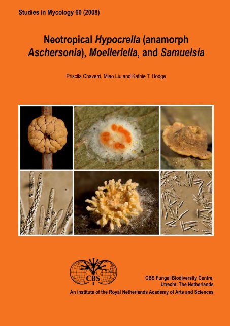
Studies in MycologyThe Studies in Mycology is an international journal which publishes systematic monographs of filamentous fungi <strong>and</strong> yeasts, <strong>and</strong> in rareoccasions the proceedings of special meetings related to all fields of mycology, biotechnology, ecology, molecular biology, pathology <strong>and</strong>systematics. For instructions for authors see www.cbs.knaw.nl.Ex e c u t i v e Ed i t o rProf. dr Robert A. Samson, <strong>CBS</strong> Fungal Biodiversity Centre, P.O. Box 85167, 3508 AD Utrecht, The Netherl<strong>and</strong>s.E-mail: r.samson@cbs.knaw.nlLay o u t Ed i t o rManon van den Hoeven-Verweij, <strong>CBS</strong> Fungal Biodiversity Centre, P.O. Box 85167, 3508 AD Utrecht, The Netherl<strong>and</strong>s.E-mail: m.verweij@cbs.knaw.nlScientific Ed i t o rsProf. dr Uwe Braun, Martin-Luther-Universität, Institut für Geobotanik und Botanischer Garten, Herbarium, Neuwerk 21, D-06099 Halle, Germany.E-mail: uwe.braun@botanik.uni-halle.deProf. dr Pedro W. Crous, <strong>CBS</strong> Fungal Biodiversity Centre, P.O. Box 85167, 3508 AD Utrecht, The Netherl<strong>and</strong>s.E-mail: p.crous@cbs.knaw.nlProf. dr David M. Geiser, Department of Plant Pathology, 121 Buckhout Laboratory, Pennsylvania State University, University Park, PA, U.S.A. 16802.E-mail: dgeiser@psu.eduDr Lorelei L. Norvell, Pacific Northwest Mycology Service, 6720 NW Skyline Blvd, Portl<strong>and</strong>, OR, U.S.A. 97229-1309.E-mail: llnorvell@pnw-ms.comDr Erast Parmasto, Institute of Zoology & Botany, 181 Riia Street, Tartu, Estonia EE-51014.E-mail: e.parmasto@zbi.eeProf. dr Alan J.L. Phillips, Faculdade de Ciências e Tecnologia, Universidade Nova de Lisboa, Quinta de Torre, 2829-516 Caparica, Portugal.E-mail: alp@mail.fct.unl.ptDr Amy Y. Rossman, Rm 304, Bldg 011A, Systematic Botany & Mycology Laboratory, Beltsville, MD, U.S.A. 20705.E-mail: amy@nt.ars-grin.govDr Keith A. Seifert, Research Scientist / Biodiversity (Mycology <strong>and</strong> Botany), Agriculture & Agri-Food Canada, KW Neatby Bldg, 960 Carling Avenue,Ottawa, ON, Canada K1A OC6.E-mail: seifertk@agr.gc.caProf. dr Jeffrey K. Stone, Department of Botany & Plant Pathology, Cordley 2082, Oregon State University, Corvallis, OR, U.S.A. 97331-2902.E-mail: stonej@bcc.orst.eduDr Richard C. Summerbell, 27 Hillcrest Park, Toronto, Ont. M4X 1E8, Canada.E-mail: summerbell@aol.comCopyright 2008 <strong>CBS</strong> Fungal Biodiversity Centre, P.O. Box 85167, 3508 AD Utrecht, The Netherl<strong>and</strong>s.You are free to share — to copy, distribute <strong>and</strong> transmit the work, under the following conditions:Attribution:You must attribute the work in the manner specified by the author or licensor (but not in any way that suggests that they endorse youor your use of the work).Non-commercial: You may not use this work for commercial purposes.No derivative works: You may not alter, transform, or build upon this work.For any reuse or distribution, you must make clear to others the license terms of this work, which can be found at http://creativecommons.org/licenses/bync-nd/3.0/legalcode.Any of the above conditions can be waived if you get permission from the copyright holder. Nothing in this license impairs or restrictsthe author"s moral rights.Publication date: 15 March 2008Published <strong>and</strong> distributed by <strong>CBS</strong> Fungal Biodiversity Centre, P.O. Box 85167, 3508 AD Utrecht, The Netherl<strong>and</strong>s. Internet: www.cbs.knaw.nl.E-mail: info@cbs.knaw.nl.ISBN/EAN : 978-90-70351-70-0Online ISSN : 1872-9797Print ISSN : 0166-0616Cover: Images of various species of Hypocrella, <strong>Moelleriella</strong> <strong>and</strong> <strong>Samuelsia</strong>. Top from left to right: <strong>Moelleriella</strong> macrostroma. Stroma; <strong>Moelleriella</strong> libera(anam. “A.” alyerodis). Conidiomata; <strong>Samuelsia</strong> geonomis. Stromata containing perithecia. Bottom from left to right: <strong>Moelleriella</strong> boliviensis. Asci withascospores; Hypocrella hirsuta. Stromata; Hypocrella viridans. Conidia.
Neotropical Hypocrella (anamorphAschersonia), <strong>Moelleriella</strong>, <strong>and</strong> <strong>Samuelsia</strong>Priscila ChaverriDepartment of Biology, Howard University, 415 College Street NW, Washington D.C. 20059, U.S.A.Miao LiuAgriculture <strong>and</strong> Agri-Food Canada/Agriculture et Agroalimentaire Canada, Biodiversity (Mycology <strong>and</strong> Botany), 960 Carling Avenue, Ottawa, Ontario K1A 0C6,CanadaKathie T. HodgeDepartment of Plant Pathology, Cornell University, 334 Plant Science Building, Ithaca, New York 14853, U.S.A.<strong>CBS</strong> Fungal Biodiversity Centre,Utrecht, The Netherl<strong>and</strong>sAn institute of the Royal Netherl<strong>and</strong>s Academy of Arts <strong>and</strong> Sciences
CONTENTSAbstract ............................................................................................................................................................................................................ 1Introduction ...................................................................................................................................................................................................... 1The genera Hypocrella/Aschersonia, <strong>Moelleriella</strong>, <strong>and</strong> <strong>Samuelsia</strong> .................................................................................................................. 2Morphology <strong>and</strong> Taxonomy ........................................................................................................................................................................ 2Taxonomic Background: Subdividing Hypocrella/Aschersonia s. l. ............................................................................................................ 4Geographical Distribution ........................................................................................................................................................................... 4Habitat ....................................................................................................................................................................................................... 5Microclimate ........................................................................................................................................................................................ 5Host specificity, co-evolution, <strong>and</strong> adaptation ...................................................................................................................................... 5Nutrition ..................................................................................................................................................................................................... 6Life cycle <strong>and</strong> epidemiology ....................................................................................................................................................................... 6Secondary metabolites produced by Hypocrella/Aschersonia <strong>and</strong> <strong>Moelleriella</strong> ......................................................................................... 7Hypocrella/Aschersonia, <strong>Samuelsia</strong>, <strong>and</strong> <strong>Moelleriella</strong> in biological control of insects ............................................................................... 8Materials <strong>and</strong> methods ..................................................................................................................................................................................... 8Isolates <strong>and</strong> herbarium specimens ............................................................................................................................................................ 8Morphological characterisation .................................................................................................................................................................. 8DNA extraction, PCR <strong>and</strong> sequencing ....................................................................................................................................................... 9Phylogenetic analyses ............................................................................................................................................................................... 9Results ........................................................................................................................................................................................................... 12Phylogenetic analyses ............................................................................................................................................................................. 12Hypocrella/Aschersonia, <strong>Samuelsia</strong>, <strong>and</strong> <strong>Moelleriella</strong> ....................................................................................................................... 12<strong>Moelleriella</strong> ........................................................................................................................................................................................ 13Hypocrella/Aschersonia s. str. ........................................................................................................................................................... 13<strong>Samuelsia</strong> .......................................................................................................................................................................................... 13Morphological analyses <strong>and</strong> geographical distribution ............................................................................................................................ 16<strong>Moelleriella</strong>, <strong>Samuelsia</strong>, <strong>and</strong> Hypocrella/Aschersonia ....................................................................................................................... 16<strong>Moelleriella</strong> morphology ..................................................................................................................................................................... 16Hypocrella/Aschersonia morphology ................................................................................................................................................. 17<strong>Samuelsia</strong> morphology ...................................................................................................................................................................... 18Discussion ...................................................................................................................................................................................................... 18Generic concept ....................................................................................................................................................................................... 18Species concept ...................................................................................................................................................................................... 18Evolutionary hypotheses in <strong>Moelleriella</strong>, Hypocrella/Aschersonia, <strong>and</strong> <strong>Samuelsia</strong> ................................................................................. 19Evolution of anamorph characters ..................................................................................................................................................... 19Evolution of teleomorph characters ................................................................................................................................................... 20Key to genera treated ..................................................................................................................................................................................... 20Synoptic keys ................................................................................................................................................................................................. 20<strong>Moelleriella</strong> (anamorphs aschersonia-like) .............................................................................................................................................. 20Hypocrella (anamorphs Aschersonia s. str.) ............................................................................................................................................ 24<strong>Samuelsia</strong> (anamorphs aschersonia-like) ................................................................................................................................................ 26Dichotomous keys .......................................................................................................................................................................................... 27<strong>Moelleriella</strong> key based on teleomorph characters .................................................................................................................................... 27<strong>Moelleriella</strong> key based on anamorph characters ..................................................................................................................................... 29Hypocrella/Aschersonia key based on teleomorph characters ................................................................................................................ 30Hypocrella/Aschersonia key based on anamorph characters .................................................................................................................. 30<strong>Samuelsia</strong> key based on teleomorph characters ..................................................................................................................................... 30<strong>Samuelsia</strong> key based on anamorph characters ....................................................................................................................................... 30Genera <strong>and</strong> species descriptions ................................................................................................................................................................... 31MOELLERIELLA ...................................................................................................................................................................................... 311. <strong>Moelleriella</strong> basicystis/“A.” basicystis ................................................................................................................................................. 312. <strong>Moelleriella</strong> boliviensis ....................................................................................................................................................................... 333. <strong>Moelleriella</strong> castanea ......................................................................................................................................................................... 344. <strong>Moelleriella</strong> colliculosa ....................................................................................................................................................................... 345. <strong>Moelleriella</strong> cornuta ........................................................................................................................................................................... 346. <strong>Moelleriella</strong> disjuncta ......................................................................................................................................................................... 367. <strong>Moelleriella</strong> epiphylla/“A.” cubensis ................................................................................................................................................... 388. <strong>Moelleriella</strong> evansii ............................................................................................................................................................................ 399. <strong>Moelleriella</strong> gaertneriana ................................................................................................................................................................... 3910. <strong>Moelleriella</strong> globosa ........................................................................................................................................................................... 39
11. <strong>Moelleriella</strong> guaranitica/“A.” caapi ...................................................................................................................................................... 4112. <strong>Moelleriella</strong> libera/“A.” aleyrodis ......................................................................................................................................................... 4113. <strong>Moelleriella</strong> macrostroma ................................................................................................................................................................... 4414. <strong>Moelleriella</strong> madidiensis .................................................................................................................................................................... 4415. <strong>Moelleriella</strong> ochracea/“A.” <strong>and</strong>ropogonis ........................................................................................................................................... 4416. <strong>Moelleriella</strong> palmae ............................................................................................................................................................................ 4617. <strong>Moelleriella</strong> phyllogena/“A.” juruensis ................................................................................................................................................ 4618. <strong>Moelleriella</strong> rhombispora ................................................................................................................................................................... 4819. <strong>Moelleriella</strong> sloaneae ......................................................................................................................................................................... 4820. <strong>Moelleriella</strong> turbinata/“A.” turbinata .................................................................................................................................................... 5021. <strong>Moelleriella</strong> umbospora ..................................................................................................................................................................... 5022. <strong>Moelleriella</strong> zhongdongii/“A.” incrassata ........................................................................................................................................... 52HYPOCRELLA ......................................................................................................................................................................................... 531. Hypocrella aurantiaca/A. aurantiaca .................................................................................................................................................. 532. Hypocrella citrina/A. blumenaviensis ................................................................................................................................................. 533. Hypocrella disciformis/A. disciformis ................................................................................................................................................. 554. Hypocrella hirsuta .............................................................................................................................................................................. 555. Hypocrella viridans/A. viridans .......................................................................................................................................................... 58SAMUELSIA ............................................................................................................................................................................................ 591. <strong>Samuelsia</strong> chalalensis ....................................................................................................................................................................... 592. <strong>Samuelsia</strong> geonomis ......................................................................................................................................................................... 593. <strong>Samuelsia</strong> intermedia/“A.” intermedia ................................................................................................................................................ 604. <strong>Samuelsia</strong> rufobrunnea ..................................................................................................................................................................... 625. <strong>Samuelsia</strong> sheikhii ............................................................................................................................................................................. 63Excluded or doubtful species ......................................................................................................................................................................... 63Acknowledgements ........................................................................................................................................................................................ 64References ..................................................................................................................................................................................................... 64Index .............................................................................................................................................................................................................. 67
available online at www.studiesinmycology.orgdoi:10.3114/sim.2008.60.01St u d i es in My c o l o g y 60: 1–66. 2008.A monograph of the entomopathogenic genera Hypocrella, <strong>Moelleriella</strong>, <strong>and</strong><strong>Samuelsia</strong> gen. nov. (Ascomycota, Hypocreales, Clavicipitaceae), <strong>and</strong> theiraschersonia-like anamorphs in the NeotropicsP. Chaverri 1 , M. Liu 2 <strong>and</strong> K.T. Hodge 31Department of Biology, Howard University, 415 College Street NW, Washington D.C. 20059, U.S.A.; 2 Agriculture <strong>and</strong> Agri-Food Canada/Agriculture et Agroalimentaire Canada,Biodiversity (Mycology <strong>and</strong> Botany), 960 Carling Avenue, Ottawa, Ontario K1A 0C6, Canada; 3 Department of Plant Pathology, Cornell University, 334 Plant Science Building,Ithaca, New York 14853, U.S.A.*Correspondence: Priscila Chaverri pchaverri@howard.eduAbstract: The present taxonomic revision deals with Neotropical species of three entomopathogenic genera that were once included in Hypocrella s. l.: Hypocrella s. str.(anamorph Aschersonia), <strong>Moelleriella</strong> (anamorph aschersonia-like), <strong>and</strong> <strong>Samuelsia</strong> gen. nov (anamorph aschersonia-like). Species of Hypocrella, <strong>Moelleriella</strong>, <strong>and</strong> <strong>Samuelsia</strong>are pathogens of scale insects (Coccidae <strong>and</strong> Lecaniidae, Homoptera) <strong>and</strong> whiteflies (Aleyrodidae, Homoptera) <strong>and</strong> are common in tropical regions. Phylogenetic analysesof DNA sequences from nuclear ribosomal large subunit (28S), translation elongation factor 1-α (TEF 1-α), <strong>and</strong> RNA polymerase II subunit 1 (RPB1) <strong>and</strong> analyses of multiplemorphological characters demonstrate that the three segregated genera can be distinguished by the disarticulation of the ascospores <strong>and</strong> shape <strong>and</strong> size of conidia. <strong>Moelleriella</strong>has filiform multi-septate ascospores that disarticulate at the septa within the ascus <strong>and</strong> aschersonia-like anamorphs with fusoid conidia. Hypocrella s. str. has filiform to longfusiformascospores that do not disarticulate <strong>and</strong> Aschersonia s. str. anamorphs with fusoid conidia. The new genus proposed here, <strong>Samuelsia</strong>, has filiform to long-fusiformascospores that do not disarticulate <strong>and</strong> aschersonia-like anamorphs with small allantoid conidia. In addition, the present study presents <strong>and</strong> discusses the evolution of species,morphology, <strong>and</strong> ecology in Hypocrella, <strong>Moelleriella</strong>, <strong>and</strong> <strong>Samuelsia</strong> based on multigene phylogenetic analyses.Key words: multilocus phylogenetics, polyphasic taxonomy, species identification, species recognition.Taxonomic novelties: New genus: <strong>Samuelsia</strong>. New species: Hypocrella disciformis, H. hirsuta, <strong>Moelleriella</strong> basicystis, M. boliviensis, M. cornuta, M. evansii, M. madidiensis,M. umbospora, S. chalalensis, S. geonomis, S. intermedia, S. rufobrunnea, <strong>and</strong> S. sheikhii. New combinations: M. castanea, M. colliculosa, M. disjuncta, M. epiphylla, M.gaertneriana, M. globosa, M. guaranitica, M. javanica, M. libera, M. macrostroma, M. ochracea, M. palmae, M. phyllogena, M. rhombispora, M. sloaneae, M. turbinata, <strong>and</strong> M.zhongdongii.INTRODUCTIONSpecies in the genus Hypocrella Sacc. s. l. (anamorph AschersoniaMont. s. l.) are insect pathogens characterised by their brightlycolouredstromata, filiform ascospores, <strong>and</strong> pycnidial to acervularanamorphs. They are common in tropical regions, particularly inmoist old-growth forests, where they often cause epizootics ofdisease in their hosts, scale insects (Coccidae <strong>and</strong> Lecaniidae,Homoptera) <strong>and</strong> whiteflies (Aleyrodidae, Homoptera). Themajority of the species are tropical, but a few are also found inthe subtropics (Petch 1921, Mains 1959a, b, Evans & Hywel-Jones 1990, Hywel-Jones & Evans 1993). Other genera, includingAscopolyporus A. Möller, Cosmospora Rabenh., HyperdermiumJ.F. White et al., Podonectria Petch, Regiocrella Chaverri & K.T.Hodge, Septobasidium Pat., <strong>and</strong> Torrubiella Boud., among others,are also found as hyperparasites of whiteflies <strong>and</strong> scale insects,but generally not as frequently as Hypocrella, <strong>Moelleriella</strong>, <strong>and</strong><strong>Samuelsia</strong>.Molecular <strong>and</strong> morphological characters support therecognition of three well-defined genera within the broad conceptof Hypocrella: Hypocrella s. str., <strong>Moelleriella</strong> Bres., <strong>and</strong> <strong>Samuelsia</strong>gen. nov. (Clavicipitaceae, Hypocreales, Hypocreomycetidae,Sordariomycetes, Pezizomycotina, Ascomycota). These newgenera <strong>and</strong> their anamorphs are presented here. Hypocrella s. str.has an Aschersonia anamorph. The anamorphs of <strong>Moelleriella</strong> <strong>and</strong><strong>Samuelsia</strong> are aschersonia-like <strong>and</strong> thus hereafter will be denotedin quotes (e.g. “Aschersonia”) to avoid confusion.Until 1897, Hypocrella s. l. species were described asphyllogenous <strong>and</strong> thought to be either parasitic or superficialcolonisers of living leaves (Evans & Hywel-Jones 1990). Theirparasitic association with scale insects <strong>and</strong> whiteflies was notrecognised until Webber (1897) concluded that several <strong>Moelleriella</strong>species (then identified as “Aschersonia” aleyrodis Webber) wereimportant factors in the biological control of whitefly pests in citrusplantations. Indeed, the first biocontrol applications in the U.S.A.were done with “A.” aleyrodis (teleom. <strong>Moelleriella</strong> libera) to controlcitrus whitefly (Dialeurodes citri Ashmead) in Florida (Berger 1921).Since then, the biocontrol potential <strong>and</strong> ubiquity of Hypocrella s.l. has been widely recognised (Parkin 1906, Morrill & Back 1912,Petch 1921, Fawcett 1936, Ferron 1978, Barua 1983, Brady 1984,Ramakers & Samson 1984, Fransen 1987, Rombach & Gillespie1988, Gerling 1992, Osborne & L<strong>and</strong>a 1992, Meekes et al. 1994,Samson 1995, Meekes et al. 1996, Lourencao et al. 1999, Meekeset al. 2000, Faria & Wraight 2001, Meekes 2001).Despite the potential of these species in insect biocontrol,few taxonomic treatments have been written after Petch’s(1921) monograph of Hypocrella s. l. Approximately 115 namesin Hypocrella <strong>and</strong> 79 names in Aschersonia have been validlypublished; however, only about 50 <strong>and</strong> 44 species, respectively,are currently accepted (Petch 1921, Dingley 1954, Mains 1959b,a, Hywel-Jones & Evans 1993). The present taxonomic treatmentdeals with Hypocrella s. str. (anamorphs Aschersonia s. str.) <strong>and</strong> itsnewly segregated sister genera <strong>Moelleriella</strong> <strong>and</strong> <strong>Samuelsia</strong> that areencountered in the Neotropics. Hundreds of freshly collected <strong>and</strong>Copyright 2008 <strong>CBS</strong> Fungal Biodiversity Centre, P.O. Box 85167, 3508 AD Utrecht, The Netherl<strong>and</strong>s.You are free to share - to copy, distribute <strong>and</strong> transmit the work, under the following conditions:Attribution:You must attribute the work in the manner specified by the author or licensor (but not in any way that suggests that they endorse you or your use of the work).Non-commercial: You may not use this work for commercial purposes.No derivative works: You may not alter, transform, or build upon this work.For any reuse or distribution, you must make clear to others the license terms of this work, which can be found at http://creativecommons.org/licenses/by-nc-nd/3.0/legalcode. Any of the above conditions can be waived if you getpermission from the copyright holder. Nothing in this license impairs or restricts the author’s moral rights.1
Ch av e r r i e t a l.herbarium specimens, including types were examined, <strong>and</strong> multigenephylogenetic analyses were conducted. Even though this isa comprehensive study, fungal biodiversity surveys in other poorlyexplored regions will probably reveal more undescribed species.The genera Hypocrella/Aschersonia,<strong>Moelleriella</strong>, <strong>and</strong> <strong>Samuelsia</strong>Morphology <strong>and</strong> TaxonomyThe genera Hypocrella, <strong>Moelleriella</strong>, <strong>and</strong> <strong>Samuelsia</strong> arerecognised by their brightly coloured stromata that form on scaleinsects or whiteflies. As will be shown in this study, Hypocrella s.str. includes species with ascospores that do not disarticulate <strong>and</strong>fusiform conidia; <strong>Moelleriella</strong> includes species with ascospores thatdisarticulate inside the ascus <strong>and</strong> fusiform conidia; <strong>and</strong> <strong>Samuelsia</strong>species have non-disarticulating ascospores <strong>and</strong> small allantoidconidia.The genus Hypocrella was erected by Saccardo (1878) toaccommodate four species previously assigned to Hypocrea Fr.(Hypocreaceae, Hypocreales): Hypocrea atramentosa Berk. & M.A.Curtis (= Myriogenospora atramentosa (Berk. & M.A. Curtis) Diehl);Hypocrea discoidea Berk. & Broome; Hypocrea semiamplexaBerk. (a Balansia species according to Petch 1921); <strong>and</strong> Hypocreabambusae Berk. & Broome (= Balansia bambusae (Berk. & Broome)Petch). Only Hypocrella discoidea (Berk. & Broome) Sacc. (type ofthe genus) remains in Hypocrella. <strong>Moelleriella</strong> was erected in 1896to accommodate <strong>Moelleriella</strong> sulphurea Bres. (= M. phyllogena). InBresadola’s original diagnosis, M. sulphurea has a pulvinate stroma<strong>and</strong> filiform ascospores that disarticulate into numerous cylindricalpart-spores.The genus Hypocrella was originally distinguished fromHypocrea by the formation of eight, filiform ascospores in eachascus (Saccardo 1878). Petch (1921) reported that the filiformascospores in H. discoidea disarticulated at the septa while stillin the asci <strong>and</strong> speculated that if whole ascospores were seenin asci, it was an indication of immaturity. Later, Petch (1939)described a new genus, Stereocrea H. Sydow & P. Sydow, wherehe placed species with non-disarticulating ascospores. At that time,Petch did not realise that H. discoidea also had non-disarticulatingascospores. Later, Mains (1959b) concluded that the presence ofnon-disarticulating ascospores in H. aurantiaca (Petch) Mains (=Stereocrea aurantiaca) was not enough evidence to separate itfrom Hypocrella. Until now, it had been accepted that Hypocrellaspecies include those with disarticulating <strong>and</strong> non-disarticulatingascospores (Petch 1939, Mains 1959a, b, Hywel-Jones & Evans1993). However, evidence presented in this study supportsspeculations by Hywel-Jones <strong>and</strong> Evans (1993) <strong>and</strong> Petch (1939)that species with disarticulating ascospores form a monophyleticgroup that should be segregated from Hypocrella. In the presentmonograph, species with disarticulating ascospores are placed in<strong>Moelleriella</strong>.Anamorphs of Hypocrella have been classified in the genusAschersonia, which was erected by Montagne (1848) based on A.tahitensis Mont. Aschersonia s. str. is characterised by pycnidiumlikeconidiomata, phialides, paraphyses, <strong>and</strong> unicellular, fusiform,hyaline conidia that are brightly coloured in mass <strong>and</strong> producedin copious slime. <strong>Moelleriella</strong>, Hypocrella <strong>and</strong> <strong>Samuelsia</strong> speciesmay or may not have paraphyses. The presence of paraphyses(sterile, hypha-like elements in the hymenium) has sometimesbeen used as a character to distinguish Aschersonia subgenera.<strong>Moelleriella</strong> <strong>and</strong> <strong>Samuelsia</strong> also have aschersonia-like anamorphsthat are similar to Aschersonia s. str. but can be distinguished byconidial size <strong>and</strong> shape. Hypocrella s. str. has fusiform conidia thatare larger than those of <strong>Moelleriella</strong> <strong>and</strong> <strong>Samuelsia</strong>. <strong>Samuelsia</strong> hasallantoid <strong>and</strong> smaller conidia. The anamorphic states of Hypocrella,<strong>Moelleriella</strong>, <strong>and</strong> <strong>Samuelsia</strong> are more commonly collected than theteleomorphs, <strong>and</strong> are rarely associated with the teleomorph in thesame stroma.As discussed in Chaverri et al. (2005a), within theClavicipitaceae, pycnidial to acervular anamorphic forms havebeen assigned to one of three anamorph genera: Aschersonia,Ephelis Fr., or Sphacelia Lév. These types of anamorphs are knownonly for the plant-associated teleomorph genera AtkinsonellaDiehl, Balansia Speg., Claviceps Tul., Epichloë (Fr.) Tul. & C. Tul.,Myriogenospora G.F. Atk. <strong>and</strong> Neoclaviceps J.F. White et al., <strong>and</strong>the scale-insect <strong>and</strong> whitefly parasites Hypocrella, <strong>Moelleriella</strong>,<strong>Samuelsia</strong>, <strong>and</strong> Regiocrella. Some studies have shown evidence ofa single evolutionary origin of the pycnidial-acervular morphologyin the Clavicipitaceae (Kuldau et al. 1997, Sullivan et al. 2001,Chaverri et al. 2005a). Regiocrella was recently described <strong>and</strong>although similar to Hypocrella, <strong>Moelleriella</strong>, <strong>and</strong> <strong>Samuelsia</strong> in itspycnidial-acervular conidiomata, brightly coloured ascomata, <strong>and</strong>its parasitism on scale insects, Regiocrella differs from those threegenera by its short-fusiform <strong>and</strong> unicellular ascospores (Chaverriet al. 2005a). Within the group of genera with pycnidial-acervularconidiomata, Hypocrella, <strong>Moelleriella</strong>, <strong>and</strong> <strong>Samuelsia</strong> are easilydistinguished by the shapes of their conidia <strong>and</strong> ascospores.The first suggestion that Aschersonia was the anamorphof Hypocrella was made by Massee (1896) in an account ofAschersonia oxyspora Berk. (= <strong>Moelleriella</strong> phyllogena = H.phyllogena (Mont.) Petch). Later, Möller (1901) reported on theoccurrence of both the perithecial <strong>and</strong> pycnidial states in thesame stromata of H. cavernosa A. Möller (= M. cavernosa = M.palmae Berk. & M.A. Curtis), <strong>and</strong> identified the pycnidial state asaschersonia-like. Although it is not common to find both statesin the same stroma, several species of Hypocrella, <strong>Moelleriella</strong>,<strong>and</strong> <strong>Samuelsia</strong> possess this characteristic (e.g. M. libera, M.mollii, M. ochracea, M. reineckiana, M. sloaneae, M. turbinata, H.disciformis, H. discoidea, H. viridans, <strong>and</strong> S. rufobrunnea, amongothers). It is now widely accepted that Aschersonia is the anamorphof Hypocrella. However, the whole life cycles of many of the 40–50species of Hypocrella, <strong>Moelleriella</strong>, <strong>and</strong> <strong>Samuelsia</strong> are not yetknown. Only about 15 species of Aschersonia s. l. have been linkedto their teleomorphs (Petch 1921, Dingley 1954, Mains 1959b,a, Hywel-Jones & Evans 1993). In the present study, previouslyunknown life cycles of Hypocrella, <strong>Moelleriella</strong>, <strong>and</strong> <strong>Samuelsia</strong> aredescribed <strong>and</strong> illustrated (see Table 1 for teleomorph-anamorphconnections).2
Ne o t r o p i c a l Hy p o c r e l l a, Mo e l l e r i e l l a, a n d Sa m u e l s i aTable 1. Teleomorph-anamorph connections to accepted species of <strong>Moelleriella</strong>, Hypocrella <strong>and</strong> <strong>Samuelsia</strong>. Old World: OW; New World:NW.Teleomorph 1 Anamorph 1,2 Source DistributionHypocrella aurantiaca A. aurantiaca Petch 1939, Mains 1959a NWH. citrina A. blumenaviensis Mains 1959b NWH. disciformis 3 A. disciformis NWH. discoidea A. samoensis Hywel-Jones & Evans 1993 OWH. hirsuta 3 Not named NWH. hypocreoidea A. hypocreoidea Petch 1924 OWH. viridans A. viridans Petch 1921 NW<strong>Moelleriella</strong> africana Not known Hywel-Jones & Samuels 1998 OWM. amomi “A.” caespiticia Petch 1921, Mains 1959a OWM. basicystis 3 “A.” basicystis NWM. bispora Not known OWM. boliviensis Not known NWM. botryosa Not known OWM. castanea Not named Mains 1959a, Petch 1932 NWM. ceramichroa Not named Petch 1921 OWM. colliculosa Not known NWM. convexa Not known Petch 1921 OWM. cornuta Not known NWM. disjuncta 3 Not named NWM. duplex “A.” duplex Petch 1921 OWM. epiphylla “A.” cubensis Mains 1959a, b NWM. evansii 3 Not named NWM. gaertneriana Not known NWM. globosa 3 Not named NWM. guaranitica 3 “A.” caapi NWM. javanica “A.” coffeae Petch 1921 OWM. libera “A.” aleyrodis Petch 1924 NWM. macrostroma Not named Chaverri et al. 2005 NWM. madidiensis 4 Not named NWM. mollii “A.” confluens Petch 1921 OWM. murrayae Not named Kobayashi 1973 OWM. ochracea “A.” <strong>and</strong>ropogonis Petch 1921, Liu et al. 2006 NWM. olivacea Not named Petch 1921 OWM. oxystoma “A.” oxystoma Petch 1921 OWM. palmae Not known Petch 1921 NWM. palmicola Not named Petch 1921 OWM. phyllogena 3 “A.” juruensis NWM. raciborskii “A.” placenta Liu et al. 2006 OWM. reineckiana “A.” marginata Petch 1921 OWM. rhombispora Not named Liu et al. 2006 NWM. schizostachyi Not named Hywel-Jones & Samuels 1998 OWM. scutata Not known OWM. sloaneae Not named Mains 1959b NWM. tubulata Not named Petch 1921 OWM. turbinata “A.” turbinata Petch 1921 NWM. umbospora 3 Not namedM. zhongdongii “A.” incrassata Liu & Hodge 2005 NW<strong>Moelleriella</strong> sp. 4 “A.” insperata OW<strong>Samuelsia</strong> chalalensis 4 Not named NWS. geonomis Not known NWwww.studiesinmycology.org3
Ch av e r r i e t a l.Table 1. (Continued).Teleomorph 1 Anamorph 1,2 Source DistributionS. intermedia 4 “A.” intermedia NWS. rufobrunnea 3 Not named NWS. sheikhii 4 Not named NWNot known A. acutispora OWNot known A. australiensis OWNot known A. badia OWNot known A. crenulata OWNot known A. flava OWNot known A. papillata OWNot known A. tahitensis OWNot known A. tamurai OW1“Not named” refers to the anamorph or teleomorph that has been described but no name was given; or “not known” when the anamorph has not been documented in theliterature.2Aschersonia names in quotes (e.g. “Aschersonia”) denotes taxa that are not in Aschersonia sensu stricto.3Indicates connections made in the present study.4Based on anamorph; no mature perithecia observed.Taxonomic Background: Subdividing Hypocrella/Aschersonia s. l.Few studies have comprehensively dealt with the taxonomy ofHypocrella <strong>and</strong> related genera or have included more than a fewspecies (Petch 1921, Mains 1959a, b, Liu et al. 2006). The firsttaxonomic treatment of the genus was made by Parkin (1906)who considered extensive collections from Sri Lanka. The seminalmonograph of Petch (1921) treated 57 species of Hypocrella,including anamorphs <strong>and</strong> teleomorphs from the New <strong>and</strong> OldWorlds. Mains (1959a, 1959b) dealt with 24 species of Hypocrella<strong>and</strong> 15 species of Aschersonia.Petch (1921) proposed Hypocrella subgenus Fleischeriafor species of Hypocrella in which the Aschersonia state lacksparaphyses (<strong>and</strong> Aschersonia subg. Leprieuria for the correspondinganamorphs). He considered that most of these species attackscale insects (Coccidae; Lecaniidae according to Petch 1921).In that same work, Petch proposed Hypocrella subg. Hypocrella(as “Euhypocrella” see Art. 21 ICBN) for species on Aleyrodidae<strong>and</strong> with paraphyses in the conidiomata of their Aschersonia subg.Aschersonia (as “Euaschersonia” see Art. 21 ICBN) anamorphs.Recent evidence reveals that, although sometimes diagnostic forindividual species, the presence of paraphyses in the conidiomais not phylogenetically informative (Liu & Hodge 2005, Liu et al.2006). Dingley (1954) also questioned this division since “A.” duplexBerk., with paraphyses in the conidioma, is consistently associatedwith Coccidae. In addition, other studies showed that cultures ofAschersonia may lack paraphyses whereas these were presentin the conidioma on the host, further indicating that the presenceof paraphyses is not a stable taxonomic character (Hywel-Jones1993, Hywel-Jones & Evans 1993, Meekes 2001).Petch (1921) also discussed the possibility of splittingHypocrella into groups characterised by the form of the stroma.However, he mentioned that these forms “…grade into one anotherto such an extent that this character cannot be relied on.” In theoriginal diagnosis of the genus, Fleischeria was distinguished fromHypocrella by its harder stroma (Penzig & Saccardo 1901). Petch(1921) synonymised the two genera because species of Hypocrellacan vary in hardness, from hard in H. schizostachyi Henn. (=M. schizostachyi) to soft in H. convexa Racib. (= M. convexa).Chaverri et al. (2005b) demonstrated the presence of three naturalgroups (i.e. clades) in Hypocrella s. l. based on DNA sequencedata that correlated with stromatal morphology. The Effuse grouphas flat, effuse stromata of loose hyphal tissue, broad hypothalli,<strong>and</strong> whitish colouration (e.g. M. evansii, M. libera, M. madidiensis,M. ochracea, M. raciborskii, M. rhombispora, <strong>and</strong> M. zhongdongii),except pale yellow to orange in the M. basicystis species complex;the Globose group includes species that have globose stromata thatare generally darker in colour (yellow to brownish), large, compacttissue, hard or coriaceous, moderately to strongly tuberculate, <strong>and</strong>without hypothalli (e.g. M. africana, M. boliviensis, M. cornuta,M. epiphylla, M. gaertneriana, M. insperata, M. macrostroma, M.turbinata, <strong>and</strong> M. schizostachyi); <strong>and</strong> the Pulvinate group (nowHypocrella <strong>and</strong> <strong>Samuelsia</strong>) comprises species that have pulvinate orcushion-like stromata, somewhat compact <strong>and</strong> flattened, yellowishor green, sometimes brownish, with or without hypothalli, generallychanging colour in 3 % KOH (e.g. H. aurantiaca, H. disciformis,H. viridans, H. discoidea, H. hirsuta, S. geonomis, S. chalalensis,S. rufobrunnea, <strong>and</strong> S. sheikhii). The groups defined by Chaverriet al. (2005b) correlate roughly with some of the groupings thatPetch (1921) discussed. Although there are distinct phenotypiccharacteristics that distinguish each of the three major groupsdefined in Chaverri et al. (2005b), a formal generic or subgenericrevision of the taxonomy to distinguish the Effuse, Globose, <strong>and</strong>Pulvinate groups was not proposed.Geographical DistributionHypocrella, <strong>Moelleriella</strong>, <strong>and</strong> <strong>Samuelsia</strong> are mainly distributed inthe Tropics, with a few species found in the Subtropics (e.g.M.colliculosa, M. duplex, M. guaranitica, S. intermedia, H. aurantiaca,<strong>and</strong> H. citrina) (Table 1). This pattern of distribution <strong>and</strong> evolutionarydynamics have been observed in many other organisms where taxapreferentially originate in the Tropics <strong>and</strong> exp<strong>and</strong> toward the poleswithout losing their tropical presence (Jablonski et al. 2006). Thismight explain the higher biodiversity of fungi in the tropics compared4
Ne o t r o p i c a l Hy p o c r e l l a, Mo e l l e r i e l l a, a n d Sa m u e l s i ato that in temperate or arctic regions. Therefore, a tropical diversitycrisis would have profound evolutionary effects at all latitudes.In Hypocrella <strong>and</strong> <strong>Moelleriella</strong> but not <strong>Samuelsia</strong>, taxa witha disjunct distribution are apparent (Lee et al. 1996, Taylor et al.1999, Wen 1999, Wu et al. 2000). A disjunct distribution is one inwhich two closely related taxa (i.e. morphologically similar) arewidely separated geographically. In Hypocrella <strong>and</strong> <strong>Moelleriella</strong>,an Old World (OW) / New World (NW) disjunction is sometimesobserved (Petch 1921, Evans 1982, Evans & Hywel-Jones 1990).Disjunctions have been observed in M. palmae (NW) vs. M.sclerotioides Höhn. (OW); M. epiphylla (NW) vs. M. reineckiana(OW); M. libera (NW) vs. M. raciborskii (OW); M. ochracea (NW)vs. M. mollii (OW); M. macrostroma (NW) vs. M. africana (OW);M. castanea Petch (NW) vs. M. palmicola Henn. (OW); <strong>and</strong> M.gaertneriana (NW) vs. M. schizostachyi (OW) (Petch 1921, Evans1982, Evans & Hywel-Jones 1990, Chaverri et al. 2005b). It ispossible that disjunct geographical distributions are common inHypocrella <strong>and</strong> <strong>Moelleriella</strong>, but, because this monograph focuseson New World species, this issue is beyond the scope of thestudy.Individual species of Hypocrella, <strong>Samuelsia</strong>, <strong>and</strong> <strong>Moelleriella</strong>may be distributed over a large region or may be endemic tosmaller areas. The presumably short dispersal distances of spores,the distributions limited to the Tropics, <strong>and</strong> the relatively lowintraspecific genetic variability suggests the majority of the speciesform geographically conserved populations (Obornik et al. 2000).For example, in the New World Tropics, M. epiphylla, M. libera,M. ochracea, M. rhombispora, M. turbinata, <strong>and</strong> M. zhongdongiiare widespread <strong>and</strong> ubiquitous. In contrast, M. gaertneriana <strong>and</strong>M. cornuta have been found only in the Amazon basin (i.e. Brazil,French Guiana, Venezuela) <strong>and</strong> are rare. <strong>Moelleriella</strong> evansii <strong>and</strong>S. chalalensis, S. geonomis, <strong>and</strong> S. rufobrunnea have been foundonly in Ecuador, Bolivia, <strong>and</strong> Peru, respectively. The phenomenonof geographically restricted species is also observed in the M.basicystis species complex: M. basicystis s. str., M. phyllogena,M. disjuncta, <strong>and</strong> M. umbospora. The present monographdemonstrates that M. basicystis has been found in Panama <strong>and</strong>Costa Rica (probably southern Central America); M. phyllogena inPanama, Brazil, Ecuador, Bolivia, <strong>and</strong> Peru (probably Panama <strong>and</strong>Amazon basin); M. umbospora in Mexico, Honduras, Guatemala(probably northern Central America); <strong>and</strong> M. disjuncta in Panama<strong>and</strong> Guyana.Increased collecting in poorly explored regions will very likelydiscover new <strong>and</strong> rare species of Hypocrella, <strong>Samuelsia</strong>, <strong>and</strong><strong>Moelleriella</strong>. However, many species are apparently restricted toundisturbed old growth tropical forests. The increased destructionof these sensitive habitats due to deforestation, forest fires, urbansprawl, <strong>and</strong> other factors, will result in the disappearance of fungalspecies, some with potential beneficial value to human societies(e.g. as biocontrols <strong>and</strong> sources of novel metabolites) (Evans 1982,Samson & Evans 1985, Evans 1988, Samson 1995, Chaverri &Vílchez 2006).HabitatMicroclimateAlthough Hypocrella, <strong>Samuelsia</strong>, <strong>and</strong> <strong>Moelleriella</strong> are widespreadthroughout the Tropics, they do not occur in all types of climates <strong>and</strong>habitats within the Tropics. Few publications report on the climateor habitat characteristics that Hypocrella or <strong>Moelleriella</strong> speciesprefer, <strong>and</strong> there is no published information on <strong>Samuelsia</strong> species.www.studiesinmycology.orgBased on studies done for other fungi, ultraviolet light <strong>and</strong> directsolar radiation may be one important factor affecting the survivalof entomopathogenic fungal spores (Fargues et al. 1996, Mooreet al. 1996), as well as scale insects <strong>and</strong> whiteflies. This explainsin part why species of Hypocrella, <strong>Samuelsia</strong>, <strong>and</strong> <strong>Moelleriella</strong> arefound mostly on the abaxial surface of leaves, where the insect <strong>and</strong>the fungus are protected from direct solar radiation. For example,during the survey of Hypocrella, <strong>Samuelsia</strong>, <strong>and</strong> <strong>Moelleriella</strong> donefor the present study, no specimens were found in open highly sunexposedareas lacking a tree/shrub canopy.Temperature may also affect survival of Hypocrella, <strong>Samuelsia</strong>,<strong>and</strong> <strong>Moelleriella</strong>. Some studies have shown that temperature mayaffect conidial germination <strong>and</strong> nymphal mortality by M. libera(anam. “A.” aleyrodis) (Fransen et al. 1987, Fransen 1995). Incontrast, Meekes et al. (2000) considered that temperature didnot greatly affect infection ability by M. libera, but found a slightlyhigher conidial germination rate at 25 °C compared to 20 °C. Inthe present study, it was observed that habitats with a combinationof suboptimum temperatures (ca. 28–38 °C or below 18 °C) <strong>and</strong>extreme continuous relative humidity conditions (e.g. dry forests,very humid forests) did not produce many fungal collections. Forexample, six weeks of surveying a lowl<strong>and</strong> very wet tropical forestin Costa Rica yielded only eight species of Hypocrella s. l. (Chaverri& Vilchez 2006). Similarly, Hywel-Jones <strong>and</strong> Evans (1993) inThail<strong>and</strong>, did not find specimens of Hypocrella s. l. during the driestmonths of the year (February, March, <strong>and</strong> April). However, highrelative humidity (ca. 80 %) may be necessary for increased conidialgermination <strong>and</strong> whitefly mortality by M. libera <strong>and</strong> M. raciborskii(Meekes 2001). The environment immediately surrounding theconidia may also influence their survival. Chemicals on the leafsurface may influence the viability of spores, but also they can havean effect on the pest <strong>and</strong> its susceptibility to pathogens (Hare &Andreadis 1983, Cooke & Rayner 1984, Ramoska & Todd 1985).Host specificity, co-evolution, <strong>and</strong> adaptationSpecies of Hypocrella, <strong>Samuelsia</strong>, <strong>and</strong> <strong>Moelleriella</strong> are scaleinsect<strong>and</strong> whitefly pathogens. The insect host is almost alwayscompletely consumed by the fungus before the stroma <strong>and</strong> fruitingstructures become evident (Evans 1988, Evans & Hywel-Jones1990, Meekes et al. 1994), at which point it is almost impossible toidentify the insect. Therefore, there is scarce information about hostspecificity in Hypocrella, <strong>Samuelsia</strong>, or <strong>Moelleriella</strong>. Petch (1921)stated that species of Hypocrella could parasitise either whiteflies(Aleyrodidae) or scale insects (Coccidae <strong>and</strong> Lecaniidae). In somecases, it may be possible to hypothesise the identity of the insecthost based on neighboring individuals that are not completelyconsumed by the fungus or not infected. However, this approachcan be misleading, because in many cases several species ofscale insects or whiteflies are found on the same leaf (Petch 1921).Some species such as M. libera/”A.” aleyrodis, M. raciborskii/”A.”placenta, <strong>and</strong> M. ochracea/”A.” <strong>and</strong>ropogonis appear to begeneralists <strong>and</strong> have been found to infect at least five scale-insect<strong>and</strong> whitefly species (Petch 1921, Meekes et al. 2000, Meekes2001, Meekes et al. 2002). In most species of Hypocrella s. l. thedegree of host specificity is completely unknown.Characteristics of the plant upon which the host insect isfeeding may also be important. A single strain of M. libera showeddifferences in persistence as a consequence of chemical <strong>and</strong>/ormorphological differences between plants (Hare & Andreadis 1983,Cooke & Rayner 1984, Ramoska & Todd 1985, Meekes et al.2000). The co-evolution of fungi <strong>and</strong> their insect hosts may reflectco-evolution between the insects <strong>and</strong> their plant hosts. In tropical5
Ch av e r r i e t a l.forests, the spatial separation between conspecific plant individualsmay have given rise to spatially separated insect populationsthat, in turn, coevolved with their fungal parasites (Evans 1988).Successful horizontal dispersal of the pathogens between coccid<strong>and</strong> whitefly colonies would have decreased proportionally withincreasing adaptation <strong>and</strong> restriction of their hosts to certain trees(Evans 1988).Epizootics caused by Hypocrella, <strong>Samuelsia</strong>, <strong>and</strong> <strong>Moelleriella</strong>on coccids <strong>and</strong> whiteflies in the Tropics are so prominent thatseveral authors have wondered how these insects survived (Petch1925, Evans 1974, 1982, 1988). As mentioned before, dispersal ofthe fungus probably played an important role in the co-evolution withits insect host, <strong>and</strong> the co-evolution of the insect with its plant host.The anamorphic forms of Hypocrella, <strong>Samuelsia</strong>, <strong>and</strong> <strong>Moelleriella</strong>produce slimy masses of conidia that are well adapted for shortdistance,water-borne movement over leaf surfaces (Parkin 1906,Chaverri & Samuels 2003, Hodge 2003). The mucilage that coversthe conidia is high in sugar content making the conidia morehygroscopic (Meekes 2001) <strong>and</strong> attractive to insects. This modeof short-distance dispersal is apparently so efficient that once acolony of insects becomes infected by the fungus, it is difficult tofind healthy individuals (Evans 1988).Evans (1988) speculated about insect adaptation to the threatfrom entomopathogenic fungi. The short-distance dispersal ofconidia that contributes to epizootics may select for discontinuouspatterns of insect distribution. On the other h<strong>and</strong>, the teleomorphicforms of Hypocrella, <strong>Samuelsia</strong>, <strong>and</strong> <strong>Moelleriella</strong> are lesscommonly encountered <strong>and</strong> have dry, discharged ascospores thatare probably wind-dispersed <strong>and</strong> so better adapted for medium tolong distance dispersal (Evans 1988, 1989). However, the abovehypothesis could be refuted because in related genera, such asSphacelia, Ephelis <strong>and</strong> Neotyphodium, the discharge of conidia inslime appears to aid dispersal by insects (Loveless 1964, Moweret al. 1973, Mower & Hancock 1975, Samways 1983, Butler et al.2001, Hodge 2003).Scale insects <strong>and</strong> whiteflies often secrete sticky honeydewthat is attractive to wasps <strong>and</strong> ants, so it is also possible thatHypocrella, <strong>Samuelsia</strong>, <strong>and</strong> <strong>Moelleriella</strong> evolved to produce slimyconidia that adhere to non-host insect vectors. Infected coccid orwhitefly alates may further transport the fungus, as shown for someaphid-Entomophthorales interactions (Wilding & Perry 1980, Evans1989). Insect vectors <strong>and</strong> infected alates could disperse the sporesacross long distances; water, such as rain splash <strong>and</strong> run-off, coulddisperse across short distances (Chaverri et al. 2005a).Plant-insect-fungus specificity is suspected but not wellknown in Hypocrella, <strong>Samuelsia</strong>, <strong>and</strong> <strong>Moelleriella</strong>, <strong>and</strong> furtherexperimental evidence will be important in developing thesefungi as biological controls. Based on observations made for thepresent study, the majority of the specimens were found on shrubs,trees, palms, <strong>and</strong> Musaceae leaves, with a few species (e.g. M.libera, M. ochracea, H. disciformis, <strong>and</strong> H. viridans) found onsmall herbaceous plants. The plant-specificity of the host insectsmay be important in determining where the fungi are found; scaleinsects <strong>and</strong> whiteflies are themselves poorly understood, with theexception of a h<strong>and</strong>ful of species that are crop pests. <strong>Samuelsia</strong>geonomis, S. chalalensis, S. rufobrunnea, M. gaertneriana <strong>and</strong> M.schizostachyi have been found only on monocotyledonous plants,such as bamboo culms <strong>and</strong> palm leaves; <strong>and</strong> the related speciesM. africana <strong>and</strong> M. macrostroma have been found only on stemsof dicotyledonous plants (Hywel-Jones & Samuels 1998, Chaverriet al. 2005b). Although most species of Hypocrella, <strong>Samuelsia</strong>, <strong>and</strong><strong>Moelleriella</strong> are found on leaves, other species such as M. epiphylla<strong>and</strong> M. turbinata are found both on leaves <strong>and</strong> stems (Petch 1921,Mains 1959a, b). A few other species that are more widespreadcan be found both on monocot <strong>and</strong> dicot leaves (e.g. M. basicystis,M. libera, M. ochracea, <strong>and</strong> M. phyllogena). The great majority ofthe species of Hypocrella, <strong>Samuelsia</strong>, <strong>and</strong> <strong>Moelleriella</strong> are foundon leaves, with a larger portion occurring on the abaxial surfaceof leaves <strong>and</strong> a smaller percentage on the adaxial surface (e.g. M.epiphylla, M. turbinata, M. reineckiana) (Petch 1921, Hywel-Jones1998). Whitefly <strong>and</strong> scale-insect nymphs are mainly present on theabaxial surface of leaves.NutritionThe mechanisms by which Hypocrella, <strong>Samuelsia</strong>, <strong>and</strong> <strong>Moelleriella</strong>species obtain enough nutrients from the host to support the relativelylarge size of the stromata are not well studied. In <strong>Moelleriella</strong>,several species have especially large stromata: M. gaertneriana,M. africana, M. schizostachyi, <strong>and</strong> M. macrostroma. Other generain the Clavicipitaceae, including Ascopolyporus A. Möller, DussiellaPat. <strong>and</strong> Hyperdermium, also parasitise scale insects <strong>and</strong> haverelatively large stromata. In these genera, the stromatal massgreatly exceeds that of the scale-insect host. Sullivan et al. (2000)hypothesised that the large size of the stromata results from a kindof secondary plant parasitism. Once the fungus has consumedthe scale-insect body, the fungus may continue to access plantnutrients through the insect’s stylet. Another hypothesis suggeststhat the mechanism of nutrient acquisition in fungal speciesparasitic on scale insects <strong>and</strong> whiteflies is through the living insectthat forms a bridge between the fungus <strong>and</strong> the plant (Couch 1938,Hywel-Jones & Samuels 1998). Koroch et al. (2006) studied hostnutrient adaptation in Balansia henningsiana (a plant pathogen)<strong>and</strong> M. phyllogena (a scale-insect pathogen). They observed thatboth fungi exhibit a restricted range of similar nutrient sources thatmay support growth <strong>and</strong> that those nutrients (such as sucrose) aremost likely from plant sources. This same study suggests that thescale-insect pathogen M. phyllogena obtains some of its nutrientsfrom the host plant rather than exclusively from the insect. In termsof its nutrition, M. phyllogena is more similar to the plant biotrophsthan to entomopathogenic fungi in the same family (Koroch et al.2006). Its close evolutionary relationship with plant pathogens hasbeen supported by previous studies (Bischoff et al. 2004, Chaverriet al. 2005a).Hypocrella, <strong>Samuelsia</strong>, <strong>and</strong> <strong>Moelleriella</strong> species can usually begrown in culture on most st<strong>and</strong>ard laboratory media. Growth ratesare relatively slow, but most species will produce conidia in culture.Typical stromata are not formed in culture, <strong>and</strong> sexual fruitingbodies have not been observed.Life cycle <strong>and</strong> epidemiologyHost <strong>and</strong> conidia may meet in two different ways: (1) directcontact, when conidia are released/introduced upon the insect host(dispersed by water, air, or other insects), or (2) indirect contact,when hatching or moulting nymphs (larvae) settle on or nearconidia already present on the leaf surface (Meekes 2001). Thesticky mucilage that covers the conidia permits them to adhere tohydrophobic surfaces, such as the insect cuticle in the presence ofwater (St.-Leger 1991, Meekes 2001). The whitefly or scale insectis infected when germinating spores penetrate the insect cuticle.Germination <strong>and</strong> appressorium formation in several aschersonialikespecies does not seem to be affected by the specific binding6
Ne o t r o p i c a l Hy p o c r e l l a, Mo e l l e r i e l l a, a n d Sa m u e l s i asite on the insect nor by a specific instar (Meekes 2001). The idealtemperature for germination seems to be ca. 25 °C, but as lowas ca. 20 °C <strong>and</strong> as high as ca. 30 °C; germination <strong>and</strong> germtubelength is severely limited at 15 °C <strong>and</strong> 35 °C (Ibrahim et al.1993). Studies also show that exposure of conidia to extendedperiods of time at 25 °C or higher reduce their viability after ca.15 d (Fransen 1995, Meekes et al. 2000, Meekes et al. 2002). Theability of Hypocrella, <strong>Samuelsia</strong>, <strong>and</strong> <strong>Moelleriella</strong> conidia to remainviable in a potential habitat of the host insect may be influenced bytemperature, relative humidity, solar radiation, characteristics of theleaf surface (chemical <strong>and</strong>/or morphological differences betweenthe plants), canopy characteristics, <strong>and</strong> the presence of othermicroorganisms on the leaf. Conidial germination capacity canremain high for at least one month. With respect to ascospores,there is no known information on the factors that influence theirgermination <strong>and</strong> viability. Conidia of most species of Hypocrella,<strong>Samuelsia</strong>, <strong>and</strong> <strong>Moelleriella</strong> germinate in favorable conditions after24–48 h (Ibrahim et al. 1993, Fransen 1995, Meekes et al. 2000,Meekes et al. 2002).Under greenhouse conditions, most infections occur during thenight following the release of conidia. In M. libera, the first sign ofinfection is a discolouration of the first instar larvae 4–10(–14) dafter inoculation (Samson & Rombach 1985, Meekes et al. 2000,Meekes et al. 2002). The rate of infection declines with increasedage of the insect; the fourth instar, prepupae, <strong>and</strong> pupae are lesssusceptible. Eggs are not infected. After infection, the fungusproliferates inside the host by first forming hyphal bodies, a yeastlikestage. Once the mycelium has fully colonised the body cavity,it emerges from the insect <strong>and</strong> forms a fringe around the insect’sbody, apparently adhering it to the plant host.Sporulation occurs early in the infection process, soon after thehyphae rupture the dorsal cuticle <strong>and</strong> produce mat-like pustulesof white mycelia on the host surface (Samson & Rombach 1985,Meekes et al. 2000, Meekes et al. 2002). Pycnidia appear to formfirst. This is supported by observations of Hywel-Jones <strong>and</strong> Evans(1993), who found the anamorph in the first months of the wetseason <strong>and</strong> the teleomorph in the last months of the wet season,right before the dry season started. In a few cases, pycnidia<strong>and</strong> perithecia may be present in the same stroma at the timeof collection. Many of the teleomorph collections for the presentmonograph were made from fallen leaves that were on the forestfloor.In culture, conidia <strong>and</strong> ascospores of several species ofHypocrella <strong>and</strong> <strong>Moelleriella</strong> (H. discoidea, M. epiphylla, M. turbinata)germinate to produce long <strong>and</strong> slender conidiogenous cells, <strong>and</strong>then secondary conidia (capilliconidia) (Hywel-Jones & Evans1993, Evans 1994, Meekes 2001). This may be a mechanism toensure secondary dispersal if the primary spores (i.e. ascosporesor conidia) do not reach the target insect. This phenomenon hasbeen observed in some entomophthoralean fungi (King & Humber1981, Keller 1991, Hywel-Jones & Evans 1993). It is thought thatthese capilliconidia are formed in response to the absence of asuitable host (King & Humber 1981, Humber 1984, Evans 1994) oras a mechanism to increase the chances of transmission to mobilehost insects (Glare et al. 1985b, a). Other types of anamorphs(i.e. synnematous or mononematous synanamorphs) have alsobeen observed in Hypocrella <strong>and</strong> <strong>Moelleriella</strong>: hirsutella-like inM. insperata Rombach et al. (Liu et al. 2005), M. turbinata, M.schizostachyi (Hywel-Jones & Samuels 1998), <strong>and</strong> Hypocrellahirsuta. This type of synanamorph is usually produced in cultureat an early stage in the development of the stroma, <strong>and</strong> is followedlater by the aschersonia-like form (Liu et al. 2005).Species of Hypocrella, <strong>Samuelsia</strong>, <strong>and</strong> <strong>Moelleriella</strong> are notknown to produce resting spores, chlamydospores, or otherstructures that persist during unfavorable environmental conditions.Carotenoids or similar pigments in their stromata <strong>and</strong> conidiamay contribute to long-term survival—most species are brightlycoloured, mostly orange, due to these pigments (Eijk et al. 1986).Pigments may enhance the ability of the spores to withst<strong>and</strong> shortperiods of exposure to solar radiation.Although sexual fruiting bodies of Hypocrella, <strong>Samuelsia</strong>, <strong>and</strong><strong>Moelleriella</strong> are found in nature, nothing is known of the stimuli orrequirements for sexual reproduction. Based on observations madefor the present study, no perithecia form in culture. It is possiblethat the genera are heterothallic or the environmental conditionsare not conducive for the formation of perithecia <strong>and</strong> ascospores.This is also the case for most other clavicipitaceous fungi, amongwhich the development of teleomorphs in culture is rare. Only afew insect pathogens have been reported to fruit in culture afterartificial manipulations. For example, Cordyceps militaris (L.) Link,Torrubiella spp., <strong>and</strong> Romanoa Thirum. have been observed toproduce perithecia in semisynthetic media (Thirumalachar 1954,Basith & Madelin 1968, Hodge 2003). Sung (1996) induced theformation of stromata <strong>and</strong> perithecia in several Cordyceps speciesusing media composed of sterilised brown rice with choppedsilkworm pupae.Secondary metabolites produced by Hypocrella/Aschersonia <strong>and</strong> <strong>Moelleriella</strong>The death of insects invaded by ascomycetous fungi is thoughtto be caused by toxins released by the fungus (Roberts 1981,Evans 1988). Evans (1988) hypothesised that toxins probably buildup in the haemocoel of the insect as the yeast-like parasitic cellscolonise the circulatory system. There is little information regardingsecondary metabolites or toxins produced by Hypocrella, <strong>Samuelsia</strong>,<strong>and</strong> <strong>Moelleriella</strong>, mostly because of the lack of available culturesfor studies (Isaka et al. 2003). Watts et al. (2003) demonstratedthat the anthraquinone dimers, rugulosin <strong>and</strong> skyrin, extracted fromH. discoidea were cytotoxic to some insect cells (i.e. Spodopterafrugiperda). Watts et al. (2003) reported that most of the isolatespositive against insect cells were from “…Hypocrella with wholeascospores…”. Destruxins (cyclohexadepsipeptides), which exhibitinsecticidal, phytotoxic, antiviral, cytotoxic, <strong>and</strong> immunodepressanceactivities, have been found in various entomopathogenic fungi,including <strong>Moelleriella</strong> <strong>and</strong> Hypocrella (Krasnoff et al. 1996). Thesefew studies support the hypothesis that secondary metabolites maybe involved in insect pathogenicity.The compounds hypocrellin A <strong>and</strong> B have been erroneouslylinked to Hypocrella (Zhang et al. 1989, Hudson et al. 1994, Diwu1995, Zhang et al. 1998, Fei et al. 2006). This compound is onlyknown from “Hypocrella” bambusae, which is actually a Balansia(Petch 1921). A few other secondary metabolites that have beenidentified but not linked to a function in vivo are triterpenes 3β, 15α,22-trihydroxyhopane from M. libera (Eijk et al. 1986) <strong>and</strong> an analogfrom M. tubulata (Boonphong et al. 2001). These compoundsexhibit activity against Mycobacterium tuberculosis. Zeorin (6α, 22-dihydroxyhopane), another triterpene, has been found in severalother species of Hypocrella <strong>and</strong> <strong>Moelleriella</strong> (Isaka et al. 2003).Interestingly, these triterpenes have been found in Hypocrella <strong>and</strong><strong>Moelleriella</strong> but not in other entomopathogenic fungi (Isaka et al.2003).www.studiesinmycology.org7
Ch av e r r i e t a l.Hypocrella/Aschersonia, <strong>Samuelsia</strong>, <strong>and</strong> <strong>Moelleriella</strong>in biological control of insectsThe ability of Hypocrella, <strong>Samuelsia</strong>, <strong>and</strong> <strong>Moelleriella</strong> to causeepizootics on whitefly <strong>and</strong> scale-insect populations makes thesethree genera potentially useful agents of biological control. The firstbiocontrol applications were done with <strong>Moelleriella</strong> libera (anam.“A.” aleyrodis) to control citrus whitefly in Florida, U.S.A. (Berger1921). In some regions, whiteflies are still effectively controlledby epizootics following these original applications (Samson &Rombach 1985). Rolfs <strong>and</strong> Fawcett (1908) included two <strong>Moelleriella</strong>species (then classified in Aschersonia) among the so-called“friendly” fungi. Their “red fungus” is M. libera. Their “yellow fungus”was thought to be Aschersonia flavocitrina; however Petch (1921)reported that it was A. goldiana Sacc. & Ellis. Aschersonia goldianais now considered a synonym of M. libera, <strong>and</strong> modern collectionsfrom Florida have also been shown to be a yellow-spored form ofM. libera (Liu et al. 2006).Interest in the use of Aschersonia species as biocontrol agentsstarted to decline around the 1920s, following doubts about theirefficacy <strong>and</strong> the increasing popularity of chemical insecticides(Evans & Hywel-Jones 1990). In the 1960s <strong>and</strong> 1970s, biologicalcontrol research on Aschersonia s. l. species was revived, especiallyin Eastern Europe <strong>and</strong> Asia (Evans & Hywel-Jones 1990). Sincethen, many studies have shown that aschersonia-like speciescan been successful biocontrol agents against several species ofwhiteflies <strong>and</strong> scale insects (Uchida 1970, Ferron 1978, Ramakers1983, Ramakers & Samson 1984, Hall 1985, Gerling 1986,Fransen 1987, Gerling 1992, Meekes et al. 1996, Faria & Wraight2001, Meekes 2001, Sengonca et al. 2006, among others). Meekes(2001) presented an extensive list of Aschersonia s. l. species<strong>and</strong> susceptible insect hosts (Table 1.1., p. 5, in Meekes 2001).Because <strong>Samuelsia</strong> is a small genus with few known collections,no information on its biocontrol potential exists.Hypocrella <strong>and</strong> <strong>Moelleriella</strong> meet some of the important criteriafor the development of a mycoinsecticide: ease of production inartificial medium, high spore yield, <strong>and</strong> high virulence againstthe target insect. In general, Hypocrella <strong>and</strong> <strong>Moelleriella</strong> speciesreadily produce conidia in artificial media. Light may enhanceconidial production (Hirte et al. 1989, Ibrahim et al. 1993, Laceyet al. 1996, Meekes 2001). However, the fact that most species ofthese two genera do not sporulate in liquid culture, or, if so, onlyat its surface, is an impediment to mass production for biocontrol(Ibrahim et al. 1993). In addition, the lack of dispersing agents ingreenhouses (e.g. wind <strong>and</strong> rain) makes repeated applicationsnecessary to maintain high levels of the pathogen in the insectpopulation (Samson & Rombach 1985).Materials <strong>and</strong> methodsIsolates <strong>and</strong> herbarium specimensHerbarium specimens were obtained from various herbaria:U.S. National Fungus Collection (BPI); Cornell University PlantPathology Herbarium (CUP); Instituto Agronômico de Campinas(IAC); Botanischer Garten und Botanisches Museum Berlin (B);Royal Botanic Gardens, Kew (K); Farlow Reference Library <strong>and</strong>Herbarium of Cryptogamic Botany (FH); Erbario Patavinum (PAD);Herbarium of the Botany Department, Swedish Museum of NaturalHistory (S); Instituto de Botánica Carlos Spegazzini (LPS); William<strong>and</strong> Lynda Steere Herbarium (NY); Herbier Cryptogamique, Dépt.Systématique et Évolution, Muséum National d’Histoire Naturelle(PC); Museum of Evolution, Botany Section (Fytoteket), UppsalaUniversity (UPS); Herbario del Departamento de Botánica, InstitutoNacional de Biodiversidad (INB); Herbarium of Kunming Instituteof Botany, Chinese Academy of Sciences (KUN); Herbariumof the Department of Plant Systematics, Polish Academy ofSciences (KRAM); National Herbarium, Department of AgricultureRoyal Botanic Gardens, Peradeniya (PDA); Herbarium of ForestMycology <strong>and</strong> Pathology, Japan (TFM); Mycological Herbarium,Institute of Microbiology, Academia Sinica (HMAS); Herbarium ofthe University of Michigan (MICH); <strong>and</strong> Herbarium, Department ofBotany, Naturhistorisches Museum Wien (W). Additional specimensexamined were collected by the authors during field expeditions toFlorida (U.S.A.), Costa Rica, Honduras, Mexico, Peru, Puerto Rico,<strong>and</strong> Bolivia, <strong>and</strong> others provided by collaborators: J.F. Bischoff (JB;Panama), R.O. Gazis (Peru), J. Hern<strong>and</strong>ez (Puerto Rico), M.C.Aime (M.C.A.; Guyana), G.F. Bills (Mexico, Panama), G.J. Samuels(GS; Ecuador, Cameroon, Ghana), <strong>and</strong> especially H. C. Evans whocontributed hundreds of specimens from all over the world; all thesespecimens are deposited at the Cornell University Plant PathologyHerbarium (CUP).Cultures were obtained by isolating asci containing ascosporesor conidial masses, <strong>and</strong> placing them on Difco potato dextrose agar(PDA) with a 1 % antibiotic solution (0.2 % Sigma StreptomycinSulfate + 0.2 % Sigma Neomycin Sulfate). Additional cultures wereobtained from ARS Collection of Entomopathogenic Fungal Cultures,New York, U.S.A. (ARSEF); Biotec Culture Collection, Bangkok,Thail<strong>and</strong> (BCC); <strong>and</strong> Centraalbureau voor Schimmelculures,Utrecht, The Netherl<strong>and</strong>s (<strong>CBS</strong>).Morphological characterisationFor morphological characterisation, the macromorphology of thestroma was observed: location of the stromata on the plant (i.e.abaxial or adaxial surface of leaves, stems, leaf veins), stroma size,colour, shape, hardness, presence/absence of hypothallus, textureof the stroma surface, distribution of perithecia <strong>and</strong> conidiomata inthe stroma, presence of perithecia <strong>and</strong> conidiomata in the samestroma, presence of projecting or embedded perithecia, colour ofostiole openings, colour of conidiomatal cavities or openings, <strong>and</strong>colour of conidial masses. Colour terminology is from Kornerup<strong>and</strong> Wansher (1967). The reaction of the stromatal tissue in 3 %potassium hydroxide (KOH) was noted. If a change in colour wasobserved then it is noted as KOH+. To observe internal characteristicsof the stromata, such as tissue type, diameter of stromal hyphae,<strong>and</strong> shape of perithecia, the stromata were rehydrated briefly indistilled water with a trace of Tween ® 80 (J.T. Baker Chemical Co.,Phillipsburg, New Jersey, U.S.A.). Then, the rehydrated stromatawere supported by Tissue-Tek O.C.T. Compound 4583 (Miles Inc.,Elkhart, Indiana, U.S.A.) <strong>and</strong> sectioned at a thickness of ca. 15 µmwith a freezing microtome. Characteristics of the asci, ascospores,phialides, paraphyses, <strong>and</strong> conidia were observed by rehydratingthe stroma in 3 % KOH or water containing Tween ® <strong>and</strong> removingpart of the centrum with a fine glass needle <strong>and</strong> placing it on a glassslide. Terminology applying to stromal tissue types (i.e. texturaangularis, intricata, oblita, epidermoidea) is used in the sense of Korf(1958). Morphological observations of the colonies <strong>and</strong> anamorphsin culture were based on cultures grown on PDA for ca. 3 wk in anincubator at 25 ºC with alternating 12 h fluorescent light <strong>and</strong> 12 hdarkness. Based on Liu & Hodge (2005), growth rate over 3 wk8
Ne o t r o p i c a l Hy p o c r e l l a, Mo e l l e r i e l l a, a n d Sa m u e l s i ahas been categorised as: fast-growing 30–35 mm diam, moderategrowth 20–30 mm diam, <strong>and</strong> slow-growing
Ch av e r r i e t a l.Table 2. Species, isolates, vouchers, geographic origin, <strong>and</strong> GenBank numbers used in the phylogenetic analyses.GenBank Accession NumbersSpecies Voucher/Isolate Origin LSU EF-1a RPB1Hypocrella citrina P.C. 597 = CUP 067825 Bolivia AY986905 AY986930 DQ000331H. citrina P.C. 598 = CUP 067826 Bolivia EU392555H. citrina P.C. 606 = CUP 067827 Bolivia EU392656 EU392640 EU392694H. disciformis M.L. 202i = ARSEF 7695 Puerto Rico AY986913 AY986939 DQ000340H. disciformis P.C. 575 = CUP 067831 Bolivia EU392557 EU392641 EU392695H. disciformis P.C. 589 = CUP 067833 Bolivia EU392559H. disciformis P.C. 585 = CUP 067832 Bolivia EU392558 EU392642 EU392696H. disciformis P.C. 659 = CUP 067835 Honduras EU392561 EU392644 EU392698H. disciformis P.C. 663.1 = CUP 067837 Honduras EU392563H. disciformis P.C. 661.1 = CUP 067836 Honduras EU392562H. disciformis P.C. 655 = CUP 067861 Honduras EU392560 EU392643 EU392697H. disciformis P.C. 676 = CUP 067840 Honduras EU392566 EU392645 EU392699H. disciformis P.C. 667 = CUP 067838 Honduras EU392564H. disciformis P.C. 673 = CUP 067839 Honduras EU392565H. cf. discoidea P.C. 737 = Afr 55 = ARSEF 7697 Ghana AY986910 AY986936 DQ000337H. cf. discoidea I93-901d = ARSEF 7663 Côte D’Ivoire EU392567 EU392646 EU392700H. cf. discoidea I95-901d = ARSEF 7668 Côte D’Ivoire EU392568 EU392647 EU392701H. cf. discoidea 4093 Thail<strong>and</strong> AF327381H. cf. discoidea 2630 Thail<strong>and</strong> AF327387H. cf. discoidea BCC 2097 Thail<strong>and</strong> AF327381 AY986945 DQ000346H. hirsuta P.C. 543 = CUP 067841 Bolivia EU392569 EU392648 EU392702H. hirsuta P.C. 436.2 = CUP 067848 Mexico AY986922 AY986949 DQ000350H. viridans P.C. 671 = CUP 067853 Honduras EU392575H. viridans P.C. 635 = CUP 067850 Honduras EU392572 EU392651 EU392705H. viridans P.C. 670 = CUP 067852 Honduras EU392574 EU392652 EU392706H. viridans P.C. 695 = CUP 067854 Honduras EU392576H. viridans P.C. 632 = CUP 067849 Honduras EU392571 EU392650 EU392704H. viridans P.C. 637 = CUP 067851 Honduras EU392573H. viridans I89-490 = IMI 346739 Mexico EU392570 EU392649 EU392703<strong>Moelleriella</strong> africana P.C. 736 = Afr 33 = ARSEF 7696 Ghana AY986917 AY986943 DQ000344M. basicystis P.C. 374 = CUP 067745 Costa Rica AY986903 AY986928 DQ000329M. basicystis F183147 = CUP 067746 Panama EU392577 EU392653M. boliviensis P.C. 603 = CUP 067747 Bolivia AY986923 AY986950 DQ000351M. disjuncta M.C.A. 2331 = CUP 067864 Guiana EU392579M. disjuncta M.C.A. 2445 = CUP 067865 Guiana EU392580 EU392655M. disjuncta J.B. 205 = CUP 067751 Panama EU392578 EU392654M. epiphylla M.L. 159 Puerto Rico EU392584 EU392658 EU392709M. epiphylla I95-958 = ARSEF 7671 Ecuador EU392581 EU392657 EU392708M. epiphylla P.C. 545 = CUP 067866 Bolivia EU392585 EU392660 EU392711M. epiphylla P.C. 552 = CUP 067752 Bolivia EU392586 EU392661 EU392712M. epiphylla P.C. 386 = CUP 067755 Costa Rica EU392582 EU392659 EU392710M. epiphylla P.C. 375 = CUP 067754 Costa Rica AY986906 AY986931 DQ000332M. epiphylla I93-813 = IMI 362282 Guiana EU392583 EU392656 EU392707M. epiphylla P.C. 440 = CUP 067763 Mexico AY986907 AY986932 DQ000333M. evansii P.C. 627 = CUP 067764 Ecuador AY986916 AY986942 DQ000343M. insperata ARSEF 2396 Philippines AY518374 DQ070029 EU392713M. libera P.C. 550 = CUP 067769 Bolivia EU392593 EU392664 EU39271610
Ne o t r o p i c a l Hy p o c r e l l a, Mo e l l e r i e l l a, a n d Sa m u e l s i aTable 2. (Continued).GenBank Accession NumbersSpecies Voucher/Isolate Origin LSU EF-1a RPB1M. libera P.C. 502 = CUP 067867 Costa Rica EU392592M. libera P.C. 321 = ARSEF 7617 Costa Rica DQ070121M. libera I93-807 = CUP 067772 Guiana EU392587M. libera M.C.A. 2335 = CUP 067870 Guiana EU392590M. libera P.C.672 = CUP 067773 Honduras EU392594 EU392665 EU392717M. libera P.C. 445 = CUP 067869 = ARSEF 7707 Mexico AY986900 AY986925 DQ000326M. libera P.C. 444 = CUP 067868 Mexico EU392591 EU392662 EU392714M. libera I93-815 = IMI 362284 = ARSEF 7654 Trinidad EU392588M. libera I94-908 = CUP 067774 Venezuela EU392589M. macrostroma P.C. 605 = CUP 067508 Bolivia AY986919 AY986946 DQ000347M. macrostroma J.B. 115 = CUP 067509 = ARSEF 7748 Costa Rica AY986920 AY986947 DQ000348M. madidiensis P.C. 569 = CUP 067776 Bolivia AY986915 AY986941 DQ000342M. madidiensis P.C. 594 = CUP 067775 Bolivia EU392595 EU392666 EU392718M. mollii Afr 64 = ARSEF 7698 Ghana EU392596M. mollii Afr 70 = ARSEF 7638 Ghana EU392597M. mollii I93-901a = ARSEF 7660 Côte D’Ivoire EU392599 EU392667 EU392719M. mollii I93-901c = ARSEF 7667 Côte D’Ivoire EU392600 EU392668 EU392720M. ochracea P.C. 535 = CUP 067777 Bolivia AY986901 AY986926 DQ000327M. ochracea P.C. 384 = CUP 067515 Costa Rica EU392603M. ochracea P.C. 626 = CUP 067778 Ecuador EU392604 EU392670 EU392722M. ochracea P.C. 661.2 = CUP 067780 Honduras EU392606M. ochracea P.C. 648 = CUP 067779 Honduras EU392605 EU392671 EU392723M. ochracea P.C. 685 = CUP 067781 Honduras EU392607M. ochracea IMI 317421 = ARSEF 7672 Jamaica EU392602M. ochracea IE 1308 = P.C. 726 Mexico EU392601 EU392669 EU392721M. ochracea I85-165 = CUP 067782 Trinidad EU392698M. phyllogena P.C. 554 = CUP 067784 Bolivia EU392609 EU392673 EU392725M. phyllogena P.C. 555 = CUP 067785 Bolivia EU392610 EU392674 EU392726M. phyllogena P.C. 628 = CUP 067790 Ecuador AY986902 AY986927 DQ000328M. phyllogena J.B. 130 = CUP 067793 Panama EU392608 EU392672 EU392724M. raciborskii Afr 28 = ARSEF 7637 Ghana DQ070113 EU392675 EU392727M. raciborskii P.C. 533 Vietnam AY986911 AY986937 DQ000338M. raciborskii I93-901b = ARSEF 7661 Côte D’Ivoire EU392611 EU392676 EU392728M. rhombispora P.C. 467 = CUP 067538 Costa Rica AY986908 AY986933 DQ000334M. rhombispora CUP 067494 Guatemala EU392612 EU392677 EU392729M. rhombispora P.C. 675 = CUP 067547 Honduras EU392615M. rhombispora P.C. 691 = CUP 067548 Honduras EU392616M. rhombispora P.C. 693 = CUP 067549 Honduras EU392617M. rhombispora P.C. 696 = CUP 067550 Honduras EU392618 EU392680 EU392732M. rhombispora P.C. 698 = CUP 067551 Honduras EU392619M. rhombispora P.C. 458 = CUP 067795 Mexico EU392613 EU392678 EU392730M. rhombispora P.C. 460 = CUP 067534 Mexico EU392614 EU392679 EU392731M. schizostachyi <strong>CBS</strong> 100067 Thail<strong>and</strong> AY986921 AY986948 DQ000349M. sloaneae I94-922c = CUP 067796 Belize EU392622 EU392683 EU392735M. sloaneae I94-920 = CUP 067802 Guatemala EU392621 EU392682 EU392734M. sloaneae I93-805 = CUP 067803 Guiana EU392620 EU392681 EU392733M. sloaneae P.C. 658 = CUP 067804 Honduras EU392623 EU392684 EU392736www.studiesinmycology.org11
Ch av e r r i e t a l.Table 2. (Continued).GenBank Accession NumbersSpecies Voucher/Isolate Origin LSU EF-1a RPB1M. sloaneae P.C. 665 = CUP 067805 Honduras EU392624M. turbinata M.C.A. 2432 = CUP 067860 Guiana AY986912 AY986938 DQ000339M. turbinata P.C. 639 = CUP 067808 Honduras EU392626 EU392686 EU392738M. turbinata P.C. 678 = CUP 067863 Honduras EU392627 EU392687 EU392739M. turbinata IMI 352838 Mexico EU392625 EU392685 EU392737M. umbospora P.C. 684 = CUP 067812 Honduras EU392629M. umbospora P.C. 457 = CUP 067816 Mexico AY986904 AY986929 DQ000330M. umbospora P.C. 461 = CUP 067817 Mexico EU392628 EU392688 EU392740M. zhongdongii I95-954 = CUP 067871 = ARSEF 7670 Bolivia EU392630M. zhongdongii P.C. 581 = CUP 067821 Bolivia EU392635M. zhongdongii P.C. 595 = CUP 067823 Bolivia AY986909 AY986934 DQ000335M. zhongdongii P.C. 561 = CUP 067820 Bolivia EU392634M. zhongdongii P.C. 549 = CUP 067818 Bolivia EU392632 EU392690 EU392742M. zhongdongii P.C. 557 = CUP 067819 Bolivia EU392633M. zhongdongii P.C. 591 = CUP 067822 Bolivia EU392636M. zhongdongii P.C. 504 = CUP 067544 Costa Rica EU392631 EU392689 EU392741<strong>Samuelsia</strong> chalalensis P.C. 560 = CUP 067856 Bolivia EU392637 EU392691 EU392743S. geonomis P.C. 614 = CUP 067857 Bolivia EU392638 EU392692 EU392744S. rufobrunnea P.C. 613 = CUP 067858 Bolivia AY986918 AY986944 DQ000345S. sheikhii P.C. 686 = CUP 067859 Honduras EU392639 EU392693 EU392745Regiocrella sinensis CUP CH-2640 China DQ118736 DQ118744 DQ127235Epichloë elymi C. Schardl 760 U.S.A. AY986924 AY986951 DQ000352Balansia henningsiana GAM 16112 U.S.A. AY489715 AY489610 AY489643ResultsPhylogenetic analysesThe total number of characters included in the phylogeneticanalyses was 2584 bp. Of those, 901 were from LSU, 942 fromEF1-α, <strong>and</strong> 741 from RPB1 (Table 3). RPB1 contributed morepolymorphic sites (46 %), than EF1-α (36 %) <strong>and</strong> LSU (25 %).The amount of homoplasious characters (i.e. parallel, convergent,reversed, or superimposed changes) was relatively high, especiallyin EF1-α <strong>and</strong> RPB1 data (Table 3). Whelan et al. (2001) reportedthat as homoplasy levels increase the likelihood of finding thecorrect evolutionary tree using MP is progressively reduced. Thiswas noticed in the individual LSU (Fig. 1) <strong>and</strong> EF1-α trees whereBP values for MP analyses were low, especially at the backboneof the tree, <strong>and</strong> fewer clades were supported by BP values >75% (EF1-α <strong>and</strong> RPB1 trees not shown; BP data indicated in Fig.2). The high homoplasy especially affects the EF1-α MP treewhere low BP values support the internal nodes, thus reducing theeffective resolution. Therefore, NJ <strong>and</strong> BI trees will add confidenceto the resulting phylogeny. RPB1 sequence data produced a wellresolvedtree with high BP values at internal <strong>and</strong> external nodes.LSU, EF1-α, RPB1, <strong>and</strong> combined data trees, show high BPsupport for the species treated in this study. However, only EF1-α<strong>and</strong> RPB1 data were able to distinguish between the disjunct sisterspecies M. ochracea (New World) <strong>and</strong> M. mollii (Old World).Hypocrella/Aschersonia, <strong>Samuelsia</strong>, <strong>and</strong> <strong>Moelleriella</strong>The results of the phylogenetic analyses illustrate three majorclades: one that includes species with non-disarticulatingascospores <strong>and</strong> fusiform conidia (i.e. Hypocrella), a second onethat includes species with non-disarticulating ascospores <strong>and</strong>allantoid small conidia (i.e. <strong>Samuelsia</strong>), <strong>and</strong> a third one that includesspecies with disarticulating ascospores <strong>and</strong> fusiform conidia (i.e.<strong>Moelleriella</strong>) (Figs 1–2). The MP <strong>and</strong> NJ analyses of LSU sequencedata supports <strong>Moelleriella</strong> with MP BP of
Ne o t r o p i c a l Hy p o c r e l l a, Mo e l l e r i e l l a, a n d Sa m u e l s i aTable 3. Results from LSU, EF1-α, <strong>and</strong> RPB1 sequence analyses.Locus LSU EF1-α RPB1 CombinedNumber of taxa included in analyses 120 98 80 82Total number of bp included 901 942 741 2584Number of polymorphic sites (%) 224 (25) 334 (36) 340 (46) 889 (34)Number of unique polymorphisms 57 34 22 111Number of informative polymorphic sites (%) 167 (19) 300 (32) 318 (43) 778 (30)Consistency Index 0.513 0.290 0.337 0.338Homoplasy Index 0.487 0.710 0.663 0.662Retention Index 0.903 0.790 0.828 0.802Number of clades supported by >75% bootstrap in MP <strong>and</strong> NJ analyses 29, 46 50, 59 45, 62 60, 64M. evansii, M. libera, M. madidiensis, M. mollii, M. ochracea,M. rhombispora, M. phyllogena, M. raciborskii, M. umbospora,<strong>and</strong> M. zhongdongii. The Globose clade includes M. africana,M. boliviensis, M. epiphylla, M. insperata, M. macrostroma, M.schizostachyi, M. sloaneae, <strong>and</strong> M. turbinata. The Pulvinate clade(i.e. Hypocrella s. str. <strong>and</strong> <strong>Samuelsia</strong>) includes two groups: PulvinateA <strong>and</strong> Pulvinate B. Pulvinate A is Hypocrella s. str. <strong>and</strong> contains H.citrina, H. hirsuta, H. disciformis, H. viridans, <strong>and</strong> H. discoidea; <strong>and</strong>Pulvinate B is <strong>Samuelsia</strong> <strong>and</strong> includes S. chalalensis, S. geonomis,S. rufobrunnea, <strong>and</strong> S. sheikhii. EF1-α data do not support thosethree groups <strong>and</strong> LSU data show relatively weak support only forthe Pulvinate clade. The Hypocrella s. str. clade, <strong>and</strong> the nodesnested within, have better resolution, BP <strong>and</strong> PP support, than theEffuse <strong>and</strong> Globose clades <strong>and</strong> nodes within <strong>Moelleriella</strong>. Somepolytomies are observed within the Effuse clade.<strong>Moelleriella</strong>In the combined analyses (Fig. 2), the close relationship betweenthe disjunct sister taxa M. ochracea (NW) vs. M. mollii (OW) <strong>and</strong>M. libera (NW) vs. M. raciborskii (OW) is clear. The M. libera cladeis supported by MP BP of 90 %, NJ BP of 100 %, <strong>and</strong> PP of 61 %.The M. raciborskii clade is supported by MP BP of 99 %, NJ BP of100 %, <strong>and</strong> PP of 100 %. The node that supports these two taxahas a MP <strong>and</strong> NJ BP, <strong>and</strong> PP of 100 %. <strong>Moelleriella</strong> evansii, whichis morphologically similar to M. libera, is basal to a robust clade thatincludes M. libera <strong>and</strong> M. raciborskii (Figs 1–2). Likewise, the M.ochracea clade is supported by high MP BP, NJ BP, <strong>and</strong> PP values(91, 99, <strong>and</strong> 100 %, respectively). A M. mollii clade is supported by100 % BP <strong>and</strong> PP values. The node that includes M. mollii <strong>and</strong> M.ochracea is supported by 100 % BP <strong>and</strong> PP values. <strong>Moelleriella</strong>mollii <strong>and</strong> M. ochracea are within a clade that includes two othermorphologically similar taxa: M. zhongdongii <strong>and</strong> M. madidiensis.This clade is supported by BP of 78 %, NJ BP of 100 %, <strong>and</strong> PPof 100 %.<strong>Moelleriella</strong> phyllogena (type species of <strong>Moelleriella</strong>), M.umbospora, M. basicystis, <strong>and</strong> M. disjuncta are morphologicallysimilar species that belong to a clade supported by high BP<strong>and</strong> PP values (Fig. 2). The structure of this clade correlateswith geographic origin: M. basicystis in Panama <strong>and</strong> Costa Rica(probably southern Central America); M. phyllogena in Panama,Brazil, Ecuador, Bolivia, <strong>and</strong> Peru (probably Panama <strong>and</strong> Amazonbasin); M. umbospora in Mexico, Honduras, Guatemala (probablynorthern Central America); <strong>and</strong> M. disjuncta in Panama <strong>and</strong>Guyana. Recognition of each of these species is supported by highBP <strong>and</strong> PP values. <strong>Moelleriella</strong> disjuncta is basal within this clade.Although M. rhombispora shares some morphological similaritiesto the M. phyllogena species complex, DNA sequence data do notsupport their close relationship.Combined DNA sequence analyses show significant supportfor most nodes (internal <strong>and</strong> external) within the Globose clade(Fig. 2). <strong>Moelleriella</strong> epiphylla <strong>and</strong> M. turbinata are morphologicallysimilar species <strong>and</strong> their close relationship is supported by MP BPof 78 %, NJ BP of 95 %, <strong>and</strong> PP of 100 %. The M. epiphylla <strong>and</strong> M.turbinata clades are closely related to a group that contains mostlyspecies with relatively large, globose <strong>and</strong> hard stromata (exceptM. insperata): M. boliviensis, M. africana, M. schizostachyi, <strong>and</strong> M.macrostroma. Species with large stromata are closely related (MPBP 78 %, NJ BP 96 %, <strong>and</strong> PP 100 %). <strong>Moelleriella</strong> sloaneae isbasal in the Globose clade <strong>and</strong> is morphologically different fromother species in the group. The inclusion of M. sloaneae in theGlobose group is weakly supported in the combined phylogeneticanalyses: MP BP
Ch av e r r i e t a l.<strong>Moelleriella</strong><strong>Samuelsia</strong>77/5974/
Ne o t r o p i c a l Hy p o c r e l l a, Mo e l l e r i e l l a, a n d Sa m u e l s i a1 Effuse clade2 Globose clade3 Pulvinate clade A4 Pulvinate clade B<strong>Moelleriella</strong>Balansia henningsiana10 changes100/88/77100/98/6399/
Ch av e r r i e t a l.Morphological analyses <strong>and</strong> geographicaldistribution<strong>Moelleriella</strong>, <strong>Samuelsia</strong>, <strong>and</strong> Hypocrella/AschersoniaThe distributions of the species presented here are generally poorlyknown. Based on the specimens collected for this study, <strong>Moelleriella</strong>is most diverse <strong>and</strong> common in the Neotropics (22 known species),followed by Hypocrella (5 known species), <strong>and</strong> <strong>Samuelsia</strong> (5known species). <strong>Moelleriella</strong> <strong>and</strong> Hypocrella species are alsomore abundant that <strong>Samuelsia</strong>. Field collections <strong>and</strong> additionalspecimens from collaborators <strong>and</strong> herbaria yielded hundredsof specimens of <strong>Moelleriella</strong> <strong>and</strong> Hypocrella from many differentcountries. On the other h<strong>and</strong>, <strong>Samuelsia</strong> species are only knownfrom one collection each. <strong>Samuelsia</strong> rufobrunnea, S. chalalensis,<strong>and</strong> S. geonomis are known from Bolivia <strong>and</strong> Peru; S. sheikhii fromHonduras; <strong>and</strong> S. intermedia from Chile.Examination of multiple morphological characters showsthat segmentation of ascospores <strong>and</strong> conidial size <strong>and</strong> shapeare the main characters that distinguish <strong>Moelleriella</strong>, <strong>Samuelsia</strong>,<strong>and</strong> Hypocrella. All known species of <strong>Moelleriella</strong> have filiformmultiseptate ascospores that disarticulate at maturity inside theascus <strong>and</strong> fusiform conidia < 15 µm long. Hypocrella specieshave filiform to long-fusiform multiseptate ascospores that do notdisarticulate <strong>and</strong> fusiform conidia > 15 µm long. <strong>Samuelsia</strong> specieshave long-fusiform, non-disarticulating ascospores <strong>and</strong> smallallantoid conidia < 10 µm long. Another character common to mostspecies of Hypocrella <strong>and</strong> <strong>Samuelsia</strong> is a reddish colour reaction ofthe stroma when 3 % KOH is applied. However, H. citrina does notchange colour. All studied species of <strong>Moelleriella</strong> lack a reaction to 3% KOH. <strong>Moelleriella</strong> gaertneriana <strong>and</strong> M. cornuta release brownishpigments when 3 % KOH is added, but the tissue of the stromatadoes not change colour. Stromal anatomy is somewhat conservedin Hypocrella. The Hypocrella species examined have pulvinatestromata, with or without hypothalli. On the other h<strong>and</strong>, stromata in<strong>Moelleriella</strong> are highly variable: from flat or effuse with loose hyphaltissue (e.g. M. libera), to somewhat pulvinate (e.g. M. madidiensis)or knob-shaped (e.g. M. phyllogena, M. basicystis, M. umbospora,M. disjuncta), to large, globose <strong>and</strong> hard stromatal tissue (e.g. M.macrostroma, M. gaertneriana).Characters of the anamorph are somewhat useful indistinguishing between Hypocrella, <strong>Samuelsia</strong>, <strong>and</strong> <strong>Moelleriella</strong>.The three genera have pycnidial-acervular conidiomata, slenderflask-shaped to cylindrical phialides arranged in a compacthymenium, with or without paraphyses, brightly coloured <strong>and</strong> slimyconidial masses, <strong>and</strong> fusoid, unicellular conidia. Only conidialshape <strong>and</strong> size are useful characters.<strong>Moelleriella</strong> morphologyThe stromatal size of <strong>Moelleriella</strong> species treated in this studyranges from 0.5–30 mm diam. <strong>Moelleriella</strong> macrostroma, M.gaertneriana, <strong>and</strong> M. cornuta have large stromata that range from2–30 mm diam. The smallest stromata are those of M. umbospora,M. basicystis, M. turbinata, M. castanea, <strong>and</strong> M. colliculosa, whichrange between 0.5 <strong>and</strong> ca. 2 mm in diam. The size of the stromais generally correlated with the number of perithecia or pycnidiaper stroma. The colours of the stromata of the species studiedare in shades of white, yellow, orange, brown, or black. White<strong>and</strong> orange are the most common colours. A few species, i.e. M.cornuta, M. turbinata, M. palmae, M. guaranitica, M. globosa, <strong>and</strong>M. castanea, have brown to almost black stromata; these speciesare phylogenetically related in the Globose clade (Figs 1–2).Most of the examined species in the Effuse clade have stromatain shades of white or orange. The shapes of the stromata arehighly variable. They can range from effuse or flat, to somewhatpulvinate, globose, cerebriform, tubular, conical, or knob-like.<strong>Moelleriella</strong> libera, M. ochracea, M. sloaneae, <strong>and</strong> M. evansii haveeffuse or thin, pulvinate stromata. <strong>Moelleriella</strong> zhongdongii, M.madidiensis, M. rhombispora, M. guaranitica, M. castanea, <strong>and</strong>M. epiphylla, have pulvinate to convex stromata. The surface ofthe stroma can be shiny or dull/matt, opaque, glabrous or scurfy,<strong>and</strong> tomentose or roughened. Most species of <strong>Moelleriella</strong> have astromatal surface that is opaque <strong>and</strong> pruinose; however, there aremany exceptions. <strong>Moelleriella</strong> libera, M. evansii, M. zhongdongii,M. madidiensis, M. ochracea, M. rhombispora, M. sloaneae, <strong>and</strong>some forms of M. basicystis, M. disjuncta, M. phyllogena, <strong>and</strong> M.umbospora, have tomentose stroma surfaces. Teleomorph stromataof M. basicystis, M. disjuncta, M. phyllogena, <strong>and</strong> M. umbospora,<strong>and</strong> stromata of M. macrostroma, M. gaertneriana, M. boliviensis,M. turbinata, M. epiphylla, M. cornuta, M. palmae, M. globosa, M.guaranitica, M. castanea, <strong>and</strong> M. colliculosa have almost glabroussurfaces. Stromata of M. macrostroma, M. gaertneriana, <strong>and</strong> M.cornuta, have somewhat shiny stromata. Hypothalli are presentin some species, except M. epiphylla, M. turbinata, M. boliviensis,M. macrostroma, <strong>and</strong> M. gaertneriana, M. castanea, M. colliculosa,M. globosa, <strong>and</strong> M. guaranitica. There is no reaction to 3 % KOHon the tissue of the stromata; only M. gaertneriana <strong>and</strong> M. cornutarelease brownish pigments when KOH is added. The tissuestructure of the stroma surface is highly conserved among theexamined species. All species studied have the tissue type referredto as textura intricata or epidermoidea. Some differences are foundin the thickness of the cell walls. Darkly pigmented hard stromataare composed of hyphae with thick cell walls (ca. 2–6 µm). Lightlypigmented stromata with loose hyphal tissues have thin-walledhyphae (ca. 1 µm). The inner tissue below the outermost layerof the stroma is generally of textura oblita, intricata, epidermoideaor a condition intergrading between these tissue types.Perithecia in some species of <strong>Moelleriella</strong> can be formed inwell-separated or gregarious tubercles, or they can be completelyembedded in the stroma. <strong>Moelleriella</strong> libera, M. ochracea, M.rhombispora, M. zhongdongii, M. evansii, <strong>and</strong> M. sloaneaeform perithecial tubercles. When the stroma contains perithecia<strong>and</strong> pycnidia, the perithecia are either towards the edges of thestroma or scattered, with pycnidia either clustered in the centre orinterspersed among the perithecia, respectively. The ostioles canrange in colour from yellow to orange yellow, to reddish brown orbrownish yellow. In longitudinal section, perithecia are generallyelongated to subglobose, sometimes globose, with walls composedof 3–4 layers of highly compacted, thin-walled cells.The asci are cylindrical <strong>and</strong> sometimes swollen in the middlewhen ascospores have disarticulated <strong>and</strong> accumulated towardsthe middle. Generally the ascus cap is thickened (ca. 2–5 µm)<strong>and</strong> capitate. <strong>Moelleriella</strong> boliviensis, M. epiphylla, M. globosa,M. guaranitica, M. colliculosa, <strong>and</strong> M. castanea have thin ascustips (< ca. 1.5 µm). Because the multiseptate filiform ascosporesdisarticulate at the septum inside the ascus upon mounting, itis difficult to know exactly the length of intact ascospores. Thepart-ascospores are always hyaline <strong>and</strong> smooth. They canvary in shape, from cylindrical, to fusoid, ventricose, or almostovoid. <strong>Moelleriella</strong> basicystis, M. phyllogena, M. umbospora,<strong>and</strong> M. disjuncta have a swollen protuberance on one side ofthe part-spore. In M. rhombispora, the part-spores have similarprotuberances in the middle (i.e. ventricose shape). For themost part, the species studied have part-spores ca. 9–14 µm16
Ne o t r o p i c a l Hy p o c r e l l a, Mo e l l e r i e l l a, a n d Sa m u e l s i along. <strong>Moelleriella</strong> gaertneriana, M. boliviensis, M. guaranitica, M.castanea, M. globosa, <strong>and</strong> M. disjuncta have small part-spores (ca.5–8 µm long); <strong>and</strong> M. phyllogena, M. umbospora, <strong>and</strong> M. basicystishave large part-spores (ca. 17–25 µm long). <strong>Moelleriella</strong> castaneahas the smallest part-spores <strong>and</strong> M. umbospora has the largest.The shape of the anamorphic stromata is highly variable,sometimes even within species. Anamorphic stromata may bepulvinate with pezizoid projections; tuberculate; hemiglobose;scutate; thin pulvinate, almost effuse; or thin pulvinate/effusewith pronounced erect or embedded tubercles. Conidiomata of<strong>Moelleriella</strong> are pycnidial to acervular, with one to several loculesper stroma. The conidiomata are more commonly found than theperithecia, but sometimes both can be found in the same stroma(e.g. M. turbinata, M. evansii, M. libera, M. sloaneae, <strong>and</strong> M.ochracea, among others). The pycnidial locules can be simpledepressions of the surface with or without distinct rims, or wideopenlocules that resemble half-embedded bowls (e.g. M. turbinata).The arrangement of the pycnidia in the stroma can be scattered(e.g. most species), circular (e.g. M. libera <strong>and</strong> M. phyllogena<strong>and</strong> related species), or central (e.g. M. epiphylla, M. evansii, <strong>and</strong>M. globosa). Filiform paraphyses are present in some species,but are not a reliable character because sometimes they are notproduced in culture. Conidiophores are formed in a compactlayer, which sometimes makes it difficult to separate phialides <strong>and</strong>characterise them. Phialides are formed from short conidiophoresthat are sometimes indistinct, branching once monochasial, twicemonochasial, monoverticillate, or two level monochasial (Seifert1985). In most cases, the same species might have more thanone type of conidiophore branching. Phialides are generallyflask-shaped to slender <strong>and</strong> cylindrical (ca. 10–15 × 2.5–3 µm).Conidial masses are in bright colours ranging from reddish brownto orange to deep or pale yellow. Conidia are hyaline, unicellular,<strong>and</strong> generally fusoid, ca. 9–13 × 2.5–4 µm, with a length/width ratio(l/w) ca. 3–4.5. Some species, such as M. epiphylla, M. turbinata,<strong>and</strong> M. globosa have ovoid conidia with acute ends (l/w 2–2.5).The extent of variation in appearance of colonies on PDA at 25ºC after ca. 3 wk of growth is somewhat significant at the specieslevel. Most of the species studied produce slow-growing spreadingcolonies, minutely tomentose or compact <strong>and</strong> leathery. The coloniesare initially white <strong>and</strong> then may turn greyish white, yellowish, ororange. The fastest growing species do not reach more than 35mm in 3 wks. Generally, phialides in PDA at 25 °C are smallerthan phialides from stromata. However, M. epiphylla has slightlylarger phialides in PDA at 25 °C. Conidia in PDA at 25 °C afterthree wk are also generally smaller than conidia from stromata.However, M. macrostroma has significantly larger conidia in PDA at25 °C. A hirsutella-like synanamorph was observed in cultures ofM. turbinata <strong>and</strong> M. epiphylla. This type of synanamorph has alsobeen observed in M. insperata (Liu et al. 2005). Capilliconidiophores<strong>and</strong> capilliconidia have been also observed developing from asci<strong>and</strong> conidia on PDA at 25 °C. Production of diffusing pigment wasnot observed in the cultures examined.Hypocrella/Aschersonia morphologyStroma morphology in Hypocrella is significantly more conservedthan in <strong>Moelleriella</strong>. The stromatal size of Hypocrella speciestreated in this study does not vary greatly <strong>and</strong> ranges from 1.5–2mm diam. The colour of the stromata of the species studiedis in shades of yellow, orange, or green. Pale yellow is the mostcommon colour. Hypocrella disciformis <strong>and</strong> H. viridans have greenstromata; these species are phylogenetically related (Figs 1–2). Thewww.studiesinmycology.orgshape of the stromata is generally pulvinate with a narrow base.The surface of the stroma can be dull/matt, opaque, glabrous,or roughened. Hypocrella hirsuta has synnematous projectionson the surface of the stroma. These projections correspond to ahirsutella-like synanamorph. Hypothalli are present in some of theexamined species, i.e. H. hirsuta, <strong>and</strong> sometimes in H. viridans<strong>and</strong> H. disciformis (but are lacking in culture). The majority of thespecies examined have a reaction to 3 % KOH on the tissue ofthe stromata. Most of the species turn brown when KOH is added,except H. citrina. Hypocrella discoidea becomes reddish when KOHis added. The tissue structure of the stroma surface is highlyconserved among the examined species. All species studied havethe tissue type referred to as textura intricata or epidermoidea. Thecell walls of the stromatal hyphae are ca. 2 µm thick. The innertissue below the outermost layer of the stroma is also texturaepidermoidea to intricata.Perithecia in Hypocrella are generally completely embedded inthe stroma or, sometimes half-embedded. When the stroma containsperithecia <strong>and</strong> pycnidia, the perithecia are towards the edges ofthe stroma, with pycnidia towards the centre. The ostioles are inshades of orange or brown. In longitudinal section, perithecia areelongated to subglobose, sometimes globose, with walls composedof 3–4 layers of highly compacted, thin-walled cells.The asci are cylindrical <strong>and</strong> sometimes swollen in the middle.The ascus cap is thickened (ca. 1–6 µm) <strong>and</strong> with a conspicuousring. Hypocrella viridans <strong>and</strong> H. citrina have thin ascus caps (ca.1–1.5 µm); <strong>and</strong> H. hirsuta <strong>and</strong> H. aurantiaca have thick ascus caps(ca. 4–6 µm). The ascospores are always filiform or long fusiform,hyaline <strong>and</strong> smooth. For the most part, the species studied haveascospores ca. 75–140 × 2–5 µm. Hypocrella citrina has shortascospores (ca. 110–115 µm long); <strong>and</strong> H. aurantiaca, <strong>and</strong> H.hirsuta have long ascospores (ca. 120–140 µm).The shape of the anamorphic stromata is pulvinate,sometimes almost discoid, like that of the stromata containing theteleomorph. Conidiomata of Hypocrella are pycnidial, with oneto few (ca. 2–5) locules per stroma. The conidiomata are morecommonly found than the perithecia, but sometimes both can befound in the same stroma (e.g. H. viridans <strong>and</strong> H. disciformis). Thepycnidial locules are flask-shaped with narrow openings. Thearrangement of the pycnidia in the stroma is circular. Paraphyseswere present in all the species studied. Conidiophores are formedin a compact layer, which sometimes makes it difficult to separatephialides <strong>and</strong> characterise them. Phialides are formed from shortconidiophores that can be indistinct, branching once monochasial,twice monochasial, monoverticillate, or two level monochasial(Seifert 1985). In most cases, the same species might have morethan one type of conidiophore branching. The phialides are flaskshapedto slender <strong>and</strong> cylindrical (ca. 12–20 × 2–2.5 µm). Exudedconidial masses are less common, but when present they arein shades of greyish yellow. Conidia are hyaline, unicellular, <strong>and</strong>fusoid, ca. 13–30 × 2–6 µm, with a length/width ratio (l/w) ca. 5–6.Hypocrella aurantiaca, H. hirsuta, <strong>and</strong> H. citrina have the largestconidia (ca. 20–30 µm long).The extent of variation in appearance of colonies on PDAat 25 ºC after ca. 3 wk of growth is somewhat significant at thespecies level. However, many of the cultures examined did notsurvive storage at 8 °C <strong>and</strong> many did not produce conidia inculture. Of the few colonies that grew, the growth rate is slow <strong>and</strong>the colonies are tomentose compact <strong>and</strong> leathery. The coloniesare initially white <strong>and</strong> then may turn greyish brown or light brown.Generally, phialides in PDA at 25 °C are smaller than phialides17
Ch av e r r i e t a l.from stromata. Conidia in PDA at 25 °C after three wk are alsogenerally smaller than conidia from stromata. Hypocrella hirsutadid not produce a pycnidial anamorph in culture, but did producea hirsutella-like synanamorph. Production of diffusing pigmentwas not observed in the cultures examined.<strong>Samuelsia</strong> morphologyThe stroma morphology in <strong>Samuelsia</strong> is somewhat conserved.The stromatal size of <strong>Samuelsia</strong> species treated in this studydoes not vary greatly <strong>and</strong> ranges from ca. 2–3 mm diam. Thecolours of the stromata are in shades of yellowish brown. Theshape of the stromata is generally pulvinate with a wide base,becoming convex. The surface of the stroma can be dull/matt,opaque, glabrous, or roughened. A hypothallus is present only inS. sheikhii. The species examined have a reaction to 3 % KOHon the tissue of the stromata. The stromata turn brown when KOHis added; S. sheikhii becomes dark reddish. The tissue structureof the stroma surface is highly conserved among the examinedspecies. All species studied have the tissue type referred to astextura epidermoidea. The cell-walls of the stromatal hyphae areca. 2 µm thick. The inner tissue below the outermost layer ofthe stroma is also textura epidermoidea.Perithecia in <strong>Samuelsia</strong> are completely embedded in thestroma. When the stroma contains perithecia <strong>and</strong> pycnidia, theperithecia are towards the edges of the stroma, with pycnidiatowards the centre. The ostioles are in shades of orange or brown.In longitudinal section, perithecia are elongated to subglobose,sometimes globose, with walls composed of 3–4 layers of highlycompacted thin-walled cells.The asci are cylindrical <strong>and</strong> sometimes swollen in the middle.The ascus cap is thickened (ca. 1.5–3 µm) <strong>and</strong> with a conspicuousring. <strong>Samuelsia</strong> geonomis has a thin ascus cap (ca. 1–1.5 µm).The ascospores are always filiform or long fusiform, hyaline <strong>and</strong>smooth. The species studied have ascospores ca. 45–50 × 2 µm.The shape of the anamorphic stromata is also pulvinate, likethat of the stromata containing the teleomorph. Conidiomata of<strong>Samuelsia</strong> are pycnidium-like, with one to few (ca. 2–5) loculesper stroma. The pycnidial locules are simple depressions of thesurface with narrow openings, sometimes slit-like. Sometimes thelocules merge to form a ring, e.g. S. rufobrunnea. The conidiomataare more commonly found than the perithecia, but sometimesboth can occur in the same stroma (e.g. S. rufobrunnea). Thearrangement of the pycnidia in the stroma can be scattered (e.g.most species) or circular. Paraphyses are somewhat circinate.Conidiophores are formed in a compact layer. Phialides areformed from short conidiophores that are sometimes indistinct,branching once monochasial, twice monochasial, monoverticillate,or two level monochasial (Seifert 1985). In most cases, the samespecies may have more than one type of conidiophore branching.The phialides are flask-shaped to slender <strong>and</strong> cylindrical (ca. 10–17 × 1.5–1.8 µm). Exuded conidial masses were not observed.Conidia are hyaline, unicellular, <strong>and</strong> allantoid, ca. 4.5–6 × 1.5–2µm, with a length/width ratio (l/w) ca. 3–4. Cultures did not survivestorage at 8 °C <strong>and</strong> thus cultural observations were not made.DiscussionGeneric conceptA key purpose of systematics is to identify taxa in a hierarchicalsystem (i.e. genera) that reflects the evolution of the organisms.The present taxonomic treatment defines genera based on acombination of phylogenetic <strong>and</strong> morphological characters. Unlikemany other genera in the Hypocreales, teleomorph phenotype in<strong>Moelleriella</strong>, <strong>Samuelsia</strong>, <strong>and</strong> Hypocrella is as diagnostic at thegenus <strong>and</strong> species levels as is anamorph morphology. Resultsfrom the present study demonstrate that Hypocrella s. l. comprisesthree distinct <strong>and</strong> well-supported clades: species with ascosporesthat disarticulate within the ascus (<strong>Moelleriella</strong>); species with nondisarticulatingascospores <strong>and</strong> fusiform conidia (Hypocrella); <strong>and</strong>species with non-disarticulating ascospores <strong>and</strong> allantoid smallconidia (<strong>Samuelsia</strong>). Previous authors have speculated that thespecies of Hypocrella s. l. with disarticulating ascospores shouldbe considered a distinct genus (Hywel-Jones & Evans 1993; Petch1939). In addition to ascospores <strong>and</strong> conidia, stroma shape <strong>and</strong>3 % KOH reaction are synapomorphies for the three genera.<strong>Moelleriella</strong> species have highly variable stromata, but are mostlyeffuse, pulvinate with wide base (almost convex), or globose,<strong>and</strong> the tissue is KOH-. Hypocrella species almost always havepulvinate stromata with a narrow base <strong>and</strong> generally KOH+ tissue.<strong>Samuelsia</strong> species have brown pulvinate stromata with a wide base<strong>and</strong> KOH+ tissue. <strong>Samuelsia</strong> is described here as new.Petch (1921) proposed subgenera of Hypocrella s. l. based onthe insect-host <strong>and</strong> presence of paraphyses in the conidiomata.Based on the results of this study, <strong>and</strong> others previously publishedworks (Hywel-Jones & Evans 1993, Liu et al. 2005, Liu et al. 2006),neither host nor paraphyses are phylogenetically informativecharacters. Therefore, Petch’s subgenera (Hypocrella subg.Fleischeria, anam. Aschersonia subg. Leprieuria <strong>and</strong> (Hypocrellasubg. Hypocrella, anam. Aschersonia subg. Aschersonia) (Petch1921) are rejected.Two clades are present within <strong>Moelleriella</strong> that could potentiallybe classified as subgeneric taxa (e.g. section, subgenus). TheEffuse <strong>and</strong> Globose clades of <strong>Moelleriella</strong> were first recognised byChaverri et al. (2005b); each has some phylogenetically informativemorphological characters. The Effuse clade includes species witheffuse to thin pulvinate stromata that are generally whitish in colour(orangish in a few species), somewhat smaller part-ascospores,<strong>and</strong> slightly larger conidia. The Globose clade includes species withharder <strong>and</strong> more globose stromata in darker shades, slightly largerpart-ascospores, <strong>and</strong> smaller conidia. However, these charactersare not unique for each clade <strong>and</strong> may overlap between clades.Therefore, a classification below the genus level is not proposed.The Pulvinate clade (Chaverri et al. 2005b) corresponds toHypocrella s. str. (Pulvinate A) <strong>and</strong> <strong>Samuelsia</strong> gen. nov. (PulvinateB). Hypocrella s. str. includes Hypocrella discoidea, H. citrina, H.hirsuta, H. disciformis, <strong>and</strong> H. viridans, which have species withlonger ascospores <strong>and</strong> longer slightly fusoid conidia than <strong>Samuelsia</strong>species.Species conceptOver the course of scientific history, taxonomists <strong>and</strong> evolutionarybiologists have attempted to define species concepts usingseveral approaches. The best-known species concepts include18
Ne o t r o p i c a l Hy p o c r e l l a, Mo e l l e r i e l l a, a n d Sa m u e l s i athe morphological (MSC) (Hawksworth et al. 1995, John & Maggs1997), Biological (BSC) (Mayr 1940), Phylogenetic (PSC) (Cracraft1983), <strong>and</strong> the Genealogical Concordance Phylogenetic SpeciesRecognition (GCPSR) (Taylor et al. 2000) [see more discussionabout this topic in Chaverri & Samuels (2003)]. In the presentmonograph, a species concept similar to that followed by Chaverri& Samuels (2003) was used. This concept is a combination of theGCPSR <strong>and</strong> MSC, which can be defined as the smallest diagnosablephylogenetic lineage (clade) that can be distinguished by discretephenotypic character or characters. The strength of GCPSR liesin its comparison of more than one gene genealogy. The speciesconcept used in the present taxonomic treatment attempts toreflect true evolutionary relationships <strong>and</strong> make species diagnosispractical to biologists, plant pathologists, <strong>and</strong> mycologists who donot have access to molecular tools.In Hypocrella, <strong>Samuelsia</strong>, <strong>and</strong> <strong>Moelleriella</strong>, phylogeneticallyinformative morphological characters can easily be assigned toeach species. Even in the closely related groups of M. phyllogena,M. basicystis, M. disjuncta, <strong>and</strong> M. umbospora; or H. disciformis<strong>and</strong> H. viridans, discrete conidial <strong>and</strong> part-ascospore characteristicscan reliably be used to distinguish species. In the case of disjunctspecies pairs, the two species may be almost indistinguishable,but the disjunct distribution (i.e. Neotropics vs. Paleotropics)may be used to separate species. For example, M. palmae vs.M. sclerotioides; M. epiphylla vs. M. reineckiana; M. libera vs.M. raciborskii; M. ochracea vs. M. mollii; M. macrostroma vs. M.africana; M. castanea vs. M. palmicola; <strong>and</strong> M. gaertneriana vs.M. schizostachyi. Further sampling in Paleotropics may well yieldfurther examples.Evolutionary hypotheses in <strong>Moelleriella</strong>, Hypocrella/Aschersonia, <strong>and</strong> <strong>Samuelsia</strong>Speciation begins with genetic isolation, then a loss of sharedpolymorphisms as the loci become fixed, <strong>and</strong> lastly the locus maybecome fixed in both daughter species (Taylor et al. 2000). Therefore,ancestral lineages are expected to have more phylogeneticallyinformative polymorphisms <strong>and</strong> better branch/node resolution(i.e. stronger bootstrap or posterior probability support) becausemore polymorphisms have become fixed in each putative species.This can be observed in the phylogeny of <strong>Moelleriella</strong>, Hypocrella,<strong>and</strong> <strong>Samuelsia</strong>, in which more basal clades (e.g. <strong>Samuelsia</strong>, thenHypocrella) have higher bootstrap <strong>and</strong> posterior probability supportat almost every node (Fig. 2, clades 3 <strong>and</strong> 4). On the other h<strong>and</strong>,<strong>Moelleriella</strong>, which appears to be more recently derived, has lowerbootstrap <strong>and</strong> posterior probability values for the deeper nodes. Wehypothesise that lineage sorting is still occurring among recentlydiverged <strong>Moelleriella</strong> species (Chaverri et al. 2003b). This can beespecially noticed in individual gene trees in which <strong>Moelleriella</strong>internal nodes are weakly supported in LSU <strong>and</strong> EF 1-α trees.In defining species based on the GCPSR, several uncertaintiesabout its applicability have arisen (Harrington & Rizzo 1999, Tayloret al. 2000). One is that species should exhibit a unique phenotype,preferably one that is related to the role of the species in nature.However, if speciation theories are followed, genetic isolationprecedes the divergence of character states, whether due to driftor selection. So, it is not expected that recently genetically isolatedspecies will show immediate phenotypic differences, althoughover time they should. In those cases it might be difficult to placephylogenetic species into formal classification systems if nophenotypic characters can be used to describe them in a practicalwww.studiesinmycology.orgor usable way. For these reasons, even though some phylogeneticstructure (i.e. sublineages below species level) was observedin species of <strong>Moelleriella</strong> <strong>and</strong> Hypocrella (i.e. M. phyllogena,M. disjuncta, M. epiphylla, M. sloaneae, M. zhongdongii, H.disciformis, <strong>and</strong> H. viridans), no distinct phenotype was correlatedto those sublineages. Therefore, no taxonomy was proposed forthose sublineages.It is possible that the limited mobility of the conidia in <strong>Moelleriella</strong>,Hypocrella, <strong>and</strong> <strong>Samuelsia</strong> may have affected the evolution <strong>and</strong>geographical distribution of lineages/species. Oborník et al. (2000)hypothesised that geographically restricted lineages in Hypocrellas. l. might have arisen due to the limited dispersal of spores, itsdistribution limited to the tropics, <strong>and</strong> the conservation of hostspecificity. These factors may have been important in determiningendemism in some of the species studied here. Some examples ofspecies with restricted geographic distributions are: M. madidiensis(Bolivia), M. gaertneriana (Brazil, French Guiana, Venezuela), M.cornuta (Brazil), M. evansii (Ecuador), M. basicystis (probablysouthern Central America), M. phyllogena (probably Panama <strong>and</strong>Amazon basin), M. umbospora (probably northern Central America),M. disjuncta in (Panama, Guyana), S. chalalensis (Bolivia), S.geonomis (Bolivia), S. intermedia (Chile), <strong>and</strong> S. rufobrunnea(Bolivia, Peru). Additional collections from Old World <strong>and</strong> poorlyexplored areas may provide more insights into the evolution ofendemism in these genera <strong>and</strong> species.Evolution of anamorph charactersA previous study of Hypocrella s. l. (Liu et al. 2006) revealedmany apomorphies that can be used to define species: e.g.,morphology of the stroma, <strong>and</strong> shape <strong>and</strong> size of conidia. Theanamorph characters that define the more recently derived generaHypocrella s. str. <strong>and</strong> <strong>Moelleriella</strong> are the shape of the stroma<strong>and</strong> the fusiform conidia. On the other h<strong>and</strong>, in <strong>Samuelsia</strong>, whichappears to be basal to Hypocrella <strong>and</strong> <strong>Moelleriella</strong>, conidial shape<strong>and</strong> size resemble anamorphs of related genera such as Epichloë,Claviceps, <strong>and</strong> Regiocrella (Chaverri et al. 2005a). The mostparsimonious explanation for the evolution of conidial morphologyis that small allantoid conidia is a plesiomorphic character withinthis group (i.e. <strong>Moelleriella</strong>/Hypocrella/<strong>Samuelsia</strong> clade), as seenby the basal position of <strong>Samuelsia</strong>. In general, taxa with small,allantoid conidia (i.e. <strong>Samuelsia</strong>) form a monophyletic group, as dospecies with large, fusiform conidia (i.e. Hypocrella), <strong>and</strong> specieswith medium-sized, fusiform conidia (i.e. <strong>Moelleriella</strong>).The presence of paraphyses in the conidiomata seems to be arecently derived character in the Clavicipitaceae that is only knownin <strong>Moelleriella</strong>, <strong>Samuelsia</strong>, <strong>and</strong> Hypocrella (Hodge 2003). AlthoughPetch (1921) thought the presence of paraphyses was diagnosticat the subgeneric level in Hypocrella s. l., results from this studyshow that paraphyses are not phylogenetically informative, <strong>and</strong>that conspecific individuals may or may not have paraphyses.When present, the paraphyses may aid in the dispersal of conidiaextruded from the conidiomata.Within the Clavicipitaceae, pycnidial to acervular anamorphicforms have been assigned to a few genera. These anamorphs areknown only for plant-associated genera Atkinsonella, Balansia,Claviceps, Epichloë, Myriogenospora <strong>and</strong> Neoclaviceps <strong>and</strong>the scale-insect <strong>and</strong> whitefly parasites <strong>Moelleriella</strong>, Hypocrella,<strong>Samuelsia</strong>, <strong>and</strong> Regiocrella. We hypothesise that this pycnidialacervularform has a single evolutionary origin in the Clavicipitaceae(Chaverri et al. 2005a). This conclusion is supported byprevious studies (Kuldau et al. 1997, Sullivan et al. 2001). In the19
Ch av e r r i e t a l.Hypocreales, in addition to the clavicipitaceous genera mentionedabove, a few species in the Nectriaceae have pycnidial anamorphs(i.e. Nectria spp.: anam. Zythiostroma Höhn. <strong>and</strong> GyrostromaNamouv; <strong>and</strong> Cosmospora kurdica (Petrak) Rossman & Samuels:anam. Botryocrea = pycnidial Fusarium) (Samuels & Seifert1987, Rossman et al. 1999). The evolution of pycnidial-acervularanamorphs <strong>and</strong> glioconidia (conidia borne in slime) is widelythought to be associated with spore dispersal by insects (Loveless1964, Mower et al. 1973, Mower & Hancock 1975, Samways 1983,Butler et al. 2001, Hodge 2003) or water (i.e. rain splash <strong>and</strong> runoff)(Parkin 1906, Chaverri et al. 2003a, Hodge 2003).The presence of hirsutella-like synanamorphs in <strong>Moelleriella</strong><strong>and</strong> Hypocrella is a character that has evolved or been lost multipletimes. It is only known in a few species that are not phylogeneticallyrelated to each other (e.g. M. turbinata, M. schizostachyi, M.insperata, <strong>and</strong> H. hirsuta) nor to Hirsutella s. str. hirsutella-likestates are mainly formed in culture; only H. hirsuta produces asynnematous hirsutella-like synanamorph on the surface of thestroma. The roles of synanamorphs <strong>and</strong> why they evolved infungi are not well-known, but various studies have proposed thatthey are an adaptation to insect dispersal (Glare et al. 1985b, a,Evans 1994, Hodge 2003). The pycnidial anamorph producesspores that may be more efficient for infecting insects withinhigh-density host populations. On the other h<strong>and</strong>, the hirsutellalikesynanamorphs <strong>and</strong> capilliconidia may increase the chancesof the fungus being transmitted between mobile host insects thatoccur as dispersed, low-density populations. Another hypothesisis that these synanamorphs, especially the capilliconidiophores<strong>and</strong> capilliconidia, evolved in response to conditions unsuitablefor vegetative growth, presumably in the absence of suitable hosts(see example for Entomophthorales in King & Humber 1981).Synanamorphs are also present in many other genera in theHypocreales (Rossman et al. 1999, Hodge 2003).Evolution of teleomorph charactersSeveral teleomorph characters are phylogenetically informative:stroma morphology, reaction to 3 % KOH, <strong>and</strong> ascosporemorphology. Two clades within <strong>Moelleriella</strong> that correlate withstroma morphology have been described: the Effuse <strong>and</strong> Globoseclades (Chaverri et al. 2005b). Each has stroma morphologiesthat are diagnostic of monophyletic groups. One exception is M.sloaneae, which seems to form a separate lineage from Globose<strong>and</strong> Effuse clades (although closer to Globose clade). <strong>Moelleriella</strong>sloaneae forms flat stromata with perithecial tubercles, similar to M.libera. It is possible to hypothesise that M. sloaneae is the ancestorto the Effuse clade because it has stromatal characteristics thatare also present in species in the Effuse clade; yet it retains somecharacters from the Globose clade (e.g. smaller conidia).Ascospore morphology is phylogenetically informative <strong>and</strong> wasused in this study to segregate <strong>Samuelsia</strong> <strong>and</strong> <strong>Moelleriella</strong> fromHypocrella s. l. Disarticulation of ascospores in <strong>Moelleriella</strong> is themain diagnostic character. The differences between Hypocrella<strong>and</strong> <strong>Samuelsia</strong> are in ascospore size <strong>and</strong> conidial size <strong>and</strong> shape.Most genera in the Clavicipitaceae have filiform ascospores thatdo not disarticulate within the ascus; only <strong>Moelleriella</strong> is known tohave filiform ascospores that disarticulate within the ascus. Thus,disarticulation of ascospores is a derived character within theClavicipitaceae, yet it is present in many other ascomycetous fungi.Disarticulation of ascospores may be a mechanism to ensure anincreased number of propagules <strong>and</strong> thus better dispersal (Evans1988).Reaction of the stroma tissue to 3 % KOH is another characterthat is phylogenetically informative, especially at the specieslevel. <strong>Moelleriella</strong> species are KOH-, <strong>and</strong> a few Hypocrella <strong>and</strong><strong>Samuelsia</strong> species are KOH-. A change in colour when KOH isadded is a characteristic present in many other Hypocrealeangenera, especially Nectriaceae (Rossman et al. 1999). It has alsobeen observed in the clavicipitaceous genus Regiocrella (Chaverriet al. 2005a). However, because traditionally the KOH reaction hasnot been evaluated in the Clavicipitaceae, assumptions about theevolution of this character cannot be made.Key to genera treated1. Filiform to long-fusiform multiseptate ascospores disarticulating at the septum within the ascus into multiple part-ascospores; stromataKOH- ...........................................................................................................................................................................................<strong>Moelleriella</strong>1’. Filiform to long-fusiform multiseptate ascospores that do not disarticulate; stromata generally KOH+ ......................................................22. Ascospores (50–)75–120(–170) µm long; conidia (10–)12–25(–30) µm long, fusoid ............................................ Hypocrella/Aschersonia2’. Ascospores (90–)100–120(–130) µm long; conidia (4–)1.5–6(–7) µm long, almost allantoid .................................................... <strong>Samuelsia</strong>Synoptic keys<strong>Moelleriella</strong> (anamorphs aschersonia-like)1. <strong>Moelleriella</strong> basicystis/“Aschersonia” basicystis2. <strong>Moelleriella</strong> boliviensis3. <strong>Moelleriella</strong> castanea4. <strong>Moelleriella</strong> colliculosa5. <strong>Moelleriella</strong> cornuta6. <strong>Moelleriella</strong> disjuncta7. <strong>Moelleriella</strong> epiphylla/“Aschersonia” cubensis20
Ne o t r o p i c a l Hy p o c r e l l a, Mo e l l e r i e l l a, a n d Sa m u e l s i a8. <strong>Moelleriella</strong> evansii9. <strong>Moelleriella</strong> gaertneriana10. <strong>Moelleriella</strong> globosa11. <strong>Moelleriella</strong> guaranitica/“Aschersonia” caapi12. <strong>Moelleriella</strong> libera/“Aschersonia” aleyrodis13. <strong>Moelleriella</strong> macrostroma14. <strong>Moelleriella</strong> madidiensis15. <strong>Moelleriella</strong> ochracea/“Aschersonia” <strong>and</strong>ropogonis16. <strong>Moelleriella</strong> palmae17. <strong>Moelleriella</strong> phyllogena/“Aschersonia” juruensis18. <strong>Moelleriella</strong> rhombispora19. <strong>Moelleriella</strong> sloaneae20. <strong>Moelleriella</strong> turbinata/“Aschersonia” turbinata21. <strong>Moelleriella</strong> umbospora22. <strong>Moelleriella</strong> zhongdongii/“Aschersonia” incrassataTeleomorph charactersStromata1. Size (diam)a. 1–2 mm: ...................................................................................................................................................... 1–4, 6, 7, 11, 12, 14, 19–22b. >2–3 mm: ......................................................................................................................................................................5, 10, 15, 17, 18c. >3–4 mm: ............................................................................................................................................................................................16d. >4 mm: ........................................................................................................................................................................................8, 9, 132. Coloura. Yellowish white to white, pale yellow ..........................................................................................................1, 2, 6, 8, 12, 14, 15, 17–22b. Yellow ...........................................................................................................................................................................................13, 17c. Greyish yellow ..................................................................................................................................................................2, 6, 7, 19, 20d. Pale orange ..............................................................................................................................................................................6, 19, 20e. Greyish orange ...........................................................................................................................................................................4, 6, 17f. Brownish orange .................................................................................................................................................................................10g. Brown ...................................................................................................................................................................................................3h. Reddish brown ......................................................................................................................................................................................7i. Buff to pale greenish .............................................................................................................................................................................9j. Greyish brown ....................................................................................................................................................................................10k. Dark brown almost black .................................................................................................................................................... 5, 10, 11, 163. Shapea. Globose with head markedly constricted at base (stud-shaped) ......................................................................................1, 2, 6, 17, 21b. Globose .............................................................................................................................................................................. 9, 11, 13, 16c. Subglobose ...................................................................................................................................................................2–4, 7, 9, 10, 14d. Pulvinate, base slightly constricted .......................................................................................................................3, 4, 7, 14, 15, 18, 22e. Pulvinate with sloping sides, ovoid or globose tubercles half-embedded .....................................................................................15, 19f. Thin pulvinate with pronounced cylindrical or ovoid tubercles ............................................................................................8, 12, 19, 20g. Obconical ..............................................................................................................................................................................................54. Hypothallusa. Present .........................................................................................................................................................1, 6, 12, 15, 17, 18, 21, 22b. Absent ....................................................................................................................................................... 2–11, 13, 14, 16, 18–20, 225. Surface texturea. Tomentose ..........................................................................................................................................................................................15b. Pruinose ...................................................................................................................................................................... 8, 12, 15, 18–22c. Smooth ............................................................................................................................................. 1, 2, 4–7, 9–11, 13, 16–18, 20–22d. Roughened ...........................................................................................................................................................................................3Perithecia1. Position in stromaa. In gregarious but well-separated tubercles .........................................................................................................................8, 12, 15, 20www.studiesinmycology.org21
Ch av e r r i e t a l.b. In crowded, gregarious tubercles .......................................................................................................................................9, 15, 18, 22c. Embedded in stroma, scattered ......................................................................................................................................1–7, 13, 19–21d. Embedded in top/central part of stroma ............................................................................................................................ 10, 11,16, 17Part-ascospores1. Shapea. Cylindrical with rounded or blunt ends ............................................................................................................2–5, 7–13, 15, 16, 19, 22b. Fusoid .................................................................................................................................................................................8, 12, 13, 20c. Ventricose with rounded or acute ends ..........................................................................................................................1, 6, 17, 18, 212. Average lengtha. 5–10 µm ....................................................................................................................................................... 2, 3, 5–7, 9–11, 16, 20, 22b. >10–15 µm .........................................................................................................................................................4, 8, 12, 13, 15, 18, 19c. >15–20 µm .....................................................................................................................................................................................1, 17d. >20 µm ...............................................................................................................................................................................................213.Average widtha. 1.5–2.5 µm ............................................................................................................................................................. 2, 3, 5, 9, 11, 15, 22b. >2.5–3.5 µm .................................................................................................................................................6, 8, 10, 12, 13, 16, 18, 19c. >3.5–4.5 µm .....................................................................................................................................................................1, 4, 7, 17, 20d. >4.5 µm ..............................................................................................................................................................................................21Anamorph charactersStromata1. Coloura. Yellowish white to white, pale yellow ..............................................................................................................1, 6–8, 12, 14, 15, 17–22b. Yellow .................................................................................................................................................................................................13c. Greyish yellow ................................................................................................................................................................................7, 20d. Pale orange ........................................................................................................................................................................................20e. Greyish .................................................................................................................................................................................................7f. Greyish brown ....................................................................................................................................................................................10g. Brown ...................................................................................................................................................................................................3h. Dark brown, black ......................................................................................................................................................................... 10, 112. Shapea. Pulvinate base with pezizoid projections ............................................................................................................................................20b. Tuberculate ...............................................................................................................................................................................7, 20, 22c. Cylindrical .................................................................................................................................................................................7, 12, 20d. Globose ..............................................................................................................................................................................................13e. Hemiglobose, subglobose .................................................................................................................................................. 7, 10, 11, 18f. Scutate (a hemisphaerical central region abruptly attenuating <strong>and</strong> extending to the edge) ..................... 1, 6, 8, 11, 14, 15, 17, 18, 21g. Thick pulvinate, obconical pulvinate .........................................................................................................................3, 7, 14, 15, 18, 22h. Thin pulvinate, almost effuse ..................................................................................................................................................12, 15, 19Conidiomata1. Approximate number of loculesa. Fewer than ten ........................................................................................................................................ 3, 7, 11, 12, 14, 15, 18, 20, 22b. More than ten ...............................................................................................................................1, 6, 8, 10, 12, 13, 15, 17–19, 21, 222. Shape of loculesa. Simple depressions of surface without distinct rims .......................................................................1, 3, 6–8, 10–13, 15, 17–19, 21, 22b. With distinct rims, like half-embedded bowls ..........................................................................................................................14, 15, 22c. Pezizoid ..............................................................................................................................................................................................203. Arrangement of conidiomata on stromaa. Scattered .............................................................................................................................................................3, 7,10–15, 18–20, 22b. Circular ...........................................................................................................................................................................1, 6, 12, 17, 21c. Central ..................................................................................................................................................................................................822
Ne o t r o p i c a l Hy p o c r e l l a, Mo e l l e r i e l l a, a n d Sa m u e l s i a4. Paraphyses in conidiomaa. Present .............................................................................................................................................................................12, 15, 19, 22b. Absent ..................................................................................................................................................1, 3, 6–8, 10–15, 17, 18, 20–22Conidia1. Colour of conidial massesa. Pale yellow .......................................................................................................................................................................13, 15, 18, 22b. Yellow ...................................................................................................................................................................1, 6, 7, 14, 17, 19, 21c. Deep yellow ........................................................................................................................................................................................18d. Greyish yellow ................................................................................................................................................................................7, 20e. Orange ..................................................................................................................................................................................6, 7, 12, 20f. Reddish orange ..................................................................................................................................................................................12g. Yellowish orange .................................................................................................................................................................................20h. Reddish brown ......................................................................................................................................................................................7i. Brownish orange .............................................................................................................................................................................7, 20j. Yellowish brown ..............................................................................................................................................................................8, 19k. Brown ...............................................................................................................................................................................................3, 82. Average length of conidiuma. 5–10 µm .....................................................................................................................................................................................3, 6, 19b. >10–15 µm ...............................................................................................................................................1, 7, 8, 10–15, 17, 18, 20, 22c. >15 µm ...............................................................................................................................................................................................213. Average width of conidiuma. 1.5–3 µm ..................................................................................................................................................3, 6, 8, 12, 13, 15, 18, 19, 22b. >3–4.5 µm .......................................................................................................................................................................... 1, 11, 14, 17c. 4.5 µm ................................................................................................................................................................................7, 10, 20, 214. Average length/width ratio of conidiuma. 2–3.5 µm .......................................................................................................................................................... 1, 3, 6, 7, 10, 11, 14, 20b. >3.5–5 µm ..........................................................................................................................................................................8, 17–19, 21c. >5 µm ...............................................................................................................................................................................12, 13, 15, 225. Shape of conidiuma. Fusoid ................................................................................................................................................................... 3, 6, 8, 11–15, 19, 22b. Ventricose ...........................................................................................................................................................................1, 17, 18, 21c. Ovoid ........................................................................................................................................................................................7, 10, 20Cultural characteristics on PDA1. Growth rate on PDA at 25 °C at 3 wka. Relatively rapid (>30 mm diam) ................................................................................................................................................12, 7, 20b. Moderate (20–30 mm diam) .....................................................................................................................................................1, 17–19c. Slow (
Ch av e r r i e t a l.c. Honduras.......................................................................................................................................................................7, 12, 15, 18–21d. Guatemala.....................................................................................................................................................................................12, 19e. Nicaragua............................................................................................................................................................................................20f. Panama.................................................................................................................................................................................1, 6, 12, 173. South America.................................................................................................................................................................2, 4–17, 19, 20, 22a. Argentina.............................................................................................................................................................................................17b. Bolivia..................................................................................................................................................................2, 7, 12–15, 17, 19, 22c. Brazil..................................................................................................................................................................2, 4, 5, 9–12, 16, 17, 19d. Colombia...................................................................................................................................................................................7, 17, 20e. Ecuador...........................................................................................................................................................................7, 8, 15, 17, 19f. French Guiana.............................................................................................................................................................................6, 9, 17g. Guyana............................................................................................................................................................................6, 7, 12, 19, 20h. Paraguay....................................................................................................................................................................................... 11, 15i. Peru...............................................................................................................................................................................7, 16, 17, 19, 20j. Surinam...............................................................................................................................................................................................16k. Venezuela............................................................................................................................................................................9, 12, 17, 194. Caribbean Isl<strong>and</strong>s....................................................................................................................................................1, 3, 6, 7, 15, 18–20, 22a. Cuba..................................................................................................................................................................................................1, 7b. Grenada...............................................................................................................................................................................................20c. Guadeloupe.........................................................................................................................................................................................19d. Puerto Rico....................................................................................................................................................................6, 12, 15, 18, 22e. Trinidad............................................................................................................................................................................3, 6, 10, 12, 15f. Saint Vincent..........................................................................................................................................................................................7Hypocrella (anamorph Aschersonia s. str.)1. Hypocrella aurantiaca/Aschersonia aurantiaca2. Hypocrella citrina/Aschersonia blumenaviensis3. Hypocrella disciformis/Aschersonia disciformis4. Hypocrella hirsuta5. Hypocrella viridans/Aschersonia viridansTeleomorph charactersStromata1. Size (diam)a. 1–1.5 mm...........................................................................................................................................................................................1, 5b.> 1.5–2 mm...........................................................................................................................................................................................2–42. Coloura. Yellowish white to white ........................................................................................................................................................................2b. Pale yellow............................................................................................................................................................................................2c. Yellow................................................................................................................................................................................................2, 4d. Greyish yellow.......................................................................................................................................................................................2e. Pale orange...........................................................................................................................................................................................1f. Orange...................................................................................................................................................................................................1g. Greyish orange......................................................................................................................................................................................2h. Brown....................................................................................................................................................................................................5i. Greenish............................................................................................................................................................................................3, 53. Shapea. Pulvinate, base slightly constricted....................................................................................................................................................1–5b. Discoid...............................................................................................................................................................................................3–54. Reaction to 3 % KOHa. KOH+ (changing colour)................................................................................................................................................................1, 3–5b. KOH- (not changing colour)...................................................................................................................................................................25. Hypothallusa. Present..............................................................................................................................................................................................2–5b. Absent...............................................................................................................................................................................................1–324
Ne o t r o p i c a l Hy p o c r e l l a, Mo e l l e r i e l l a, a n d Sa m u e l s i a6. Surface texturea. Pruinose............................................................................................................................................................................................2–5b. Smooth..................................................................................................................................................................................................2c. Roughened............................................................................................................................................................................................1Perithecia1. Position in stromaa. Gregarious but well-separated half-embedded tubercles......................................................................................................................3b. In crowded, gregarious tubercles......................................................................................................................................................2–4c. Embedded in stroma, scattered.........................................................................................................................................................1, 5d. Embedded in stroma, in somewhat circular fashion towards the periphery...............................................................................1, 2, 4, 5Ascospores1. Shapea. Almost filiform....................................................................................................................................................................................3–5b. Long fusiform.....................................................................................................................................................................................1–52. Average lengtha. 70–100 µm........................................................................................................................................................................................3, 5b. >100–130 µm........................................................................................................................................................................................2c. >130 µm............................................................................................................................................................................................1, 43. Average widtha. 3.5–5 µm.............................................................................................................................................................................................1c. >5 µm....................................................................................................................................................................................................4Anamorph charactersStromata1. Coloura. Yellowish white to white ........................................................................................................................................................................2b. Yellow ...............................................................................................................................................................................................2, 4c. Greyish yellow ......................................................................................................................................................................................2d. Pale orange ..........................................................................................................................................................................................1e. Orange ..................................................................................................................................................................................................1f. Greyish orange .....................................................................................................................................................................................2g. Greenish ...........................................................................................................................................................................................3, 5h. Greyish brown ......................................................................................................................................................................................5i. Brown ...................................................................................................................................................................................................52. Shapea. Subglobose ...........................................................................................................................................................................................1b. Pulvinate .......................................................................................................................................................................................1–3, 5c. Pulvinate, almost discoid ..................................................................................................................................................................3, 4Conidia1. Average length of conidiuma.
Ch av e r r i e t a l.a. Compact, leathery ............................................................................................................................................................................3–5b. Compact, floccose/tomentose ..........................................................................................................................................................4–52. Synanamorpha. Present, hirsutella-like or capilliconidiophores <strong>and</strong> capilliconidia .........................................................................................................4Known geographical distribution1. North America .....................................................................................................................................................................................1, 3–5a. Florida, U.S.A. ......................................................................................................................................................................................1b. Mexico ..........................................................................................................................................................................................1, 3–52. Central America ..................................................................................................................................................................................2, 3, 5a. Costa Rica ............................................................................................................................................................................................2b. Honduras ..........................................................................................................................................................................................3, 53. South America ........................................................................................................................................................................................2–5a. Bolivia ...............................................................................................................................................................................................2–4b. Brazil .............................................................................................................................................................................................2, 4, 5c. Colombia ..............................................................................................................................................................................................3d. Ecuador ............................................................................................................................................................................................3, 44. Caribbean Isl<strong>and</strong>s .......................................................................................................................................................................................5a. Cuba .....................................................................................................................................................................................................5<strong>Samuelsia</strong> (anamorphs aschersonia-like)1. <strong>Samuelsia</strong> chalalensis2. <strong>Samuelsia</strong> geonomis3. <strong>Samuelsia</strong> intermedia/“Aschersonia” intermedia4. <strong>Samuelsia</strong> rufobrunnea5. <strong>Samuelsia</strong> sheikhiiTeleomorph charactersStromata1. Coloura. Pale yellow .......................................................................................................................................................................................2, 4b. Brownish yellow ................................................................................................................................................................................2, 4c. Reddish brown ......................................................................................................................................................................................42. Shapea. Pulvinate with a wide base, convex, not extending ..............................................................................................................................4b. Pulvinate to cylindrical, with a wide base, extending ............................................................................................................................23. Reaction to 3 % KOHa. KOH+ (changing colour) ...................................................................................................................................................................2, 4b. KOH- (not changing colour)...........................................................................................................................................................1, 3, 5Anamorph charactersStromata1. Size (diam)a.
Ne o t r o p i c a l Hy p o c r e l l a, Mo e l l e r i e l l a, a n d Sa m u e l s i a3. Shapea. Pulvinate with a wide base, convex ..................................................................................................................................................1, 4b. Pulvinate with a narrow base ............................................................................................................................................................3, 54. Reaction to 3 % KOHa. KOH+ (changing colour) ...............................................................................................................................................................1, 4, 5b. KOH- (not changing colour) ..................................................................................................................................................................35. Hypothallusa. Present .............................................................................................................................................................................................3, 5b. Absent ..........................................................................................................................................................................................1, 3, 46. Surface texturea. Slightly pruinose ...............................................................................................................................................................................1, 5b. Smooth .........................................................................................................................................................................................1, 3, 5Conidiomata1. Shape of loculesa. Simple depressions of stroma, irregular, shallow .................................................................................................................................1b. Simple depressions of stroma, elongated, slit-like, or irregular, deep ..............................................................................................3–52. Arrangement of conidiomata in stromaa. Scattered ..........................................................................................................................................................................................1, 3b. Arranged in a circular fashion ...........................................................................................................................................................4, 53. Paraphysesa. Present .........................................................................................................................................................................................3, 4, 5b. Absent ..................................................................................................................................................................................................1Conidia1. Average length of conidiuma.
Ch av e r r i e t a l.2’. Part-ascospores >20 µm long (24–27 µm); conidia ventricose almost rhomboid............................................................21. M. umbospora3. Part-ascospores 16–18 µm long; conidia 13–14 × 4–4.2 µm; distributed mostly in Southern Central America <strong>and</strong>Caribbean Isl<strong>and</strong>s (Costa Rica, Panama, Cuba)...........................................................................................1. M. basicystis/“A.” basicystis3’. Part-ascospores 18–20 µm; conidia 14–15 × 3.8–4 µm; distributed mostly in South America(Panama through Argentina)...................................................................................................................... 17. M. phyllogena/ “A.” juruensis4. Part-ascospores 6.5 µm long; stromata in various shapes <strong>and</strong> textures...................................................................................................85. Stromata large, 6–30 mm diam, buff to pale greenish, tuberculate; part-ascospores 4–6.5 × 1.5–2 µm.........................9. M. gaertneriana5’. Stromata small, 1–2 mm diam, in darker colours, smooth...........................................................................................................................66. Part-ascospores 5–5.7 × 1.7–2 µm; stromata brown; conidia fusoid almost allantoid 5.5–6 × 1.8–2 µm;known only from Trinidad.........................................................................................................................................3. <strong>Moelleriella</strong> castanea6’. Part-ascospores > 5.8 µm long; stromata in various colours; conidia fusoid; known from tropical <strong>and</strong> subtropical South America...........77. Stromata dark brown, almost black; part-ascospores 6–6.5 × 2.3–2.5 µm; known only from subtropical South America(S. Brazil, Paraguay)........................................................................................................................................11. M. guaranitica/ “A.” caapi7’. Stromata greyish yellow to dull yellow; part-ascospores 5.8–6.4 × 2.3–2.5 µm; known only from Bolivia.......................... 2. M. boliviensis8. Stromata almost black, hard, globose or obconical with 1–3 cone-like projections; part-ascospores cylindrical 8–10 µm long.................98’. Stromata in various shapes <strong>and</strong> colours, not black; part-ascospores not as above..................................................................................109. Stromata globose; part-ascospores 8.5–10 × 2.5–2.7 µm..................................................................................................... 16. M. palmae9’. Stromata obconical with cone-like projections; part-ascospores 9–9.5 × 2.2–2.5 µm..............................................................5. M. cornuta10. Stromata whitish, generally pulvinate with pronounced tubercles, each tubercle corresponding to one perithecium............................... 1110’. Stromata not in the above combination.....................................................................................................................................................1511. Stromata whitish to yellowish white, thick pulvinate, somewhat constricted at the base, slightly tuberculate;part-ascospores 5–10 × 1–2 µm........................................................................................................... 22. M. zhongdongii/ “A.” incrassata11’. Part-ascospores >10 µm long...................................................................................................................................................................1212. Stromata 7 –10 mm diam, thin pulvinate almost effuse, with perithecia generally arranged near the periphery of the stroma,tuberculate; part-ascospores 10–12 × 2.7–3 µm; known only from Ecuador........................................................................... 8. M. evansii12’. Teleomorph characteristics not as above...................................................................................................................................................1313. Stromata pulvinate, tuberculate; part-ascospores 10–14 × 2–3 µm; conidia ventricose almost rhomboidal,9–14 × 2.5–3 µm............................................................................................................................................................18. M. rhombispora13’. Stromata pulvinate or thin pulvinate; part-ascospores 12–15 × 1.5–3 µm; conidia fusiform, 8–14 × 1.5–2.5 µm....................................1414. Stromata thin pulvinate to pulvinate with wide base, tuberculate, whitish, greyish yellow, pale orange;ascus caps thick 4.5–5.5 µm; conidia 8–10 × 2–2.3 µm..................................................................................................... 19. M. sloaneae14’. Stromata thin pulvinate, tuberculate, whitish (sometimes orange white), ascus caps 3–3.5 µm;8–14 × 1.5–2 µm.................................................................................................................................. 15. M. ochracea/ “A.” <strong>and</strong>ropogonis15. Part-ascospores 10 µm long...................................................................................................................................................................1816. Part-ascospores
Ne o t r o p i c a l Hy p o c r e l l a, Mo e l l e r i e l l a, a n d Sa m u e l s i a18’. Stromata small, ca. 1–2 mm diam, in various shapes; part-ascospores 11–16 × 2.5–4.5 µm..................................................................1919. Stromata pulvinate with constricted base, orangish; part-ascospores 11–12 × 4–4.5 µm;known only from southern Brazil.......................................................................................................................................... 4. M. colliculosa19’. Stromata in various shapes; part-ascospores 13–16 µm long..................................................................................................................2020. Stromata pale orange or greyish yellow, generally with 1–2 pezizoid conidiomata present;part-ascospores 14.5–16 × 3.3–3.5 µm; conidia ovoid.................................................................................20. M. turbinata/ “A.” turbinata20’. Stromata in various colours, without pezizoid conidiomata; part-ascospores 13–16 × 2.5–3 µm; conidia fusiform..................................2121. Stromata thin pulvinate to pulvinate with wide base, tuberculate, whitish, greyish yellow, pale orange;conidia 8–10 × 2–2.3 µm..................................................................................................................................................... 19. M. sloaneae21’. Stromata thin pulvinate, sometimes almost effuse, strongly tuberculate, whitish, sometimes yellowish to orange-white;conidia 10–16 × 1.5–2 µm..................................................................................................................................12. M. libera/ “A.” aleyrodis<strong>Moelleriella</strong> key based on anamorph characters1. Conidia 20–24 × 5.3–6 µm, ventricose almost rhomboid.................................................................................................21. M. umbospora1’. Conidia 6 µm long; stromata in various colours........................................................................................................................................33. Stromata almost black, sometimes greyish brown, hard.............................................................................................................................43’. Stromata not as above.................................................................................................................................................................................54. Conidia 10–11.5 × 3.5–3.5 µm, fusoid; known only from southern Brazil <strong>and</strong> Argentina (subtropical)............ 11. M. guaranitica/ “A.” caapi4’. Conidia 12–13 × 5–5.5 µm, ovoid; known only from Brazil <strong>and</strong> Trinidad (tropical)................................................................ 10. M. globosa5. Conidia ovoid, width >4.5 µm......................................................................................................................................................................65’. Conidia mostly fusoid, width 3.8 µm; stromata thick pulvinate, obconical pulvinate, sometimes stud-shaped...............................................................87’. Conidial width 2 µm; stromata in various shapes.....................................................................................................................................1210. Conidial masses generally orange; conidia 10–16 × 1.5–20 µm; stromata short cylindrical,thin pulvinate almost effuse; part-ascospores 13–16 × 2.5–3 µm .....................................................................12. M. libera/ “A.” aleyrodis10’. Conidial masses generally pale yellow to yellow; stromata <strong>and</strong> part-ascospores not as above................................................................ 1111. Stromata tuberculate or thick pulvinate (obconical/convex); conidia 10–18 × 1.5–2 µm;part-ascospores 5–10 × 1–2 µm........................................................................................................... 22. M. zhongdongii/ “A.” incrassata11’. Stromata thick pulvinate, scutate or thin pulvinate; conidia 8–14 × 1.5–2 µm;part-ascospores 12–15 × 1.5–2 µm..................................................................................................... 15. M. ochracea/ “A.” <strong>and</strong>ropogonis12. Conidia 7.2–8.2 × 2.5–2.8 µm; stromata stud-shaped...........................................................................................................6. M. disjuncta12’. Conidia >8.2 µm long; stromata not stud-shaped.....................................................................................................................................1313. Stromata large, 3–22 mm diam, yellowish, globose, cerebriform, tuberculate; conidia 11.7–13 × 2.3–2.7 µm.............13. M. macrostroma13’. Stromata smaller, in various colours (mostly whitish); conidia not as above.............................................................................................14www.studiesinmycology.org29
Ne o t r o p i c a l Hy p o c r e l l a, Mo e l l e r i e l l a, a n d Sa m u e l s i aGenera <strong>and</strong> Species descriptionsMOELLERIELLA Bres., Hedwigia 35: 298. 1896.= Fleischeria Penz. & Sacc., Malpighia 15: 230. 1902.Anamorph: aschersonia-like.Type species: <strong>Moelleriella</strong> sulphurea Bres. now considered asynonym of <strong>Moelleriella</strong> phyllogena (Mont.) Chaverri & K.T. Hodge(basionym Hypocrella phyllogena Mont., Ann. Sci. Nat. Ser. II 13:340. 1840).Stromata mostly globose, thick pulvinate, convex, tuberculate,scutate, thin pulvinate almost effuse, (0.5–)1.5–2(–30) mm diam;whitish, yellowish, orange, brown, <strong>and</strong> black; stromatal surfaceglabrous, tomentose, pruinose, or roughened; stromatic tissuenot changing colour in 3 % KOH; perithecia completely embeddedin stroma, half-embedded, or forming strong tubercles; stromatalouter tissue of textura angularis, epidermoidea or intricata, cellwalls (1–)2–2.3(–5) µm thick; stromatal internal tissue generallyof textura epidermoidea or intricata, cell walls (0.8–)2.2–2.7(–4.7)µm thick. Perithecia, obpyriform to subglobose, (265–)325–375(–450) × (115–)135–200(–450) µm. Asci mostly cylindrical, (120–)180–200(–325) × (5.2–)9.5–11(–18) µm, capitate, cap thickness(0.8–)3.5–4.5(–6.5) µm. Ascospores hyaline, smooth, filiform,multiseptate, disarticulating at septa, part-ascospores fusoid,cylindrical, subcylindrical, or ventricose, (4.5–)11.5–12.5(–30) ×(1.5–)3(–5.5) µm.Conidiomata from original substrate on host aschersonia-like;pycnidium-like conidiomata irregularly shaped; phialides formedin a thick compact palisade, in short conidiophores, sometimesindistinct, branching once monochasial, twice monochasial,monoverticillate, or two level monochasial; phialides flask-shapedto slender, (5.2–)11–12.5(–24.5) × (1.3–)2.2–2.5(–6.3) µm; conidiahyaline, smooth, unicellular, fusoid, ventricose or ovoid, generallywith acute ends, (5–)12–12.7(–25.5) × (1.5–)4–4.2(–7.8) µm,length/width ratio (2–)3.1–3.2(–7.7). Paraphyses sometimespresent.Habitat: Parasitic on scale-insects (Lecaniidae <strong>and</strong> Coccidae,Homoptera) <strong>and</strong> whiteflies (Aleyrodidae, Homoptera).Distribution: Pantropical, <strong>and</strong> a few species in the subtropics.Notes: <strong>Moelleriella</strong> is considered to include hypocrella-like specieswith multiseptate ascospores that disarticulate within the ascus.Species with non-disarticulating ascospores are included inHypocrella s. str. <strong>and</strong> <strong>Samuelsia</strong> gen. nov.Bresadola erected <strong>Moelleriella</strong> based on his observation thatthe asci contained many spores from earliest development, thusdiffering from Hypocrella in which the ascospores were initiallyfiliform <strong>and</strong> later break up. Later Petch (1921) examined the typematerial <strong>and</strong> observed that the tight spiraling of the ascosporesfrom early stages gave the illusion of many spores. This spiralingis typical of M. phyllogena, an earlier synonym of M. sulphurea.Although the concept upon which Bresadola based <strong>Moelleriella</strong>appears faulty, <strong>Moelleriella</strong> is the oldest name available for the cladeof phylogenetically related species with disarticulating ascospores<strong>and</strong> fusoid conidia, which is distinct from Hypocrella s. str. <strong>and</strong> from<strong>Samuelsia</strong>. For the purpose of nomenclatural stability, teleomorphnames will be used. Therefore, a new genus for the anamorph of<strong>Moelleriella</strong> will not be used.www.studiesinmycology.org<strong>Moelleriella</strong> was described based on M. sulphurea, nowconsidered a synonym of M. phyllogena (≡ H. phyllogena); thus, thetype species of the genus is M. phyllogena. The type specimen of<strong>Moelleriella</strong> (M. sulphurea) was deposited in herbarium Berlin (B),but the specimen was lost in a fire during World War II; M. sulphureais neotypified herein. A specimen from Brazil was selected as theneotype of M. sulphurea.The name <strong>Moelleriella</strong> has been erroneously used forspecies in the Elsinoaceae. However, based on observations <strong>and</strong>measurements Petch (1921) made of the type specimen in B, it isundoubtedly in the Clavicipitaceae.The genus Fleischeria was described in 1902 <strong>and</strong> is considereda synonym of <strong>Moelleriella</strong>. In the original description of the genus,based on F. javanica, Penzig <strong>and</strong> Saccardo (1902) distinguishedFleischeria from Hypocrella by the harder stroma of Fleischeria.Only two species have been placed in Fleischeria: F. javanica <strong>and</strong>F. paulensis Höhn (= M. palmae). Species with hard <strong>and</strong> coriaceousstromata on living branches are in the Globose clade, probablyclosely related to M. epiphylla, M. turbinata, M. africana, or M.schizostachyi. The original diagnosis of F. javanica also clearlymentions the disarticulating ascospores. Examination of typematerial of F. javanica confirms it is congeneric with <strong>Moelleriella</strong>.Fleischeria javanica is transferred to <strong>Moelleriella</strong> herein.<strong>Moelleriella</strong> javanica (Penz. & Sacc.) Chaverri & K.T. Hodge,comb. nov. [≡ Fleischeria javanica Penz. & Sacc., Malpighia 15: 230. 1901 ≡Hypocrella javanica (Penz. & Sacc.) Petch, Ann. Perad. 4: 231. 1910].1. <strong>Moelleriella</strong> basicystis Chaverri & K.T. Hodge, sp. nov.MycoBank MB511361. Fig. 3A–I.Anamorph: “Aschersonia” basicystis Berk. & M.A. Curtis, J. Linn.Soc. 10: 352.1869.Teleomorphosis: Stromatibus globosis, 1–1.5(–2) mm diam, flavidus; ascosporaemulticellulares, ad septum disarticulatae, incolora, partis subcylindrici vel fusiformes,ventricosus ad medium, (11.5–)16–18(–26) × (2.7–)3.5–4(–4.5) µm. Holotypus:CUP 067746. Anamorphosis: “Aschersonia” basicystis.Teleomorphic stromata with a subglobose head <strong>and</strong> a markedlyconstricted base (stud-shaped), whitish when immature becomingpale yellow when mature, 1–1.5(–2) mm diam, surface smooth,opaque, with hypothallus (0.5–)0.7–1 mm. Hyphae of stromataforming compact textura intricata to epidermoidea. Peritheciacompletely embedded <strong>and</strong> on top part of stroma, numerousperithecia per stroma (>20), perithecia flask-shaped, 300–450×160–250 µm. Asci cylindrical, (190–)210–298 × (9.3–)11–18 µm,caps (3.2–)4–5(–5.3) µm thick. Ascospores filiform, multi-septate,disarticulating into part-spores that are cylindrical or curved withrounded ends <strong>and</strong> usually inflated in the middle, (11.5–)16–18(–26)× (2.7–)3.5–4(–4.5) µm.Teleomorph <strong>and</strong> anamorph present or not in same stromata;anamorphic conidiomata on natural substrata only on constrictedpart of stroma when teleomorph present. Exclusively anamorphicstromata pulvinate with sloping sides (convex), whitish to paleyellow, conidiomata simple depressions of surface withoutdistinct rims, numerous conidiomata per stroma (>20), fusingwith neighboring ones, widely open. Conidial masses yellow. Insection, conidioma U-shaped, shallow. Conidioma with hymeniumlining inner surface; phialides flask-shaped, generally straight,sometimes curved, (5.5–)8.2–9.5 (–13.5) × (2–)2.3–2.5(–3) µm.Conidia hyaline, unicellular, ventricose, with acute ends, (11–)13–31
Ch av e r r i e t a l.Fig. 3. A–I. <strong>Moelleriella</strong> basicystis. A–C. Stromata. A. Young stroma containing only the anamorph. B. Top view of mature stroma showing ostioles. C. Sectioned stromashowing perithecia <strong>and</strong> anamorph (arrow). D–E. Ascus containing part-ascospores. F. Part-ascospores. G–H. Phialides. I. Conidia. A–B, D–I: CUP 067746 (holotype of M.basicystis); C: CUP 67340. Bars: A–C = 1 mm; D–I = 100 µm.13.5(–15.5) × (3–)4–4.2(–5) µm, l/w (2.5–)3–3.3(–4.2), produced incopious slime. No paraphyses observed.Colonies on PDA at 25 °C with moderate growth. Colonieswhitish, compact, forming a thick stroma, surface minutely velvety.Conidial masses usually not abundant, pale yellow to yellow.Phialides (5–)7.5–8(–12.2) × (2–)2.5(–3) µm. Conidia (10–)12–13(–14.2) × (3–)4(–4.3) µm, l/w ca. 3. No paraphyses observed.Habitat: On scale insects <strong>and</strong> whiteflies on leaves of Citrus,Dendropanax, or unidentified.Known distribution: Costa Rica, Cuba, <strong>and</strong> Panama i.e. southernCentral America <strong>and</strong> the Caribbean Isl<strong>and</strong>s.Specimens examined: Costa Rica, Guanacaste, Guanacaste Conservation Area,Rincon de la Vieja National Park, Las Pailas, on leaf, 30 Nov. 2003, P. Chaverri (P.C.374), J. Hern<strong>and</strong>ez (CUP 067745); Heredia, La Selva Biological Station, SenderoOriental, on leaf of Citrus sp., 20 Jun. 2002, M. Liu (CR 17) (CUP 067340). Cuba,C. Wright 776 (lectotype of A. basicystis designated here K(M) 130013!). Panama,Chiriqui, Quebrada Hacha, San Juan Oriente, Besiko, on leaf of Dendropanaxarboreus, 29 Sep. 2003, G. F. Bills (F-183.147) (holotype of M. basicystis P.C. 740= CUP 067746).Notes: <strong>Moelleriella</strong> basicystis is similar to M. disjunta, M.phyllogena, <strong>and</strong> M. umbospora. The differences are in the size<strong>and</strong> shape of the part-spores <strong>and</strong> conidia, <strong>and</strong> the geographicaldistribution. <strong>Moelleriella</strong> basicystis belongs in the Effuse clade. In K<strong>and</strong> FH there are three collections labeled as types of Aschersoniabasicystis (C. Wright 766); K(M) 130013 is designated here as thelectotype. The specimen K(M) 130014 bears no stromata. Culturesdid not survive storage at 8 °C.32
Ne o t r o p i c a l Hy p o c r e l l a, Mo e l l e r i e l l a, a n d Sa m u e l s i aFig. 4. A–F. <strong>Moelleriella</strong> boliviensis. A–C. Stromata. A. Immature stromata over scale-insects (scale-insect indicated by the arrow). B–C. Stroma showing ostioles. D–F. Asciwith ascospores. G–M. <strong>Moelleriella</strong> castanea. G–I. Stromata. G–H. Stromata containing the teleomorph. I. Stroma containing the anamorph. J. Asci. K. Part-ascospores. L.Phialides (arrow) <strong>and</strong> conidia. M. Conidia. A–F: CUP 067747 (holotype of M. boliviensis); G–M: K(M)120353 (type of M. castanea). Bars: A–C, G–I = 1 mm; D–F, J–M = 10µm.2. <strong>Moelleriella</strong> boliviensis Chaverri & K.T. Hodge, sp. nov.MycoBank MB511362. Fig. 4A–F.Anamorph: Not known.Teleomorphosis: Stromatibus subglobosus, 1.2–2 mm diam, cinereoflavidus;ascosporae multicellulares, ad septum disarticulatae, incolora, partis cylindrici,(4.5–)6–6.5(–7.5) × (2–)2.3–2.5 µm. Holotypus: CUP 067747.www.studiesinmycology.orgTeleomorphic stromata irregularly subglobose to cylindrical,narrowing towards base <strong>and</strong> then widening slightly, almost knobshaped,greyish yellow to dull yellow, sometimes pale yellowish,(0.8–)1.2–2(–3) mm diam, surface smooth, opaque. Hyphae ofstromata forming compact textura epidermoidea. Perithecia fullyembedded, numerous perithecia per stroma (>20), ostioles greyishorange, perithecia subglobose to ovoid, 300–400 × 250–300 µm.Asci cylindrical, (134–)138–147(–150) × 8–8.5 µm, caps (0.8–)1–1.5(–1.7) µm thick. Ascospores filiform, multi-septate, disarticulatinginto part-spores that are short-cylindrical with rounded or bluntends, (4.5–)6–6.5(–7.5) × (2–)2.3–2.5 µm.33
Ch av e r r i e t a l.Habitat: On scale insects on leaves.Known distribution: Bolivia, Brazil.Specimens examined: Bolivia, Dpto. La Paz, San Jose de Uchipiamonas, MadidiNational Park, on trail up the mountain along Eslabon River (“Senda RAP”), elev.400–600 m, on scale-insects on leaves, 12 Jun. 2004, P. Chaverri (P.C. 603), D.Quintana, M. Sogonov, A. Alvarez (holotype CUP 067747). Brazil, Manaus,Amazon River, Boa Vista, INPA f.r., on leaves, 4 Oct. 1992, H.C. Evans (I92-785)(CUP 067748).Notes: <strong>Moelleriella</strong> boliviensis is similar to M. globosa <strong>and</strong> M.palmae. <strong>Moelleriella</strong> boliviensis can be distinguished by themorphology <strong>and</strong> size of the stromata <strong>and</strong> smaller part-ascospores.<strong>Moelleriella</strong> boliviensis belongs in the Globose clade. Cultures didnot survive storage at 8 °C.3. <strong>Moelleriella</strong> castanea (Petch) Chaverri & K.T. Hodge,comb. nov. MycoBank MB511363. Fig. 4G–M.≡ Hypocrella castanea Petch, Trans. Br. Mycol. Soc. 16: 224. 1931.Anamorph: aschersonia-like.Teleomorphic stromata pulvinate to subglobose, hard, brown,1–1.5(–2) mm diam, surface smooth to scurfy, opaque, withouthypothallus. Hyphae of stromata forming compact textura intricatato epidermoidea. Perithecia completely embedded in the stroma,numerous perithecia per stroma (>30), perithecia flask-shaped, ca.200–250 × 110–130 µm. Asci cylindrical, with a thin ascus cap.Ascospores filiform, multi-septate, disarticulating into cylindricalwith rounded ends part-ascospores, (4.5–)5–5.7(–6.3) × (1.5–)1.7–2(–2.2) µm.Teleomorph <strong>and</strong> anamorph not present in same stroma.Anamorphic conidiomata on natural substrata pulvinate with awide base (i.e. convex), brown, conidiomata simple depressionsof surface without distinct rims, few conidiomata per stroma (ca. 2),conidial masses in dry herbarium specimen dark brown. In section,conidioma U-shaped, shallow. Conidioma with hymenium lininginner surface; phialides flask-shaped, slender, generally straight,(7–)11.5–17(–17.5) × 1.5–2 µm. Conidia hyaline, unicellular,fusoid, straight or slightly curved, (5–)5.5–6(–6.7) × 1.5–2 µm, l/w(2.5–)3–3.3(–4.2). No paraphyses observed.Habitat: On whiteflies on leaves of Adiantum.Known distribution: Trinidad (type locality).Specimen examined: Trinidad, Port of Spain, St. Ann’s, on an aleyrodid on Adiantumsp., 1939(?), R. Thaxter 9 (holotype K(M) 120353!).Notes: Petch (1921) originally treated M. castanea as a synonymof Hypocrella palmicola Henn., but later (1931) recognised themas two distinct species. <strong>Moelleriella</strong> palmicola is distributed in theOld World Tropics. Because genetic evidence suggests that othermorphologically similar disjunct taxa are distinct (M. ochracea vs. M.mollii, M. libera vs. M. raciborskii), in the present study M. castaneais kept distinct from M. palmicola. <strong>Moelleriella</strong> castanea probablybelongs in the Globose clade, based on the characteristics of thestroma.4. <strong>Moelleriella</strong> colliculosa (Speg.) Chaverri & K.T. Hodge,comb. nov. MycoBank MB511364. Fig. 5A–C.≡ Hypocrella colliculosa Speg., Bol. Acad. Nac. Cien. Córdoba 11: 533.1889.Anamorph: Not known.Teleomorphic stromata pulvinate to subglobose, somewhattuberculate, (1.5–)1.7–2 mm diam, greyish orange, ostioles palebrown, surface smooth, opaque, without hypothallus. Hyphaeof stromata forming compact textura intricata to epidermoidea.Perithecia completely embedded in stroma, few perithecia perstroma (ca. 4–10), perithecia flask-shaped to subglobose, ca. 250–300 × 150 µm. Asci cylindrical, caps 1.5–2 µm thick. Ascosporesfiliform, multiseptate, disarticulating into cylindrical almost ellipsoidalwith rounded ends part-ascospores, (10.2–)11–12(–13.7) × (3.5–)4–4.5(–5) µm.Habitat: On scale insects or whiteflies on living leaves ofRubiaceae.Known distribution: Brazil (type locality). Subtropical.Specimen examined: Brazil, Sao Paolo, Apiahy, on insects on living leaves ofRubiaceae, Apr. 1888, C. Spegazzini 301 (holotype LPS No. 902!).Notes: <strong>Moelleriella</strong> colliculosa is one of the few species that occursin subtropical regions. Based on the characteristics of the stroma,this species probably belongs in the Globose clade.5. <strong>Moelleriella</strong> cornuta Chaverri & K.T. Hodge, sp. nov.MycoBank MB 511365. Fig. 5D–M.≡ Hypocrella cornuta H.P. Krug in Viegas, nom. nud., Bragantia 4: 93.1944.Anamorph: Not known.Teleomorphosis: Stromatibus obconicus, cornutus, fuscus vel nigricans, durus,1–3 mm diam, 2–9 mm altus; ascosporae multicellulares, ad septum disarticulatae,incolora, partis subcylindrici vel fusiformes, (7.8–)9–9.5(–10.5) × (2–)2.2(–2.5) µm.Holotypus: CUP 067749.Teleomorphic stromata obconical, sometimes with two cone-likeprojections, 1–3 mm diam at base, 2–9 mm high, dark brownto black, hard, surface smooth, glossy, without hypothallus;yellow-brown pigment released when 3 % KOH added. Hyphaeof stromata forming compact textura epidermoidea. Peritheciacompletely embedded in stroma, numerous perithecia per stroma(>30), perithecia flask-shaped, (275–)297–362(–430) × (135–)145–165(–180) µm. Asci cylindrical, (175–)183–193(–196) × (8.5 –)8.8–10.3(–12.2) µm, caps (4.5–)5–5.5(–6) µm thick. Ascosporesfiliform, multi-septate, disarticulating into cylindrical with roundedends part-ascospores, (7.8–)9–9.5(–10.5) × (2–)2.3(–2.5) µm.Habitat: On scale insects or whiteflies on leaves of palms, Guarea,<strong>and</strong> unidentified.Known distribution: Brazil.Specimens examined: Brazil, Ilha do Tatú, Amazon river basin, on insect on livingleaves, 7 Oct. 1992, H.C. Evans (P.C. 744) (holotype CUP 067749); Sao Paolo,S. Vicente, next to main bridge, on aleyrodid (?) on palm leaves, 20 Feb. 1941,H.P. Krug (IAC 3692!); road to Pariguera, Assú, on Eucalymnatus sp. coccid (?) onleaves of Guarea tuberculata, 16 Jun. 1939, H.P. Krug (IAC 2900); Chara Morrinhos,Capital, on insect on leaves, 1 Sep. 1942, D.B. Pickel (IAC 5129).34
Ne o t r o p i c a l Hy p o c r e l l a, Mo e l l e r i e l l a, a n d Sa m u e l s i aFig. 5. A–C. <strong>Moelleriella</strong> colliculosa. A. Stromata showing ostioles. B–C. Asci <strong>and</strong> part-ascospores. D–M. <strong>Moelleriella</strong> cornuta. D–F. Stromata. G. Section of stroma. H.Section showing outermost layer of stroma. I. Section showing inner layer of stroma. J–L. Asci <strong>and</strong> part-ascospores. M. Part-ascospores. A–C: LPS 902 (type of M. colliculosa);D: IAC 2900; E, G–M: CUP 067749 (holotype of M. cornuta); F: IAC 3692. Bars: A, D–F = 1 mm; G = 100 µm; B–C, H–M = 10 µm.Notes: Two specimens of H. cornuta were included by Viégas(1944) in a listing of Brazilian fungi but the species was neverformally described. The original specimens have few stromata, <strong>and</strong>therefore a collection made by H.C. Evans with many stromata isdesignated as the holotype. Mains (1959b) considered M. cornutato be conspecific with H. palmae, but it differs in its conoid stromata.The collections made by H.P. Krug <strong>and</strong> D.B. Pickel deposited inIAC are from the State of Sao Paolo, Brazil, which is subtropical.Based on the characteristics of the stroma, this species probablybelongs in the Globose clade.www.studiesinmycology.org35
Ch av e r r i e t a l.Fig. 6. A–N. <strong>Moelleriella</strong> disjuncta. A–F. Stromata. A, D–F. Stromata containing perithecia. B–C. Young stromata containing only anamorph. G–J. Asci <strong>and</strong> part-ascospores.K. Part-ascospores. L–M. Phialides (arrows). N. Conidia. A, H–J: NY 3370 (epitype); B–C, L–N: NY 1640 (type); D–E: CUP 067864; F–G, K: CUP 067865. Bars: A–F = 1 mm;G–N = 10 µm.6. <strong>Moelleriella</strong> disjuncta (Seaver) Chaverri & K.T. Hodge,comb. nov. MycoBank MB511366. Fig. 6A–N.≡ Hypocrella disjuncta Seaver, Mycologia 12: 97. 1920.Anamorph: aschersonia-like.Teleomorphic stromata with irregularly globose head <strong>and</strong> markedlyconstricted base (stud-shaped), sometimes tuberculate, whitishwhen immature becoming pale greyish orange to pale orange,or pale greyish yellow to pale yellow when mature, (1–)1.5–2(–3)mm diam, surface smooth, opaque, with hypothallus (0.5–)1(–1.5)36
Ne o t r o p i c a l Hy p o c r e l l a, Mo e l l e r i e l l a, a n d Sa m u e l s i aFig. 7. A–P. <strong>Moelleriella</strong> epiphylla. A–G. Stromata. A–B. Stromata containing perithecia. C–G. Stromata containing only the anamorph. H. Ascus with part-ascospores. I.Part-ascospores. J. Part-ascospores germinating within the ascus <strong>and</strong> forming capilliconidiophores <strong>and</strong> capilliconidia (arrow) on PDA. K. Section showing irregularly-shapedconidioma in the center of the stroma. L–M. Phialides. L. Compact palisade of conidiophores with phialides. M. Phialides (arrow). N. Conidia. O–P. Colonies on PDA at 25 °Cafter ca. 3 wk. A, H, I: K(M)120332 (type of M. epiphylla); B: CUP 067757; C: CUP 067755; D: CUP 067752; E, K–N: IMI 362282; F: S-F22397; G, J, O–P: CUP 067754. Bars:A–G = 1 mm; H–J, L–N: 10 µm; K: 100 µm.www.studiesinmycology.org37
Ch av e r r i e t a l.mm. Hyphae of stromata forming compact textura intricata toepidermoidea. Perithecia completely embedded <strong>and</strong> on top part ofstroma, ostioles deep yellow to pale orange, numerous peritheciaper stroma (>30), perithecia flask-shaped, 350–400 × 200–250µm. Asci cylindrical, (145–)212–188(–231) × (9.5–)11.5–14.5(–18.3) µm, caps (1.8–)2.2–3(–3.3) µm thick. Ascospores filiform,multi-septate, disarticulating into part-spores that are cylindrical orcurved with rounded ends <strong>and</strong> usually inflated in middle, ventricose,(6.8–)8–8.5(–11.5) × (2.5–)3(–3.5) µm.Teleomorph <strong>and</strong> anamorph generally present in same stromata.Anamorphic conidiomata on natural substrata only on constrictedpart of stroma when teleomorph present. Conidiomata as simpledepressions of surface without distinct rims, numerous conidiomataper stroma (>20), but difficult to count due to fusion with neighboringones, widely open. Conidial masses orange. In section, conidiomaU-shaped, shallow. Conidioma with hymenium lining inner surface;phialides flask-shaped, generally straight, (9.5–)10.7–14(–16) ×(1.5–)2–2.2(–2.5) µm. Conidia hyaline, unicellular, fusoid, withacute ends, (6.8–)7.2–8.2(–9.3) × (2.3–)2.5–2.8(–3) µm, l/w(2.5–)2.8–3(–3.3), produced in copious slime. No paraphysesobserved.Conidia from one dry culture (I93-806 = CUP 067750) (5–)5.8–6(–7) × 2.5–2.7(–3) µm, l/w (1.8–)2.2(–2.5); conidial masses yellowto greyish orange. No paraphyses observed.Habitat: On scale insects <strong>and</strong> whiteflies on leaves of Bignonia <strong>and</strong>unidentified.Known distribution: French Guiana, Guyana, Panama, Puerto Rico,<strong>and</strong> Trinidad.Specimens examined: French Guiana, Cayenne, on insect on leaves, Leprieur (#1121) (PC!); Esequibo River, on leaf, 27 Feb. 1993, H.C. Evans (I93-806) (CUP067750). Guyana. Pakaraima Mountains, Upper Potaro River, on leaf, 6 Jul. 2003,M.C. Aime (M.C.A. 2331, M.C.A. 2445) (CUP 067864, CUP 067865). Panama,Fortuna, along trail 1 km West of STRI Biological Station, on dicot leaf, 14 Jul.2002, J.F. Bischoff (J.B. 205) (P.C. 739 = CUP 067751). Puerto Rico, Naguabo,on whiteflies on Bignonia unguis, 9 Mar. 1914, J.R. Johnston & J.A. Stevenson(holotype NY 1640!). Trinidad, Vicinity of Tabaquite, on insect on dead leaf, 26 Mar.1921, F.J. Seaver (epitype designated here NY 3370!).Notes: <strong>Moelleriella</strong> disjuncta is similar to M. phyllogena, M.umbospora, <strong>and</strong> M. basicystis. The differences are in the size<strong>and</strong> shape of the part-spores <strong>and</strong> conidia, <strong>and</strong> the geographicaldistribution. The holotype of M. disjuncta (NY 1640) has immatureperithecia. A specimen from Trinidad (NY 3370), which has matureperithecia <strong>and</strong> ascospores, is designated here as the epitype. Nocultures survived storage at 8 °C. <strong>Moelleriella</strong> disjuncta belongs inthe Effuse clade.7. <strong>Moelleriella</strong> epiphylla (Massee) Chaverri & K.T. Hodge,comb. nov. MycoBank MB511367. Fig. 7A–P.≡ Hypocrea epiphylla Massee, J. Bot. 333: 164. 1892.≡ Hypocrella epiphylla (Massee) Sacc., Sylloge Fungorum 11: 368. 1895.Anamorph: “Aschersonia” cubensis Berk. & M.A. Curtis, J. Linn.Soc. 10: 351. 1869.= Aschersonia chaetospora Sacc., Bol. Soc. Broter. Coimbra 11: 69. 1893, fidePetch 1921.= Aschersonia amazonica Henn., Hedwigia 43: 338. 1904.= Aschersonia consociata Henn., Hedwigia 43: 338. 1904, fide Petch 1921.Teleomorphic stromata when young subglobose to pulvinate, withslightly constricted base, yellowish gray with a whitish tomentum,stromata when mature subglobose, slightly tuberculate, greyishyellow, becoming brown when old, sometimes brownish red togreyish red, (0.8–)1.3–2(–3) mm diam, surface smooth, opaque,without hypothallus. Hyphae of stromata forming compact texturaepidermoidea. Perithecia completely embedded in stroma, ostiolesbrownish orange, dark brown, or reddish brown, numerousperithecia per stroma (>20), perithecia flask-shaped, ca. 400–550× 200 µm. Asci cylindrical, caps thin. Ascospores filiform, multiseptate,disarticulating into part-spores that are short-cylindricalwith rounded or blunt ends, (7.8–)8.3–9.5(–12) × (3.5–)3.8–4.2(–4.5) µm.Teleomorph <strong>and</strong> anamorph usually not present in samestroma. Anamorphic stromata subglobose, subconoid (convex), orsubcylindrical, greyish, greyish yellow, or pale yellow, conidiomatasimple depressions of surface without distinct rims, few conidiomataper stroma (
Ne o t r o p i c a l Hy p o c r e l l a, Mo e l l e r i e l l a, a n d Sa m u e l s i aNotes: The <strong>Moelleriella</strong> epiphylla clade appears to include morethan one phylogenetic species (Figs 1–2). However, no discretemorphological characters or geography separate the lineages <strong>and</strong>thus no names were given to those phylogenetic species. Thisspecies belongs in the Globose clade. Evans <strong>and</strong> Samson (1982)reported this fungus from the Galapagos, although they found onlyanamorphic specimens.8. <strong>Moelleriella</strong> evansii Chaverri & K.T. Hodge, sp. nov.MycoBank MB511368. Fig. 8A–G.Anamorph: aschersonia-like.Etymology: In honour of Harry C. Evans, who donated hundredsof specimens of Hypocrella s. l. to Herbarium CUP <strong>and</strong> who hascontributed immensely to the knowledge of entomopathogenicfungi.Teleomorphosis: Stromatibus effusus, planus, tuberculatus, ca. 7–10 mm diam,albidus vel flavidus; ascosporae multicellulares, ad septum disarticulatae, incolora,partis subcylindrici vel fusiformes, (6–)10.3–12(–17.2) × (2–)2.7–3(–4.2) µm.Anamorphosis: Aschersonia similis. Phialide 10–17 × 1.5–2 µm; conidii fusiforme,incolora, eseptatus, (11.2–)13–14(–15) × (2.5–)2.7–3(–3.2) µm, longitudo/crassitudo(4–)4.5–5(–6); paraphysis absens. Holotypus: CUP 067764.Teleomorphic stromata effuse to thin pulvinate with pronouncedcylindrical or ovoid tubercles on periphery of stroma, whitish to paleyellow, ca. 7–10 mm diam, surface pruinose, opaque. Hyphae ofstromata forming loose textura intricata to epidermoidea. Peritheciafully embedded, one perithecium per tubercle, ostioles yellow,numerous perithecia per stroma (>20), perithecia subgloboseto ovoid, ca. 300–400 × 300-500 µm. Asci cylindrical, (121–)126–200(–325) × (8.5–)9–11(–13.7) µm, caps thick ca. 4.5 µm.Ascospores filiform, multi-septate, disarticulating into part-sporesthat are short-cylindrical to fusoid with rounded ends, (6–)10.3–12(–17.2) × (1.8–)2.7–3(–4.2) µm.Teleomorph <strong>and</strong> anamorph usually not present in same stroma;teleomorph stroma more common. When anamorph present,conidiomata aggregated in centre of stroma, forming a pulvinatebulge with sloping edges, conidiomata simple depressions of thesurface without distinct rims, numerous conidiomata per stroma(>20). Conidial masses yellowish brown to brown. In section,conidioma generally U-shaped or irregular, shallow. Conidiomawith hymenium lining inner surface; conidiophores in a compactpalisade, phialides flask-shaped, slender, straight, 10–17 × 1.5–2µm. Conidia hyaline, unicellular, fusoid, with acute ends, (11.2–)13–14(–15) × (2.5–)2.7–3(–3.2) µm, l/w (4–)4.5–5(–6), producedin copious slime. No paraphyses were observed.Habitat: On scale insects or whiteflies on leaves.Known distribution: Ecuador.Specimens examined: Ecuador, Manabi. Y de la Laguna, Reserva Bilsa, primaryforest, 00°24’N, 79° 49’E, elev. 500-600 m, on leaf, 2 May 2004, G.J. Samuels(GS 9504), H.C. Evans, M.C. Aime (holotype P.C. 627 = CUP 067764); Napo,Cuyabeno, on leaves of forest shrubs, 24 Jul. 1993, H.C. Evans (I93-877) (CUP067765); Napo River, Panacocha, on leaf, 26 Mar. 1999, H.C. Evans (I99-1164)(CUP 067766); Quito, Puerto Quito, Km 106.5, on leaf, 13 Apr. 2002, H.C. Evans(P.C. 743 = CUP 067767).Notes: <strong>Moelleriella</strong> evansii is similar to M. libera <strong>and</strong> the two arephylogenetically related. They can be distinguished by the shapesof the stroma <strong>and</strong> the conidia. <strong>Moelleriella</strong> libera generally haswww.studiesinmycology.orgperithecia <strong>and</strong> conidiomata scattered throughout the stroma withreddish orange conidial masses. On the other h<strong>and</strong>, M. evansii hasperithecia arranged on the periphery of the white to pale yellowstroma <strong>and</strong> conidiomata in a pulvinate bulge in the centre of thestroma. The stromata of M. libera are significantly smaller thanthose of M. evansii. <strong>Moelleriella</strong> libera has more slender conidia(l/w ca. 7) than M. evansii (l/w 4.5–5). Ascospores <strong>and</strong> conidia didnot germinate. <strong>Moelleriella</strong> evansii belongs in the Effuse clade.9. <strong>Moelleriella</strong> gaertneriana (A. Möller) Chaverri & K.T.Hodge, comb. nov. MycoBank MB511369. Fig. 8H–I.≡ Hypocrella gaertneriana A. Möller, Phycom. & Ascomyc., p. 299. 1901.Anamorph: Not known.Teleomorphic stromata large, globose to subglobose, 6–30 × 6–20mm, buff to pale greenish, surface smooth, shiny, composed ofsterile base from which many closely aggregated tubercles arise.Stroma releases a red-orange pigment when 3 % KOH added.Hyphae of stromata forming compact textura epidermoidea tointricata. Perithecia completely embedded in stroma, ostiolesgolden yellow, numerous perithecia per stroma (>50), peritheciaflask-shaped, (247–)290–358(–378) × (142–)175–221(–378) µm.Asci cylindrical, (120–)139–175(–178) × (4–)5–6(–6.5) µm, caps3.3–4 thick. Ascospores filiform, multi-septate, disarticulating intopart-spores that are oblong to cylindrical with rounded or bluntends, (4–)4.3–6.5(–8) × 1.5–2 µm.Habitat: On scale insects on bamboos.Known distribution: Brazil, French Guiana, Venezuela.Specimen examined: French Guiana, Saül, ca. 20 km SW of Saul toward Mt.Galbao, Camp 3, elev. 650 m, on living bamboo culm (?Guadua), 22 Jan. 1986,G.J. Samuels <strong>and</strong> J. Boise 3255 (NY). Venezuela, Río Negro Department, Cerro dela Neblina, along Río Mawarinuma, just outside Cañon Gr<strong>and</strong>e, near Neblina BaseCamp, ca. 140 m elev., N 00º 50’, W 66º 10’, on bamboo culms, Apr.–May 1984, G.J.Samuels 1776 (epitype designated here, NY!).Notes: The type specimen of H. gaertneriana was deposited inthe Berlin Botany Museum herbarium (B), but was lost in a firein 1943. For that reason the illustrations in Möller (1901), whichwere based on a specimen from Brazil on bamboo culms weredesignated as lectotype by Chaverri et al. (2005b). The specimenof M. gaertneriana GS 1776 deposited in NY is designated here asthe epitype. Based on the characteristics of the stroma, this speciesprobably belongs in the Globose clade.Additional illustrations: Möller (1901): Taf. III: fig. 51, Taf. IV: fig. 62;Hywel-Jones & Samuels (1998): figs 8–14.10. <strong>Moelleriella</strong> globosa (Syd.) Chaverri & K.T. Hodge,comb. nov. MycoBank MB511370. Fig. 8J–O.≡ Hypocrella globosa Syd., Ann. Mycol. 5: 359. 1907.Anamorph: aschersonia-like.Teleomorphic stromata sub-globose, brownish orange, greyishbrown, dark brown, (1–)1.5–2.5(–5.5) mm diam, hard, surfacesmooth to slightly pruinose, opaque. Hyphae of stromata formingcompact textura intricata to epidermoidea. Perithecia fullyembedded, mostly on top part of stroma, numerous perithecia perstroma (>20), ostioles pale brown, perithecia flask-shaped, 300–39
Ch av e r r i e t a l.Fig. 8. A–G. <strong>Moelleriella</strong> evansii. A–B. Stroma showing perithecia (black arrow) <strong>and</strong> conidiomata (white arrow). C–E. Asci with part-ascospores. F. Part-ascospores. G.Conidia. H–I. <strong>Moelleriella</strong> gaertneriana. H. Stroma. I. Asci. J–O. <strong>Moelleriella</strong> globosa. J–L. Stromata. J–K. Stromata containing only perithecia. L. Stromata containing onlyconidiomata. M. Asci. N. Part-ascospores. O. Conidia. A–B, D–G: CUP 067764; C: CUP 067766; H–I: NY specimen from French Guiana; J–K, M–N: W 00435; L, O: FH 4241.Bars: A–B, H, J–L = 1 mm; C–G, I, M–O = 10 µm.400 × 200–250 µm. Asci cylindrical, 150–160 × 8–10 µm, capsthin. Ascospores filiform, multi-septate, disarticulating into partsporesthat are cylindrical with rounded ends, (6.5–)7–8.2(–9) ×(2–)2.3–2.5(–3) µm.Teleomorph <strong>and</strong> anamorph usually not observed in samestroma. Anamorphic stromata sub-globose, greyish brown to darkbrown, conidiomata simple depressions of surface without distinctrims, embedded in stroma, openings narrow, numerous conidiomataper stroma (>20), pycnidial openings olive brown to greyish yellow.Conidial masses not visible. In section, the conidioma generally U-,40
Ne o t r o p i c a l Hy p o c r e l l a, Mo e l l e r i e l l a, a n d Sa m u e l s i aor V-shaped or irregular, deep. Conidioma with hymenium lininginner surface; phialides not seen, probably degraded due to ageof specimen. Conidia hyaline, unicellular, ovoid, with pointed ends,(11.5–)12–13(–14.3) × (4.5–)5–5.5(–6) µm, l/w (2–)2.2–2.5(–3). Noparaphyses were observed.Habitat: On scale insects or whiteflies on leaves.Known distribution: Brazil, Trinidad.Specimens examined: Brazil, Brasilia, San Francisco dos Campos, on insects onleaves of Melastomaceae, Jan. 1897 (type W 00435!; isotype S-F10588!); RioGr<strong>and</strong>e do Sul, Cascata de Caracol, on twig, 19 May 1994, H.C. Evans (I94-928)(CUP 067768). Trinidad, Aripo Savanna, on insects on leaves, 1912–13, R. Thaxter(as “Aschersonia” cavernosa, FH 4241!, FH 4242!; as Hypocrella cavernosa FH4102!).Notes: <strong>Moelleriella</strong> globosa resembles M. palmae <strong>and</strong> M.guaranitica. <strong>Moelleriella</strong> guaranitica has smaller ascospores <strong>and</strong>conidia than M. globosa. <strong>Moelleriella</strong> palmae has larger ascosporesthan M. globosa. Based on the characteristics of the stroma, thisspecies probably belongs in the Globose clade.The specimens in FH labeled “Hypocrella cavernosa” <strong>and</strong>“Aschersonia cavernosa,” are not Hypocrella cavernosa A. Möllers. str. Based on the morphology of their stromata, these FHspecimens are considered to be M. globosa.11. <strong>Moelleriella</strong> guaranitica (Speg.) Chaverri & K.T. Hodge,comb. nov. MycoBank MB511371. Fig. 9A–J.≡ Hypocrella guaranitica Speg., An. Soc. Cient. Argent. 19: 47. 1885.= Hypocrella spegazzini Sacc. Syll. Fung. 2: 579. 1888.= Hypocrella phyllophila Theiss., Ann. Mycol. 9: 66. 1911, fide Petch 1921.Anamorph: “Aschersonia” caapi Viégas, Bragantia 5: 721. 1945.Teleomorphic stromata globose to pulvinate, with a wide base, darkbrown almost black (1–)1.5–2(–4) mm diam, hard, surface smooth,generally shiny. Hyphae of stromata forming compact texturaintricata to epidermoidea. Perithecia fully embedded, mostly oncentral portion of top part of stroma, numerous perithecia perstroma (>15), perithecia flask-shaped, 400–450 × 200–250 µm.Asci cylindrical, 150–200 × 7.5–10 µm, caps thin. Ascosporesfiliform, multi-septate, disarticulating into part-spores that are shortcylindricalwith rounded or blunt ends, (5.5–)6–6.5(–8.2) × (2–)2.3–2.5(–3) µm.Teleomorph <strong>and</strong> anamorph usually not observed in same stroma.Anamorphic stromata sub-globose to pulvinate with sloping edges(convex), dark brown to black, conidiomata simple depressionsof surface without distinct rims, few conidiomata per stroma(1–5), pycnidial openings dark brown to black. Conidial massesnot visible. In section, the conidioma generally U-, or V-shaped,deep. Conidioma with hymenium lining inner surface, phialides in acompact palisade. Phialides flask-shaped, slender, straight, (14.2–)15–24.5(–25.2) × (2–)2.3–3 µm. Conidia hyaline, unicellular,fusoid, with pointed ends, (9–)10.2–11.5(–13.5) × (3–)3.5(–4) µm,l/w (2.5–)3–3.5(–4). No paraphyses were observed.Habitat: On insects on monocot stems <strong>and</strong> dicot leaf veins.Known distribution: Southern Brazil, Paraguay. Probably subtropical.Specimens examined: Brazil, Sao Paolo, Apiahy, on insects on leaf veins of Coutareamollis, May 1881, C. Spegazzini? (J. Puiggari No. 1483) (type of H. spegazzini LPS903!). Minas Gerais, Falcao Ouro Preto, on insects on stems of a Graminae, 29 Jun.1941, A.P. Viegas (holotype of A. caapi IAC 1977!). Paraguay, Santa Barbara, footof Villa Rica, on leaf veins of Euphorbiaceae, Feb. 1882, C. Spegazzini? (lectotypeof H. guaranitica designated here: PAD 3546!; isotype in NY!).Notes: <strong>Moelleriella</strong> guaranitica resembles M. palmae <strong>and</strong> M.globosa. The part-spores <strong>and</strong> conidia of M. guaranitica are smallerthan those of M. palmae <strong>and</strong> M. globosa. <strong>Moelleriella</strong> guaranitica ismostly found in subtropical South America (Paraguay <strong>and</strong> southernBrazil). Based on the characteristics of the stroma, this speciesprobably belongs in the Globose clade.12. <strong>Moelleriella</strong> libera (Syd.) Chaverri & M. Liu, comb. nov.MycoBank MB511372. Fig. 10A–G.≡ Hypocrella libera Syd., Ann. Mycol. 14: 85. 1916.= Hypocrella nectrioides Petch, Ann. Roy. Bot. Gard. Peradeniya 7: 225.1921.Anamorph: “Aschersonia” aleyrodis Webber, Bull. USDA Div. Veg.Phy. Path. 13: 21. 1897.= Aschersonia goldiana Sacc. & Ellis, Syll. Fung. 14: 990. 1899.= Aschersonia paraensis Henn., Hedwigia 41: 17. 1902.= Aschersonia columnifera Petch, Ann. Roy. Bot. Gard. Peradeniya 7: 258.1921.Stroma white, yellowish to orange white, composed of fewto numerous gregarious tubercles arising from pulvinate tohemispherical base; sometimes surrounded by thin hypothallus0.7–2 mm wide; surface of tubercles <strong>and</strong> base pruinose due toloosely woven, thick-walled hyphae that form stroma. Tuberclesstrongly projecting <strong>and</strong> aggregated, hemispherical, cylindricalor slightly narrowing apically, 0.3–0.5 mm diam, 0.3–0.7 mm inheight; ostioles reddish orange in fresh specimens, fading toyellow when dry. Perithecia fully embedded, one perithecium pertubercle, perithecia nearly globose to ovoid, 300–400 × 300–600µm; stromatal tissue around perithecium textura intricata. Ascicylindrical, with a cap ca. 5 µm thick. Ascospores filiform, slightlyshorter than perithecia, septate, sometimes helically twisted inascus, disarticulating into oblong oval or cylindrical spores, slightlytapering towards ends, with somewhat rounded ends, 13–16 ×2.5–3 µm.Separate from sexual stroma, anamorphic stroma thinpulvinate or scutate, 1–2(–3) mm diam, white, minutely tomentose;hypothallus usually present in pulvinate stromata, 0.3–1 mm inwidth, present or absent in scutate stromata; centre of stromatausually covered with conidial masses reddish orange or paleyellow. Conidiomata generally scattered, sometimes circularlyarranged; 3–20 per stroma; shape of conidiomatal ostiolescircular, or irregular due to fusion of adjacent ostioles; widely open,appearing as simple depressions of stromatic surface without adifferentiated rim; conidioma shape in section globose or irregular.Phialides flask-shaped to cylindrical, 10–20 × 1–1.5 µm. Conidiafusiform, unicellular, hyaline, ends acute but not prolonged, (9–)10–16(–18) × 1.5–2(–2.5) µm, produced in copious slime. Paraphysesabundant in the hymenium, especially in thick stromata, hyaline,filiform, 50–90(–113) × 1–1.5 µm.Colonies on PDA fast-growing, tomentose, white to yellowishwhite. Conidial masses pale orange, deep orange to reddish orange,abundant, confluent. Most conidiogenous cells, 10–19 × 1–1.5 µm,some phialides elongated to the length of paraphyses. Conidiafusoid, 9–13 × 1.5–2 µm, paraphyses not ordinarily observed in allisolates, 40–123 µm when present.Habitat: On scale insects <strong>and</strong> whiteflies on leaves of variousplants.www.studiesinmycology.org41
Ch av e r r i e t a l.Fig. 9. A–J. <strong>Moelleriella</strong> guaranitica. A–C. Stromata. A–B. Stromata containing only perithecia (perithecia location shown by the arrow). C. Stromata containing conidiomataonly. D–E. Asci <strong>and</strong> part-ascospores. F. Part-ascospores. G–H. Phialides. I–J. Conidia. A–B, D–F: NY 3546 (type of M. guaranitica); C, G–J: IAC 1977. Bars: A–C = 1 mm;D–J = 10 µm.Known distribution: Neotropical, widespread.Specimens examined: Bolivia, Dpto. La Paz, San Jose de Uchipiamonas, MadidiNational Park, trail Tapacare, on insects on leaves, 10 Jun. 2004, P. Chaverri (P.C.550), D. Quintana, M. Sogonov, A. Alvarez (CUP 067769). Cobija, Rio Acre, oncoccids on fallen leaves, Jan. 1912, E. Ule 3413 (type of H. libera, W 00939). Brazil,Para, on leaves of Psidium pomiferum, May 1901, J. Huber 50 (S F22444, as A.paraensis); Manaus, River Solimoes, on leaf, 30 Jul. 1991, H.C. Evans (P.C. 745= CUP 067770). Costa Rica, Heredia: OTS La Selva Biological Station, laboratoryarea, 17 Jun. 2002, M. Liu (culture CR01 = CUP 067280); Camino Cantarrana,19 Jun. 2002, M. Liu (CUP 067298, CUP 067303 = ARSEF 7512, culture CR11 =CUP 067310); plantation beside entrance RCC, 20 Jun. 2002, M. Liu (culture CR19= CUP 067341, culture CR20 = CUP 067342); Sendero Oriental 450 m, 20 Jun.2002, M. Liu (CUP 067325); succession plots, 20 Aug. 2003, P. Chaverri (P.C. 321)(culture ARSEF 7617); Lindero El Peje, on leaf, 8–13 Jan. 2004, P. Chaverri (P.C.502, P.C. 518) (CUP 067867, CUP). Guatemala, Lake Peten, on leaf of fern, 28 Apr.1994, H.C. Evans (I94-919) (CUP 067771). Guyana, Esequibo River, Sharkl<strong>and</strong>s,on insect on leaves, 27 Feb. 1993, H.C. Evans (I93-807) (CUP 067772). Kamarang,on line to old Ayanganna Airstrip, west Pakaraima mountains, upper Potaro river, 20km east of Mount Ayangana, near confluence of Potaro <strong>and</strong> Alukyadongbaru Creek,general area N5°160’, W59°540’, ca. 650 m elev., 8 Jan. 2004, M.C. Aime (M.C.A.2465) (CUP 067565, culture ARSEF 7642). Pakaraima Mountains, Upper PotaroRiver, on leaf, 6 Jul. 2003, M. C. Aime (M.C.A. 2335) (CUP 067870). Honduras,Dpto. Copan, Santa Rita, Rio Amarillo, Peña Quemada Reserve, elev. 800 m, oninsects on leaves, 9 Sep. 2004, P.A. Sheikh, P. Chaverri (P.C. 672) (CUP 067773).Mexico, Veracruz, Catemaco, Ejido Lopez Mateo town, project ‘Cielo, Tierra ySelva’, trail to mountain, 200–300 m elev., 13 Dec. 2003, P. Chaverri (P.C. 434,P.C. 439), J. Garcia-Alvarado, C. Mena-Jiles, (CUP 067525, culture ARSEF 7706;<strong>and</strong> CUP 067528); on leaf of Piper, 13 Dec. 2003, P. Chaverri (P.C. 444, P.C. 445),J. Garcia-Alvarado, C. Mena-Jiles (CUP 067868, CUP 067869 = ARSEF 7707).Panama, Fortuna, behind field station, 14 Jul. 2002, J. F. Bischoff (J.B. 133) (cultureARSEF 7641). Puerto Rico, Guillarte, RD 388 trail, 16 Dec. 2003, M. Liu & Z. D.Wang (cultures ML175-1; ML175-2; ML175-3; ML175-4 = CUP-PR 4421 = ARSEF7393; ARSEF 7394); On Psidium guajaba, 1912, (FH 360 as A. goldiana). Trinidad,Hollis Reserve, N. Range., on insects on leaves, 10 Mar. 1993, H.C. Evans (I93-815) (IMI 362284 = ARSEF 7654). Port of Spain, St. Ann’s Valley, on scale insecton Pentaclethra sp., R. Thaxter (K(M)120325 as H. nectrioides); Maraval Valley,1912–1913, R Thaxter (FH); Anne’s valley, on leaves of Adiantum, Feb. 1913, R.Thaxter, (FH). U.S.A., Florida, Manatee County, on whitefly on Citrus, Dec. 1896, H.J. Webber (lectotype of A. aleyrodis, BPI 0389438); H.J. Webber (BPI 0389440);Mar. 1896, H. J. Webber (BPI 0389439); Micanopy, Cross Creek, Majorie KinnanRowlings’ residence, on Citrus, 22 Aug. 2003, M. Liu & Z.D. Wang (CUP 067435= ARSEF 7339; ARSEF 7340; ARSEF 7343; ARSEF 7344); Lake Alfred, on citrusleaves, 9 Jan. 1980, R.S. Soper (culture ARSEF 430); on scale insects on citrusleaves, M.A. Wolf, (M.H. gift from M.A. Wolf); Inverness, 14 Feb. 1923, E.W. Berger34 (FH 6298); Gainesville, 1923, E.W. Berger (FH 6301); on Aleyrodes on leavesof Ilex dahoon, H.S. Fawcett, Apr. 1910 (holotype of A. columnifera FH 3966!).Venezuela, Bolivar, Canaima, on insects on leaves, 23 Jan. 1994, H.C. Evans (I94-908) (CUP 067774).Notes: <strong>Moelleriella</strong> libera (anam. “A.” aleyrodis) was extensivelydiscussed in Liu et al. (2006). <strong>Moelleriella</strong> libera is distinct in having42
Ne o t r o p i c a l Hy p o c r e l l a, Mo e l l e r i e l l a, a n d Sa m u e l s i aFig. 10. A–G. <strong>Moelleriella</strong> libera. A–D. Stromata. A–B. Stromata containing perithecia. C–D. Stromata containing conidiomata. E. Asci <strong>and</strong> part-ascospores. F. Conidia. G.Colony on PDA at 25 °C after ca. 3 wk. H–K. <strong>Moelleriella</strong> macrostroma. H. Stroma. I. Asci. J. Part-ascospores. K. Conidia. A–B: CUP 067870; C, E: CUP 067341; D: P.C. 321;F–G: CUP-PR 4421; H–J: CUP 67508 (holotype of M. macrostroma); K: CUP 67509. Bars: A–B, D = 1 mm; D, F, I–K = 10 µm.a neotropical <strong>and</strong> subtropical distribution, orange (or sometimesyellow), narrow fusoid conidia, <strong>and</strong> a tuberculate teleomorph. Asdiscussed earlier in the introduction, historically, there has beentaxonomic confusion regarding the relationship between M. libera,commonly called the ‘red fungus,’ <strong>and</strong> the ‘yellow fungus’ “A.”goldiana. Petch (1921) <strong>and</strong> Mains (1959a, b) observed that the twospecies were only distinguished by the colour of the conidial mass,that of the former being red to reddish orange, <strong>and</strong> the latter yellowto orange. Based on results from Liu et al. (2006) <strong>and</strong> this study, themorphological characters that were in the past used to distinguish“A.” aleyrodis from “A.” goldiana are not useful in distinguishingspecies. The connection between M. libera <strong>and</strong> “A.” aleyrodiswas first inferred by Petch (1925) based on two specimens fromPanama. Mains (1959a) cast doubt on the connection due to theinconsistency of the host: M. libera was described on coccids,whereas “A.” aleyrodis was described on Aleyrodes. Examinationof the type specimens demonstrate that M. libera is clearly theteleomorph of “A.” aleyrodis, <strong>and</strong> that is possible that the hosts wasmisidentified or that M. libera has a wide host range.<strong>Moelleriella</strong> libera resembles M. raciborskii <strong>and</strong> M. evansii.<strong>Moelleriella</strong> raciborskii is distributed in the Old World. Manyspecimens found in the Old World that were identified as M. libera(anam. “A.” aleyrodis) are actually M. raciborskii (Liu et al. 2006).<strong>Moelleriella</strong> libera is restricted to the New World. <strong>Moelleriella</strong> liberagenerally has perithecia <strong>and</strong> conidiomata scattered throughout thestroma with reddish orange conidial masses. On the other h<strong>and</strong>,M. evansii has perithecia arranged on the periphery of the stroma<strong>and</strong> conidiomata in a pulvinate bulge in the centre of the stroma.The stromata of M. libera are significantly smaller than M. evansii.<strong>Moelleriella</strong> libera has more slender conidia (l/w ca. 7) than M.evansii (l/w 4.5–5). This species belongs in the Effuse clade.Additional illustrations: figs 4D–F, 6A–K, in Liu et al. (2006).www.studiesinmycology.org43
Ch av e r r i e t a l.13. <strong>Moelleriella</strong> macrostroma (Chaverri & K.T. Hodge)Chaverri & K.T. Hodge, comb. nov. MycoBank MB511373. Fig.10H–K.≡ Hypocrella macrostroma Chaverri & K.T. Hodge, Mycol. Res. 109: 1273.2005.Anamorph: aschersonia-like.Stromata large, 3–22 mm diam, yellow, somewhat globose,cerebriform, surface smooth, somewhat glossy, ostiolar openingsvisible, flush with surface, brownish-yellow, stromata formed ofclosely aggregated tubercles, tubercles 1–4 mm diam, stromatissue compact, of textura angularis to epidermoidea. Peritheciacompletely immersed in stroma, obpyriform, (377–)389–441(–451)× (137–)138–149(–152) µm. Asci cylindrical, (140–)146–189(–197) × (5.2–)5.5–6.7(–7) µm, caps 3–4 µm thick. Ascosporeshyaline, smooth, filiform, multiseptate, disarticulating at septa,part-ascospores fusiform to subcylindrical, (8.7–)10.5–11.5(–18) ×(2.2–)2.5–2.7(–3) µm.Conidiomata sometimes in same stroma as teleomorph.Conidiomata from original substrata, with pycnidium-like depressionsin stroma, lacking well-defined walls, irregular in shape. Phialidesnot observed, probably degraded. Conidia fusiform, hyaline,smooth, unicellular, (10.7–)11.7–13(–14.5) × (2–)2.3–2.7(–3) µm,l/w (4–)4.5–5.7(–7). Paraphyses not observed.Colony on PDA at 25 ºC slow-growing, floccose or fluffy,compact, pale yellow, forming irregular pycnidium-like concavedepressions or cavities in colony <strong>and</strong> lacking a differentiated wall;conidial masses pale yellow. Conidiophores aggregated into acompact hymenium-lined cavities. Phialides slender, taperingtowards tip, somewhat irregular not straight, (23.7–)25–27(–27.5) ×(2.5–)2.7–3(–3.5) µm. Conidia fusiform, hyaline, (13.3–)15.5–17(–19) × (2.2–)2.5–2.7(–3.2) µm, l/w (4.5–)6–6.7(–7.7). Paraphysesnot observed.Habitat: On scale insects on living dicotyledonous plants.Known distribution: Bolivia, Costa Rica.Specimens examined: Bolivia, La Paz Department, Province Franz Tamayo, SanJosé de Uchipiamonas, Madidi National Park, Chalalán Ecolodge, trail to the top ofthe mountain, along Eslabón River starting at “Senda RAP” camp, elev. 400–600 m,on insects on living vine of dicotyledonous plant, 12 Jun. 2004, D. De La Quintana,M. Sogonov, A. Alvarez, P. Chaverri (P.C. 605) (holotype CUP 67508!). CostaRica, Heredia, Sarapiquí, La Selva Biological Station, Oriental Trail at 350 m fromthe beginning, stromata found on ground, substrate unknown, 26 Jun. 2002, J.F.Bischoff (J.B. 115) (CUP 67509 = culture ARSEF 7748).Notes: <strong>Moelleriella</strong> macrostroma belongs in the Globose clade.Additional illustrations: Chaverri et al. (2005b), figs 2–18.14. <strong>Moelleriella</strong> madidiensis Chaverri & K.T. Hodge, sp.nov. MycoBank MB511374. Fig. 11A–F.Anamorph: aschersonia-like.Anamorphosis: Aschersonia similis. Stromatibus lutescentia vel albidus, pulvinatavel conico-pulvinata, 1–2 mm diam; Phialide 10–20 × 2–2.5 µm; conidii fusiforme,8.5–10.5 × 2–2.5 µm, longitudo/crassitudo 3.5–4.2; paraphysis absens. Holotypus:CUP 067775.The stromata containing immature perithecia, subglobose, 1–2 mmdiam, whitish to pale yellow, surface tomentose. Strictly anamorphicstroma pulvinate to scutate, 1–2 mm diam, whitish to pale yellow,surface tomentose, with hypothallus ca. 1 mm; tissue of stroma ofsomewhat loose textura intricata to epidermoidea; few conidiomataper stroma (3–5), scattered, conidiomata irregular, with distinct rims,resembling irregular half-embedded bowls, generally V-shaped,conidial masses yellow; phialides flask-shaped, slender, straight,10–20 × 2.5–3 µm. Conidia fusiform, with acute ends, 8.5–10.5 ×2–2.5 µm, l/w 3–4. Paraphyses not observed.Habitat: On scale insects or whiteflies on leaves.Known distribution: Bolivia.Specimens examined: Bolivia, Dpto. La Paz, San Jose de Uchipiamonas, MadidiNational Park, Chalalan, trail to mountain along Eslabon River (“Senda RAP”), oninsect on leaves, 12–13 Jun. 2004, P. Chaverri (P.C. 594), D. Quintana, M. Sogonov,A. Alvarez (holotype CUP 067775); Almendrillo trail, on insect on leaves, 11 Jun.2004, P. Chaverri (P.C. 569), D. Quintana, M. Sogonov, A. Alvarez (CUP 067776).Notes: Although no teleomorph is known for M. madidiensis, theteleomorph name is used for this new species because DNAsequence data <strong>and</strong> anamorph morphology undoubtedly places itin <strong>Moelleriella</strong>. <strong>Moelleriella</strong> madidiensis is similar to M. zhongdongii<strong>and</strong> M. rhombispora. <strong>Moelleriella</strong> madidiensis has smaller conidiathan the latter species. This species belongs in the Effuse clade.15. <strong>Moelleriella</strong> ochracea (Massee) M. Liu & Chaverri,comb. nov. MycoBank MB511375. Fig. 11G–N.≡ Hypocrella ochracea Massee, J. Bot. 34: 150. 1896.= Hypocrella <strong>and</strong>ropogonis Petch, Ann. Roy. Bot. Gard. Peradeniya 7: 247.1921.Anamorph: “Aschersonia <strong>and</strong>ropogonis” Henn., Hedwigia 39: 139.1900.= Aschersonia parasitica Henn., Hedwigia 43: 149. 1904.Teleomorphic stromata flattened pulvinate with subglobosetubercles, some tubercles fused together, but more often discrete,surface minutely tomentose, white or orange-white, 1–3 mm diam,with hypothallus. Perithecia develop singly in tubercles, flaskshaped,250–450 × 160–300 µm. Asci cylindrical, 138–180 ×5–8 µm, caps 3–3.5 µm thick. Ascospores filiform, multi-septate,disarticulating into part-spores that are cylindrical with roundedends, 12–15 × 1.5–2 µm. Teleomorph <strong>and</strong> anamorph may or maynot be present in same stromata.Strictly anamorphic stromata on natural substrate usuallypulvinate, white to pale yellow, 1–4 mm diam, 0.5–1 mm thick.Hyphae of stromata forming compact textura intricata. Conidiomatascattered in stromata, 1–6 per stroma, widely open, orifice circular,0.1–0.6 mm diam, rim of conidiomata sometimes distinctly elevated,conidioma resembles a half-immersed bowl. Conidial massesyellow, yellowish orange to orange, usually contained in conidioma,but sometimes erumpent or forming a column-like cirrus. In section,the conidioma U- or V-shaped. Conidioma with hymenium lininginner surface; phialides cylindrical, slightly narrower near theirtruncate ends, unicellular, 7–16 × 1–1.5 µm. Conidia fusoid, apicesmore or less blunt, 8–14 × 1.5–2 µm, l/w ca. 6, produced in copiousslime. Paraphyses present in some specimens.Colonies on PDA at 25 °C slow-growing. Stromatic colonieswhite to greyish white, compact, forming a thick pulvinate structure,surface minutely velvety, wrinkled. Conidial masses usuallynot abundant, pale yellow to yellow. Phialides 10–17 × 1.5–2µm, conidia fusoid, apices acute, 8–12 × 1.5–2 µm, l/w ca. 5.6.Paraphyses usually long <strong>and</strong> abundant, up to 190 µm long.Habitat: On scale-insects <strong>and</strong> whiteflies generally on the abaxialsurface of leaves of various monocot <strong>and</strong> dicot plants.44
Ne o t r o p i c a l Hy p o c r e l l a, Mo e l l e r i e l l a, a n d Sa m u e l s i aFig. 11. A–F. <strong>Moelleriella</strong> madidiensis. A–B. Stromata. A. Stroma containing immature perithecia. B. Stromata containing conidiomata. C. Section of conidioma. D. Sectionshowing outermost layer of stroma. E. Section showing inner layer of stroma. F. Conidia. G–N. <strong>Moelleriella</strong> ochracea. G. Stroma containing perithecia. H–I. Stromata containingconidiomata. J. Asci. K. Part-ascospores L. Phialides. M. Conidia. N. Colony on PDA at 25 °C after ca. 3 wk. A–F: CUP 067775; G: CUP-PR 4438, H: CUP-PR 4407; I: CUP067781; J–K: K(M) 120354; L, N: CUP 067526; M: CUP 067436. Bars: A–B, G–I = 1 mm; C = 100 µm; D–F, J–M = 10 µm.Known distribution: Neotropical, widespread.Specimens examined: Bolivia, Dpto. La Paz, San Jose de Uchipiamonas, MadidiNational Park, Chalalan, near Chalalan lodge, 9 Jun. 2004, P. Chaverri (P.C. 535),D. Quintana, M. Sogonov, A. Alvarez (CUP 067777). Brazil, On leaves, A. Glaziou18812 (K(M) 120324!, isotype of H. ochracea). Costa Rica, Heredia. OTS La SelvaBiological Station, Sendero Holdridge, 18 Jun. 2002, M. Liu (CUP 067291); Caminowww.studiesinmycology.orgCantarrana, 19 Jun. 2002, M. Liu (CUP 067307); beside entrance to plantation, 20Jun. 2002, M. Liu (CUP 067343, 067345); Guanacaste, Guanacaste ConservationArea, Rincon de la Vieja National Park, Las Pailas, on insects on ferns, 30 Nov.2003, P. Chaverri (P.C. 384), J. Hern<strong>and</strong>ez (CUP 067515). Ecuador, Manabi. Yde la Laguna, Reserva Bilsa, primary forest, 00°24’N, 79°49’E, elev. 500–600 m,on scale-insect on citrus leaf, 2 May 2004, G.J. Samuels (GS 9474), H.C. Evans,M.C. Aime (P.C. 626 = CUP 067778). Honduras, Dpto. Atlántida, Tela, Pico Bonito45
Ch av e r r i e t a l.National Park, loop trail, on insects on leaves, P.A. Sheikh, P. Chaverri (P.C. 648)(CUP 067779); Dpto. Copan, Copan Ruinas, Natural trail, elev. 700 m., on insectson leaves, 31 Aug. 2004, P.A. Sheikh, P. Chaverri (P.C. 661.2) (CUP 067780); Yojoa,Los Pinos, Cerro Azul/Meambar National Park, elev. 850 m, on insects on leaves, 3Sep. 2004, P.A. Sheikh, P. Chaverri (P.C. 685) (CUP 067781). Jamaica, RichmondVale, St. Thomas, on Citrus scale insect, 8 Apr. 1987, C. Prior (IMI 317421). Mexico,Veracruz. Municipio Emiliano Zapata, Plan Chico, N19° 26.7350’, W 96° 49.865’,elev. 900 m, 11 Dec. 2003, P. Chaverri (P.C. 431), J. Hern<strong>and</strong>ez, J. Garcia-Alvarado,(CUP 67523); behind Instituto Genetica Forestal Universidad Veracruzana building,12 Dec. 2003, P. Chaverri (P.C. 432), & J. Garcia-Alvarado (CUP 67524); Catemaco,Ejido: Lopez Mateo town, project ‘Cielo, Tierra Y Selva,’ trail to mountain, 200–300melev., 13 Dec. 2003, P. Chaverri (P.C. 436, P.C. 452), J. Garcia-Alvarado & C. Mena-Jiles (CUP 67526, CUP 67532); culture provided by G.F. Bills IE 1308 (= P.C. 726).Paraguay, Cerro Coche, on insects on leaves of Andropogon sp., K. Fiebrig 770(type of A. parasitica, B 70 0005658). Puerto Rico, Guajataca, trail no. 9, on ferns,M Liu & ZD Wang (CUP-PR 4438); Mayaguez, beside Road 105, on ferns, 15 Dec.2003, M. Liu & Z.D. Wang (CUP-PR 4407 epitype of H. ochracea designated here).Trinidad, Mt. Harris, on insects on leaves of grapefruit tree, 1 Dec. 1985, H.C. Evans(I85-165) (CUP 067782); on leaves, R. Thaxter 18 (K(M) 120354, holotype of H.<strong>and</strong>ropogonis). U.S.A., Florida: Micanopy, cross creek, Marjorie Kinnan Rawling’sresidence, on Citrus, 22 Aug. 2003, M. Liu & Z.D. Wang (CUP 67436).Notes: Hypocrella ochracea was described based on anamorphicspecimens. Other specimens examined here contain bothanamorph <strong>and</strong> teleomorph, <strong>and</strong> therefore an epitype for thisspecies has been designated here. <strong>Moelleriella</strong> mollii is almostidentical to M. ochracea. <strong>Moelleriella</strong> mollii has smaller part-sporesthan M. ochracea, <strong>and</strong> M. mollii is distributed in the Old World. Liuet al. (2006) discussed M. ochracea further. <strong>Moelleriella</strong> ochraceabelongs in the Effuse clade.Additional illustrations: figs 4A–C, 5A–I, in Liu et al. (2006). Plate II,fig. 20; plate III, fig. 53, in Petch (1921).16. <strong>Moelleriella</strong> palmae (Berk. & M.A. Curtis) Chaverri & K.T.Hodge, comb. nov. MycoBank MB511376. Fig. 12A–C.≡ Hypocrea palmae Berk. & M.A. Curtis, J. Acad. Nat. Sci. Philad. 2: 285.1854.≡ Hypocrella palmae (Berk. & M.A. Curtis) Sacc., Syll. Fung. 2: 580.1883.= Hypocrella luteo-olivacea G. Winter, Grevillea 15: 86. 1988?, fide Petch(1921)= Hypocrella filicina Rehm, Hedwigia 37: 200. 1898, fide Petch (1921)= Hypocrella cavernosa A. Möller, Phycomyc. & Ascomyc.: 299. 1901., fidePetch (1921)= Fleischeria paulensis Höhn., Ergeb. Bot. Exped. K. Akad. Wissensch. SudBrasilien 2: 21. 1907, fide Petch (1921)= Hypocrella orbicularis Syd., Ann. Mycol. 9: 67. 1911, fide Petch (1921)= Hypocrella ambiens Theiss., Ann. Mycol. 9: 68. 1911, fide Petch (1921),Mains (1959)= Hypocrella sydowii Sacc. & Trott., Syll. Fung. 22: 503. 1913, fide Petch(1921)Anamorph: Not known.Teleomorphic stromata globose, dark brown almost black (0.6–)2–5(–6.5) mm diam, hard, surface smooth, generally glossy.Hyphae of stromata forming compact textura globosa, angularisto epidermoidea. Perithecia fully embedded, mostly on top part ofstroma, numerous perithecia per stroma (>20), perithecia flaskshaped,400–450 × 200–250 µm. Asci cylindrical, 160–200 ×7–9.5 µm, caps ca. 1 µm thick. Ascospores filiform, multi-septate,disarticulating into part-spores that are short-cylindrical withrounded or blunt ends, (8–)8.5–10(–11.2) × (2.2–)2.5–2.7(–3) µm.No anamorph seen.Habitat: On scale insects or whiteflies on leaves <strong>and</strong> twigs.Known distribution: Brazil, Peru, Surinam.Specimens examined: Peru, Huanuco Province, Huallaga Valley, Tingo MariaForest, on leaf, 16 Apr. 1996, H.C. Evans (I96-971) (CUP 067783). Surinam, oninsects on living leaves of palm (type of H. palmae, K(M) 52653!).Notes: <strong>Moelleriella</strong> palmae is similar to M. globosa <strong>and</strong> M.guaranitica. They can be distinguished by the morphology of thestroma <strong>and</strong> ascospores. <strong>Moelleriella</strong> palmae has larger stromata<strong>and</strong> ascospores than the latter species. Based on observationsmade by Petch (1921), H. cavernosa, H. orbicularis <strong>and</strong> H. filicinaare conspecific with M. palmae. Although type specimens of H.ambiens, H. sydowii, <strong>and</strong> M. paulensis were not examined here (theywere deposited in Herbarium Berlin (B), where many collectionswere lost in a fire during World War II), based on Petch’s (1921)observations, these species are also conspecific with M. palmae.None of the former specimens included the anamorph, accordingto Petch (1921). The anamorph of M. palmae described in Petch(1921) was based on one unidentified collection from Trinidad.Petch’s description of the anamorph better fits that of M. globosa.<strong>Moelleriella</strong> palmae probably belongs to the Globose clade.17. <strong>Moelleriella</strong> phyllogena (Mont.) Chaverri & K.T. Hodge,comb. nov. MycoBank MB511377. Fig. 12D–K.≡ Hypocrea phyllogena Mont., Ann. Sci. Nat. Ser. II 13: 340. 1840.≡ Hypocrella phyllogena (Mont.) Petch, Ann. Roy. Bot. Gard. Peradeniya7: 228. 1921.= Hypocrella edwalli Henn., Hedwigia 36: 223. 1897.= Hypocrella weberbaueri Henn., Engler’s Bot. Jahrb. 40: 226. 1907.= Hypocrella abnormis Henn., Hedwigia 34: 106. 1895, fide Petch (1921)≡ <strong>Moelleriella</strong> sulphurea Bres., Hedwigia 35: 298. 1896.= Hypocrella sulphurea (Bres.) Arx & E. Müll., Beitr. Kryptogamenfl. Schweiz11: 393. 1954)Anamorph: “Aschersonia” juruensis Henn., Hedwigia 47: 388.1908.= Aschersonia lauricola Speg., An. Mus. Nac. Buenos Aires 20: 457. 1910.= ?Aschersonia puttemansii Henn., Hedwigia 48: 1–20. 1908, fide Petch(1921)= ?Aschersonia cheloniae Speg., An. Mus. Nac. Buenos Aires 20: 456. 1910,fide Petch (1921)= ?Aschersonia jacar<strong>and</strong>ae Speg., An. Mus. Nac. Buenos Aires 20: 456. 1910,fide Petch (1921)Teleomorphic stromata with a globose head <strong>and</strong> a markedlyconstricted base (stud-shaped), sometimes almost cylindrical,whitish to cream-coloured when immature becoming pale yellow,vivid yellow, deep yellow to greyish orange when mature, (1–)2–2.5(–3) mm diam, surface smooth, opaque, with hypothallus(0.5–)0.7–1.2(–0.5) mm. Hyphae of stromata forming compacttextura intricata to epidermoidea. Perithecia completely embedded<strong>and</strong> on top part of stroma, numerous perithecia per stroma (>30),perithecia flask-shaped, 300–450 ×160–250 µm. Asci cylindrical,(190–)210–300 × (9.5–)13–20(–21.5) µm, caps (2–)4.5–6.5 µmthick. Ascospores filiform, multi-septate, disarticulating into partsporesthat are cylindrical or curved with rounded ends <strong>and</strong> usuallyinflated in the middle, (13.7–)18.5–20(–25) × (2.5–)3.5–3.7(–4.5)µm.Teleomorph <strong>and</strong> anamorph may or may not be present in samestromata. Anamorphic conidiomata on natural substrata only onconstricted part of stroma when teleomorph present. Exclusivelyanamorphic stromata pulvinate with sloping sides (convex), whitishto pale yellow, conidiomata simple depressions of surface withoutdistinct rims, numerous conidiomata per stroma (>20), but difficultto count because they fuse with neighboring ones, widely open.Conidial masses yellow. In section, conidioma U-shaped, shallow.Conidioma with hymenium lining inner surface; phialides flask-46
Ne o t r o p i c a l Hy p o c r e l l a, Mo e l l e r i e l l a, a n d Sa m u e l s i aFig. 12. A–C. <strong>Moelleriella</strong> palmae. A. Stroma. B. Asci. C. Part-ascospores. D–K. <strong>Moelleriella</strong> phyllogena. D–G. Stromata. D. Stromata containing perithecia. E–G. Stromatacontaining conidiomata (arrow indicates where conidiomata are located). H. Ascus showing thickened cap. I. Part-ascospores. J. Palisade of phialides (arrow indicates aphialide). K. Conidia. L–O. <strong>Moelleriella</strong> rhombispora. L–M. Stromata. L. Stroma containing perithecia. M. Stroma containing conidiomata. N. Part-ascospores. O. Conidia.A–C: K(M) 52653 (type of M. palmae); D: PC No. 580; E–G: CUP 067784; H–I: CUP 067785; J–K: CUP 067793; L, N: CUP 067548 (holotype of M. rhombispora); M: CUP067551; O: CUP-PR 4437. Bars: A, D–F, L–M = 1 mm; B–C, G–K, N–O = 10 µm.shaped, generally straight, 8–10.5(–10.7) × 2–3 µm. Conidiahyaline, ventricose, with acute ends, (12–)14.5–15.5(–18.5) ×(3.2–)3.7–4(–5) µm, l/w (3–)3.7–4(–5), produced in copious slime.No paraphyses observed.Colonies on PDA at 25 °C with moderate growth. Colonieswhitish, compact, forming a thick stroma, surface minutely velvety.Conidial masses usually not abundant, light yellow to yellow.Phialides 6.5–8 × 2.5–3 µm. Conidia (11.2–)13.5–15 × 3.5–4 µm,l/w 3.5–3.8. No paraphyses observed.Habitat: On scale insects <strong>and</strong> whiteflies generally on the abaxialsurface of leaves of various plants.Known distribution: Argentina, Bolivia, Brazil, Colombia, Ecuador,French Guiana, Panama, Peru, Venezuela; probably widespreadin the Neotropics.Specimens examined: Bolivia, Dpto. La Paz, San Jose de Uchipiamonas, MadidiNational Park, Chalalan, trail Tapacare, on insect on Musa sp. leaf, 10 Jun. 2004, P.www.studiesinmycology.org47
Ch av e r r i e t a l.Chaverri (P.C. 554, P.C. 555), D. Quintana, M. Sogonov, A. Alvarez (CUP 067784;CUP 067785). Brazil, On leaves of Lauraceae, Aug. 1895, Edwalli (holotype of H.edwallii, S-F10585); Itaimbezinho, on leaf, 22 May 1994, H.C. Evans (I94-927) (CUP067786); Rio de Janeiro, Tijuca National Park, on leaf, 15 Jun. 1989, H.C. Evans(neotype of M. sulphurea P.C. 756 = CUP 067787). Colombia, Choco, North of RioJobi-Coqui, forest, on leaf, 29–31 May 1993, H.C. Evans (I93-856 = CUP 067788;P.C. 753 = CUP 067789). Ecuador, Manabi. Y de la Laguna, Reserva Bilsa, primaryforest, 00°24’N, 79° 49’E, elev. 500–600 m, 2 May 2004, on insect on leaf, G.J.Samuels (GS 9513, GS 9508, GS 9516), H.C. Evans, M.C. Aime (P.C. 628 = CUP067790; CUP 067791; CUP 067792). French Guiana, Cayenne, on insect on leaf,Mar. 1839, Leprieur 580 (lectotype of H. phyllogena PC!) Panama, Fortuna, onleaf of Costa, 14 Jul. 2002, J. F. Bischoff (J.B. 130 = P.C. 738) (CUP 067793).Venezuela, Bolivar, Canaima, on leaf, 23 Jan. 1994, H.C. Evans (I94-909) (CUP067794).Notes: <strong>Moelleriella</strong> phyllogena is similar to M. basicystis, M.umbospora, <strong>and</strong> M. disjuncta. The differences are in the size<strong>and</strong> shape of the part-spores <strong>and</strong> conidia, <strong>and</strong> the geographicaldistribution. The type of “A.” juruensis was deposited in herbariumB <strong>and</strong> was lost in WW II. According to Petch (1921), the type of A.juruensis contained only one stroma that was poorly developed <strong>and</strong>in bad condition. The type of A. lauricola in LPS does not includeany stromata. However, based on a drawing on the envelope <strong>and</strong>the original description of the species, this species appears to bea synonym of M. phyllogena. The types of H. weberbauri <strong>and</strong> M.sulphurea in herbarium B were also lost in WWII. According toPetch’s (1921) measurements of these specimens, they seem tobe synonyms of M. phyllogena. <strong>Moelleriella</strong> sulphurea is neotypifiedherein with a specimen from Brazil. Cultures did not survive storageat 8 °C. <strong>Moelleriella</strong> phyllogena belongs in the Effuse clade.18. <strong>Moelleriella</strong> rhombispora (M. Liu & K.T. Hodge) M. Liu& Chaverri, comb. nov. MycoBank MB511378. Fig. 12L–O.≡ Hypocrella rhombispora M. Liu & K.T. Hodge, Mycol. Res. 110: 551.2006.Anamorph: aschersonia-like.Stromata pale yellow to pale orange, pulvinate <strong>and</strong> slightlytuberculate, 2–2.5 mm diam, slightly constricted at base, sometimessurrounded by hypothallus. Stromatal tissue dense texturaintricata. If present, hypothallus narrow, 0.6 mm wide, <strong>and</strong> minutelytomentose. Perithecia densely arranged in stroma, embedded,ostioles not projecting, brownish yellow; 300–450 × 210–300µm. Asci cylindrical, 148–296 × 6–14 µm, caps 5–8 µm thick.Ascospores initially filiform, dividing into part-spores. Part sporesfusoid, acute at both ends, 10–14 × 2–3 µm, or others cylindricalwith blunt ends, usually swollen at midpoint, 7–12 × 1.5–2.5 µm.Anamorphic stromata white, thin pulvinate, hemi-globose orscutate with a hemispheric central region abruptly attenuating<strong>and</strong> towards edge, 1–3 mm diam; surface minutely pruinose.Hypothallus, if present, 0.2–0.8 mm wide. Conidiomata >4, arrangedconcentrically, scattered, or forming a reticulum on conical part ofstroma. Conidial masses pale yellow, not confluent. ConidiomataU-shaped or convolute in section, hymenium lining inner surface ofconidioma. Phialides flask-shaped, slender, tapering near truncateapices, 8–12 × 1.5–2 µm. Conidia 9–14 × 2.5–3 µm, inflated atmidpoint <strong>and</strong> tapering at both ends, l/w ca. 3.5–4.5. Paraphysesabsent.Colonies on PDA at 25 °C slow-growing, thick pulvinate,moderately compact, firm <strong>and</strong> leathery, greyish white to yellowishwhite, surface minutely tomentose, smooth to radially wrinkled,covered with deep yellow conidial masses. No discrete conidiomataformed, conidial masses directly produced from surface of colony.Phialides 8–12(–15) × 2–2.5 µm. Conidia markedly inflated at themidpoint <strong>and</strong> tapering at both ends, 8.5–12(–17) × 2–3 µm, l/w ca.4–4.5. No paraphyses observed.Habitat: On scale insects or whiteflies on leaves of Cyclanthus,Guarea, <strong>and</strong> unidentified.Distribution: Costa Rica, Guatemala, Honduras, Mexico, <strong>and</strong>Puerto Rico.Specimens examined: Costa Rica, Heredia, La Selva Biological Station, CaminoCantarrana, on Cyclanthus bipartitus, 19 Jun. 2002, M. Liu (CR 07) (CUP 67296 =culture ARSEF 7511); 5 Jan. 2004, P. Chaverri, (P.C. 466, P.C. 467) (CUP 067537,CUP 067538); beside entrance to Plantation RCC, 20 Jun. 2002, M. Liu (ML44-3),(culture CR32 = CUP 067346); Puntarenas, Las Cruces Biological Reserve, WilsonBotanical Garden, large loop of jungle trail, on Guarea rhopalocaipa, 4 Jul. 2002,M. Liu (ML64) (culture CR34 = CUP 67369). Guatemala, Tikal National Park, onleaf, 26 Jun. 2004, M.G. Milgroom (CUP 067494). Honduras, Yojoa, Los Pinos,Parque Nacional Cerro Azul-Meambar, 850 m elev., 3 Sep. 2004, P. Chaverri (P.C.691, P.C. 693, P.C. 696, P.C. 698), P.A. Sheikh, (holotype CUP 067548; CUP067549, CUP 067550; CUP 067551); Copan, Santa Rita, Reserva Peña Quemada,9 Sep. 2004, P. Chaverri (P.C. 675), P. A. Sheikh (CUP 067547). Mexico, Veracruz,Amayaga, Catemaco, 500 m elev., 14 Dec. 2003, P. Chaverri (P.C. 458, P.C. 460),J. García-Alvarado (CUP 067795, CUP 067534). Puerto Rico, between Mayaguez<strong>and</strong> Maricao, beside road 105, 15 Dec. 2003, M. Liu & Z.D. Wang, ML164 (CUP-PR4406; ex-type culture ML164 = ARSEF 7390); Guajataca Forest, trail no. 9, on fern,18 Dec. 2003, M. Liu (ML201-1, ML201-3, ML201-5a), Z.D. Wang (CUP-PR 4437 =ARSEF 7395, ARSEF 7399, ARSEF 7400).Notes: The most distinctive characters of this species are the shapeof the part-spores <strong>and</strong> conidia, both of which are distinctly inflatedin the middle. These characters are shared by M. phyllogena, M.basicystis, <strong>and</strong> M. umbospora. <strong>Moelleriella</strong> rhombispora belongs inthe Effuse clade.Additional illustrations: figs 4J–L, 8A–M, in Liu et al. (2006).19. <strong>Moelleriella</strong> sloaneae (Pat.) Chaverri & K.T. Hodge,comb. nov. MycoBank MB511379. Fig. 13A–I.≡ Hypocrella sloaneae Pat., Enum. Champ. Guadeloupe, p. 80. 1903.= Hypocrella amazonica Henn., Hedwigia 43: 246. 1904.Anamorph: aschersonia-likeStromata containing teleomorph, (1.5–)1.7–2(–2.2) mm diam,whitish, greyish yellow, pale orange, thin-pulvinate to pulvinate withwide base, composed of few to numerous gregarious tuberclesarising from a pulvinate to hemispherical base; when present,conidiomata scattered throughout; surface of tubercles <strong>and</strong> basepruinose due to loosely woven, thick-walled hyphae that formstroma. Tubercles projecting <strong>and</strong> aggregated, hemispherical,cylindrical or slightly narrowing apically; ostioles brownish yellow tobrownish orange. Perithecia embedded in stroma, one peritheciumper tubercle, perithecia nearly flask-shaped to ovoid, 400–500 ×250–300 µm. Asci cylindrical, (163–)175–240(–248) × 7.2–9.7(–10.5) µm, caps thick, (3.3–)4.7–5.5(–6.3) µm. Ascospores filiform,disarticulating into cylindrical part-spores, with somewhat roundedends, (9–)13.5–15(–18.3) × (2–)2.8–3(–4) µm.Teleomorph <strong>and</strong> anamorph sometimes in same stromata.Exclusively anamorphic stromata when young, effuse, thin, whitish,tomentose, with irregular <strong>and</strong> shallow conidiomata that may besomewhat circular or irregular <strong>and</strong> confluent, sometimes formingsterile cylindrical finger-like projections. Conidiomata when maturegenerally scattered, 2 to numerous per stroma, appearing as simpleshallow depressions of stromatic surface without a differentiated48
Ne o t r o p i c a l Hy p o c r e l l a, Mo e l l e r i e l l a, a n d Sa m u e l s i aFig. 13. A–I. <strong>Moelleriella</strong> sloaneae. A–C. Stromata. A. Stroma containing only perithecia. B. Stroma containing perithecia <strong>and</strong> conidiomata. C. Stroma containing onlyconidiomata. D–E. Asci. F. Part-ascospores. G. Palisade of phialides. H. Conidia (note that some conidia are still covered with mucilage). I. Colony on PDA at 25 °C after 3 wk.A–E: CUP 067802; F–G: CUP 067796; H–I: CUP 067804. Bars: A–C = 1 mm; D–H = 10 µm.rim, conidial masses brownish yellow or yellow. Phialides flaskshapedto cylindrical, (5.2–)11.2–14.5(–19.7) × (1.3–)1.7–2(–2.5)µm. Conidia fusiform, unicellular, hyaline, ends acute, (6.3–)8.2–10(–14.7) × (1.8–)2–2.3(–2.8) µm, l/w (3–)3.5–4(–4.5). Paraphysesabundant in the hymenium.Colonies on PDA at 25 °C, with moderate-growth, compact,elevated, strongly attached to agar, wrinkled, greyish yellow;conidiomata abundant, scattered. Conidial masses yellow,abundant, confluent. Phialides (7.5–)12.3–14.5(–18.2) × (1.3–)1.7–2 µm. Conidia fusiform, (6.5–)7.8–8.3(–9.7) × (1.8–)2–2.2(–2.5) µm, l/w (3–)3.7–4(–4.5). No paraphyses seen in culture.Habitat: On scale insects or whiteflies on leaves.Known distribution: Belize, Bolivia, Brazil, Ecuador, Guadeloupe,Guatemala, Guyana, Honduras, Peru, Venezuela; probablywidespread in the Neotropics.de Uchipiamonas, Madidi National Park, Chalalan, Almendrillo Trail, on leaf, 11Jun. 2004, P. Chaverri (P.C. 577, P.C. 590, P.C. 592), D. Quintana, M. Sogonov, A.Alvarez (CUP 067797, CUP 067798, CUP 067799). Brazil, Amazonas, Manaus,Ducke Forest Reserve, on leaf, 3 Oct. 1992, H.C. Evans (I92-784) (CUP 067800).Ecuador, Cuyabeno, Puerto Bolivar, on leaf, 23 Jul. 1993, H.C. Evans (P.C. 747= CUP 067801). Guadeloupe, Bois des Bains-Jaunes, on leaves of Sloanea sp.,Feb. 1903 (type of H. sloaneae FH 6774!). Guatemala, Tikal, on leaf, 27 Apr. 1994,H.C. Evans (I94-920) (CUP 067802 = culture ARSEF 7667). Guyana, EsequiboRiver, on leaf, 26 Feb. 1993, H.C. Evans (I93-805) (CUP 067803 = culture ARSEF7680). Honduras, Dpto. Copan, Copan Ruinas, Natural trail, elev. 700 m, on leaf,31 Aug. 2004, P. Chaverri (P.C. 658, P.C. 665), P.A. Sheikh (CUP 067804, CUP067805). Peru, Iquitos, on leaves of Sterculiaceae, Jul. 1902, E. Ule 3198 (type ofH. amazonica Henn., FH!). Venezuela, Isla Margarita, on leaf, 16 Jan. 1994, H.C.Evans (I94-911) (CUP 067806).Notes: Although M. sloaneae resembles species in the Effuseclade, phylogenetic analyses place M. sloaneae closer to theGlobose clade.Specimens examined: Belize, Cayo, Rio Frio, on leaf, 26 Apr. 1994, H.C. Evans(I94-922) (CUP 067796 = culture ARSEF 7669). Bolivia, Dpto. La Paz, San Josewww.studiesinmycology.org49
Ch av e r r i e t a l.20. <strong>Moelleriella</strong> turbinata (Petch) Chaverri & K.T. Hodge,comb. nov. MycoBank MB511380. Fig. 14A–N.≡ Hypocrella turbinata Petch, Ann. Roy. Bot. Gard. Peradeniya 5: 535.1914.Anamorph: “Aschersonia” turbinata Berk., Ann. Nat. Hist. 9: 192.1852.= Aschersonia pittieri Henn., Hedwigia 41: 104. 1902.Teleomorph rarely found without anamorph. Conidiomata generallyon top of stroma, resembling pezizoid projections, <strong>and</strong> peritheciabelow conidiomata <strong>and</strong> towards base. Stromata 1–2.5(–5) mmdiam, subcylindrical, sometimes with 2–3 finger-like projections instrictly anamorphic stromata, pale orange, greyish yellow, whenyoung pulvinate with wide base, whitish to cream-coloured, surfacesmooth, opaque, slightly pruinose in younger specimens, almostblack in some very old specimens, without hypothallus. Hyphaeof stromata forming compact textura epidermoidea. Peritheciacompletely embedded in stroma, slightly tuberculate, ostiolesbrownish orange or brownish yellow, numerous perithecia perstroma (>20), perithecia flask-shaped to obpyriform, (264–)270–320 × 116–127(–130) µm. Asci cylindrical, 210–275 × 8–14 µm,caps thick (2.8–)3.2–4.5 µm. Ascospores filiform, multi-septate,disarticulating into part-spores that are fusoid with rounded ends,(13–)14.5–16(–21) × (2.8–)3.3–3.5(–4.2) µm.Anamorphic stromata more common. Conidiomata pezizoid,few conidiomata per stroma (1–3). Conidial masses pale greyishyellow, orange yellow, deep orange, or brownish orange. In section,conidioma U-shaped, shallow. Conidioma with hymenium lininginner surface; conidiophores in compact palisade, phialides shortflask-shaped, (9–)10.7–16.5(–18) × (2.7–)3–5(–6.3) µm. Conidiahyaline, unicellular, ovoid, with acute ends, (8–)10.5–11.2(–13.5) ×(3.8–)4.5–5(–6.2) µm, l/w (2–)2.3–2.5(–2.8), produced in copiousslime. No paraphyses observed.Colonies on PDA at 25 °C fast-growing. Stromatic colonies,greyish yellow, with few tinges of green, compact but spreading,surface minutely velvety, wrinkled. Conidiomata scattered. Conidialmasses abundant, orange, not confluent. Phialides in a compactpalisade. Phialides (5.7–)8.5–10(–11.5) × (2.5–)2.7–3(–3.7) µm.Conidia (8.3–)10–10.5(–12.5) × (3–)3.8–4(–5) µm, l/w (2–)2.5–2.7(–3.5). No paraphyses observed. A hirsutella-like anamorphseen in one culture (M.C.A. 2432 = CUP 067860). Phialides ofsynanamorph flask-shaped, (17.5–)26.5–46.7(–48) × (3.3–)3.5–4µm, with a long thin neck (10–)15.5–27.5(–27.5) µm long.Habitat: On scale insects <strong>and</strong> whiteflies on twigs <strong>and</strong> abaxial <strong>and</strong>adaxial surfaces of leaves of various plants, including Citrus.Known distribution: Widespread throughout the Neotropics.Specimens examined: Grenada, Gr<strong>and</strong> Etang, on leaves, 1912–13, R. Thaxter(lectotype of H. turbinata FH 4272!). Colombia, Choco, Rio Jobi-Coqui, on leaf, 29May 1993, H.C. Evans (I93-858) (CUP 067807). Costa Rica, Cocos Isl<strong>and</strong>, WasserBay, on Lecanium hesperidum? on leaves of Citrus aurantium, Jan. 1902, Pittier(isotypes of A. pittieri S-F22557! <strong>and</strong> FH 4003!). Guyana, Pakaraima Mountains,Upper Potaro River, Jan. 2004, M.C. Aime (M.C.A. 2432) (CUP 067860). Honduras,Dpto. Atlántida, Tela, Pico Bonito National Park, loop trail, on leaf, 26 Aug. 2004,P. Chaverri (P.C. 639), P.A. Sheikh (CUP 067808); Dpto. Copan, Santa Rita, RioAmarillo, Peña Quemada Reserve, elev. 800 m, on leaf, 9 Sep. 2004, P. Chaverri(P.C. 677, P.C. 678), P.A. Sheikh (CUP 067809, CUP 067863). Mexico, EstadoVeracruz, Xalapa, on Citrus leaves, 22 Feb. 91, H.C. Evans (I91-598) (IMI 352838 =culture ARSEF 7675). Nicaragua, on leaves, C. Wright 5479 (isotype of A. turbinataK(M) 120254!); Matagalpa, Selva Negra, 1600 m elev., on leaf, 24 May 1992, H. C.Evans (P.C. 757 = CUP 067810). Peru, San Martin, Tarapoto, Rio Shilcayo, on leaf,28 Aug. 2002, H.C. Evans (P.C. 758 = CUP 067811).21. <strong>Moelleriella</strong> umbospora Chaverri & K.T. Hodge, sp.nov. MycoBank MB511381. Fig. 14O–U.Anamorph: aschersonia-like.Teleomorphosis: Stromatibus globosis, (0.5–)1–1.5(–2.5) mm diam, flavidus;ascosporae multicellulares, ad septum disarticulatae, incolora, partis subcylindricivel fusiformes, ventricosus ad medium, (21.5–)24.5–26.2(–30.1) × (4–)4.5–5(–5.5)µm. Anamorphosis: Aschersonia similis. Stromatibus lutescentia vel albidus,pulvinata vel subglobosis. Phialide 8.5–10 × 2–2.5 µm; conidii ventricosus, (16.5–)20.2–22(–25.5) × (4.5–)5.3–6(–7.7) µm, longitudo/crassitudo (3–)3.7–4(–4.5);paraphysis absens. Holotypus: CUP 067817.Teleomorphic stromata with globose head <strong>and</strong> markedly constrictedbase (stud-shaped), whitish when immature becoming pale yellowwhen mature, (0.5–)1–1.5(–2.5) mm diam, surface smooth toslightly pruinose, opaque, with hypothallus (0.1–)0.5–0.7( –1)mm. Hyphae of stromata forming compact textura intricata toepidermoidea. Perithecia completely embedded <strong>and</strong> on top part ofstroma, numerous perithecia per stroma (>30), ostioles yellowishorange; perithecia flask-shaped, 375–500 ×200–280 µm. Ascicylindrical, (274–)300–330(–340) × (21.2–)25–30(–31.5) µm, caps3–4 µm thick. Ascospores filiform, multiseptate, disarticulating intopart-spores that are cylindrical or curved with rounded ends <strong>and</strong>strongly inflated in the middle, (21.5–)24.5–26.2(–30.1) × (4–)4.5–5(–5.5) µm.Teleomorph <strong>and</strong> anamorph may or may not be present in samestromata. The anamorphic conidiomata on natural substrate onlyon constricted part of stroma when teleomorph present. Exclusivelyanamorphic stromata pulvinate with sloping sides (convex), whitishto pale yellow, conidiomata simple depressions of surface withoutdistinct rims, numerous conidiomata per stroma (>20), but difficultto count due to fusion with neighboring ones, widely open. Conidialmasses yellow, yellowish orange, or dark pale yellow. In section,the conidioma U-shaped, shallow. Conidioma with hymenium lininginner surface; phialides flask-shaped, generally straight, sometimescurved, 8.5–10 × 2–2.5 µm. Conidia hyaline, unicellular, ventricosealmost rhomboid, with acute ends, (11–)13–13.5(–15.5) × (3–)4–4.2(–5) µm, l/w (2.5–)3–3.3(–4.2), produced in copious slime. Noparaphyses observed.Habitat: On scale insects or whiteflies on leaves.Known distribution: Honduras, Mexico.Specimens examined: Honduras, Yojoa, Los Naranjos Eco-Archeological Park,elev. 750 m, on leaf, 2 Sep. 2004, P. Chaverri (P.C. 684), P.A. Sheikh (CUP 067812);Yuscaran, Biological Station, elev. 1780 m, on leaf, 4 Sep. 2004, P. Chaverri (P.C.703), P.A. Sheikh, P. Arneson (CUP 067813). Mexico, Chiapas, Palenque, on leaf,24 Apr. 1994, H.C. Evans (I94-923) (CUP 067814); Cozumel, Coba, on leaf, 30 Apr.1994, H.C. Evans (I94-918) (CUP 067815); Veracruz, Catemaco, Amayaga, on leaf,14 Dec. 2003, P. Chaverri (P.C. 457, P.C. 461), J. Garcia-Alvarado, A. Ibars-Vera(CUP 067816; holotype CUP 067817).Notes: <strong>Moelleriella</strong> umbospora can be distinguished from the similarspecies M. basicystis, M. phyllogena, <strong>and</strong> M. disjuncta by thesignificantly larger part-ascospores <strong>and</strong> conidia of M. umbospora.In addition, M. umbospora part-spores have a pronounced swellingin the middle, <strong>and</strong> almost rhomboid conidia. Cultures did notsurvive storage at 8 °C. <strong>Moelleriella</strong> umbospora belongs in theEffuse clade.Notes: <strong>Moelleriella</strong> turbinata belongs in the Globose clade.50
Ne o t r o p i c a l Hy p o c r e l l a, Mo e l l e r i e l l a, a n d Sa m u e l s i aFig. 14. A–N. <strong>Moelleriella</strong> turbinata. A–D. Stromata. A. Stromata containing perithecia (black arrow) <strong>and</strong> conidiomata (white arrow). B. Stroma containing perithecia. C–D.Stromata containing conidiomata. E–F. Section of stroma showing perithecia. G. Ascus. H–I. Part-ascospores. J. Palisade of conidiophores <strong>and</strong> phialides. K. Phialides. L.Conidia. M. Synanamorph formed in PDA at 25 °C. N. Colony on PDA at 25 °C after ca. 3 wk. O–U. <strong>Moelleriella</strong> umbospora. O–P. Stromata. O. Stroma containing conidiomata.P. Stroma containing perithecia. Q–R. Asci. S–T. Part-ascospores. U. Conidia. A–B: FH 4272 (lectotype of M. turbinata); C: CUP 067863; D, J–N: CUP 067860; E–I: P.C. 361;O–T: CUP 067817 (holotype of M. umbospora); U: CUP 067816. Bars: A–D, O–P = 1 mm; E, Q = 100 µm; F–M, R–U = 10 µm.www.studiesinmycology.org51
Ch av e r r i e t a l.Fig. 15. A–H. <strong>Moelleriella</strong> zhongdongii. A–C. Stromata. A. Stroma containing perithecia. B. Stroma containing perithecia <strong>and</strong> conidiomata. C. Stroma containing conidiomata.D. Ascus. E. Part-ascospores. F. Phialides. G. Conidia. H. Colony on PDA at 25 °C after ca. 3 wk. A–H: CUP-PR 4394 (holotype of M. zhongdongii). Bars: A–C = 1 mm; D–G= 10 µm.22. <strong>Moelleriella</strong> zhongdongii (M. Liu & K.T. Hodge) M. Liu &Chaverri, comb. nov. MycoBank MB511382. Fig. 15A–H.≡ Hypocrella zhongdongii M. Liu & K.T. Hodge, Mycol. Res. 109: 820.2005.Anamorph: “Aschersonia” incrassata Mains, J. Insect Pathol. 1: 46.1959.Stromata on leaf surfaces, circular in outline, white to yellowishwhite, thick pulvinate, somewhat constricted at base, 1–2 mm diamSurface slightly tuberculate <strong>and</strong> minutely tomentose to pruinose.In some specimens stroma surrounded by narrow hypothallus, ca.0.5 mm wide. Perithecia flask-shaped, embedded in slightly bulgingtubercles, 350–450 × 125–200 µm. Ostioles pale yellow whenfresh, turning yellow brown when dried, not projecting, scattered,1–40 ostioles per stroma. Asci cylindrical, to 300 × 6–10 µm, caps4–5 µm thick. Ascospores filiform, part-spores cylindrical to oblong,ends obtuse or truncate 5–10 × 1–2 µm.Anamorphic conidiomata sometimes present in same stroma asteleomorph. When two stages present in same stroma, conidiomataoccur in various positions in stroma; often 1–2 conidiomata present.When teleomorph absent, anamorphic stroma pulvinate, slightlysmaller than teleomorphic stromata, white. Conidiomata 1–4 perstroma, usually as simple depressions of surface, cupulate <strong>and</strong>widely open, sometimes with raised edges. Conidia produced inpale yellow to yellow masses, never confluent. Phialides short orlong; short phialides most abundant, 10–20 × 1–1.5 µm, slender,cylindrical; long phialides 40–70 × 1–1.5 µm; long conidiogenouscells at first resemble paraphyses, but careful observation revealsthey bear conidia. Conidia fusoid, 10–18 × 1.5–2 µm, l/w ca. 7.5.Paraphyses sometimes present.Stromatic colonies on PDA at 25 °C slow growing, compact,surface minutely tomentose, hyphae congregating sometimes toform numerous tubercle-like tufts. Conidia produced in scanty, paleyellow to yellow masses. Conidia 10–14 × 1.5–2 µm. Coloniesliable to die after 2–3 transfers. Paraphyses not observed.Habitat: On scale insects or whiteflies on leaves of Synechanthus,Piper, Annonaceae, Doliocarpus, <strong>and</strong> unidentified.Known distribution: Bolivia, Costa Rica, Puerto Rico.Specimens examined: Bolivia, Yungas, on leaf, 3 May 1995, H.C. Evans (I95-954)(CUP 067871 = ARSEF 7670); Dpto. La Paz, San Jose de Uchipiamonas, MadidiNational Park, Chalalan, on leaves, 10–13 Jun. 2004, P. Chaverri (P.C. 549, P.C.557, P.C. 561, P.C. 581, P.C. 591, P.C. 595), D. Quintana, M. Sogonov, A. Alvarez(CUP 067818, CUP 067819, CUP 067820, CUP 067821, CUP 067822, CUP067823). Costa Rica, Heredia, La Selva Biological Station, Sendero Cantarrana,on leaves of Synechanthus warscewiczianus, 19 Jun. 2002, M. Liu (ML-25-3) (CUP067313); Camino Cantarrana, on leaves of Piper multiplinervium, 19 Jun. 2002, M.Liu (ML-27-1) (CUP 067316); Sendero Holdridge ab<strong>and</strong>oned plantation, on leavesof Annonaceae, 18 Jun. 2002, M. Liu (ML-6-1) (CUP 067288); Sendero Oriental,400 m, on leaves of Piper holdridgeianum, 20 Jun. 2002, M. Liu (ML-32-2) (CUP067322); Camino Circular Lejano, on Aleyrodidae larvae on leaves of Doliocarpusdentatus, 21 Jun. 2002, M. Liu (ML-46) (CUP 067350); Lindero El Peje, on leaf, 8Jan. 2004, P. Chaverri (P.C. 504) (CUP 067544). Puerto Rico, Adjuntas, on whiteflyon a fern, 13 Mar. 1915, Britton, Cowell & Brown 5250 (holotype of A. incrassataMICH); El Yunque: Caribbean National Forest, western side of the mountain, onlarva of Homoptera on ferns, 5 Jul. 2002, K.T. Hodge (PR-8) (CUP-PR 4314); 12Dec. 2003, M. Liu (ML149) (holotype of M. zhongdongii CUP-PR 4394 = ARSEF7369).Notes: <strong>Moelleriella</strong> zhongdongii belongs in the Effuse clade.Additional illustrations: figs 2–15 in Liu & Hodge (2005).52
Ne o t r o p i c a l Hy p o c r e l l a, Mo e l l e r i e l l a, a n d Sa m u e l s i aHYPOCRELLA Sacc., Michelia 1: 322. 1878.Anamorph: Aschersonia Mont. (type Aschersonia tahitensis Mont.)Type species: Hypocrella discoidea (Berk. & Broome) Sacc.(basionym Hypocrea discoidea Bark. & Broome, J. Linn. Soc. 14:113. 1873.Stromata generally pulvinate, with or without hypothallus, (0.5–)1–2(–3) mm diam; in various shades of whitish, yellow, orange,<strong>and</strong> green; stroma surface generally minutely pulverulent orpruinose; stromatic tissue generally changing colour in 3 % KOH,to reddish or brown; perithecia generally embedded in stroma orhalf-embedded; stroma outer tissue of textura epidermoidea, cellwalls ca. (0.5–)1–2.5(–3) µm thick; stroma internal tissue generallyof textura intricata, cell walls generally thin, (0.5–)1–2(–3.5) µm.Perithecia, obpyriform to subglobose, 250–350 × 110–200 µm.Asci mostly cylindrical or clavate, 110–180(–250) × 8–12(–20)µm, generally capitate. Ascospores hyaline, multiseptate, smooth,filiform to long fusiform, non-disarticulating, (50–)75–120(–170) ×(1–)2–5(–7) µm.Conidiomata from original substrata pycnidial generally flaskshaped;phialides formed in a thick compact palisade, in shortconidiophores sometimes indistinct, branching once monochasial,twice monochasial, monoverticillate, or two level monochasial;phialides cylindrical, slightly tapering towards tip, (9–)12–20(–23) ×2–2.5 µm; conidial masses mucilaginous; conidia hyaline, smooth,unicellular, fusoid, generally with acute ends, (10–)12–25(–30)× (1.5–)2–5(–6) µm, l/w ratio 5–6(–7). Paraphyses generallypresent.Habitat: Parasitic on scale insects (Lecaniidae <strong>and</strong> Coccidae,Homoptera) <strong>and</strong> whiteflies (Aleyrodidae, Homoptera).Known distribution: Pantropical.Notes: Hypocrella can be distinguished from <strong>Moelleriella</strong> by thenon-disarticulating ascospores in Hypocrella. Hypocrella can bedistinguished from <strong>Samuelsia</strong> by the smaller <strong>and</strong> allantoid conidiain <strong>Samuelsia</strong>; Hypocrella has fusiform conidia with acute ends. Thegenus Stereocrea Syd. & P. Syd. resembles Hypocrella. Illustrations<strong>and</strong> descriptions of the type species, S. schizostachyi, in Rossmanet al. (1999) reveal that the ascospores of Stereocrea s. str. areclavate, yellowish green, multiseptate, <strong>and</strong> slightly constricted ateach septum.Aschersonia tahitensis Mont. is the type of Aschersonia.Although we did not examine the type specimen, observationsmade by Petch (1921) suggest that A. tahitensis is close to H.discoidea. In addition, Petch states that A. tahitensis becomesreddish in “potash”; a characteristic that is only present in Hypocrella<strong>and</strong> <strong>Samuelsia</strong>. Therefore, we conclude that Aschersonia is theanamorph of Hypocrella s. str.1. Hypocrella aurantiaca (Petch) Mains, Mycopathol. &Mycol. Appl. 11: 318. 1959. Fig. 16A–F.≡ Stereocrea aurantiaca Petch, Trans. Br. Mycol. Soc. 23: 135. 1939.Anamorph: Aschersonia aurantiaca Petch, Trans. Br. Mycol. Soc.23: 135. 1939.Stromata pulvinate, slightly tuberculate, without hypothallus, 1–2mm diam; orange, scattered; stroma surface scurfy somewhatglossy; KOH+, perithecia embedded in stroma; stroma outer tissuewww.studiesinmycology.orgof textura epidermoidea; stroma internal tissue of textura intricata.Perithecia, obpyriform to subglobose, 300–350 × 160–200 µm.Asci mostly clavate, 145–180(–250) × (8–)15–20 µm, with a thickascus cap 3.5–4 µm. Ascospores hyaline, multiseptate, smooth,long fusiform, 120–180 × 4–5 µm.Teleomorph generally present with anamorph in same stroma.Conidiomata generally circularly arranged towards centre of stroma,<strong>and</strong> perithecia near periphery of tstroma. Conidiomata pycnidial,generally subglobose, ca. 410 × 400, with phialides formed in thickcompact palisade. Phialides cylindrical, slightly tapering towardstip, 11–13(–15) × 2–2.5 µm. Conidia hyaline, smooth, unicellular,fusoid, with acute ends, (23.5–)27–30(–32) × (4.5–)5.3–6(–7) µm,l/w (3.8–)4.5–5.7(–6.8). Paraphyses present.Habitat: On whiteflies on leaves of palms <strong>and</strong> Tamala.Known distribution: Mexico, U.S.A. (Florida).Specimens examined: Mexico, Veracruz, Catemaco, town of Ejido Lopez-Mateo,project “Cielo, Tierra y Selva”, trail to the mountain, elev. 200–300 m, on leaf ofpalm, 13 Dec. 2003, P. Chaverri (P.C. 435), J. Garcia-Alvarado, C. Mena-Jiles (CUP067824). U.S.A., Florida, Gainesville, Sugarfoot, on whitefly Paraleyrodes perseaeon Tamala bortonia, 7 Oct. 1936, E. West, R.K. Voorhus (no. 11876) (type K(M)120355!).Notes: Petch originally placed this species in Stereocrea for its fusoidasci <strong>and</strong> ascospores. The conidia of the anamorph are unusuallylarge (28–33 x 4–5 µm), <strong>and</strong> the anamorph <strong>and</strong> teleomorph mayoccur on the same stroma (Petch 1939).Rossman et al. (1999) provided a description of the type, <strong>and</strong>noted that the spores reported by Mains (1959) as belonging to theanamorph are in fact fusoid part spores that have not formed septa.Hywel-Jones <strong>and</strong> Evans (1993) considered this species closelyrelated to H. discoidea.Additional illustrations: Plate 54: figs d–j, in Rossman et al. (1999).2. Hypocrella citrina Speg., Bol. Acad. Nac. Cien. Córdoba11: 534. 1889. Fig 16G–P.Anamorph: Aschersonia blumenaviensis Henn., Hedwigia 41: 27.1902.= Aschersonia flavocitrina Henn., Hedwigia 41: 307. 1902.= Aschersonia abnormis Henn., Hedwigia 43: 93. 1904.Stromata strictly teleomorphic, pulvinate, yellow, pale yellow,greyish yellow, greyish orange, or yellowish white, sometimes witha hypothallus (
Ch av e r r i e t a l.Fig. 16. A–F. Hypocrella aurantiaca. A–B. Stromata containing perithecia (white arrow) <strong>and</strong> conidiomata (black arrow). C–E. Asci containing ascospores. F. Conidia. G–P.Hypocrella citrina. G–I. Stromata. G, I. Stromata containing conidiomata. H. Stroma containing perithecia (white arrow) <strong>and</strong> conidiomata (black arrow). J–K. Asci <strong>and</strong>ascospores. L. Ascospores. M. Section of stroma showing conidiomata. N–O. Phialides <strong>and</strong> paraphyses. P. Conidia. A–F: K(M)120355 (type of H. aurantiaca); G–H: LPS 904(type of H. citrina); I, N–O: CUP 067825; J–M, P: CUP 067827. Bars: A–B, J–I = 1 mm; C–F, J–P = 10 µm.54
Ne o t r o p i c a l Hy p o c r e l l a, Mo e l l e r i e l l a, a n d Sa m u e l s i a22) × (2–)2.5(–3) µm. Conidia hyaline, smooth, unicellular, fusoid,with acute ends, (12–)20–22(–29) × (2–)3.5–3.7(–4.5) µm, l/w(4.2–)5.5–6(–7.7). Paraphyses present.Habitat: On scale insects or whiteflies on leaves.Known distribution: Bolivia, Brazil, Costa Rica.Specimens examined: Bolivia, Dpto. La Paz, San Jose de Uchipiamonas, MadidiNational Park, Chalalan, trail to mountain along Eslabon River (“Senda RAP”),on leaf, 12–13 Jun. 2004, P. Chaverri (P.C. 597, P.C. 598, P.C. 606, P.C. 610) D.Quintana, M. Sogonov, A. Alvarez (CUP 067825, CUP 067826, CUP 067827, CUP067828). Brazil, Blumenau, Santa Katharina, on leaves, P. Hennings? (Möller 67)(type of A. blumenaviensis B 70 0007291); Rio de Janeiro, Macae, on leaf of tree/shrub in Restinga forest, 13 Oct. 1992, H.C. Evans (I92-786) (CUP 067829); Riode Janeiro, Palmiras, on monocot leaf, Apr. 1900, E. Ule 2640 (type of A. abnormisS-F22380); Sao Paolo, Apiahy, on leaves of Myrtaceae or Zanthoxylea, 1888(type of H. citrina LPS 904!); on leaves of Psidium, Oct. 1901, No. 362 (type ofA. flavocitrina S-F22399). Costa Rica, Heredia, San Isidro, Calle Zurqui, near P.Chaverri’s property, on leaf, May 2004, P. Chaverri (P.C. 622) (CUP 067830).Notes: Cultures did not survive storage at 8 °C.3. Hypocrella disciformis Chaverri & K.T. Hodge, sp. nov.MycoBank MB511383. Fig. 17A–O.Anamorph: Aschersonia disciformis Pat., Bull. Soc. Mycol. France8: 136. 1892.= Aschersonia viridula Sacc. Ann. Myc. 11: 547. 1913.Teleomorphosis: Stromatibus pulvinatus vel disciformis (1–)1.2–1.5(–2) mm diam,viridans vel cinereoviridans; ascosporae multicellulares, macrofusiformes, (65–)78–105(–110) × 2.5–3 µm. Holotypus: CUP 067838. Anamorphosis: Aschersoniadisciformis Pat.Teleomorph rare. Stromata generally discoid, pulvinate <strong>and</strong>tuberculate, each tubercle containing one perithecium whenteleomorph present, greyish green or olive brown, sometimes with anarrow hypothallus (5)synnematous projections that correspond to a hirsutella-likesynanamorph, without projections when stroma over-mature,stromata sometimes with hypothallus 0.5–1 mm, stroma surfacepruinose; stroma outer tissue of textura epidermoidea; stromainternal tissue of textura intricata. Perithecia embedded in stroma,sometimes protruding from stroma surface. Perithecia flaskshaped,(305–)315–355(–360) × (110–)113–150(–160) µm. Ascimostly cylindrical, 200–245 × 18–22 µm, caps 5–6.5 µm thick.Ascospores hyaline, multiseptate, smooth, filiform to long fusiform,(120–)135–150(–170) × (5–)6–6.5(–7.2) µm.When anamorph present, stromata pulvinate generally witha flattened top, conidiomata circularly arranged towards centreof stroma, <strong>and</strong> perithecia near periphery of stroma. Conidiomatapycnidial, flask-shaped, 280–310 × 265–270 µm, with phialidesformed in a thick compact palisade. Phialides cylindrical, slightlytapering towards tip, (9.5–)12–13.5(–16.5)× (2–)2.2–2.5(–3) µm.Conidia hyaline, smooth, unicellular, fusoid, with acute ends,(24–)30–31(–36.5) × (4–)5.5–5.7(–6.5) µm, l/w (4.3–)5.3–5.5(–7).Paraphyses abundant. The synnematous synanamorph hirsutellalike,indeterminate synnema, sometimes branched, with phialideswith a long thin neck, sometimes with more than one neck perphialide.Germinating conidia forming capilliconidiophores <strong>and</strong>capilliconidia. Colonies on PDA at 25 °C slow-growing, pulvinate,compact, hard, tomentose, greyish yellow. No pycnidia formed inculture, only a hirsutella-like synanamorph on surface of colony,no synnemata formed. Conidia from synanamorph ellipsoidal toobovoid, unicellular.Habitat: On scale insects or whiteflies on leaves.Known distribution: Bolivia, Brazil, Ecuador, Mexico.55
Ch av e r r i e t a l.Fig. 17. A–O. Hypocrella disciformis. A–D. Stromata. A–B. Stromata containing perithecia. C–D. Stromata containing conidiomata. E. Section of stroma showing perithecia.F–G. Asci. H. Ascospore. I–J. Section of stroma showing conidioma. K–M. Phialides <strong>and</strong> paraphyses. N. Conidia. O. Colony on PDA at 25 °C after ca. 3 wk. A–B, I–J: CUP067836; C: CUP 067835; D: CUP 067833; E: CUP 067861; F–H: CUP 067838; K–M, O: CUP 067837; N: FH 7929 (isotype of A. disciformis). Bars: A–D = 1 mm; E, I = 100µm; F–H, J–N = 10 µm.Specimens examined: Bolivia, Dpto. La Paz, San Jose de Uchipiamonas, MadidiNational Park, Chalalan, near Chalalan lodge, on leaf, 9 Jun. 2004, P. Chaverri (P.C.543), D. Quintana, M. Sogonov, A. Alvarez (holotype CUP 067841); trail Tapacare,on leaf, 10 Jun. 2004, P. Chaverri (P.C. 559), D. Quintana, M. Sogonov, A. Alvarez(CUP 067842); Almendrillo trail, on leaf, 11 Jun. 2004, P. Chaverri (P.C. 574, P.C.587), D. Quintana, M. Sogonov, A. Alvarez (CUP 067843, CUP 067844). Brazil,Manaus, Ducke Forest Reserve, on leaf, 3 Oct. 1992, H.C. Evans (P.C. 746 = CUP067845); Rio Negro, Forest, on leaf, 21 May 1985, H.C. Evans (P.C. 752 = CUP067846). Ecuador, Cuyabeno, on leaf, 24 Jul. 1993, H.C. Evans (P.C. 742 = CUP067847). Mexico, Veracruz, Catemaco, town of Ejido Lopez-Mateo, project “Cielo,Tierra y Selva”, trail to the mountain, elev. 200–300 m, on leaf, 13 Dec. 2003, P.Chaverri (P.C. 436.2), J. Garcia-Alvarado, C. Mena-Jiles (CUP 067848).Notes: Cultures did not survive storage at 8 °C.56
Ne o t r o p i c a l Hy p o c r e l l a, Mo e l l e r i e l l a, a n d Sa m u e l s i aFig. 18. A–L. Hypocrella hirsuta. A–C. Stromata. D–E. Ascus <strong>and</strong> ascospores. F. Section of stroma showing conidioma. G–H. Phialides <strong>and</strong> paraphyses. I. Conidia. J.Synnematous synanamorph. K. Synanamorph phialides. L. Colony on PDA at 25 °C after ca. 3 wk. A: CUP 067848; B–I: CUP 067841; J–L: CUP 067843. Bars: A–C = 1 mm;D–E, G–K = 10 µm; F = 100 µm.www.studiesinmycology.org57
Ch av e r r i e t a l.Fig. 19. A–J. Hypocrella viridans. A–C. Stromata. A–B. Stromata containing conidiomata. C. Stromata containing perithecia. D–E. Section of stroma showing perithecia. F–G.Asci <strong>and</strong> ascospores. H. Phialides <strong>and</strong> paraphyses. I. Conidia. J. Colony on PDA at 25 °C after ca. 3 wk. A: K(M)120253 (type); B: CUP 067849; C–G: IMI 346739 (epitype);H–I: CUP 067850, J: CUP 067851. Bars: A–C = 1 mm; D–E = 100 µm; F–I = 10 µm.5. Hypocrella viridans (Berk. & M.A. Curtis) Petch, Ann. Roy.Bot. Gard. Peradeniya 7: 236. 1921. Fig. 19A–J.≡ Hypocrea viridans Berk. & M.A. Curtis in Berkeley, J. Linn. Soc. Bot.10: 376. 1868.= Hypocrella glaziovii Henn., Engler’s Bot. Jahrb. 17: 524. 1893.Anamorph: Aschersonia viridans (Berk. & M.A. Curtis) Pat., Bull.Soc. Myc. France 7: 48. 1891.= Aschersonia brunnea Petch, Ann. Roy. Bot. Gard. Peradeniya 7: 251. 1921.Teleomorph rare. Stromata (0.5–)1.2–1.5(–2.5) mm diam,generally discoid, pulvinate <strong>and</strong> sometimes tuberculate, eachtubercle containing one perithecium when teleomorph present,greyish green or olive brown, dark brown when old, sometimes witha narrow hypothallus (
Ne o t r o p i c a l Hy p o c r e l l a, Mo e l l e r i e l l a, a n d Sa m u e l s i aSpecimens examined: Brazil, 1894, Glaziou (type of H. glaziovii S!); Para, on leaves,Spruce 528 (type of A. brunnea, K(M)130012 in part, under Hypocrea amazonicaCooke). Cuba, on leaf of Gesneriaceae, 1857, Wright 756 (type K(M)120253! <strong>and</strong>isotype FH! of Hypocrella viridans <strong>and</strong> Aschersonia viridans). Honduras, Dpto.Atlantida, Tela, Lancetilla Natural Reserve, elev. 0 m, on leaf, 24 Aug. 2004, P.Chaverri (P.C. 632, P.C. 635, P.C. 637), P.A. Sheikh (CUP 067849, CUP 067850,CUP 067851); Dpto. Copan, Santa Rita, Rio Amarillo, Peña Quemada Reserve,elev. 800 m, on leaf, 9 Sep. 2004, P. Chaverri (P.C. 670, P.C. 671), P.A. Sheikh (CUP067852, CUP 067853); Yojoa, Los Pinos, Cerro Azul/Meambar National Park, elev.850 m, on leaf, 3 Sep. 2004, P. Chaverri (P.C. 695), P.A. Sheikh (CUP 067854).Mexico, Guerrero, Cuajimicuilapa, on leaf <strong>and</strong> rachis of Mimosa pigra, 30 Nov.1989, H.C. Evans (I89-490) (epitype of H. viridans designated here IMI 346739 =culture ARSEF 7674).Notes: Hypocrella viridans can be distinguished from H. disciformisby the smaller conidia of the former species. The type of H. viridansonly contains conidiomata. Therefore, this species is epitypifiedhere with a specimen from Mexico (IMI 346739), which is the onlyavailable specimen that contains perithecia. The type of H. glazioviiin herbarium S is in bad condition <strong>and</strong> includes only a half-stroma;however, it resembles an old H. viridans. The type of A. brunneain K is in the same packet as the type of Hypocrea amazonicaCooke. Aschersonia brunnea is an old over-mature specimen ofH. viridans.SAMUELSIA Chaverri & K.T. Hodge, gen. nov. MycoBankMB511385.Anamorph: aschersonia-like.Type: <strong>Samuelsia</strong> rufobrunnea Chaverri & K.T. Hodge.Etymology: In honour of Gary J. Samuels, for his great contribution tosystematics of hypocrealean fungi <strong>and</strong> for mentoring P. Chaverri.Teleomorphosis: Stromatibus convexus vel pulvinatus, 1.5–3(–4) mm diam,subaurianticus, brunneus, sub-brunneus, brunneoluteus; ascosporae multicellulares,macrofusiformes, (30–)40–60 × 1.5–2.5 µm. Anamorphosis: Stromatibus pulvinatus,pycnidium. Conidii allantoideus, subelliposideus, (4–)4.5–6(–7) × 1.5–2 µm,longitudo/crassitudo 3–4; paraphysis prescens vel abscens, circinatus aliqu<strong>and</strong>o.Stromata pulvinate, 1.5–3(–4) mm diam, with a wide base,sometimes cylindrical <strong>and</strong> extending towards base, generallyhard, pale yellow, pale orange, brownish orange, brownish yellow,brown, or greyish yellow, no hypothallus, stroma surface generallysmooth or slightly pruinose, opaque or glossy; generally KOH+;stroma tissue of compact textura intricata, cell walls 1–3 µm thick.Perithecia completely embedded in stroma, scattered. Perithecia,subglobose, 320–380 × 110–120 µm. Asci mostly cylindrical toclavate, (90–)100–120(–130) × 5–7 µm, caps 1–3 µm. Ascosporeshyaline, multiseptate, smooth, long fusiform to filiform, (30–)40–60× 1.5–2.5 µm.Conidiomata as simple depressions of stroma surface, shallowor deep, U-shaped, from top view elongated <strong>and</strong> generally fusingwith neighboring conidiomata, sometimes arranged in a circularfashion; conidiophores in a compact palisade, short, monoverticillatewith 2–5 phialides per verticil, once monochasial, sometimes twicemonochasial; phialides cylindrical, long <strong>and</strong> slender, taperingtowards apices, 8–20 × 1–2 µm; conidia small, hyaline, unicellular,smooth, mostly allantoid, (4–)4.5–6(–7) × 1.5–2 µm, l/w 3–4.Paraphyses present or absent, circinate when present. Cultures donot produce conidiomata or conidia in culture; slow-growing.Habitat: On scale insects <strong>and</strong> whiteflies on leaves.Known distribution: Bolivia, Chile, Honduras, Peru.www.studiesinmycology.org1. <strong>Samuelsia</strong> chalalensis Chaverri & K.T. Hodge, sp. nov.MycoBank MB511386. Fig. 20A–D.Anamorph: aschersonia-like.Stromatibus convexus vel pulvinatus, (1–)1.5–2.5(–4) mm diam, subaurianticus;phialides (12.3–)13–19.5(–22.7) × 1.3–1.5 µm; conidii allantoideus, (5.3–)5.5–5.7(–6) × (1–)1.5(–2) µm, longitudo/crassitudo (3–)3.5–4.2(–5.2); paraphysisabscens. Holotypus: CUP 067856.Perithecia not observed. Stromata irregularly pulvinate, highlyaggregated, sometimes fusing with neighboring stromata, (1–)1.5–2.5(–4) mm diam, with a wide base, pale orange with yellowishtinges, no hypothallus, stroma surface generally smooth, slightlypruinose; changing to brownish when KOH added; stroma tissueof compact textura intricata. Conidiomata as simple depressionsof stroma surface, shallow, U-shaped, scattered, conidial massesbrown; conidiophores in a compact palisade, short; phialidescylindrical, tapering towards apices, (12.3–)13–19.5(–22.7) × 1.3–1.5 µm; conidia small, hyaline, unicellular, smooth, allantoid, (5.3–)5.5–5.7(–6) × (1–)1.5(–2) µm, l/w (3–)3.5–4.2(–5.2). Paraphysesabsent.Habitat: On whiteflies on bamboo.Known distribution: Bolivia (type locality).Specimen examined: Bolivia, Dpto. La Paz, San Jose de Uchipiamonas, MadidiNational Park, Chalalan, trail Tapacare, on bamboo, 10 Jun. 2004, P. Chaverri (P.C.560), D. Quintana, M. Sogonov, A. Alvarez (holotype CUP 067856).Notes: Although no perithecia were observed, a teleomorph nameis given to this species because the link to <strong>Samuelsia</strong> is proven byDNA sequence data <strong>and</strong> the morphology of the anamorph. Culturedid not survive storage at 8 °C.2. <strong>Samuelsia</strong> geonomis Chaverri & K.T. Hodge, sp. nov.MycoBank MB511387. Fig. 20E–I.Anamorph: Not known.Teleomorphosis: Stromatibus convexus vel pulvinatus, centralis levatum (3–)3.2–3.5(–4) mm diam, subaurianticus, subbrunneus; asci (107–)112–122(–130) × (5.5–)6–6.7(–7) µm; ascosporae multicellulares, macrofusiformes, (35–)40.5–51(–53) ×2–2.5 µm. Holotypus: CUP 067857.Stromata pulvinate, elevated at central part <strong>and</strong> tapering towardsbase, base wide, with a sunken circular area in centre of top partof stroma, (3–)3.2–3.5(–4) mm diam, hard, pale yellow to brownishyellow, no hypothallus, stroma surface smooth <strong>and</strong> slightly glossy;generally KOH+ mostly on top central part of stroma; perithecialopenings brown; stroma tissue of compact textura intricata.Perithecia completely embedded in stroma, scattered. Perithecia,subglobose, 320–350 × 110–120 µm. Asci mostly cylindrical,(107–)112–122(–130) × (5.5–)6–6.7(–7) µm, caps (0.8–)1–1.3(–1.7) µm. Ascospores hyaline, multiseptate, smooth, long fusiform,(35–)40.5–51(–53) × 2–2.5 µm.Colonies on PDA at 25 °C, slow-growing, pulvinate, compact,brownish yellow, tomentose. No conidiomata or conidia formed.Habitat: On scale insects or whiteflies on leaves of palm Geonomissp.Known distribution: Bolivia (type locality).59
Ch av e r r i e t a l.Fig. 20. A–D. <strong>Samuelsia</strong> chalalensis. A–B. Stromata containing conidiomata. D. Phialides. D. Conidia. E–I. <strong>Samuelsia</strong> geonomis. E–F. Stromata containing perithecia. G–I.Asci <strong>and</strong> ascospores. J–P. <strong>Samuelsia</strong> intermedia. J–L. Stromata containing conidiomata. M–O. Phialides <strong>and</strong> paraphyses. P. Conidia. A–D: CUP 067856 (holotype of S.chalalensis); E–I: CUP 067857 (holotype of S. geonomis); J–L: FH 3990; M–P: FH 3991 (lectotype of A. intermedia). Bars: A–B, E–F, J–L = 1 mm; C–D, G–I, M–P = 10 um.Specimen examined: Bolivia, Dpto. La Paz, San Jose de Uchipiamonas, MadidiNational Park, Chalalan, trail to mountain along Eslabon River (“Senda RAP”), onleaves of Geonomis sp., 12–13 Jun. 2004, P. Chaverri (P.C. 614), D. Quintana, M.Sogonov, A. Alvarez (holotype CUP 067857 = ARSEF 7656).3. <strong>Samuelsia</strong> intermedia Chaverri & K.T. Hodge, sp. nov.MycoBank MB511388. Fig. 20J–P.Anamorph: “Aschersonia” intermedia Petch, Ann. Roy. Bot. Gard.Peradeniya 7: 325. 1922.Stromatibus pulvinatus, (1–)1.5–2 mm diam, subaurianticus vel sub-brunneus;phialides (13.5–)16–19(–20.3) x (1.3–)1.7–2(–2.2) µm; conidii allantoideus, (5.5–)6–6.5(–7.7) × (1.8–)2(–2.5) µm, longitudo/crassitudo (2.5–)3(–3.8); paraphysiscircinatus. Holotypus: FH 3991.60
Ne o t r o p i c a l Hy p o c r e l l a, Mo e l l e r i e l l a, a n d Sa m u e l s i aFig. 21. A–O. <strong>Samuelsia</strong> rufobrunnea. A–D. Stromata at different stages of development. A–B. Young stromata containing conidiomata. C. Stroma containing conidiomata<strong>and</strong> perithecia. D. Stroma containing perithecia. E–F. Section of stroma showing perithecia (white arrow) <strong>and</strong> conidiomata (black arrow). G. Section showing outermost layer ofstroma. H. Section showing inner layer of stroma. I–J. Asci. K. Ascospores. L. Phialides <strong>and</strong> paraphyses. M–N. Phialides. O. Conidia. A–O: CUP 067858 (holotype). Bars: A–D= 1 mm; E–F = 100 µm; G–O = 10 µm.Perithecia not observed. Stromata pulvinate, (1–)1.5–2 mmdiam, with a narrow base, greyish yellow to dark brown, nohypothallus or very thin, cream-coloured, stroma surface generallysmooth, glossy; KOH-; stroma tissue of compact textura intricata.www.studiesinmycology.orgConidiomata as simple depressions of stroma surface, openingswide <strong>and</strong> elongated, irregular, deep, U-shaped, scattered, conidialmasses not seen; conidiophores in a compact palisade, short;phialides cylindrical, tapering towards apices, (13.5–)16–19(–20.3)61
Ch av e r r i e t a l.Fig. 22. A–J. <strong>Samuelsia</strong> sheikhii. A–C. Stromata. B–C. Stromata after adding 3 % KOH. D–E. Section of stroma showing conidiomata. F. Section showing outermost layer ofstroma. G. Section showing inner layer of stroma. H. Phialides <strong>and</strong> paraphyses. I. Phialides. J. Conidia. A–J: CUP 067859 (holotype). Bars: A–C = 1 mm; D–E = 100 µm; F–J= 10 µm.× (1.3–)1.7–2(–2.2) µm; conidia small, hyaline, unicellular, smooth,allantoid, (5.5–)6–6.5(–7.7) × (1.8–)2(–2.5) µm, l/w (2.5–)3(–3.8).Paraphyses present, circinate.Habitat: On scale-insects on leaves.Known distribution: Chile (type locality).Specimens examined: Chile, Corral, on scale-insects on leaves, Dec. 1905, R.Thaxter (lectotype of A. intermedia designated here, <strong>and</strong> holotype of S. intermediaFH 3991!); Jan. 1906, R. Thaxter (FH 3990!).Notes: Micromorphologically, S. intermedia is indistinguishablefrom S. rufobrunnea. The species are distinguished by the distinctmacromorphologies of the stromata. In addition, S. rufobrunnea istropical (Bolivia <strong>and</strong> Peru) while S. intermedia is subtropical (Chile).There are several specimens in FH labeled as “A. intermedia”(Petch 1921), but only one reads “co-type.” Therefore, this speciesis lectotypified here with FH 3991. Although there are no DNAsequences of S. intermedia, the micro <strong>and</strong> macro characteristics ofthe anamorph place this species within <strong>Samuelsia</strong>.4. <strong>Samuelsia</strong> rufobrunnea Chaverri & K.T. Hodge, sp. nov.MycoBank MB511389. Fig. 21A–O.Anamorph: aschersonia-likeTeleomorphosis: Stromatibus pulvinatus, (2–)2.2–3.3(–4) mm diam, sub-brunneusvel brunneoluteus; ascosporae multicellulares, macrofusiformes, (42–)48–55(–57)× 1.5–2.5 µm. Anamorphosis: Stromatibus pulvinatus, pycnidium; phialides (14.5–)15.3–19.2(–22.5) × 1.3–1.5 µm; conidii allantoideus vel subelliposideus, (5.5–)6–6.2(–6.7) × (1.3–)1.7–2 µm, longitudo/crassitudo (3–)3.5–3.7(–4.5); paraphysisprescens, circinatus. Holotypus: CUP 067858.Stromata pulvinate, (2–)2.2–3.3(–4) mm diam, with a wide base,hard, pale yellow when young, then brownish yellow with a reddishtinge when perithecia are fully mature, no hypothallus, stromasurface generally smooth <strong>and</strong> glossy, KOH+, ostioles brownishorange; stroma tissue of compact textura intricata. Peritheciacompletely embedded in stroma, scattered. Perithecia, subglobose,320–380 × 110–120 µm. Asci mostly cylindrical to clavate,(98–)100–118(–120) × 5–6.5(–7) µm, caps 2–3 µm. Ascosporeshyaline, multiseptate, smooth, long fusiform to filiform, (42–)48–55(–57) × 1.5–2.5 µm.Conidiomata as deep depressions of stromal surface, from topview elongated <strong>and</strong> generally fusing with neighboring conidiomata,62
Ne o t r o p i c a l Hy p o c r e l l a, Mo e l l e r i e l l a, a n d Sa m u e l s i aarranged in circular fashion; in longitudinal section conidiomatasubglobose; phialides cylindrical, tapering towards apices, (14.5–)15.3–19.2(–22.5) × 1.3–1.5 µm; conidia allantoid to somewhatellipsoidal, (5.5–)6–6.2(–6.7) × (1.3–)1.7–2 µm, l/w (3–)3.5–3.7(–4.5). Paraphyses present, circinate. Culture did not survivestorage at 8 °C.Habitat: On insects on leaves of palm Geonomis sp.Known distribution: Bolivia, Peru.Specimen examined: Bolivia, Dpto. La Paz, San Jose de Uchipiamonas, MadidiNational Park, Chalalan, trail to mountain along Eslabon River (“Senda RAP”), onleaves of Geonomis sp., 12–13 Jun. 2004, P. Chaverri (P.C. 613), D. Quintana, M.Sogonov, A. Alvarez (holotype CUP 067858). Peru, Madre de Dios, Tambopata,Picaflor Research Station, on leaves of Geonomis sp., 16 Jun. 2007, R. Gazis, H.C.Evans, P. Chaverri (P.C. 814) (CUP 067862).Notes: Micromorphologically, S. rufobrunnea is indistinguishablefrom S. intermedia. The species are distinguished by the distinctivemacromorphology of the stromata. In addition, S. rufobrunnea istropical (Bolivia, Peru) <strong>and</strong> S. intermedia is subtropical (Chile).5. <strong>Samuelsia</strong> sheikhii Chaverri & K.T. Hodge, sp. nov.MycoBank MB511390. Fig. 22A–J.Teleomorph: Not known.Etymology: In honour of P. Chaverri’s husb<strong>and</strong>, Pervaze A. Sheikh,who helped the author with collecting in Honduras <strong>and</strong> who alsofound this new species.Stromatibus pulvinatus, subtuberculatus, (1.5–)2–2.2(–2.5) mm diam, brunneus;phialides (8–)9.5–10.5(–12) × (1.3–)1.5–1.7(–2) µm; conidii allantoideus velsubellipsoideus, (3.7–)4.2–4.5(–5) × (1–)1.5(–2) µm, longitudo/crassitudo (2.2–)2.8–3(–4); paraphysis prescens, circinatus. Holotypus: CUP 067859.Stromata pulvinate, (1.5–)2–2.2(–2.5) mm diam, with a narrowbase, brownish yellow or brown, tuberculate, with a thin hypothallus0.5–0.8 mm, conidiomata arranged in a circular fashion towardsperiphery of stroma, centre of stroma generally sterile <strong>and</strong> slightlysunken, stroma surface smooth to slightly pruinose, opaque;becoming dark purple in KOH; stroma tissue of compact texturaintricata. Conidiomata as simple depressions of stroma surface,openings wide <strong>and</strong> elongated, slit-like, deep, U-shaped or flaskshapedin longitudinal section, scattered, conidiomatal openingsdark brown; conidiophores in a compact palisade, short; phialidescylindrical, tapering towards apices, (8–)9.5–10.5(–12) × (1.3–)1.5–1.7(–2) µm; conidia small, allantoid to somewhat ellipsoidal, (3.7–)4.2–4.5(–5) × (1–)1.5(–2) µm, l/w (2.2–)2.8–3(–4). Paraphysespresent, circinate.Habitat: On scale insects or whiteflies on leaves.Known distribution: Honduras (type locality).Specimen examined: Honduras, Yojoa, Los Pinos, Cerro Azul/Meambar NationalPark, elev. 850 m, on leaf, 3 Sep. 2004, P. Chaverri (P.C. 686), P.A. Sheikh (holotypeCUP 067859).Notes: Culture did not survive storage at 8 °C.Excluded or doubtful species1. Hypocrella abnormis P. Henn., Hedwigia: 93.1904.Notes: The type specimen of “H.” abnormis from Brazil in Herb. S(S-F 22380) is immature <strong>and</strong> there is only one stroma. The stromais ca. 4 mm diam, bright yellow, discoid, flat, surface smooth <strong>and</strong>glossy, KOH-. It is possible that this species belongs in <strong>Moelleriella</strong>,but more <strong>and</strong> mature collections are necessary.2. Hyperdermium caulium (Berk. & M.A. Curtis) Chaverri &K.T. Hodge, comb. nov. MycoBank MB511391.≡ Corticium caulium Berk. & M.A. Curtis, J. Acad. Nat. Sci. Philad. 2: 279.1854.≡ Hypocrella caulium (Berk. & M.A. Curtis) Pat., Bull. Soc. Mycol. France30: 346. 1915.= Hypocrella camerunensis Henn., Engler’s Bot. Jahrb. 23: 540. 1897.= Hypocrella brasiliana (Henn.) Mains, Mycopath. Myc. Appl. 11: 311. 1959.≡ Stigmatea brasiliana Henn., Hedwigia 36: 230. 1897.≡ Hypocrella camerunensis var. brasiliana Henn., Hedwigia 43: 85. 1904.= Hyperdermium bertonii J.F. White, R.F. Sullivan, Bills & Hywel-Jones,Mycologia 92: 910. 2000.≡ Epichloë bertonii Speg., An. Mus. Nac. Hist. Nat. Buenos Aires 31: 416.1922.Notes: According to observations made by the authors oftype specimens of Hypocrella caulium, H. brasiliana, <strong>and</strong> H.camerunensis, all of these are conspecific with a geographicallywidespread species Hyperdermium bertonii. However, because theoldest epithet is caulium, a new combination is proposed here.3. Hypocrella chusqueae Petrak, Sydowia 2: 334. 1948.Notes: The type of this species from Ecuador (Herb. W 32179!)is not Hypocrella, <strong>Moelleriella</strong>, or <strong>Samuelsia</strong>. The stromata is flat,extensive, almost subicular. The perithecia are obpyriform <strong>and</strong>the ascospores are very thin, filiform, resembling more the genusHyperdermium.4. Hypocrella tamoneae Earle, Mycologia 2: 87. 1910.Notes: We did not examine the type of H. tamoneae. However,Petch (1921) did examine the specimen <strong>and</strong> concluded that it isnot a Hypocrella s. l. because it has paraphyses in the perithecium.Until now, we have not observed paraphyses in the perithecium inany Hypocrella, <strong>Moelleriella</strong>, or <strong>Samuelsia</strong> species.5. Hypocrella tenuispora P.G. Liu & Z.Q. Li, Mycotaxon78:71. 2001.Notes: We did not examine the type of this species, but basedon the original description <strong>and</strong> illustrations (Liu et al. 2001), weconclude that this species belongs in Balansia. In addition, Liu etal. (2001) mention the similarities with H. bambusae, which is alsoa Balansia.www.studiesinmycology.org63
Ch av e r r i e t a l.6. <strong>Moelleriella</strong> verruculosa (Möller) Chaverri & K.T. Hodge,comb. nov. MycoBank MB511392.≡ Hypocrella verruculosa Möller, Phycom. & Ascom., p. 157. 1910.Notes: Based on descriptions <strong>and</strong> illustrations in Möller (1910),this species from Brazil seems to be distinct from other describedspecies of Hypocrella, <strong>Moelleriella</strong>, <strong>and</strong> <strong>Samuelsia</strong>. However, thetype that was deposited in Herbarium B was lost during World WarII. Illustrations <strong>and</strong> descriptions of the disarticulating ascosporesmade by Möller, clearly places this species in <strong>Moelleriella</strong>. A newcombination is made herein.AcknowledgementsWe thank the following people <strong>and</strong> institutions for helping us with collecting, permits,<strong>and</strong> logistics in each country: Bolivia: Emilia Garcia (LPS), Diego De la Quintana(LPS); Costa Rica: Luis D. Gomez (OTS), Giselle Tamayo (INBio), LoengrinUmaña (INBio), Alvaro Herrera (INBio); Honduras: Phil Arneson (Zamorano); Peru:Romina O. Gazis; Mexico: Juventino García-Alvarado (Inst. Ecología Forestal, U.Veracruzana); Puerto Rico: B. Jean Lodge (USDA), Sharon Cantrell (U. Turabo),S<strong>and</strong>ra Maldonado (UPR), Miriam Salgado; Florida, U.S.A.: Clayton McCoy, PhilipStansly, Lee Mitchell, Stephan Brown, Ronald Cave, Lavern W. Timmer, JamesKimbrough. We also acknowledge several people who contributed specimens <strong>and</strong>cultures: Harry C. Evans (CABI), Gary J. Samuels (USDA), Joseph F. Bischoff (NCBI),M. Catherine Aime (LSU), Christopher L. Schardl (U.K.), José Hernández (USDA).We thank the staff at the different herbaria for loaning specimens <strong>and</strong> the differentculture collections (ARSEF, <strong>CBS</strong>, BCC) for providing cultures. We are grateful toAmy Y. Rossman <strong>and</strong> M. Catherine Aime for letting the senior author, P. Chaverri,use the lab at the Systematic Botany <strong>and</strong> Mycology Laboratory (SBML-ARS-USDA,Beltsville, Maryl<strong>and</strong>). Lisa Castlebury, David Farr, Cindy Park, Aimee Hyten, <strong>and</strong>Adnan Ismaiel were of great help at SBML. Kent Loeffler (Cornell U.) prepared someof the digital images of stromata. Lastly, we thank A.Y. Rossman for her commentson the manuscript. This project was supported by the National Research Initiative ofthe USDA Cooperative State Research, Education, <strong>and</strong> Extension Service, grant no.2002-35316-12263; <strong>and</strong> by the National Science Foundation, grant no. 0212719;both to K.T. Hodge.REFERENCESBarua GCS (1983). Fungi in biological control of tea pest <strong>and</strong> disease in north-eastIndia. Two <strong>and</strong> a Bud 30: 5–7.Basith M, Madelin MF (1968). Studies on the production of perithecial stromata byCordyceps militaris in artificial culture. Canadian Journal of Botany 46: 473–480.Berger EW (1921). Natural enemies of scale insects <strong>and</strong> whiteflies in Florida. FloridaState Plant Board Quarterly Bulletin 5: 141–154.Bischoff JF, Sullivan RF, Kjer KM, White JFJ (2004). Phylogenetic placement of theanamorphic tribe Ustilaginoideae (Hypocreales, Ascomycota). Mycologia 96:1088–1094.Boonphong S, Kittakoop P, Isaka M, Palittapongarnpim P, Jaturapat A,Danwisetkanjana K, Tanticharoen M, Thebtaranonth Y (2001). A newantimycobacterial, 3beta-acetoxy-15alpha,22-dihydroxyhopane, from theinsect pathogenic fungus Aschersonia tubulata. Planta Medica 67: 279–281.Brady BLK (1984). Aschersonia aleyrodis. CMI Descriptions of Pathogenic Fungi<strong>and</strong> Bacteria 811.Butler MD, Alderman SC, Hammond PC, Berry RE (2001). Association of insects<strong>and</strong> ergot (Claviceps purpurea) in Kentucky bluegrass seed production fields.Journal of Economic Entomology 94:1471–1476.Carbone I, Kohn LM (1999). A method for designing primer sets for speciationstudies in filamentous ascomycetes. Mycologia 91: 553–556.Castlebury LA, Rossman AY, Sung G-H, Hyten AS, Spatafora JW (2004). Multigenephylogeny reveals new lineage for Stachybotrys chartarum, the indoor airfungus. Mycological Research 108: 1–9.Chaverri P, Bischoff JF, Evans HC, Hodge KT (2005a). Regiocrella, a newentomopathogenic genus with a pycnidial anamorph <strong>and</strong> its phylogeneticplacement in the Clavicipitaceae. Mycologia 97: 1225–1237.Chaverri P, Bischoff JF, Hodge KT (2005b). A new species of Hypocrella, H.macrostroma, <strong>and</strong> its relationship to other species with large stromata.Mycological Research 109: 1268–1275.Chaverri P, Castlebury LA, Overton BE, Samuels GJ (2003a). Hypocrea/Trichoderma: species with conidiophore elongations <strong>and</strong> green conidia.Mycologia 95: 1100–1140.Chaverri P, Castlebury LA, Samuels GJ, Geiser DM (2003b). Multilocus phylogeneticstructure of Trichoderma harzianum/Hypocrea lixii complex. MolecularPhylogenetics <strong>and</strong> Evolution 27: 302–313.Chaverri P, Samuels GJ (2003). Hypocrea/Trichoderma (Ascomycota, Hypocreales,Hypocreaceae): Species with green ascospores. Studies in Mycology 48:1–116.Chaverri P, Vilchez B (2006). Hypocrealean (Hypocreales, Ascomycota) fungaldiversity in different stages of succession in a tropical forest in Costa Rica.Biotropica 38: 531–543.Cooke RC, Rayner ADM (1984). Ecology of saprotrophic fungi. Longman, London.Couch JN (1938). The genus Septobasidium. University of North Carolina Press,Chapel Hill, North Carolina.Cracraft J (1983). Species concepts <strong>and</strong> speciation analysis. Current Ornithology1: 159–187.Dingley JM (1954). The Hypocreales of New Zeal<strong>and</strong>. VI. The genera Hypocrella,Barya, Claviceps <strong>and</strong> Podonectria. Transactions of the Royal Society of NewZeal<strong>and</strong> 81: 489–499.Diwu Z (1995). Novel therapeutic <strong>and</strong> diagnostic applications of hypocrellins <strong>and</strong>hypericins. Photochemistry <strong>and</strong> Photobiology 61: 529–539.Eijk GW van, Roeijmans HJ, Seykens D (1986). Hopanoids from the entomogenousfungus Aschersonia aleyrodis. Tetrahedron Letters 27: 2533–2534.Evans HC (1974). Natural control of arthropods, with special reference to ants(Formicidae), by fungi in the tropical high forest of Ghana. Journal of AppliedEcology 11: 37–49.Evans HC (1982). Entomogenous fungi in tropical forest ecosystems: an appraisal.Ecological Entomology 7: 47–60.Evans HC (1988). Coevolution of entomogenous fungi <strong>and</strong> their insect hosts. In:Coevolution of fungi with plants <strong>and</strong> animals. Pirozynski KA, Hawksworth DL,eds. Academic Press, New York: 149–171.Evans HC (1989). Mycopathogens of insects of epigeal <strong>and</strong> aerial habitats. In:Insect-Fungus Interactions. Wilding N, Collins NM, Hammond PM, Webber JF,eds. Academic Press, London: 205–238.Evans HC (1994). Spore germination in the entomopathogenic genus Aschersonia.Mycological Research 98: 165–168.Evans HC, Hywel-Jones N (1990). Aspects of the genera Hypocrella <strong>and</strong>Aschersonia as pathogens of coccids <strong>and</strong> whiteflies. In: Vth InternationalColloquium on Invertebrate Pathology <strong>and</strong> microbial Control. Society forInvertebrate Pathology, Adelaide, Australia: 111–115.Evans HC, Samson RA (1982). Entomogenous fungi from the Galápagos Isl<strong>and</strong>s.Canadian Journal of Botany 60: 2325–2333.Fargues J, Goettel MS, Smits N, Ouedraogo A, Vidal C, Lacey LL, Lomer CJ,Rougier M (1996). Variability in susceptibility to simulated sunlight of conidiaamong isolates of entomopathogenic Hyphomycetes. Mycopathologia 135:171–181.Faria M, Wraight SP (2001). Biological control of Bemisia tabaci with fungi. CropProtection 20: 767–778.Fawcett HS (1936). Citrus diseases <strong>and</strong> their control, 2 nd edition. McGraw-Hill, NewYork.Fei XF, Jie C, Zheng KY, Wei W, Sun SJ, Li W, Lan M, Cong L, Teng LR (2006).Apoptotic effects of hypocrellin A on HeLa cells. Chemical Research in ChineseUniversities 22: 772–775.Ferron P (1978). Biological control of insect pests by entomogenous fungi. AnnualReview of Entomology 23: 09–442.Fransen JJ (1987). Control of greenhouse whitefly, Trialeurodes vaporariorum, bythe fungus Aschersonia aleyrodis. Bulletin SROP 10: 57–61.Fransen JJ (1995). Survival of spores of the entomopathogenic fungus Aschersoniaaleyrodis (deuteromycotina: coelomycetes) on leaf surfaces. Journal ofInvertebrate Pathology 65: 73–75.Fransen JJ, Winkelman K, Lenteren JC van (1987). The differential mortality atvarious life stages of the greenhouse whitefly, Trialeurodes vaporariorum(Homoptera: Aleyrodidae), by infection with the fungus Aschersonia aleyrodis(deuteromycotina: coelomycetes). Journal of Invertebrate Pathology 50: 158–165.Gerling D (1986). Natural enemies of Bemisia tabaci, biological characteristics <strong>and</strong>potential as biological control agents: A review. Agriculture, Ecosystems <strong>and</strong>Environment 17: 99–110.Gerling D (1992). Approaches to the biological control of whiteflies. FloridaEntomologist 75: 446–456.Glare TR, Chilvers GA, Milner RJ (1985a). Capilliconidia as infective sporesin Zoopthora phalloides (Entomophthorales). Transactions of the BritishMycological Society 85: 463–470.Glare TR, Chilvers GA, Milner RJ (1985b). A simple method for inoculating aphidswith capilliconidia. Transactions of the British Mycological Society 85: 353–354.64
Ne o t r o p i c a l Hy p o c r e l l a, Mo e l l e r i e l l a, a n d Sa m u e l s i aHall RA (1985). Whitefly control by fungi. In: Biological pest control: the glasshouseexperience. Hussey NW, Scopes N, eds: 116–118.Hare JD, Andreadis TG (1983). Variation in the susceptibility of Leptinotarsadecemlineata (Coleoptera: Chrysomelidae) when reared on the different hostplants to the fungal pathogen Beauveria bassiana in the field <strong>and</strong> laboratory.Environmental Entomology 12: 1892–1897.Harrington TC, Rizzo DM (1999). Defining species in the fungi. In: Structure <strong>and</strong>Dynamics of Fungal Populations. Worral JJ, ed. Kluwer Academic, Dordrecht:43–70.Hawksworth DL, Kirk PM, Sutton BC, Pegler DN (1995). Ainsworth <strong>and</strong> Bisby’sdictionary of the fungi, 8 th edition. CAB International, Oxon, U.K.Hirte WF, Glathe I, Adam H (1989). Production <strong>and</strong> application of a microbialpreparation with Aschersonia spores. Zentralblatt Fur Mikrobiologie 144:151–162.Hodge KT (2003). Clavicipitaceous anamorphs. In: Clavicipitalean fungi. Evolutionarybiology, chemistry, biocontrol, <strong>and</strong> cultural impacts. White JF, Bacon CW,Hywel-Jones NL, Spatafora JW, eds. Marcel Dekker, Inc., New York: 75–123.Hudson JB, Zhou J, Chen J, Harris L, Yip L, Towers GHN (1994). Hypocrellin,from Hypocrella bambusae, is phototoxic to human immunodeficiency virus.Photochemistry <strong>and</strong> Photobiology 60: 253–255.Huelsenbeck JP (2000). MrBayes: Bayesian inferences of phylogeny (software).University of Rochester, New York.Huelsenbeck JP, Ronquist F, Nielsen ES, Bollback JP (2001). Bayesian inference ofphylogeny <strong>and</strong> its impact on evolutionary biology. Science 294: 2310–2314.Humber RA (1984). Foundations for an evolutionary classification of theEntomophthorales (Zygomycetes). In: Fungus-insect relationships: perspectivesin ecology <strong>and</strong> evolution. Wheeler Q, Blackwell M, eds. Columbia UniversityPress, New York: 166–183.Hywel-Jones NL (1993). A systematic survey of insect fungi from natural, tropicalforest in Thail<strong>and</strong>. In: Aspects of Tropical Mycology. Isaac S, Frankl<strong>and</strong> JC,Watling R, eds. Cambridge University Press, Cambridge: 300–301.Hywel-Jones NL (1998). The biological diversity of invertebrate pathogenic fungi. In:Biodiversity of tropical microfungi. Hyde KD, ed. Hong Kong University Press,Hong Kong: 107–119.Hywel-Jones NL, Evans HC (1993). Taxonomy <strong>and</strong> ecology of Hypocrella discoidea<strong>and</strong> its anamorph, Aschersonia samoensis. Mycological Research 97: 871–876.Hywel-Jones NL, Samuels GJ (1998). Three species of Hypocrella with largestromata pathogenic on scale insects. Mycologia 90: 36–46.Ibrahim YB, Lim TK, Tang MK, Teng HM (1993). Influence of temperature, pH <strong>and</strong>selected growth media on germination, growth <strong>and</strong> sporulation of Aschersoniaplacenta <strong>and</strong> Hypocrella raciborskii. Biocontrol Science <strong>and</strong> Technology 3:55–61.Isaka M, Kittakoop P, Thebtaranonth Y (2003). Secondary metabolites ofClavicipitalean fungi. In: Clavicipitalean Fungi: Evolutionary Biology, Chemistry,Biocontrol <strong>and</strong> Cultural Impacts. White JF, Bacon CW, Hywel-Jones NL,Spatafora JW, eds. Marcel Dekker, Inc., New York: 355–397.Jablonski D, Roy K, Valentine JW (2006). Out of the tropics: Evolutionary dynamicsof the latitudinal diversity gradient. Science 314: 102–106.John DM, Maggs CA (1997). Species problems in eukaryotic algae: a modernperspective. In: Species: The units of biodiversity. Claridge MF, Dawah HA,Wilson MR, eds. Chapman & Hall, London.Keller S (1991). Arthropod-pathogenic Entomophthorales of Switzerl<strong>and</strong>. II.Erynia, Eryniopsis, Neozygites, Zoophthora <strong>and</strong> Tarichium. Sydowia, AnnalesMycologici Ser. II 43: 39–122.King DS, Humber RA (1981). Identification of Entomophthorales. In: MicrobialControl of Pests <strong>and</strong> Plant Diseases 1970–1980. Burges HD, ed. AcademicPress, London & New York: 107–127.Kobayashi T (1973). Notes on new or little-known fungi inhabiting woody plants inJapan V. Transactions of the Mycological Society of Japan 14: 266–279.Korf RP (1958). Japanese discomycetes. Notes I-VIII. Science Reports of theYokahama National University 2: 7–35.Kornerup A, Wanscher JH (1967). Methuen h<strong>and</strong>book of colour, 2 nd revised edition.Methuen, London.Koroch AR, Torres MS, Juliani HR, Simon JE, White JF (2006). Host nutrientadaptation in two symbiotic fungi: Balansia henningsiana <strong>and</strong> Hypocrellaphyllogena (Clavicipitaceae; Ascomycetes). Symbiosis 42: 39–44.Krasnoff SB, Gibson DM, Belofsky GN, Gloer KB, Gloer JB (1996). New destruxinsfrom the entomopathogenic fungus Aschersonia sp. Journal of Natural Products59: 485–489.Kuldau GA, Liu J-S, White JFJ, Diegel MR, Schardl CL (1997). Molecular systematicsof Clavicipitaceae supporting monophyly of genus Epichloe <strong>and</strong> form genusEphelis. Mycologia 89: 431–441.Lacey LA, Fransen JJ, Carruthers R (1996). Global distribution of naturally occurringfungi of Bemisia, their biologies <strong>and</strong> use as biological control agents. In: Bemisia1995: Taxonomy, Biology, Damage Control <strong>and</strong> Management. Gerling D, MayerRT, eds. Intercept Ltd., Andover, U.K.: 401–433.Lee NS, Sang T, Crawford DJ, Yeau SH, Kim SC (1996). Molecular divergencebetween disjunct taxa in eastern Asia <strong>and</strong> eastern North America. AmericanJournal of Botany 83: 1373–1378.Liu M, Chaverri P, Hodge KT (2006). A taxonomic revision of the insect biocontrolfungus Aschersonia aleyrodis, its allies with white stromata <strong>and</strong> their Hypocrellasexual states. Mycological Research 110: 537–554.Liu M, Hodge KT (2005). Hypocrella zhongdongii sp. nov., the teleomorph ofAschersonia incrassata. Mycological Research 109: 818–824.Liu M, Rombach MC, Humber RA, Hodge KT (2005). What’s in a name? Aschersoniainsperata: a new pleoanamorphic fungus with characteristics of Aschersonia<strong>and</strong> Hirsutella. Mycologia 97: 246–253.Liu PG, Wang XH, Li ZQ, Yang ZL, Zhang DC (2001). Notes on the bambusicolousspecies of the genus Hypocrella from southwestern China. Mycotaxon 78:67–74.Lourencao AL, Yuki VA, Alves SB (1999). Epizootics of Aschersonia cf. goldiana onBemisia tabaci (Homoptera: Aleyrodidae) biotype B in the State of Sao Paulo,Brazil. Anais da Sociedade Entomologica do Brasil 28: 343–345.Loveless AR (1964). Use of the honeydew state in the identification of ergot species.Transactions of the British Mycological Society 47: 205–213.Mains EB (1959a). Species of Aschersonia (Sphaeropsidales). Lloydia 22: 215–221.Mains EB (1959b). Species of Hypocrella. Mycopathologia et Mycologia Applicata11: 311–326.Mason-Gamer RJ, Kellogg EA (1996). Testing for phylogenetic conflict amongmolecular data sets in the tribe Triticeae (Gramineae). Systematic Biology 45:524–545.Massee G (1896). New or critical fungi. Journal of Botany (London) 34: 145–154.Mau B, Newton M, Larget B (1999). Bayesian phylogenetic inference via Markovchain Monte Carlo methods. Biometrics 55: 1–12.Mayr E (1940). Speciation phenomena in birds. American Naturalist 74: 249–278.Meekes ETM (2001). Entomopathogenic fungi against whiteflies: tritrophicinteractions between Aschersonia species, Trialeurodes vaporariorum<strong>and</strong> Bemisia argentifolii, <strong>and</strong> glasshouse crops. Ph.D.Thesis. WageningenUniversity.Meekes ETM, Fransen JJ, Lenteren JC van (1996). Pathogenicity ofentomopathogenic fungi of the genus Aschersonia against whitefly. BulletinOILB/SROP 19: 103–106.Meekes ETM, Fransen JJ, Lenteren JC van (1994). The use of entomopathogenicfungi for the control of whiteflies. Mededelingen Faculteit L<strong>and</strong>bouwkundige enToegepaste Biologische Wetenschappen Universiteit Gent 59: 371–377.Meekes ETM, Fransen JJ, Lenteren JC van (2002). Pathogenicity of Aschersoniaspp. against whiteflies Bemisia argentifolii <strong>and</strong> Trialeurodes vaporariorum.Journal of Invertebrate Pathology 81: 1–11.Meekes ETM, Voorst S van, Joosten NN, Fransen JJ, Lenteren JC van (2000).Persistence of the fungal whitefly pathogen, Aschersonia aleyrodis, on threedifferent plant species. Mycological Research 104: 1234–1240.Möller A (1901). Phycomyceten und Ascomyceten. Botanischen Mittheilungen ausden Tropen 9: 1–319.Montagne JPFC (1848). Sixième centurie de plantes cellulaires exotiques nouvelles.Cryptogamae Taitenses. Annales des Sciences Naturelles Botanique, Série 3,10: 106–136.Moore D, Higgins PM, Lomer CJ (1996). Effects of simulated <strong>and</strong> natural sunlight onthe germination of conidia of Metarrhizium flavoviride Gams <strong>and</strong> Rozysypal <strong>and</strong>interactions with temperature. Biocontrol Science <strong>and</strong> Technology 6: 63–76.Morrill AW, Back EA (1912). Natural control of whiteflies in Florida. US Departmentof Agriculture Bureau of Entomology Bulletin 102: 1–78.Mower RL, Gray GR, Ballou CE (1973). Sugars from Sphacelia sorghi honeydew.Carbohydrate Research 27: 119–134.Mower RL, Hancock JG (1975). Sugar composition of ergot honeydews. CanadianJournal of Botany 53: 2813–2825.Nicholas KB, Nicholas HB, Deerfield DW (1997). GeneDoc: Analysis <strong>and</strong>Visualization of Genetic Variation. EMBNEW.NEWS 4: 14.Obornik M, Klic M, Zizka L (2000). Genetic variability <strong>and</strong> phylogeny inferred fromr<strong>and</strong>om amplified polymorphic DNA data reflect life strategy of entomopathogenicfungi. Canadian Journal of Botany 78: 1150–1155.Osborne LS, L<strong>and</strong>a Z (1992). Biological control of whiteflies with entomopathogenicfungi. Florida Entomologist 75: 456–471.Parkin J (1906). Fungi parasitic upon scale-insects (Coccidae <strong>and</strong> Aleurodidae): ageneral account with special reference to Ceylon forms. Annals of the RoyalBotanic Gardens Peradeniya 3: 11–82.Penzig O, Saccardo PA (1901). Diagnoses fungorum novorum in insula Javacollectorum. Malpighia 15: 201–260.Penzig O, Saccardo PA (1902). Diagnoses fungorum novorum in insula Javacollectorum. Malpighia 15: 201–260.Petch T (1921). Studies in entomogenous fungi. II. The genera of Hypocrella <strong>and</strong>Aschersonia. Annals of the Royal Botanic Gardens Peradeniya 7: 167–278.www.studiesinmycology.org65
Ch av e r r i e t a l.Petch T (1925). Entomogenous fungi <strong>and</strong> their use in controlling insects pests.Bulletin of the Department of Agriculture, Ceylon 71: 1–40.Petch T (1931). Notes on entomogenous fungi. Transactions of the BritishMycological Society 16: 55–75.Petch T (1932). Notes on entomogenous fungi. Transactions of the BritishMycological Society 16: 209–245.Petch T (1939). Notes on entomogenous fungi. Transactions of the BritishMycological Society 23: 127–148.Posada D, Cr<strong>and</strong>all KA (1998). Modeltest: testing the model of DNA substitution.Bioinformatics 14: 917–918.Ramakers PMJ (1983). Aschersonia aleyrodis, a selective biological insecticide.Bulletin SROP 6: 167–171.Ramakers PMJ, Samson RA (1984). Aschersonia aleyrodis, a fungal pathogen ofwhitefly. II. Application as a biological insecticide in glasshouses. Zeitschrift fürangew<strong>and</strong>te Entomologie 97: 1–8.Rambaut A, Drummond A ((2007). TRACER v 1.4. Available from: http://beast.bio.ed.ac.uk/Tracer.Ramoska WA, Todd T (1985). Variation in efficacy <strong>and</strong> viability of Beauveriabassiana in the chinch bug (Hemiptera: Lygaeidae) as a result of feeding onselected host plants. Environmental Entomology 14: 146–148.Rannala B, Yang ZH (1996). Probability distribution of molecular evolutionary trees:A new method of phylogenetic inference. Journal of Molecular Evolution 43:304–311.Reeb V, Lutzoni F, Roux C (2004). Contribution of RPB2 to multilocus phylogeneticstudies of the euascomycetes (Pezizomycotina, Fungi) with special emphasison lichen-forming Acarosporaceae <strong>and</strong> evolution of polyspory. MolecularPhylogenetics <strong>and</strong> Evolution 32: 1036–1060.Rehner SA (2001). Primers for Elongation Factor 1-alpha (EF1-alpha). Availablefrom: http://ocid.nacse.org/research/deephyphae/EF1primer.pdf.Roberts DW (1981). Toxins of entomopathogenic fungi. In: Microbial control of pests<strong>and</strong> plant diseases 1970–1980. Burges HD, ed. Academic Press, London:441–464.Rolfs PH, Fawcett HS (1908). Fungus diseases of scale insects <strong>and</strong> whitefly. Bulletinof the Florida Agricultural Experiment Station 94: 1–17.Rombach MC, Gillespie AT (1988). Entomogenous hyphomycetes for insect <strong>and</strong>mite control on greenhouse crops. Biocontrol News <strong>and</strong> Information 9: 7–18.Rossman AY, Samuels GJ, Rogerson CT, Lowen R (1999). Genera of Bionectriaceae,Hypocreaceae <strong>and</strong> Nectriaceae (Hypocreales, Ascomycetes). Studies inMycology 42: 1–248.Saccardo PA (1878). Enumeratio Pyrenomycetum Hypocreaceorum. Michelia 1:277–325.Samson RA (1995). Constraints associated with taxonomy of biocontrol fungi.Canadian Journal of Botany 73: S83–S88.Samson RA, Evans HC (1985). New <strong>and</strong> rare entomogenous fungi from Amazonia.Proceedings of the Indian Academy of Sciences (Plant Sciences) 94: 309–317.Samson RA, Rombach MC (1985). Biology of the fungi Verticillium <strong>and</strong> Aschersonia.In: Biological pest control: the glasshouse experience. Hussey NW, Scopes N,eds. Cornell University Press, Ithaca, New York: 34–42Samuels GJ, Seifert KA (1987). Kinds of pleoanamaorphy in the Hypocreales. In:Pleomorphic fungi: the diversity <strong>and</strong> its taxonomic implications. Sugiyama J,ed. Kodansha, Ltd., Tokyo: 29–56.Samways MJ (1983). Interrelationship between an entomogenous fungus <strong>and</strong> twoant-homopteran (Hymenoptera: Formicidae—Hemiptera: Pseudococcidae &Aphididae) mutualisms on guava trees. Bulletin of Entomological Research 73:321–331.Seifert KA (1985). A monograph of Stilbella <strong>and</strong> some allied hyphomycetes. Studiesin Mycology 27: 1–235.Sengonca C, Thungrabeab M, Blaeser P (2006). Potential of different isolates ofentomopathogenic fungi from Thail<strong>and</strong> as biological control agents againstwestern flower thrips, Frankliniella occidentalis (Perg<strong>and</strong>e) (Thysanoptera :Thripidae). Journal of Plant Diseases <strong>and</strong> Protection 113: 74–80.St.-Leger RJ (1991). Integument as a barrier to microbial infections. In: Physiologyof the Insect Epidermis. Binnington K, Retnakaran A, eds. CSIRO Publications,Melbourne: 284–306.Sullivan R, Bergen MS, Patel R, Bills GF, Alderman SC, Spatafora JW, White JF(2001). Features <strong>and</strong> phylogenetic status of an enigmatic clavicipitalean fungusNeoclaviceps monostipa gen. et sp nov. Mycologia 93: 90–99.Sullivan RF, Bills GF, Hywel-Jones NL, White JFJ (2000). Hyperdermium: a newclavicipitalean genus for some tropical epibionts of dicotyledonous plants.Mycologia 92: 908–918.Sung J-M (1996). The insects-born fungus of Korea in colour. Kyohak Publishing,Seoul, Korea.Swofford D (2002). PAUP*: Phylogenetic analysis using parsimony (*<strong>and</strong> othermethods), version 4. Sinauer Associates, Sunderl<strong>and</strong>, Massachusetts.Taylor JE, Hyde KD, Jones EBG (1999). Endophytic fungi associated withthe temperate palm, Trachycarpus fortunei, within <strong>and</strong> outside its naturalgeographic range. New Phytologist 142: 335–346.Taylor JW, Jacobson DJ, Kroken S, Kasuga T, Geiser DM, Hibbett DS, Fisher MC(2000). Phylogenetic species recognition <strong>and</strong> species concepts in fungi. FungalGenetics <strong>and</strong> Biology 31: 21–32.Thirumalachar MJ (1954). Romanoa, a new genus of soil fungus with antibacterialactivity. Rendiconti Istituto Superiori di Sanitá Rome (English translation) 17:206–212.Thompson JD, Gibson TJ, Plewniak F, Jeanmougin F, Higgins DG (1997). TheClustal X windows interface: flexible strategies for multiple sequence alignmentaided by quality analysis tools. Nucleic Acids Research 24: 4876–4882.Uchida M (1970). Studies on the use of the parasitic fungus Aschersonia sp.for controlling citrus whitefly, Dialeurodes citri. Bulletin of the KanagawaHorticultural Experiment Station 18: 66–74.Viégas AP (1944). Alguns fungos do Brasil II. Bragantia 4: 1–392.Vilgalys R, Hester M (1990). Rapid genetic identification <strong>and</strong> mapping ofenzymatically amplified ribosomal DNA from several Cryptococcus species.Journal of Bacteriology 172: 4238–4246.Watts P, Kittakoop P, Veeranondha S, Wanasith S, Thongwichian R, SaisahaP, Intamas S, Hywel-Jones NL (2003). Cytotoxicity against insect cells ofentomopathogenic fungi of the genera Hypocrella (anamorph Aschersonia):possible agents for biological control. Mycological Research 107: 581–586.Webber HJ (1897). Sooty mold of the orange <strong>and</strong> its treatment. Bulletin of the U.S.Department of Agriculture Division of Vegetable Physiology <strong>and</strong> Pathology 13:1–34.Wen J (1999). Evolution of eastern Asian <strong>and</strong> eastern North American disjunctdistributions in flowering plants. Annual Review of Ecology <strong>and</strong> Systematics30: 421–455.Whelan S, Lio P, Goldman N (2001). Molecular phylogenetics: state-of-the-artmethods for looking into the past. Trends in Genetics 17: 262–272.Wilding N, Perry JN (1980). Studies on Entomophthora in populations of Aphisfabae on field beans. Annals of Applied Biology 94: 367–378.Wu QX, Mueller GM, Lutzoni FM, Huang YQ, Guo SY (2000). Phylogenetic <strong>and</strong>biogeographic relationships of eastern Asian <strong>and</strong> eastern north Americ<strong>and</strong>isjunct Suillus species (Fungi) as inferred from nuclear ribosomal RNA ITSsequences. Molecular Phylogenetics <strong>and</strong> Evolution 17: 37–47.Zhang J, Cao EH, Li JF, Zhang TC, Ma WJ (1998). Photodynamic effects ofhypocrellin A on three human malignant cell lines by inducing apoptotic celldeath. Journal of Photochemistry <strong>and</strong> Photobiology B: Biology 43: 106–111.Zhang MH, Chen S, An JY, Jiang LJ (1989). Separation <strong>and</strong> identification ofHypocrellin-B <strong>and</strong> fatty-Acids as ingredients in Hypocrella bambusae (B et Br)Sacc. Chinese Science Bulletin 34: 1008–1014.66
INDEXAschersoniasubg. Aschersonia 4, 18subg. Euaschersonia 4subg. Leprieuria 4, 18abnormis 53, 55acutispora 4aleyrodis 1, 3, 5, 8, 14, 15, 21, 29, 41–43amazonica 38<strong>and</strong>ropogonis 3, 5, 14, 15, 21, 28, 29, 44aurantiaca 3, 24, 30, 53australiensis 4badia 4basicystis 3, 14, 15, 20, 28, 29, 31, 32blumenaviensis 3, 14, 15, 24, 30, 53, 55brunnea 58, 59caapi 3, 21, 28, 29, 41caespiticia 3cavernosa 41chaetospora 38cheloniae 46coffeae 3columnifera 41, 42confluens 3, 14, 15consociata 38crenulata 4cubensis 3, 14, 15, 20, 28, 29, 38disciformis 3, 14, 15, 24, 30, 55, 56duplex 3, 4flava 4flavocitrina 8, 53, 55goldiana 8, 41–43hypocreoidea 3incrassata 3, 14, 15, 21, 28, 29, 52insperata 3, 4, 7, 10, 13–15, 17, 20intermedia 4, 26, 30, 60, 62jacar<strong>and</strong>ae 46juruensis 3, 21, 28, 29, 46, 48lauricola 46, 48marginata 3oxyspora 2oxystoma 3papillata 4paraensis 41,, 42parasitica 44, 46pittieri 50placenta 3, 5, 14, 15puttemansii 46samoensis 3tahitensis 2, 4, 53tamurai 4turbinata 3, 14, 15, 21, 29, 50viridans 3, 14, 15, 24, 30, 58, 59viridula 55Ascopolyporus 1, 6Atkinsonella 2, 19Balansia 2, 6, 7, 9, 12, 14, 15, 19, 63bambusae 2, 7, 63henningsiana 6, 9, 12, 14, 15Claviceps 2, 19Cordyceps 7militaris 7Corticium caulium 63Cosmospora1, 20kurdica 20Dussiella 6Entomophthorales 6, 20Ephelis 2, 6Epichloë 2, 9, 12, 14, 15, 19, 63bertonii 63elymi 9, 12, 14, 15Fleischeria 4, 18, 31, 46javanica 31paulensis 31, 46Hyperdermium 1, 6, 63bertonii 63caulium 63Hypocrea 2, 38, 46, 53, 58, 59amazonica 59atramentosa 2bambusae 2discoidea 2, 53epiphylla 38palmae 46phyllogena 46semiamplexa 2viridans 58Hypocrellasubg. Euhypocrella 4subg. Fleischeria 4, 18subg. Hypocrella 4, 18abnormis 46, 63amazonica 48, 49ambiens 46<strong>and</strong>ropogonis 44, 46aurantiaca 2–4, 17, 24, 30, 53, 54bambusae 7, 63brasiliana 63camerunensis 63camerunensis var. brasiliana 63castanea 34caulium 63cavernosa 2, 41, 46chusqueae 63citrina 3, 4, 10, 13–18, 24, 30, 53, 54colliculosa 34convexa 3, 4cornuta 34disciformis 1–4, 6, 10, 13–15, 17–19, 24, 30, 55, 56, 59discoidea 2–4, 7, 10, 13–15, 17, 18, 53disjuncta 36edwalli 46, 48epiphylla 38filicina 46gaertneriana 39glaziovii 58, 59globosa 38www.studiesinmycology.org67
guaranitica 41hirsuta 1, 4, 7, 10, 13–15, 17, 18, 20, 24, 30, 55, 57hypocreoidea 3javanica 31libera 41, 42luteo-olivacea 46macrostroma 44nectrioides 41, 42ochracea 44–46orbicularis 46palmae 46palmicola 34phyllogena 2, 31, 46, 48phyllophila 41rhombispora 48schizostachyi 4sloaneae 48, 49spegazzini 41sulphurea 46sydowii 46tamoneae 63tenuispora 63turbinata 50verruculosa 64viridans 2–4, 10, 13–15, 17–19, 24, 30, 55, 58, 59weberbaueri 46zhongdongii 52<strong>Moelleriella</strong>africana 3–6, 10, 13–15, 19, 31amomi 3basicystis 1, 3–6, 10, 12–17, 19, 20, 28, 29, 31, 32, 38, 48, 50bispora 3boliviensis 1, 3, 4, 10, 13–17, 20, 28, 33, 34botryosa 3castanea 1, 2, 5, 16, 17, 19, 20, 28, 29, 33, 34ceramichroa 3colliculosa 1, 3, 4, 16, 20, 29, 34, 35convexa 4cornuta 1, 3–5, 16, 19, 20, 28, 34, 65disjuncta 1, 5, 10, 12–17, 19, 20, 28, 29, 36, 38, 48, 50duplex 3, 4epiphylla 1, 3–7, 10, 13–17, 19, 20, 28, 29, 31–39evansii 1, 4, 5, 10, 13–17, 19, 21, 28, 30, 39, 40, 43gaertneriana 1, 3–6, 16, 17, 19, 21, 28, 39, 40globosa 1, 16, 17, 21, 28, 29, 34, 39–41, 46guaranitica 1, 4, 16, 17, 21, 28, 29, 41, 42, 46javanica 1, 3, 31insperata 4, 7, 10, 13–15, 17, 20libera 1, 3–8, 10, 11, 13–17, 19–21, 29, 34, 39, 41–43macrostroma 1, 3–6, 11, 13–17, 19, 21, 28, 29, 43, 44madidiensis 1, 4, 11, 13–16, 19, 21, 30, 44, 45mollii 2, 3, 5, 11–15, 19, 34, 46murrayae 3ochracea 1–6, 11–17, 19, 21, 28, 29, 34, 44–46olivacea 3oxystoma 3palmae 1–3, 5, 16, 19, 21, 28, 31, 34, 35, 41, 46, 47palmicola 3, 5, 19, 34phyllogena 1, 2, 5, 6, 11, 13–17, 19, 21, 28, 29, 31, 32, 38,46–48, 50raciborskii 3–5, 11, 13–15, 19, 34, 43reineckiana 2, 3, 5, 6, 19rhombispora 1, 3–5, 11, 13–16, 21, 28, 30, 44, 47, 48schizostachyi 3–7, 11, 13–15, 19, 20, 31sclerotioides 5, 19scutata 3sloaneae 1–3, 11–17, 19–21, 28–30, 48, 49sulphurea 2, 31, 46, 48tubulata 3, 7turbinata 1–7, 12–17, 20, 21, 29, 31, 50, 51umbospora 1, 5, 12–17, 19, 21, 28, 29, 32, 38, 48, 50, 51verruculosa 64zhongdongii 1, 3–5, 12–16, 19, 21, 28, 29, 44, 52Myriogenospora 2, 19atramentosa 2Neoclaviceps 2, 19Neotyphodium 6Podonectria 1Regiocrella 1, 2, 9, 12, 14, 15, 19, 20sinensis 9, 12, 14, 15Romanoa 7<strong>Samuelsia</strong>chalalensis 1, 4–6, 12–16, 19, 26, 30, 59, 60geonomis 1, 3–6, 12–16, 18, 19, 26, 30, 59, 60intermedia 1, 4, 16, 19, 26, 30, 60, 62, 63rufobrunnea 1, 2, 4–6, 12–16, 18, 19, 26, 30, 59, 61–63sheikhii 1, 4, 12–16, 18, 26, 30, 62, 63Septobasidium 1Sphacelia 2, 6Stereocrea 2, 53aurantiaca 2, 53schizostachyi 53Stigmatea brasiliana 63Torrubiella 1, 768


