

Cylindrocarpon root rot: multi-gene analysis reveals novel ... - CBS
Cylindrocarpon root rot: multi-gene analysis reveals novel ... - CBS
Cylindrocarpon root rot: multi-gene analysis reveals novel ... - CBS

You also want an ePaper? Increase the reach of your titles
YUMPU automatically turns print PDFs into web optimized ePapers that Google loves.
Mycol ProgressTable 1 (continued)Species Strain number a Collected/isolatedby, yearIsolated from Location GenBank accession numbersITS TUB H3 EF1“<strong>Cylindrocarpon</strong>” sp.4 Cy262 C. Rego, 2008 Vitis vinifera, basal end of a 2-year-old plant;scion Cabernet Sauvignon; <strong>root</strong>stock 110RPortugal, Vidigueira JF735371 JF735501 JF735690 JF735879Neonectria major, type strain <strong>CBS</strong> 240.29; IMI 113909 H.W. Wollenweber Alnus incana, canker Norway JF735308 DQ789872 JF735593 JF735782Neonectria ditissima,authentic strain ofC. willkommiiNeonectria ditissima,representative strainof N. galligena<strong>CBS</strong> 226.31; IMI 113922 H.W. Wollenweber Fagus sylvatica Germany, Tharandt JF735309 DQ789869 JF735594 JF735783<strong>CBS</strong> 835.97 W. Gams, 1997 Salix cinerea, dead branch of still living tree Belgium, Marais de Sampant JF735310 DQ789880 JF735595 JF735784Neonectria ditissima Cy169 H. Oliveira, 1997 Malus domestica Portugal, Alcobaça AM419080 AM419113 JF735596 JF735785Neonectria ditissima Cy172 T. Nascimento, 2004 Malus domestica; scion Oregon; <strong>root</strong>stock MM107 Portugal, Caldas da Rainha AM419081 AM419114 JF735597 JF735786Neonectria neomacrospora,representative strain<strong>Cylindrocarpon</strong> cylindroides,representative strain<strong>CBS</strong> 118984; GJS 03-28 L. Reitman, 2005 Arceuthobium tsugense, parasiting Abies balsams Canada, British Columbia,Vancouver Island, SpiderLake<strong>CBS</strong> 324.61; DSM 62489;IMB 9628JF735311 DQ789882 JF735598 JF735787J.A. von Arx Abies concolor Netherlands, Zwolle JF735312 DQ789875 JF735599 JF735788<strong>Cylindrocarpon</strong> cylindroides <strong>CBS</strong> 503.67 F. Roll-Hansen Abies alba, wood Norway, Hordaland, Fana AY677261 JF735436 JF735600 JF735789<strong>Cylindrocarpon</strong> sp. CPC 13545; DAOM185212; # 5Neonectria ramulariae,authentic strain of C.obtusiusculum (=C.magnusianum)<strong>CBS</strong> 151.29; IMI 113894;MUCL 28083; MUCL28094Neonectria ramulariae <strong>CBS</strong> 182.36; IMI 113893;UPSC 1903J.A. Traquair & B.Harrison, 1982Pyrus sp. Canada, Ontario, Harrow AY295303 JF735437 JF735601 JF735790H.W. Wollenweber Malus sylvestris, fruit UK, England, Cambridge JF735313 JF735438 JF735602 JF735791H.W. Wollenweber Malus sylvestris, fruit - JF735314 JF735439 JF735603 JF735792<strong>Cylindrocarpon</strong> sp. CR21 P. Axelrood Pseudotsuga menziesii Canada, British Columbia JF735315 JF735440 JF735604 JF735793<strong>Cylindrocarpon</strong> sp. CPC 13530; DAOM 185722;JAT 1591<strong>Cylindrocarpon</strong> sp. CPC 13531; CCFC 226722;DAOM 226722; CR6J.A. Traquair, 1983 Pyrus sp., lesions on seedlings Canada, Ontario, Harrow AY295302 JF735441 JF735605 JF735794P. Axelrood Pseudotsuga menziesii, <strong>root</strong> Canada, British Columbia AY295301 JF735442 JF735606 JF735795<strong>Cylindrocarpon</strong> obtusisporum <strong>CBS</strong> 183.36; IMI 113895 H.W. Wollenweber, 1936 Solanum tuberosum, tuber Germany AM419061 AM419085 JF735607 JF735796<strong>Cylindrocarpon</strong> obtusisporum CPC 13544; DAOM 182772;JAT 1366J.A. Traquair, 1982 Prunus armenica, twigs Canada, Ontario, Ruthven AY295306 JF735443 JF735608 JF735797<strong>Cylindrocarpon</strong> obtusisporum 94-1356 R. C. Hamelin, 1994 Picea mariana Canada, Quebec AY295304 JF735444 JF735609 JF735798a ATCC American Type Culture Collection, USA; <strong>CBS</strong> <strong>CBS</strong>-KNAW Fungal Biodiversity Centre (Centraalbureau voor Schimmelcultures), Utrecht, The Netherlands; CCFC Canadian Collection ofFungal Cultures, Agriculture and Agri-Food Canada, Ottawa, Canada; CPC Culture collection of Pedro Crous, housed at <strong>CBS</strong>; Cy <strong>Cylindrocarpon</strong> collection housed at Laboratório de PatologiaVegetal “Veríssimo de Almeida” - ISA, Lisbon, Portugal; DAOM Agriculture and Agri-Food Canada National Mycological Herbarium, Canada; DSM Deutsche Sammlung von Mikroorganismenund Zellkulturen GmbH, Braunschweig, Germany; GJS Gary J. Samuels collection; HJS Hans-Josef Schroers collection; IAFM Instituto Agroforestal Mediterráneo, Universidad Politécnica deValencia, Spain; IAM Institute of Molecular and Cellular Biosciences, The University of Tokyo, Japan; ICMP International Collection of Microorganisms from Plants, Auckland, New Zealand;IFFF Institute of Forest Entomology, Forest Pathology and Forest P<strong>rot</strong>ection, Austria; IMI International Mycological Institute, CABI-Bioscience, Egham, Bakeham Lane, U.K.; JAT J. A. Traquaircollection; JCM Japan Collection of Microorganisms, Japan; KIS Agricultural Institute of Slovenia, Ljubljana, Slovenia; LYN Lynchburg College, Biology Department, USA; MUCL Mycothèquede l’Université Catholique de Louvain, Belgium; NBRC NITE Biological Resource Center, Japan; NRRL Agricultural Research Service Culture Collection, USA; STE-U Stellenbosch University,South Africa; UPSC Fungal Culture Collection at the Botanical Museum, Uppsala University, Uppsala, Sweden; VKM All-Russian Collection of Microorganisms, Russia.
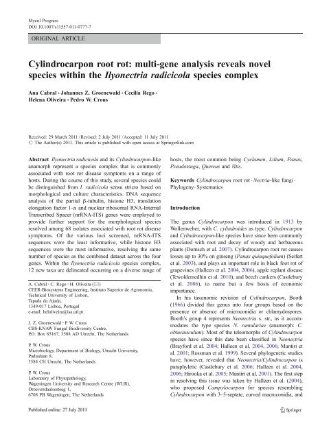
Mycol ProgressTable 2 Statistical information on the individual datasets and numberof equally most parsimonious trees for each locus [Internal TranscribedSpacers (ITS) of the nuclear ribosomal RNA <strong>gene</strong> operon, andpartial β-tubulin (TUB), histone H3 (HIS) and translation elongationfactor 1-α (TEF) <strong>gene</strong>s]ITS TUB HIS TEF CombinedAligned characters (including gaps) 475 502 440 696 2,113Parsimony-informative characters 122 212 215 364 913Variable and parsimony-uninformative characters 31 38 11 43 123Constant characters 322 252 214 289 1,077Equally most parsimonious trees obtained 136 384 1 60 1,000Tree length 294 603 1,095 1,149 3,259Consistency index (Cl) 0.718 0.660 0.468 0.611 0.559Retention index (RI) 0.978 0.972 0.946 0.966 0.959Rescaled Consistency index (RC) 0.702 0.642 0.442 0.590 0.537didyma, species 5, I. liliigena and I. pseudodesctructanswere supported by low bootstrap values, and <strong>CBS</strong> 120370clustered apart from the remaining isolates of I. crassa. Ofall loci screened, ITS proved to be the least informative,being unable to resolve 22 of the species in this study.Neighbour-Joining (NJ) analyses using the three substitutionmodels, as well as the parsimony <strong>analysis</strong>, yielded treeswith similar topology and bootstrap support values for theindividual and combined <strong>gene</strong> analyses. The trees obtainedsupported the same clades, sometimes with rearrangementsin the order of these clades between the different analyses(data not shown). The results of the phylo<strong>gene</strong>tic analysesare highlighted below under the taxonomic notes or in theDiscussion, where applicable.TaxonomyThe present study treats isolates that have been freshlycollected, or previously identified and maintained in culturecollections as “<strong>Cylindrocarpon</strong> destructans”, meaningcylindrical, rarely curved, 3-septate macroconidia withobtuse apices, abundant microconidia and chlamydospores(Samuels and Brayford 1990). The latter species has in thepast been acknowledged as anamorph of I. radicicola(Booth 1966; Chaverri et al. 2011; Samuels and Brayford1990). However, an examination of the neotype of “C.”destructans in this study [CUP-011985, conidia (18.0)23.0–30.0(35.0) × (6.0)6.5(7.0) μm], found conidia to beconsiderably smaller than those of I. radicicola (24.0)33.1(47.0) × (4.9)6.4(7.8) μm (Gerlach and Nilsson 1963) (alsoconfirmed in the present study by examination of <strong>CBS</strong>264.65, ex-type), revealing them to represent two distinctspecies. Furthermore, based on the phylo<strong>gene</strong>tic andmorphological data obtained in the present study, several<strong>novel</strong> species could be distinguished that are phylo<strong>gene</strong>ticallydistinct from I. radicicola, and morphologicallydistinct based on a range of characters linked to culturecharacteristics, conidiophores, macro- and microconidiummorphology. Some of these could be linked to oldernames, or taxa long regarded as potential syonyms of“destructans”, which could now be resurrected. Thesetaxa are treated below:Ilyonectria anthuriicola A. Cabral & Crous, sp. nov.(Fig. 2)MycoBank 560108.Etymology: Named after its host, Anthurium.Cylindrocarpi destructantis morphologice simile, sedlongitudine media conidiorum longiore, 29.5–32.2 μm,distinguitur.Conidiophores simple or complex to sporodochial.Simple conidiophores arising laterally or terminally fromaerial mycelium, solitary to loosely aggregated, unbranchedor sparsely branched bearing up to three phialides, 1–3-septate, 40–95 μm long; phialides monophialidic, more orless cylindrical but slightly tapering towards the tip, 10.5–20.5 μm long, 2.5–3.5 μm wide at the base, 3.0–4.5 μm atwidest point, 1.5–2.5 μm near the aperture. Conidiophoresgiving rise to microconidia, formed on mycelium at agarsurface, penicillately mono- or bi-verticillate; phialidesmonophialidic, narrowly flask-shaped, typically with widestpoint near the middle, 8–15 μm long, 2.0–3.0 μm wide atthe base, 2.5–4.5 μm at widest point, 1.0–2.0 μm near theapex. Sporodochial conidiophores irregularly branched;phialides cylindrical, mostly widest near the middle.Macroconidia formed in flat domes of slimy masses, (1–)Fig. 1 The first of 1,000 equally most parsimonious trees obtained from the combined ITS, TUB, HIS and TEF sequence alignment of<strong>Cylindrocarpon</strong> isolates and relatives with a heuristic search usingPAUP v. 4.0b10. The tree was <strong>root</strong>ed using Campylocarpon isolates asoutgroup sequences and bootstrap support values are indicated nearthe nodes, where”ns” designates not supported. Ex-type strains areindicated in bold. Newly described species are indicated by blueboxes. Scale bar shows 10 changes
Mycol Progress<strong>CBS</strong> 112613 Vitis, South Africa<strong>CBS</strong>112679 Vitis, South Africa1001001009910099n.s.759010010090<strong>CBS</strong> 564.95 Anthurium, NetherlandsCy228 Ficus, Portugal<strong>CBS</strong> 129082 Vitis, Portugal<strong>CBS</strong> 100819 Erica, New Zealand<strong>CBS</strong> 113550 Vitis, New ZealandCy217 Vitis, Portugal<strong>CBS</strong> 120497 Vitis, Slovenia<strong>CBS</strong> 120171 Vitis, Slovenia<strong>CBS</strong> 120172 Vitis, Slovenia<strong>CBS</strong> 120173 Vitis, Slovenia<strong>CBS</strong> 120499 Vitis, Slovenia<strong>CBS</strong> 120498 Vitis, SloveniaCy238 Vitis, PortugalCy196 Vitis, Portugal<strong>CBS</strong> 162.89 Hordeum, Netherlands1009185100100911008610090949786Cy108 Vitis, PortugalCy200 Vitis, Portugal<strong>CBS</strong> 159.34 unknown, Germany<strong>CBS</strong> 173.37 Pinus, UKCy135 Vitis, PortugalCy144 Vitis, Portugal<strong>CBS</strong> 129085 Vitis, PortugalCy146 Vitis, PortugalCy147 Vitis, PortugalCy243 Vitis, PortugalCy148 Vitis, PortugalCy149 Vitis, PortugalCy150 Vitis, PortugalCy151 Vitis, PortugalCy152 Vitis, PortugalCy153 Vitis, PortugalCPC 13539 Picea, Canada<strong>CBS</strong> 129087 Vitis, PortugalCy134 Vitis, PortugalCy133 Vitis, Spain<strong>CBS</strong> 112608 Vitis, South Africa<strong>CBS</strong> 113552 Vitis, New ZealandCy117 Vitis, USACy125 Vitis, USACy230 Festuca, Portugal<strong>CBS</strong> 112593 Vitis, South AfricaCy130 Vitis, USACy116 Vitis, USACy129 Vitis, USACy115 Vitis, USACy119 Vitis, USACy124 Vitis, USA<strong>CBS</strong> 112603 Vitis, South Africa<strong>CBS</strong> 112594 Vitis, South Africa<strong>CBS</strong> 112601 Vitis South AfricaCy128 Vitis, USA<strong>CBS</strong> 112605 Vitis, South Africa<strong>CBS</strong> 112615 Vitis, South AfricaCy140 Vitis, PortugalCy175 Vitis, PortugalCy181 Vitis, PortugalCy216 Vitis, PortugalCy244 Vitis, PortugalCy258 Vitis, PortugalCy123 Vitis, USACy139 Vitis, PortugalCy260 Vitis, PortugalCy132 Vitis, PortugalCy235 Vitis, Portugal<strong>CBS</strong> 112604 Vitis, South AfricaCy237 Vitis, PortugalCy214 Vitis, PortugalCy72 Vitis, PortugalCy142 Vitis, PortugalCy143 Vitis, Portugal<strong>CBS</strong> 112609, Vitis Australia<strong>CBS</strong> 112598, Vitis South AfricaCy246 Vitis, Portugal<strong>CBS</strong> 129086 Vitis, PortugalCPC13533 Vitis, CanadaCy157 Vitis, PortugalCy221 Fragaria, USACy69 Vitis, PortugalCy118 Vitis, USACy120 Vitis, USACy222 Fragaria, USACy71 Vitis, PortugalCy240 Vitis, PortugalCy96 Quercus, AustriaCy262 Vitis, Portugal<strong>CBS</strong> 119.41 Fragaria, NetherlandsCy223 Fragaria, USACy136 Vitis, PortugalCy137 Vitis, PortugalCy138 Vitis, PortugalCy141 Vitis, Portugal<strong>CBS</strong> 113555 Vitis, New Zealand<strong>CBS</strong> 188.49 Abies, NetherlandsCy75 Vitis, PortugalCy97 Quercus, AustriaCampyl. fasciculareCampyl. pseudofasciculareI. anthuriicola“<strong>Cylindrocarpon</strong>” sp.I. vitisC. pauciseptatum“<strong>Cylindrocarpon</strong>” sp. 1“<strong>Cylindrocarpon</strong>”sp.2“<strong>Cylindrocarpon</strong>”sp. 3“<strong>Cylindrocarpon</strong>”sp. 5“<strong>Cylindrocarpon</strong>” sp.6I. macrodidyma“<strong>Cylindrocarpon</strong>”sp. 4
Mycol ProgressFig. 1 (continued)106886661009710085100<strong>CBS</strong> 240.29 Alnus, Norway<strong>CBS</strong> 226.31 Fagus, GermanyCy169 Malus, Portugal<strong>CBS</strong> 835.97 Salix, BelgiumCy172 Malus, Portugal<strong>CBS</strong> 118984 Abies, Canada<strong>CBS</strong> 503.67 Abies, Norway<strong>CBS</strong> 324.61 Abies, NetherlandsCPC13545 Pyrus, Canada100 100100<strong>CBS</strong> 151.29 Malus, UK98100 <strong>CBS</strong> 182.36 Malus, unknownCPC13530 Prunus, Canada70100 CPC 13531 Pseudotsuga, Canada91 CR21 Pseudotsuga, Canada100n.s.100n.s.94-1356 Picea, CanadaCPC 13544 Prunus, Canada<strong>CBS</strong> 183.36 Solanum, Germany<strong>CBS</strong> 119596 Astelia, New Zealand<strong>CBS</strong> 119606 Metrosideros, New Zealand<strong>CBS</strong> 307.35 Panax, Canada<strong>CBS</strong> 120359 Panax, Canada<strong>CBS</strong> 120361 Panax, Canada<strong>CBS</strong> 120364 Panax, Canada<strong>CBS</strong> 120365 Panax, Canada<strong>CBS</strong> 120366 Panax, Canada<strong>CBS</strong> 120367 Panax, Canada<strong>CBS</strong> 120368 Panax, Canada<strong>CBS</strong> 120369 Panax, CanadaCPC13535 Panax, CanadaCPC13537 Panax, Canada<strong>CBS</strong> 306.35 Panax, Canada<strong>CBS</strong> 124662 Panax, Japan<strong>CBS</strong> 129080 Vitis, Portugal<strong>CBS</strong> 264.65 Cyclamen, Sweden100n.s.99100n.s.100n.s.<strong>CBS</strong> 117526 Vitis, Portugal<strong>CBS</strong> 117527 Vitis, Portugal<strong>CBS</strong> 117640 Vitis, Portugal<strong>CBS</strong> 112596 Vitis, South Africa<strong>CBS</strong> 112607 Vitis, South AfricaCy190 Vitis, Portugal<strong>CBS</strong> 110.81 Liriodendron, USACy232 Quercus, PortugalCy164 Malus, PortugalCy122 Vitis, USA<strong>CBS</strong> 302.93 Cyclamen, Netherlands<strong>CBS</strong> 102032 Bark, Venezuela869196<strong>CBS</strong> 129078 Vitis, PortugalCy131 Actinidia, France<strong>CBS</strong> 537.92 Aesculus, Belgium<strong>CBS</strong> 102892 Phragmites, GermanyCy155 Vitis, PortugalCD1666 Panax, CanadaCy231 Thymus, Portugal<strong>CBS</strong> 605.92 Tilia, Germany<strong>CBS</strong> 117818 Quercus, Austria<strong>CBS</strong> 117821 Quercus, Austria<strong>CBS</strong> 308.35 Panax, CanadaCPC13532 Prunus, Portugal<strong>CBS</strong> 321.34 Loroglossum, Tunisia<strong>CBS</strong> 129084 Vitis, Portugal<strong>CBS</strong> 773.83 Water, Netherlands<strong>CBS</strong> 117823 Quercus, Austria<strong>CBS</strong> 117814 Quercus, Austria<strong>CBS</strong> 117820 Quercus, AustriaCy158 Vitis, PortugalCy23 Vitis, Portugal9686<strong>CBS</strong> 117815 Quercus, Austria100 <strong>CBS</strong> 117822 Quercus, Austria<strong>CBS</strong> 129079 Panax, Canada94-1628 Picea, Canada<strong>CBS</strong> 640.77 Abies, France<strong>CBS</strong> 153.37 Dune sand, France95 <strong>CBS</strong> 156.47 Azalea, Belgium<strong>CBS</strong> 120371 Pseudotsuga, Canada93 CPC13536 Pseudotsuga, Canadan.s. <strong>CBS</strong> 120372 Pseudotsuga, Canada87<strong>CBS</strong> 120370 Pseudotsuga, CanadaNSAC-SH 2.5 Panax, Canada100 NSAC-SH 2 Panax, Canada<strong>CBS</strong> 129083 Panax, Canada<strong>CBS</strong> 139.30 Lilium, Netherlands88 <strong>CBS</strong> 158.31 Narcissus, Netherlands98 CPC 13534 Poa, Canada<strong>CBS</strong> 129081 Vitis, Portugal99Cy22 Vitis, Portugal78<strong>CBS</strong> 117824 Quercus, Austria<strong>CBS</strong> 117812 Quercus, Austria<strong>CBS</strong> 940.97 Soil, Netherlands<strong>CBS</strong> 189.49 Lilium, Netherlands88<strong>CBS</strong> 732.74 Lilium, Netherlands100<strong>CBS</strong> 305.85 Lilium, Netherlands85 <strong>CBS</strong> 304.85 Lilium, NetherlandsN. majorN. ditissimaN. neomacrosporaC. cylindroides<strong>Cylindrocarpon</strong> sp.N. ramulariae<strong>Cylindrocarpon</strong> sp.C. obtusisporumN. macroconidialisI. coprosmaeI. mors-panacisI. lusitanicaI. radicicolaI. liriodendriI. cyclaminicolaI. venezuelensisI. europaeaI. robustaI. panacisI. rufa“<strong>Cylindrocarpon</strong>” sp.I. crassaI. pseudodestructansI. gamsiiI. liliigena
Mycol ProgressFig. 2 Ilyonectria anthuriicola (<strong>CBS</strong> 564.95). a–c Simple conidiophoreson aerial mycelium. d–g Conidiophores giving rise to microconidia,formed on mycelium at agar surface, penicillately mono- orbi-verticillate. h–l Micro- and macroconidia. m Chlamydospores inmycelium. Bars 10 μm3-septate, straight or minutely curved, cylindrical with bothends more or less obtusely rounded, mostly without avisible hilum; 1-septate, (20.0)23.5–26.7(29.0)×(5.5)5.9–6.8(7.0) μm (average=25.1×6.4 μm), with a length:widthratio of 3.6–4.8; 2-septate, (25.0)26.6–29.3(32.0)×(6.5)6.8–7.8(8.5) μm (av. = 27.9×7.3 μm), with a length:widthratio of 3.2–4.8; 3-septate, (25.0)29.5–32.2(38.0)×(6.0)7.5–8.1(9.0) μm (av. = 30.8×7.8 μm) with a length:widthratio of 3.1–5.2. Microconidia 0(−1)-septate, subglobose toovoid, rarely ellipsoid, mostly with a visible centrallylocated or slightly laterally displaced hilum; aseptatemicroconidia, (4.9)5.0–8.1(12.0)×(4.0)4.3–5.5(6.5) μm(av. = 6.5×4.9 μm), with a length:width ratio of 1.0–1.8;1-septate, (11.0)11.6–16.7(18.0)×(5.0)5.4–6.1(6.0) μm(av. = 14.1×5.8 μm), with a length:width ratio 1.8–3.0.Chlamydospores globose to subglobose to ellipsoid, 8–14×7–12 μm, smooth, but often appearing rough due todeposits, thick-walled, formed intercalary in chains or inclumps and also in the cells of macroconidia, hyaline,becoming golden-brown.Holotype: Netherlands, Bleiswijk, <strong>root</strong> <strong>rot</strong> of Anthuriumsp., 1995, coll./isol. R. Pieters, holotype <strong>CBS</strong> H-20555,culture ex-type <strong>CBS</strong> 564.95.Culture characteristics: Mycelium felty with averagedensity. Surface on OA chestnut, with aerial mycelium sparse,saffron; margin pure yellow to orange. Surface on PDA,chestnut with saffron aerial mycelium, growth at marginluteous; zonation absent, transparency homo<strong>gene</strong>ous, margin
Mycol Progresseven; reverse similar to surface, but chestnut to cinnamon onOA, and chestnut on PDA. Colonies on PDA do not grow at4°C after 7 days. Optimum temperature 20°C when coloniesreach 25–27 mm, after 7 days. Colony diam was 20–22 mm at25°C, after 7 days. Hardly grows at 30°C (2 mm colony diamafter 7 days).Isolate studied: <strong>CBS</strong> 564.95 (Table 1).Host and distribution: Roots of Anthurium sp.(Netherlands).Ilyonectria crassa (Wollenw.) A. Cabral & Crous, comb. etstat. nov. (Fig. 3)MycoBank 560109.Basionym: <strong>Cylindrocarpon</strong> radicicola var. crassumWollenw., Z. Parasitenkunde 3: 495. 1931.≡ <strong>Cylindrocarpon</strong> destructans var. crassum (Wollenw.) C.Booth, Mycol. Pap. 104: 37. 1966.Conidiophores simple or complex, to sporodochial.Simple conidiophores arising laterally or terminally fromaerial mycelium, solitary to loosely aggregated, unbranchedor sparsely branched bearing up to two phialides, rarelyconsisting only of phialides, 1–4-septate, 40–180 μm long;phialides monophialidic, cylindrical to subulate, 20–55 μmlong, 2.5–4.0 μm wide at the base, 1.5–2.0 μm near theapex. Complex conidiophores aggregated in small sporodochia(on carnation leaf), repeatedly and irregularlybranched; phialides more or less cylindrical, but taperingslightly in the upper part towards the apex, or narrowlyflask-shaped, mostly with widest point near the middle, 17–24 μm long, 2.0–3.0 μm wide at the base, 2.5–3.5 μm atthe widest point, and 1.5–2.5 μm wide near the apex.Macroconidia predominating, formed on both type ofconidiophores, on SNA formed in flat domes of slimymasses, 1–3-septate, straight, cylindrical, but may narrowtowards the tip, more or less broadly rounded, and the baseappearing somewhat acute due to the presence of the hilum,mostly centrally located; 1-septate, (21.0)25.7–27.3(34.0)×(4.5)5.0–5.3(6.5) μm (av. = 26.5×5.1 μm), with a length:width ratio of 3.8–6.7; 2-septate, (23.0)28.5–30.3(37.0)×(4.5)5.3–5.6(6.5) μm (av. = 29.4×5.4 μm)with a length:width ratio of 4.2–6.7; 3-septate, (29.0)34.1–36.0(49.0)×(5.0)5.6–5.8(7.0) μm (av. = 35.1×5.7 μm), with a length:width ratio of 4.8–8.9. Microconidia 0–1-septate, ellipsoidto subcylindrical, more or less straight, with a visible,truncate hilum; aseptate microconidia, (7.0)9.7–10.9(15.0)×(3.0)3.3–3.6(4.5) μm (av. = 10.3×3.5 μm), with alength:width ratio of 1.8–4.3; 1-septate, (12.0)14.2–15.2(19.0)×(3.0)3.8–4.2(5.0) μm (av. = 14.7×4.0 μm), with alength:width ratio 2.7–5.0. Conidia formed in heads onsimple conidiophores or as white (OA) or unpigmented(SNA) masses. Chlamydospores globose to subglobose tocylindrical, 7–15×6–10 μm, smooth, but often appearingrough due to deposits, thick-walled, terminal on shortFig. 3 Ilyonectria crassa (<strong>CBS</strong> 129083). a–c Simple conidiophores on aerial mycelium. d–g Micro- and macroconidia. h–i Chlamydospores andmacroconidia. Bars 10 μm
Mycol Progresslateral branches, rarely intercalary, single, in chains or inclumps, and also in the cells of the macroconidia, hyaline,becoming pale brown.Lectotype: The Netherlands, on Lilium bulbs, Dec. 1930,coll./isol. W.F. van Hell, lectotype designated here <strong>CBS</strong> H-20556, culture ex-lectotype <strong>CBS</strong> 139.30.Culture characteristics: Mycelium cottony to felty withaverage to strong density. Surface on OA cinnamon, withaerial mycelium sparse, buff. Surface on PDA saffron withaerial mycelium sparse buff to saffron to pale luteous. Nozonation was observed, transparency was homo<strong>gene</strong>ous andgrowth at margin even. Reverse similar to surface, except incolour, saffron to cinnamon on OA, and chestnut to siennaon PDA. Colonies on PDA grow 5–8 mm diam at 4°C after7 days. Optimum temperature at 20°C, when colonies reach31–46 mm diam, after 7 days. Colony diam was 19–34 mmat 25°C, after 7 days. No growth was observed at 30°C.Isolates studied: <strong>CBS</strong> 139.30; <strong>CBS</strong> 158.31; <strong>CBS</strong>129083; NSAC-SH-2; NSAC-SH-2.5 (Table 1).Hosts and distribution: Lilium sp. (bulbs), Narcissus sp.(<strong>root</strong>s) (Netherlands), Panax quinquefolium (<strong>root</strong>s) (Canada).Notes: In the original description, Wollenweber (1931)cites <strong>Cylindrocarpon</strong> radicicola var. crassum as occurringon <strong>root</strong>s of Ulmus, Taxus and Lilium in Europe (Germanyand the Netherlands). He did not designate any typespecimen. However, he specifically refers to a culture sentto him by Prof. J. Westerdijk on Lilium from the <strong>CBS</strong> in theNetherlands in 1930, which was regarded as authentic forthe species. This culture is represented by <strong>CBS</strong> 139.30(accessioned in 1930, from Lilium, the Netherlands), andthus we designate a dried, sporulating culture as lectotypefor the species.Ilyonectria cyclaminicola A. Cabral & Crous, sp. nov.(Fig. 4)MycoBank 560110.Etymology: Named after the host from which it wasisolated, Cyclamen sp.Cylindrocarpi destructantis morphologice simile, sedlongitudine media conidiorum longiore, 26.9–31.9 μm,distinguitur.Conidiophores simple or complex to sporodochial. Simpleconidiophores arising laterally or terminally from aerialmycelium, solitary to loosely aggregated, unbranched orsparsely branched, bearing up to two phialides, 1–3-septate,60–120 μm long; phialides monophialidic, more or lesscylindrical but slightly tapering towards the tip, 20–60 μmlong, 2.0–4.0 μm wide at the base, 3.0–4.5 μm atwidestpoint, 1.5–2.5 μm near the aperture. Conidiophores givingrise to microconidia formed by mycelium at agar surface,penicillate to mono-verticillate; phialides monophialidic,more or less cylindrical, but with slight taper towards thetip, 19–34 μm long, 1.5–2.5 μm wide at the base, 2.0–3.0 μmFig. 4 Ilyonectria cyclaminicola (<strong>CBS</strong>302.93). a, b Simple conidiophoreson aerial mycelium. c Pennicilate conidiophores with aseptatemicroconidia. d Sporodochial conidiophore on carnation leaf agar. eIsolated chlamydospores formed in lateral branches. f–j Micro- andmacroconidia. Bars 10 μm
Mycol Progressat widest point, 1.0–2.0 μm near the apex. Sporodochialconidiophores irregularly branched; phialides more or lesscylindrical, but slightly tapering towards the tip, or narrowlyflask-shaped, with widest point near the base, 14–26 μmlong, 2.5–3.5 μm wide at the base 3.0–4.0 μm atwidestpoint, 1.0–2.0 μm near the apex. Macroconidia formed inflat domes of slimy masses, 1(−3)-septate, straight orminutely curved, cylindrical with both ends more or lessbroadly rounded, sometimes with a constriction at the septa,mostly without a visible hilum; 1-septate, (19.2)21.3–23.6(29.8)×(4.4)5.4–6.0(7.3) μm (av. = 22.5×5.7 μm), with alength:width ratio of 3.4–5.5; 2-septate, (23.8)24.0–28.4(29.8)×(5.0)5.5–7.3(8.0) μm (av. = 26.2×6.4 μm), with alength:width ratio of 3.1–5.1; 3-septate, (25.3)26.9–31.9(33.6)×(5.8)5.9–6.5(6.9) μm (av. = 29.4×6.2 μm), with alength:width ratio of 3.7–5.6. Microconidia formed in headsor on the agar surface, 0–1-septate, subglobose to ovoid tosubcylindrical, mostly with a visible, centrally located orslightly laterally displaced hilum; aseptate microconidia,(3.9)7.6–8.9(12.9)×(2.2)3.6–3.9(5.4) μm (av. = 8.2×3.7 μm), with a length:width ratio of 1.2– 3.4; 1-septate,(11.5)13.8–15.2(17.5)×(3.7)4.6–4.9(5.5) μm (av. = 14.5×4.7 μm), with a length:width ratio of 2.3–3.9. Chlamydosporesglobose to subglobose, 10–18×10–16 μm, smooth,but often appearing rough due to deposits, thick-walled,formed in lateral branches, rarely intercalary, mostly isolated,hyaline, becoming medium brown.Holotype: Netherlands, Roelofarendsveen, NAKS laboratory,Cyclamen bulb, May 1993, coll./isol. M. Hooftman,iden. E.J. Hermanides-Nijhof, holotype <strong>CBS</strong> H-20557,culture ex-type <strong>CBS</strong> 302.93.Culture characteristics: Mycelium felty with averagedensity. Surface on OA sepia to chestnut. Surface on PDAsepia to chestnut, with sparse, rust, aerial mycelium; nozonation was observed, and transparency was homo<strong>gene</strong>ous;margins predominantly even. Reverse similar to surface,except in colour, sepia to dark brick on OA and chestnut onPDA. Colonies on PDA do not grow at 4°C after 7 days.Optimum temperature at 22°C, when colonies reach 68–70 mm diam, after 7 days. Colony diam was 63–64 mm at25°C, after 7 days. No growth was observed at 30°C.Isolate studied: <strong>CBS</strong> 302.93 (Table 1).Host and distribution:BulbofCyclamen sp. (Netherlands).Ilyonectria europaea A. Cabral, Rego & Crous, sp. nov.(Fig. 5)MycoBank 560103.Etymology: Named after the European continent, wherethis fungus appears to be widely distributed.Fig. 5 Ilyonectria europaea (<strong>CBS</strong> 129078). a–c Simple conidiophores on aerial mycelium. d Sporodochial conidiophore on carnation leaf agar. eChlamydospores in aerial mycelium. f–i Micro- and macroconidia. Bars 10 μm
Mycol ProgressIlyonectriae robustae morphologice similis, sed longitudinemedia macroconidiorum breviore, 29.7–31.5 μm,distinguitur.Conidiophores simple or complex to sporodochial.Simple conidiophores arising laterally or terminally fromaerial mycelium, solitary to loosely aggregated, unbranchedor sparsely branched, bearing up to threephialides, 1–3-septate, 50–120 μm long; phialides monophialidic,cylindrical to subulate, 26–60 μm long, 2.5–3.5 μm wide at the base, 1.5–2.5 μm near the apex.Complex conidiophores aggregated in small sporodochia(on carnation leaf), repeatedly and irregularly branched.Macroconidia predominating, formed on both type ofconidiophores, on SNA formed in flat domes of slimymasses, 1(−3)-septate, straight or minutely curved, cylindricalwith both ends more or less broadly rounded, butmay narrow towards the tip, mostly without a visiblehilum; 1-septate, (16.4)21.9–23.4(34.0)×(4.0)5.2–5.6(7.8)μm (av.=22.7×5.4μm), with a length:width ratio of 3.2–5.4; 2-septate, (22.0)26.4–28.1(34.0)×(4.4)5.9–6.4(8.0)μm (av. = 27.2×6.1 μm), with a length:width ratio of3.4–6.4; 3-septate, (22.0)29.7–31.5(40.0)×(5.0)6.5–6.9(8.6) μm (av.=30.6×6.7μm), with a length:width ratioof 3.5–6.0. Microconidia 0–1-septate, ellipsoid to ovoid,more or less straight, without a visible hilum; aseptatemicroconidia sometimes curved towards one end, (3.0)8.5–9.8(17.0)×(1.7)3.3–3.5(5.0) μm (av.=9.1×3.4μm),with a length:width ratio of 1.5–3.4; 1-septate, (9.2)13.4–14.6(18.9)×(3.0)4.0–4.4(5.9) μm (av. = 14.0×4.2 μm),with a length:width ratio 2.6–4.0. Conidia formed in headsor on simple conidiophores as white (OA) or unpigmented(SNA) masses. Chlamydospores globose to subglobose, 9–14×7–14 μm, smooth, but often appearing rough due todeposits, thick-walled, terminal on short or long lateralbranches or intercalary, single, in chains or in clumps,golden-brown.Holotype: Portugal, Vidigueira, at basal end of a 2-yearoldVitis vinifera plant; scion Petit Verdot, <strong>root</strong>stock 110R,2008, coll./isol. C. Rego, holotype <strong>CBS</strong> H-20558, cultureex-type <strong>CBS</strong> 129078=Cy241=CPC 19165.Culture characteristics: Mycelium felty with averagedensity. Surface on OA chestnut, with saffron aerialmycelium. Sienna to saffron on PDA, with luteous aerialmycelium. Concentric zonation, with homo<strong>gene</strong>ous transparency,margins predominantly even. Reverse similar tosurface, except in the colour; sepia on OA, and chestnut toumber on PDA. Colonies on PDA grow poorly, 1–5 mmdiam at 4°C after 7 days. Optimum temperature for growthis 22°C, when colonies reach 43–57 mm diam, after 7 days.Colony diam was 37–47 mm at 25°C, after 7 days. Nogrowth was observed at 30°C.Isolates studied: Cy131; Cy155; <strong>CBS</strong> 537.92; <strong>CBS</strong>102892; <strong>CBS</strong> 129078 (Table 1).Hosts and distribution: Actinidia chinensis ‘Hayward’(internal lesion of stem) (France), Aesculus hippocastanum(wood) (Belgium), Phragmites australis (stem) (Germany),Vitis vinifera (Portugal).Ilyonectria gamsii A. Cabral & Crous, sp. nov. (Fig. 6)MycoBank 560112.Etymology: Named after Prof. dr. Walter Gams, who hasmade a major contribution to our knowledge of Hypocrealeansoil fungi.Ilyonectriae panacis morphologice similis, sed longitudinemedia macroconidiorum breviore, 34.3–38.5 μm,distinguitur.Conidiophores simple or complex to sporodochial.Simple conidiophores arising laterally or terminally fromaerial mycelium, solitary to loosely aggregated, unbranchedor sparsely branched, bearing up to two phialides, 1–3-septate, 50–150 μm long; phialides monophialidic, cylindricalto subulate, 30–60 μm long, 2.5–3.5 μm wide at thebase, 1.5–2.0 μm near the aperture. Sporodochial conidiophoresirregularly branched; phialides cylindrical, mostlywidest near the base. Macroconidia predominating, formedon simple conidiophores, on SNA formed in flat domes ofslimy masses, 1–3-septate, straight, cylindrical with bothends broadly rounded, with mostly visible, centrally locatedhilum; 1-septate, (22.0)25.7–27.9(33.0)×(4.0)5.1–5.5(6.0)μm (av. = 26.8×5.3 μm), with a length:width ratio of 4.3–6.2; 2-septate, (25.0)28.2–31.7(39.0)×(5.0)5.5–5.9(6.5) μm(av. = 29.9×5.7 μm), with a length:width ratio of 4.2–7.1; 3-septate, (24.0)34.3–38.5(44.0)×(5.0)5.9–6.3(7.0) μm (av.=36.4×6.1 μm), with a length:width ratio of 4.3–7.3. Microconidia0–1-septate, ellipsoid to subcylindrical, more or lessstraight, mostly with a visible hilum; aseptate microconidia(4.0)6.9–8.0(10.0)×(3.0)4.0–4.5(5.0) μm (av. = 7.4×4.3 μm), with a length:width ratio of 1.3–2.9; 1-septate,(8.0)12.9–15.7(18.0)×(4.0)4.2–4.7(5.5) μm (av. = 14.3×4.4 μm), with a length:width ratio 1.8–4.0. Chlamydosporesglobose to subglobose to ellipsoidal, 8–14×7–12 μm,smooth, but often appearing rough due to deposits, thickwalled,mostly intercalary, rarely terminal on short lateralbranches, single, in chains or in clumps, hyaline, becomingmedium brown.Holotype: Netherlands, Lelystad, soil, June 1997, coll./isol. J.T. Poll, iden. W. Gams, holotype <strong>CBS</strong> H-20559,culture ex-type <strong>CBS</strong> 940.97.Culture characteristics: Mycelium cottony, dense. Surfaceon OA cinnamon, with sparse, buff aerial mycelium,on PDA umber to chestnut, with buff to saffron aerialmycelium; zonation absent, transparency homo<strong>gene</strong>ous,margin even; reverse similar to surface, but chestnut onPDA. Colonies on PDA grow 6–7 mm diam at 4°C after7 days. Optimum temperature at 22°C when colonies reach
Mycol ProgressFig. 6 Ilyonectria gamsii (<strong>CBS</strong> 940.97). a–c Simple conidiophores on aerial mycelium. d–h Micro- and macroconidia. i Chlamydospores onmycelium. Bars 10 μm44–45 mm diam, after 7 days. Colony diam is 22–24 mm at25°C, after 7 days. No growth observed at 30°C.Isolate studied: <strong>CBS</strong> 940.97 (Table 1).Habitat and distribution: Soil (Netherlands).Ilyonectria liliigena A. Cabral & Crous, sp. nov. (Fig. 7)MycoBank 560114.Etymology: Named after its host, Lilium regale.Ilyonectriae panacis morphologice similis, sed longitudinemedia macroconidiorum 3-septatorum breviore, 27.9–29.8 μm, distinguitur.Conidiophores simple or complex or sporodochial.Simple conidiophores arising laterally or terminally fromaerial mycelium, solitary to loosely aggregated, unbranchedor sparsely branched, bearing up to two phialides, 1–4-septate, 50–170 μm long; phialides monophialidic, cylindricalto subulate, 30–65 μm long, 2.0–3.5 μm wide at thebase, 1.5–2.0 μm near the apex. Sporodochial conidiophoresirregularly branched; phialides cylindrical, mostlywidest near the base. Macroconidia predominating, formedon simple conidiophores, on SNA formed in flat domes ofslimy masses, 1(−3)-septate, straight or frequently minutelycurved, cylindrical or sometimes typically minutely wideningtowards the tip, therefore appearing somewhat clavate,mostly without a visible hilum; 1-septate, (19.0)22.9–24.6(30.0)×(3.3)4.2–4.5(5.2) μm (av. = 23.8×4.3 μm), with alength:width ratio of 4.0–7.0; 2-septate, (21.0)26.1–27.7(32.1)×(4.0)4.7–5(5.7) μm (av. = 26.9×4.9 μm)with alength:width ratio of 3.8–7.0; 3-septate, (23.9)27.9–29.8(35.0)×(3.9)4.7–5.1(6.0) μm (av. = 28.9×4.9 μm), with alength:width ratio of 4.0–8.3. Microconidia 0–1-septate,ellipsoidal to subcylindrical, more or less straight, mostlywith a visible hilum; aseptate, microconidia (5.9)8.9–10.3(17.0)×(2.5)3.0–3.2(4.4) μm (av. = 9.6×3.1 μm), with alength:width ratio of 2.0–4.6; 1-septate, (10.0)12.9–14.3(18.0)×(2.5)3.3–3.6(4.5) μm (av. = 13.6×3.4 μm), with alength:width ratio 2.8–5.6. Conidia formed in heads onsimple conidiophores or as white (OA) or unpigmented(SNA) masses. Chlamydospores globose to subglobose, 6–14×5–12 μm, smooth but often appearing rough due todeposits, thick-walled, mostly in terminal on short lateralbranches or rarely intercalary, single, in chains or in clumps,hyaline, becoming slightly brown at margins.Holotype: Netherlands, Hoorn, bulb <strong>rot</strong> of Lilium regale,1949, coll./isol. M.A.A. Schipper, holotype <strong>CBS</strong> H-20560,culture ex-type <strong>CBS</strong> 189.49.Culture characteristics: Mycelium felty, with an averageto strong density. Surface on OA sienna, with sparse,saffron, aerial mycelium. Surface on PDA sepia tocinnamon, with saffron to buff aerial mycelium. Zonationabsent or concentric, with homo<strong>gene</strong>ous transparency.Margins were even, or sometimes slightly uneven. Reversesimilar to surface, except in colour; on OA pale vinaceous
Mycol ProgressFig. 7 Ilyonectria liliigena (<strong>CBS</strong> 189.49). a–d Simple conidiophores on aerial mycelium. e Chlamydospores on mycelium. f–i Micro- andmacroconidia. Bars 10 μmto cinnamon; on PDA buff to saffron to chestnut.Colonies on PDA grew poorly (1–4 mm diam) at 4°Cafter 7 days. Optimum temperature at 22°C, whencolonies reach 34–45 mm diam, after 7 days. Colonydiam was 16–29 mm at 25°C, after 7 days. No growthwas observed at 30°C.Isolates studied: <strong>CBS</strong> 189.49; <strong>CBS</strong> 732.74; <strong>CBS</strong> 304.85;<strong>CBS</strong> 305.85 (Table 1).Host and distribution: Lilium regale bulbs (Netherlands).Ilyonectria lusitanica A. Cabral, Rego & Crous, sp. nov.(Fig. 8)MycoBank 560105.Etymology: Named after the Latin name for the countryfrom where it was collected, Portugal.Ilyonectriae europaeae morphologice similis, sed longitudinemedia macroconidiorum breviore, 25–28.4 μm,distinguitur.Conidiophores simple or complex, sporodochial. Simpleconidiophores arising laterally or terminally from aerialmycelium, solitary to loosely aggregated, unbranched orsparsely branched, bearing up to two phialides, 1–4-septate,60–220 μm long; phialides monophialidic, cylindrical tosubulate, 20–70 μm long, 2.5–3.5 μm wide at the base, 1.5–2.5 μm near the aperture. Complex conidiophores aggregatedin small sporodochia, repeatedly and irregularly branched.Macroconidia predominating, formed by both type ofconidiophores, on SNA formed in flat domes of slimymasses, 1(−3)-septate, straight or minutely curved, cylindricalwith both ends more or less broadly rounded, but may narrowtowards the tip, without a visible hilum, and may have aconstriction on the septa in older cultures; 1-septate, (14.0)17.3–18.8(21.0)×(4.0)4.6–5(5.5) μm (av. = 18.1×4.8 μm),with a length:width ratio of 2.8–4.8; 2-septate, (18.0)20.5–22.1(27.0)×(4.0)4.9–5.2(6.0) μm (av.=21.3×5.1μm), witha length:width ratio of 3.5–5.4; 3-septate, (18.0)25.0–28.4(38.0)×(4.5)5.2–5.5(6.0) μm (av. = 26.7×5.4 μm), with alength:width ratio of 3.6–6.8. Microconidia 0–1-septate,ellipsoid to ovoid, more or less straight, without a visiblehilum, and may have a constriction at the septum; aseptate,(5.0)6.9–8.2(10.0)×(2.5)3.0–3.3(4.0) μm (av. = 7.6×3.2 μm),with a length:width ratio of 1.7–3.3; 1-septate, (8.0)10.0–11.0(14.0)×(3.0)3.4–3.7(4.0) μm (av. = 10.5×3.6 μm), with alength:width ratio 2.0–3.7. Conidia formed in heads onsimple conidiophores or as white (OA) or unpigmented(SNA) masses. Chlamydospores rarely observed, globose tosubglobose to cylindrical, 9–13×7–11 μm, smooth, but oftenappearing rough due to deposits, thick-walled, intercalary,hyaline, becoming slightly brown at the margin.
Mycol ProgressFig. 8 Ilyonectria lusitanica (<strong>CBS</strong> 129080). a–c Simple conidiophores of the aerial mycelium. d Chlamydospores on mycelium. e–g Micro- andmacroconidia. Bars (a) 20μm, (b–g) 10μmHolotype: Portugal, Melgaço, Alvaredo, on Vitis vinifera,below grafting zone, 6–year-old plant; scion Alvarinho on<strong>root</strong>stock 196–17, 2005, coll./isol. N. Cruz, holotype <strong>CBS</strong> H-20563, culture ex-type <strong>CBS</strong> 129080=Cy197=CPC 19166.Culture characteristics: Mycelium felty with averagedensity. Surface on OA cinnamon, with aerial myceliumsparse, buff. Surface on PDA, cinnamon, with sparse,ochreous to buff aerial mycelium. Zonation absent, transparencyhomo<strong>gene</strong>ous, margin even. Reverse similar tosurface but buff to cinnamon on OA, and chestnut tocinnamon on PDA. Colonies on PDA grow 5–6 mm at 4°Cafter 7 days. Optimum temperature between 20 and 22°C,with colonies reaching 42–46 mm and 43–46 mm, respectively,after 7 days. Colony diam was 31–32 mm at 25°C,after 7 days. No growth observed at 30°C.Isolate studied: <strong>CBS</strong> 129080 (Table 1).Host and distribution: Vitis vinifera (Portugal).Ilyonectria mors-panacis (A.A. Hildebr.) A. Cabral &Crous, comb. nov. (Fig. 9)MycoBank 560115.Basionym: Ramularia mors-panacis A.A. Hildebr., Can.J. Res. 12: 101. 1935.= <strong>Cylindrocarpon</strong> panacis Matuo & Miyaz., Trans.Mycol. Soc. Japan 9: 111. 1969.≡ <strong>Cylindrocarpon</strong> destructans f.sp. panacis Matuo &Miyaz., Ann. Phytopath. Soc. Japan 50: 390. 1984.Conidiophores simple or complex, sporodochial. Simpleconidiophores arising laterally or terminally from aerialmycelium, solitary to loosely aggregated, unbranched orsparsely branched, rarely consisting only of phialides, 1–3-septate, 45–170 μm long; phialides monophialidic, cylindricalto subulate, 23–55 μm long, 2.0–3.0 μm wide at thebase, 1.5–3.0 μm near the apex. Complex conidiophoresaggregated in small sporodochia, repeatedly and irregularlybranched. Macroconidia predominating, formed on simpleconidiophores, on SNA formed in flat domes of slimymasses, 1(−3)-septate, straight, cylindrical with both endsmore or less broadly rounded, mostly without a hilum; 1-septate, (21.0)28.2–31.6(40.0)×(5.0)5.8–6.3(7.5) μm (av. =29.9×6.1 μm), with a length:width ratio of 3.3–7.0; 2-septate, (28.0)30.5–38.4(42.0)×(5.0)5.9–6.4–7.0(7.1) μm(av. = 34.4×6.4 μm), with a length:width ratio of 4.0–6.0; 3-septate, (37.8)39.0–44.2(45.0)×(6.9)7.0–7.5(7.5)μm (av. = 41.0×7.2 μm), with a length:width ratio of5.3–6.0. Microconidia 0–1-septate, ellipsoid to subcylindrical,more or less straight, without a visible hilum;aseptate, (5.0)8.9–10.4(17.0)×(2.5)3.6–3.9(5.0) μm (av.=9.6×3.8 μm), with a length:width ratio of 1.3–3.4; 1-septate, (9.0)12.5–14.1(19.0)×(3.5)4.4–4.8(5.5) μm (av.=13.3×4.6 μm), with a length:width ratio 2.0–4.0. Conidia
Mycol ProgressFig. 9 Ilyonectria mors-panacis (<strong>CBS</strong>120363). a, b Simple conidiophores on aerial mycelium. c–g Micro- and macroconidia. h–jChlamydospores on mycelium. Bars 10 μmformed in heads on simple conidiophores or as white,creamy (OA) or hyaline (SNA) masses. Chlamydosporesglobose to subglobose, 8–16×7–15 μm, smooth, but oftenappearing rough due to deposits, thick-walled, terminal onshort lateral branches or intercalary, single, in chains or inclumps, hyaline, becoming medium brown.Lectotype: Canada, Ontario, on living <strong>root</strong>s of Panaxquinquefolium, June 1935, A.A. Hildebrand, lectotype designatedhere <strong>CBS</strong> H-20561, culture ex-lectotype <strong>CBS</strong> 306.35.Culture characteristics: Mycelium felty with an averagedensity. Surface on OA and PDA chestnut, with sparse, buff torosy-buff to cinnamon or saffron aerial mycelium. Concentriczonation, with homo<strong>gene</strong>ous transparency, and even margins.Reverse similar to surface, ochreous to fulvous, or sepia todark vinaceous on OA, and chestnut to sienna on PDA.ColoniesonPDAgrow3–9 mmdiamat4°Cafter7days.Optimum temperature for growth is 18°C, when colonies reach22–40 mm diam, after 7 days. Colony diam was 31–40 mm at25°C after 7 days. No growth was observed at 30°C.Isolates studied: <strong>CBS</strong> 306.35; <strong>CBS</strong> 307.35; <strong>CBS</strong>120359; <strong>CBS</strong> 120360; <strong>CBS</strong> 120361; <strong>CBS</strong> 120362; <strong>CBS</strong>120363; <strong>CBS</strong> 120364; <strong>CBS</strong> 120365; <strong>CBS</strong> 120366; <strong>CBS</strong>120367; <strong>CBS</strong> 120368; <strong>CBS</strong> 120369; <strong>CBS</strong> 124662; CPC13535; CPC 13537 (Table 1).Hosts and distribution: Panax ginseng (Japan), P.quinquefolium (Canada).Notes: Ilyonectria mors-panacis is distinct from “C.”destructans (anamorph: “C”. destructans, neotype CUP-011985, conidia (18.0)23.0–30.0(35.0) × (6.0)6.5(7.0) μm)in having larger conidia, and indistinct hila (being prominent,flat, 2 μm diam in I. radicicola; see also Samuels andBrayford 1990, Fig. 1). “Ramularia” panacicola is distinctby also having shorter conidia than I. mors-panacis, 5.5–34.2 × 2.5–7.2 μm (Zinssmeister 1918), and appears to beanother potential synonym of “C.” destructans. However,no authentic material could be located of “R.” panacicola,and the only isolate deposited under this name was aCanadian strain collected by Hildebrand (1935), which infact represented I. mors-panacis (Fig. 1). The oldest namefor the species on Panax treated here, therefore, is R. morspanacis(<strong>CBS</strong> 306.35), with the Japanese collections (“C.”panacis ≡ “C.” destructans f.sp. panacis, <strong>CBS</strong> 124662=NBRC 31881) being later synonyms (see Fig. 1).Ilyonectria panacis A. Cabral & Crous, sp. nov. (Fig. 10)MycoBank 560104.Etymology: Named after its host, Panax quinquefolium.Ilyonectriae liliigenae morphologice similis, sed longitudinemedia macroconidiorum longiore, 31–35 μm,distinguitur.Conidiophores simple or complex, sporodochial. Simpleconidiophores arising laterally or terminally from aerialmycelium, solitary to loosely aggregated, unbranched orsparsely branched bearing up to three phialides, 1–5-septate,60–220 μm long; phialides monophialidic, cylindrical to
Mycol ProgressFig. 10 Ilyonectria panacis (<strong>CBS</strong> 129079). a–c Simple, unbranched or sparsely branched conidiophores on aerial mycelium. d, eChlamydospores on mycelium. f–i Micro- and macroconidia. Bars 10 μmsubulate, 20–65 μm long, 2.5–3.0 μm wide at the base,1.5–2.0 μm near the aperture. Complex conidiophoresaggregated in small sporodochia, repeatedly and irregularlybranched. Macroconidia predominating, formed on bothtype of conidiophores, on SNA formed in flat domes ofslimy masses, 1(−3)-septate, straight, cylindrical with bothends more or less broadly rounded, mostly with a visiblecentrally located hilum; 1-septate, (20.0)23.7–25.9(32.0)×(4.0)4.7–5.0(5.5) μm (av.=24.8×4.8μm), with a length:width ratio of 4.0–6.0; 2-septate, (23.0)27.0–30.3(37.0)×(4.8)5.0–5.4(6.0) μm (av.=28.7×5.2μm), with a length:width ratio of 4.6–6.7; 3-septate, (27.0)31.2–35.0(49.0)×(5.0)5.4–5.8(6.0) μm (av.=33.1×5.6μm), with a length:width ratio of 4.9–8.2. Microconidia 0–1-septate, ellipsoidto ovoid to subcylindrical, more or less straight, mostlywith a visible hilum; aseptate, (6.0)8.0–9.8(13.0)×(3.5)3.7–3.9(4.0) μm (av. = 8.9×3.8 μm), with a length:widthratio of 1.7–3.3; 1-septate, (8.0)11.3–13.7(16.0)×(3.5)3.8–4.2(4.5) μm (av. = 12.5×4.0 μm), with a length:width ratio1.8–4.3. Conidia formed in heads on simple conidiophoresor as white (OA) or unpigmented (SNA) masses. Chlamydosporesglobose to subglobose to ellipsoidal, 8–14×6–10 μm, smooth, but often appearing rough due to deposits,thick-walled, terminal on short lateral branches or intercalary,single, in chains or in clumps, hyaline, becomingmedium brown.Holotype: Canada, Alberta, Panax quinquefolium,1998, coll./isol. K. F. Chang, holotype <strong>CBS</strong> H-20562,culture ex-type <strong>CBS</strong> 129079=CDC-N-9A=CPC 19167.Culture characteristics: Mycelium felty with strongdensity. Surface on OA chestnut to sienna, with aerialmycelium sparse, vinaceous-buff. Surface on PDA chestnutto cinnamon, with aerial mycelium sparse, buff to saffron.No zonation was observed, and transparency was homo<strong>gene</strong>ous;margins predominantly even. Reverse similar tosurface, except in the colour, fawn to cinnamon on OA, andchestnut on PDA. Colonies on PDA grow 5 mm diam at4°C after 7 days. Optimum temperature at 20°C, withcolonies reaching 40–42 mm diam, after 7 days. Colonydiam was 15 mm at 25°C after 7 days. No growth observedat 30°C.Isolate studied: <strong>CBS</strong> 129079 (Table 1).Host and distribution: Panax quinquefolium (Canada).Notes: Several species have in the past been described onPanax in the <strong>gene</strong>ra Ramularia and <strong>Cylindrocarpon</strong>. Theonly unresolved species is “C.” destructans (and itspotential synonym, “Ramularia” panacicola, see above).“<strong>Cylindrocarpon</strong>” destructans is clearly different from I.
Mycol Progresspanacis, which has larger conidia, (27.0)31.2–33.1–35.0(49.0)×(5.0)5.4–5.6–5.8(6.0) μm.Ilyonectria pseudodestructans A. Cabral, Rego & Crous,sp. nov. (Fig. 11)MycoBank 560106.Etymology: Named after its morphological similarity to“<strong>Cylindrocarpon</strong>” destructans.Ilyonectriae crassae morphologice similis, sed macroconidiisclavatis distinguitur.Conidiophores simple or complex, sporodochial. Simpleconidiophores arising laterally or terminally from aerialmycelium, solitary to loosely aggregated, unbranched orsparsely branched, bearing up to two phialides, 1–3-septate,50–180 μm long; phialides monophialidic, cylindrical tosubulate, 30–58 μm long, 2.5–3.5 μm wide at the base,1.5–2.0 μm near the aperture. Complex conidiophoresaggregated in small sporodochia, repeatedly and irregularlybranched. Macroconidia predominating, formed by simpleconidiophores, on SNA formed in flat domes of slimymasses, 1–3(−4)-septate, straight, typically clavate, mostlycentrally located hilum; 1-septate, (19.0)25.8–27.5(35.0)×(4.0)5.0–5.3(6.5) μm (av. = 26.7×5.2 μm), with a length:width ratio of 3.8–6.6; 2-septate, (23.0)30.0–31.7(38.0)×(5.0)5.3–5.5(6.0) μm (av. = 30.9×5.4 μm), with a length:width ratio of 4.3–7.4; 3-septate, (28.0)34.2–36.2(48.0)×(5.0)5.9–6.2(7.0) μm (av. = 35.2×6.0 μm), with a length:width ratio of 4.6–7.4. Microconidia 0–1-septate, ellipsoidto ovoid to subcylindrical, more or less straight, with avisible, centrally located hilum; aseptate (6.0)10.5–11.8(15.0)×(3.0)3.6–3.8(4.5) μm (av. = 11.2×3.7 μm), with alength:width ratio of 1.5–4.3; 1-septate, (10.0)14.6–15.6(18.0)×(3.0)4.1–4.4(5.0) μm (av. = 15.1×4.2 μm), with alength:width ratio of 2.4–5.0. Conidia formed in heads onsimple conidiophores or as white (OA) or unpigmented(SNA) masses. Chlamydospores globose to subglobose toellipsoid, 9–18×8–14 μm, smooth but often appearingrough due to deposits, thick-walled, terminal on shortlateral branches or intercalary, in chains or in clumps, andalso in the cells of macroconidia, hyaline, becomingmedium brown.Holotype: Portugal, São Paio, Gouveia, Vitis vinifera, 4-year-old, showing decline symptoms, scion Malvasia fina;<strong>root</strong>stock 1103P, 1996, coll./isol. C. Rego, holotype <strong>CBS</strong> H-20564, culture ex-type <strong>CBS</strong> 129081=Cy20=CPC 19164.Culture characteristics: Mycelium felty, with average tostrong density. Surface on OA cinnamon, with sparse, buffFig. 11 Ilyonectria pseudodestructans (all from <strong>CBS</strong> 129081, except g and e from <strong>CBS</strong>117824). a–d Simple, unbranched or sparsely branchedconidiophores on aerial mycelium. e–g Chlamydospores on mycelium and macroconidia. h–l Micro- and macroconidia. Bars 10 μm
Mycol Progressto saffron or chestnut to sienna aerial mycelium. Surface onPDA cinnamon to vinaceous, with sparse, saffron to buff orchestnut to sienna aerial mycelium. Zonation absent, withhomo<strong>gene</strong>ous transparency; margins even. Reverse similarto surface, except in colour, sepia to cinnamon on OA andchestnut to cinnamon on PDA. Colonies on PDA growpoorly (4–6 mm diam), at 4°C after 7 days. Optimumtemperature between 20–22°C, when colonies reach 32–44 mm and 37–41 mm diam, respectively, after 7 days.Colony diam was 22–29 mm at 25°C after 7 days. Nogrowth was observed at 30°C.Isolates studied: CPC 13534; <strong>CBS</strong> 117812; <strong>CBS</strong>117824; <strong>CBS</strong> 129081; Cy22 (Table 1).Hosts and distribution: Poa pratensis (Canada), Quercussp. (Austria), Vitis vinifera (Portugal).Notes: Ilyonectria pseudodestructans is reminiscent of“<strong>Cylindrocarpon</strong>” destructans, in having a similar conidialmorphology (3-septate, with central, truncate hilum).However, conidia of I. pseudodestructans are somewhatlonger than those of I. radicicola.Ilyonectria robusta (A.A. Hildebr.) A. Cabral & Crous,comb. nov. (Figs. 12 and 13)MycoBank 560113.Basionym: Ramularia robusta A.A. Hildebr. Can. J. Res.12: 102. 1935.Perithecia formed hete<strong>rot</strong>hallically in vitro, disposedsolitarily or in groups, developing directly on the agarsurface or on sterile pieces of birch wood, ovoid toobpyriform, with a flattened apex, up to 70 μm wide,orange to red, becoming purple-red in 3 % KOH (positivecolour reaction), smooth to warted, up to 250 μm diam andhigh; perithecial wall consisting of two regions; outerregion 11–36 μm thick, composed of 1–3 layers of angularto subglobose cells, 10–30×6–24 μm; cell walls up to 1 μmthick; inner region 8–14 μm thick, composed of cells thatare flat in transverse optical section and angular to oval insubsurface optical face view, 5–11 × 2.5–5 μm; Ascinarrowly clavate to cylindrical, 40–50×4.5–6 μm, 8-spored; apex subtruncate, with a minutely visible ring.Fig. 12 Ilyonectria robusta (a,b from CPC 13532×<strong>CBS</strong>308.35; c–k from CPC 13532×<strong>CBS</strong> 117813). a, b Developmentof perithecia on the surface of abirch toothpick or agar. c–ePerithecium mounted in lacticacid. d Ostiolar area. e Surfaceview of perithecium wall region.f–h Longitudinal sections ofperithecia showing details ofostiole and wall. i–k Asci andascospores. Bars (a–c) 50μm;(d, f) 20μm; (e, g–k) 10μm
Mycol ProgressFig. 13 Ilyonectria robusta (All from <strong>CBS</strong> 129084, except f from <strong>CBS</strong> 605.92). a–c Simple conidiophores on aerial mycelium. d Sporodochialconidiophore on carnation leaf agar. e Chlamydospores on mycelium f–i Micro- and macroconidia. Bars 10 μmAscospores medianly 1-septate, ellipsoid to oblongellipsoid,somewhat tapering towards both ends, smoothto finely warted, frequently guttulate, hyaline, (8.2)9.4–9.7–10.0(11.5)×(2.5)2.9–3.0–3.1(3.7) μm. Conidiophores simpleor complex or sporodochial. Simple conidiophoresarising laterally or terminally from aerial mycelium, solitaryto loosely aggregated, unbranched or sparsely branched,bearing up to three phialides, 1–4-septate, 55–160 μm long;phialides monophialidic, cylindrical to subulate, 20–60 μmlong, 2.0–3.0 μm wide at the base, 1.5–2.0 μm near theapex. Complex conidiophores aggregated in small sporodochia(on carnation leaf agar; Crous et al. 2009b),repeatedly and irregularly branched; phialides more or lesscylindrical, but tapering slightly in the upper part towardsthe apex, or narrowly flask-shaped, mostly with widestpoint near the middle, 15–20 μm long, 2.5–3.5 μm wide atthe base, 3.0–4.0 μm at the widest point, and 1.0–2.0 μmwide near the apex. Macroconidia predominating, formedon simple conidiophores, on SNA formed in flat domes ofslimy masses, 1–3-septate, straight, minutely curved orsometimes distorted, cylindrical with both ends more or lessbroadly rounded, but may narrow towards the tip, mostlywithout a visible hilum; 1-septate, (15.0)22.8–24.6(35.0)×(4.5)6.3–6.7(8.0) μm (av. = 23.7×6.5 μm), with a length:width ratio of 2.7–5.2; 2-septate, (20.0)26.2–28.1(38.0)×(5.0)6.9–7.2(8.0) μm (av. = 27.2×7.0 μm), with a length:width ratio of 2.9–5.2; 3-septate, (24)32.3–34.7(58)×(6.0)7.2–7.5(9.0) μm (av. = 33.5×7.4 μm), with a length:widthratio of 3.1–7.3. Microconidia 0–1-septate, ellipsoid to ovoidto subcylindrical, more or less straight, without a visiblehilum; aseptate, (4.0)8.0–9.3(14.0)×(2.5)3.6–4.0(5.5) μm(av. = 8.7×3.8 μm), with a length:width ratio of 1.3–4.0;1-septate, (9.0)13.5–14.7(18.0)×(3.5)4.7–5.1(6.0) μm (av.=14.1×4.9 μm), with a length:width ratio 1.5–4.5. Conidiaformed in heads on simple conidiophores or as white (OA)or unpigmented (SNA) masses. Chlamydospores globose tosubglobose, 7–14×6–13 μm, smooth, but often appearingrough due to deposits, thick-walled, mostly occurringintercalary in chains, hyaline, becoming golden-brown.Lecto- and teleotype: Canada, Ontario, on living <strong>root</strong>s ofPanax quinquefolium, 1935, A.A. Hildebrand, lectotypedesignated here <strong>CBS</strong> H-20565, as dried culture of <strong>CBS</strong>308.35; teleotype designated here <strong>CBS</strong> H-20566, includingfertile perithecia of the teleomorph (CPC 13532×<strong>CBS</strong>308.35), culture ex-lectotype <strong>CBS</strong> 308.35.Fertile matings: Perithecia observed after 4 wk incrossings of strains: CPC 13532×<strong>CBS</strong> 308.35, CPC13532×<strong>CBS</strong> 773.83, CPC 13532×<strong>CBS</strong> 605.92, CPC13532×<strong>CBS</strong> 117813, <strong>CBS</strong> 129084×<strong>CBS</strong> 308.35, <strong>CBS</strong>129084×<strong>CBS</strong> 605.92, <strong>CBS</strong> 129084×<strong>CBS</strong> 117813.
Mycol ProgressCulture characteristics: Mycelium felty with an averagedensity. Surface on OA sienna to sepia with aerialmycelium sparse, buff. Surface on PDA cinnamon, withaerial mycelium buff to cinnamon, or rosy buff on PDA.Zonation absent to concentric, with homo<strong>gene</strong>ous transparency;margins predominantly even, but sometimes uneven.Reverse similar to surface, except in the colour, sienna onOA and chestnut at the centre, and sienna to orange towardsthe margin on PDA. Colonies on PDA grow 4–7 mm at 4°Cafter 7 days. Optimum temperature at 22°C when coloniesreach 40–52 mm diam, after 7 days. Colony diam was 35–48 mm at 25°C after 7 days. No growth to slight growth (0–2 mm) was observed at 30°C.Isolates studied: <strong>CBS</strong> 321.34; <strong>CBS</strong> 308.35; <strong>CBS</strong> 773.83;<strong>CBS</strong> 605.92; <strong>CBS</strong> 117813; <strong>CBS</strong> 117814; <strong>CBS</strong> 117815;<strong>CBS</strong> 117817; <strong>CBS</strong> 117818; <strong>CBS</strong> 117819; <strong>CBS</strong> 117820;<strong>CBS</strong> 117821; <strong>CBS</strong> 117822; <strong>CBS</strong> 117823; <strong>CBS</strong> 129084;CD1666; CPC 13532; Cy23; Cy158; Cy231 (Table 1).Hosts and distribution: Loroglossum hircinum (<strong>root</strong>)(Tunisia), Panax quinquefolium (<strong>root</strong>) (Canada), Prunuscerasus, Thymus sp., Vitis vinifera (basal end of <strong>root</strong>stock)(Portugal), Quercus robur (<strong>root</strong>), Quercus sp. (<strong>root</strong>)(Austria), Tilia petiolaris (<strong>root</strong>stock) (Germany), water (inaquarium with Anodonta sp.) (Netherlands).Notes: When Hildebrand (1935) described Ramulariarobusta from living <strong>root</strong>s of Panax quinquefolium inOntario, Canada, he did not indicate a type specimen.However, he deposited an original culture in the <strong>CBS</strong>. Asporulating, dried-down culture is thus herewith designatedas lectotype, and a new name proposed in Ilyonectria, witha teleotype represented by a fertile mating between CPC13532×<strong>CBS</strong> 308.35.Ilyonectria rufa A. Cabral & Crous, sp. nov. (Fig. 14)MycoBank 560116.Etymology: The epithet “rufa” referring to “Coleomycesrufus”, a provisional name proposed for this species byMoreau and Moreau (1937).Ilyonectriae crassae morphologice similis, sed macroconidiisbrevioribus, 28–31.2 μm longis, distinguitur.Conidiophores simple or complex, sporodochial. Simpleconidiophores arising laterally or terminally from aerialmycelium, solitary to loosely aggregated, unbranched orsparsely branched, bearing up to two phialides, 1–5-septate,55–210 μm long; phialides monophialidic, cylindrical tosubulate, 20–57 μm long, 2.5–3.5 μm wide at the base,1.5–2.0 μm near the aperture. Complex conidiophoresaggregated in small sporodochia, repeatedly and irregularlybranched. Macroconidia predominating, formed on bothtypes of conidiophores, on SNA formed in flat domes ofslimy masses, 1(−3)-septate, straight, cylindrical with bothFig. 14 Ilyonectria rufa (All from <strong>CBS</strong> 156.47, except c from <strong>CBS</strong> 120372). a–c Simple, sparsely branched conidiophores on aerial mycelium.d–f Chlamydospores in mycelium and in macroconidia. g–k Micro- and macroconidia. Bars 10 μm
Mycol Progressends broadly round, mostly centrally located hilum; 1-septate, (17.0)22.3–23.8(29.0)×(4.0)5.1–5.4(6.0) μm (av. =23.1×5.3 μm), with a length:width ratio of 3.1–5.6; 2-septate, (19.0)24.5–26.6(32.0)×(4.0)5.2–5.5(6.5) μm (av.=25.5×5.4 μm), with a length:width ratio of 3.4–6.0; 3-septate,(23.0)28.6–31.2(37.0)×(5.0)5.5––5.9(7.0) μm (av. = 29.9×5.7 μm), with a length:width ratio of 3.4–7.2. Microconidia0–1-septate, ellipsoid to subcylindrical, more or less straight,with a visible, centrally located hilum; aseptate, (4.0)8.4–9.8(15.0)×(3.0)3.5–3.8(5.0) μm (av. = 9.1×3.6 μm), with alength:width ratio of 1.3–4.0; 1-septate, (9.0)12.1–13.3(17.0)×(3.0)4.2–4.6(5.5) μm (av. = 12.7×4.4 μm), with alength:width ratio 2.2–3.8. Conidia formed in heads onsimple conidiophores or as white (OA) or unpigmented(SNA) masses. Chlamydospores globose to subglobose tocylindrical, 7–12×6–9 μm, smooth, but often appearingrough due to deposits, thick-walled, terminal on short, lateralbranches, or intercalary, single, in chains or in clumps, andalso in the cells of the macroconidia, hyaline, becomingslightly brown in the outer wall.Holotype: France, dune sand, Feb. 1937, coll./isol. F.Moreau, holotype <strong>CBS</strong> H-20567, culture ex-type <strong>CBS</strong>153.37.Culture characteristics: For <strong>CBS</strong> 153.37, <strong>CBS</strong> 156.47,CPC 13536 and 94–1628: Mycelium felty with average tostrong density. Surface on OA buff to saffron, aerialmycelium sparse, buff. On PDA rosy-buff to cinnamon,with aerial mycelium sparse, buff to rosy-buff or pale luteusin the centre. For <strong>CBS</strong> 640.77, <strong>CBS</strong> 120371 and <strong>CBS</strong>120372: Mycelium felty, with low to average density.Surface on OA cinnamon to sienna, aerial mycelium sparse,saffron to cinnamon. On PDA saffron to cinnamon, withaerial mycelium cinnamon to rust. Zonation absent orconcentric, with homo<strong>gene</strong>ous transparency; margins evenor sometimes uneven. Reverse similar, except in colour,saffron on OA, and cinnamon to rosy-buff on PDA, orsienna with pigments, pale vinaceous in OA and umber tochestnut on PDA. Colonies on PDA grow poorly, (2–7 mmdiam) at 4°C, after 7 days. Optimum temperature between20–22°C, when colonies reach 28–42 mm, 31–46 mmdiam, respectively, after 7 days. Colony diam was 19–24 mm at 25°C after 7 days. No growth observed at 30°C.Isolates studied: <strong>CBS</strong> 153.37; <strong>CBS</strong> 156.47; <strong>CBS</strong>640.77; <strong>CBS</strong> 120371; <strong>CBS</strong> 120372; CPC 13536; 94–1628 (Table 1).Hosts and distribution: Azalea indica (Belgium), dunesand (France), Picea glauca, Pseudotsuga menziesii(Canada).Notes: The genus Coleomyces represents a later synonymof <strong>Cylindrocarpon</strong> (Booth 1966). However, Coleomyces,which is based on C. rufus (Moreau and Moreau 1937),was published as “ad interim”, suggesting that Moreau andMoreau were planning to validate the name later, which wasnot the case. Based on the International Code of BotanicalNomenclature (Art. 34.1, Ex. 6), Chaverri et al. (2011)correctly chose to ignore the name. However, an originalstrain of C. rufus was deposited in the <strong>CBS</strong> (<strong>CBS</strong> 153.37),and the species epithet is herewith validated for the species.Ilyonectria venezuelensis A. Cabral & Crous, sp. nov.(Fig. 15)MycoBank 560117.Etymology: Named after the country from where it wascollected, Venezuela.Ilyonectriae robustae morphologice similis, sed conidiophoriscum verticillo terminali phialidum distinguitur.Conidiophores simple or complex, sporodochial. Simpleconidiophores arising laterally or terminally from aerialmycelium or from agar surface, solitary to looselyaggregated, unbranched, or bearing terminal, penicillatephialides, 1–4-septate, 35–200 μm long; phialides monophialidic,cylindrical to subulate, 40–60 μm long, 2.5–3.5 μm wide at the base, 1.5–2.5 μm near the apex, ornarrowly flask-shaped, 16–40 μm long, 2.0–3.0 μm wide atthe base, 3.0–4.0 μm at the widest point, 1.5–2.5 μm nearthe apex. Complex conidiophores aggregated in smallsporodochia, repeatedly and irregularly branched. Macroconidiapredominating, formed by both types of conidiophores,on SNA formed in flat domes of slimy masses, 1–3-septate, straight or minutely curved, cylindrical with bothends more or less broadly rounded, but may narrowtowards the tip, mostly without a visible hilum; 1-septate,(22.0)24.6–27.3(35.0)×(5.0)5.3–5.7(6.5) μm (av. = 26.0×5.5 μm), with a length:width ratio of 3.8–7.0; 2-septate,(25.0)26.3–37.4(44.0)×(5.9)6.0–6.6(7.0) μm (av. = 31.9×6.3 μm), with a length:width ratio of 4.2–6.8; 3-septate,(28.0)36.5–41.7(50.0)×(6.0)6.9–7.5(8.0) μm (av. = 39.1×7.2 μm), with a length:width ratio of 4.0–6.7. Microconidia0–1-septate, ellipsoid to ovoid, more or less straight,without a visible hilum; aseptate, (5.0)8.4–10.5(13.0)×(3.0)3.3–3.7(4.0) μm (av. = 9.5×3.5 μm), with a length:width ratio of 1.7–3.4; 1-septate, (11.0)14.5–16.3(19.0)×(3.5)3.9–4.3(5.0) μm (av. = 15.4×4.1 μm), with a length:width ratio 2.8–4.8. Conidia formed in heads on simpleconidiophores or as white (OA) or unpigmented (SNA)masses. Chlamydospores ovoid to ellipsoidal, 6–13×5–7 μm, smooth, but often appearing rough due to deposits,thick-walled, terminal on short lateral branches or intercalary,single, in chains or in clumps, hyaline, becomingslightly brown at the margin.Holotype: Venezuela, Amazonas, Cerro de la Neblina,tree bark, 1985, coll./isol. A. Rossman, holotype <strong>CBS</strong> H-20568, culture ex-type <strong>CBS</strong> 102032.Culture characteristics: Mycelium cottony with averageto strong density. Surface on OA saffron, with aerial
Mycol ProgressFig. 15 Ilyonectria venezuelensis (<strong>CBS</strong> 102032). a, b Simple conidiophores on aerial mycelium. c–e Conidiophores bearing terminal, penicillatephialides. f–j Micro- and macroconidia. Bars 10 μmmycelium sparse, buff, on PDA buff to saffron, with aerialmycelium saffron to pale luteous; zonation absent, transparencyhomo<strong>gene</strong>ous, margin even; reverse similar tosurface, but saffron to cinnamon on PDA. Colonies on PDAgrow poorly (2–3 mm) at 4°C, after 7 days. Optimumtemperature at 20°C, with colonies reaching 49 mm diam,after 7 days. Colony diam was 35–36 mm at 25°C after7 days. No growth was observed at 30°C.Isolate studied: <strong>CBS</strong> 102032 (Table 1).Host and distribution: Tree bark (Venezuela).Ilyonectria vitis A. Cabral, Rego & Crous, sp. nov.(Fig. 16)MycoBank 560107.Etymology: Named after the host from which it wascollected, Vitis vinifera.Ilyonectriae anthuriicolae morphologice similis, sedlongitudine media macroconidiorum longiore, 41.6–43.5 μm, distinguitur.Conidiophores simple or complex or sporodochial.Simple conidiophores arising laterally or terminally fromaerial mycelium, solitary to loosely aggregated, unbranchedor sparsely branched, bearing up to three phialides, 1–3septate, 30–70 μm long; monophialides more or lesscylindrical, but tapering slightly towards the tip, 11–21 μm long, 2.0–3.0 μm wide at the base, 3.0–4.5 μm atwidest point, 1.5–2.5 μm near the apex. Conidiophoresforming microconidia arising from mycelium at agarsurface, reduced to monophialides, or a stipe with aterminal arrangement of phialides, ranging from 2 to adense cluster; sparsely branched or penicillate; monophialidesnarrowly flask-shaped, typically with widest pointnear the middle, 10–17 μm long, 1.5–3.0 μm wide at thebase, 2.5–4.0 μm at widest point, 1.0–2.0 μm near theapex. Sporodochial conidiophores irregularly branched;phialides more or less cylindrical but slightly tapering towardsthe tip, or narrowly flask-shaped, with widest point near themiddle, 14–20 μm long, 2.5–3.5 μm wide at the base, 3.0–4.5 μm at widest point, 1.5–2.5 μm near the apex. Macroconidiaformed in flat domes of slimy masses, predominantly3-septate, rarely 1–2- or 4-septate, straight or minutelycurved, cylindrical with both ends more or less broadlyrounded, mostly without a visible hilum; 3-septate conidia(34.9)41.6–43.5(51.6)×(6.2)7.9–8.2(9.5) μm (av. =42.5×8.0 μm), with a length:width ratio of 3.9–6.7. Microconidiaon SNA formed in heads, aseptate, subglobose to ovoid,rarely ellipsoid, mostly with a visible, centrally located orslightly laterally displaced hilum, (3.7)4.9–5.4(6.7)×(3.2)3.7–4.0(4.6) μm (av. = 5.1×3.9 μm), with a length:widthratio of 1.1–1.7. Chlamydospores globose to subglobose toellipsoid, 9–18×6–13 μm, smooth, but often appearing
Mycol ProgressFig. 16 Ilyonectria vitis (<strong>CBS</strong> 129082). a–c Simple conidiophores onaerial mycelium. d–g Conidiophores forming microconidia arisingfrom mycelium at agar surface, reduced to a stipe with a terminalarrangement of phialides, ranging from 2 to a dense cluster; sparselybranched or penicillate. i–l Micro- and macroconidia. m Chlamydosporeson mycelium. Bars 10 μmrough due to deposits, thick-walled, formed intercalary inchains or in clumps, and also in the cells of macroconidia,hyaline, becoming golden-brown.Holotype: Portugal, Vidigueira, Vitis vinifera, basal endof a 2-year-old plant; scion Touriga Nacional; <strong>root</strong>stock110R, 2008, coll./isol. C. Rego, holotype <strong>CBS</strong> H-20569,culture ex-type <strong>CBS</strong> 129082=Cy233=CPC 19168.Culture characteristics: Mycelium felty with density lowto average. Surface on OA sienna, with sparse, saffronaerial mycelium, and luteous growth at margin. Surface onPDA chestnut, with sienna aerial mycelium, with luteousmargin. Zonation was absent (OA) or concentric (PDA),transparency was homo<strong>gene</strong>ous (PDA) or not (OA).Growth at margin even to uneven. Reverse similar tosurface, except in colour, sienna to saffron on OA, andchestnut to umber on PDA. Colonies on PDA do not growat 4°C after 7 days. Optimum temperature at 20°C, whencolonies reach 29–30 mm diam, after 7 days. Colony diamwas 39–40 mm at 25°C and 8–9 mm at 30°C after 7 days.No growth was observed at 35°C.Isolate studied: <strong>CBS</strong> 129082 (Table 1).Host and distribution: Vitis vinifera (Portugal).Key to species treated1 Growth at margin on OA after 14 days at 20°C,lacking yellow pigmentation2 Colony diameter on PDA after 7 days at 25°C
Mycol Progress3 Macroconidia forming chlamydospores4 Macroconidia 1–3-septate, 3-septate macroconidiamean range 34.1–36.2 μm long5 Macroconidia cylindrical, with the base appearingsomewhat acute I. crassa5* Macroconidia clavate I. pseudodestructans4* Macroconidia predominantly 1-septate; 3-septate macroconidiasmaller, mean range 28–31.2 μm long I. rufa3* Macroconidia lacking chlamydospores6 Macroconidia predominantly curved I. liliigena6* Macroconidia straight7 Macroconidia lacking visible hilum I. mors-panacis7* Macroconidia with a visible, centrally located hilum8 Three-septate macroconidia mean range 31.0–35.0 μmlong I. panacis8* Three-septate macroconidia mean range 34.3–38.5 μmlong I. gamsii2* Colony diameter after 7 days at 25°C>30 mm9 Colony diameter after 7 days at 25°C, >50 mm I.cyclaminicola9* Colony diameter after 7 days at 25°C, 30–50 mm10 Conidiophores bearing a terminal whorl of phialides I.venezuelensis10* Conidiophores unbranched, or different from above11 Teleomorph known, and can be induced in culture12 Three-septate macroconidia mean range 32.3–34.7 μmlong; ascospores mean range 9.4–10.0 μm long I.robusta12* Three-septate macroconidia mean range 30.0–36.0 μm long; ascospores mean range longer, 10–13 μm long I. radicicola a11* Teleomorph unknown13 Mean range of 3-septate macroconidia, 29.7–31.5 ×6.5–6.9 μm I. europaea13* Mean range of 3-septate macroconidia smaller, 25.0–28.4 × 5.2–5.5 μm I. lusitanica1* Yellow pigmentation present at margin14 Macroconidia 3-septate, mean range 29.5–32.2 μmlong I. anthuriicola14* Macroconidia 3-septate, mean range 41.6–43.5 μmlong I. vitisa No authentic cultures of “C.” destructans, conidia(18.0–)23.0–30.0(−35.0) × (6.0–)6.5(−7.0) μm, are presentlyavailable.Discussion“<strong>Cylindrocarpon</strong>” destructans is a cosmopolitan soil-bornepathogen causing disease on a wide number of herbaceousand woody plant species (Samuels and Brayford 1990). Thewide morphological and pathogenic amplitude of “C.”destructans makes it a commonly identified species, withmany diseases from the <strong>Cylindrocarpon</strong>-complex beingattributed to it, and ranking at the top of all “<strong>Cylindrocarpon</strong>”spp. deposited in the NCBI nucleotide database.In this study, “C.” destructans isolates from the <strong>CBS</strong>culture collection (deposited under the wider concept of thespecies) were analysed using a <strong>multi</strong><strong>gene</strong> approach in orderto clarify taxonomic aspects of this species complex.Molecular analyses show that these isolates cluster invarious clades supported by high bootstrap support values.A previous study (Seifert et al. 2003) included a subset ofthe strains used here, and already highlighted the existenceof unexpected divergence in “C.” destructans, as opposedto a large homo<strong>gene</strong>ity in e.g. Neonectria ditissima. Severalspecies have in recent years been separated from the “C.”destructans complex, including “C.” macroconidialis, “C.”coprosmae and “C.” austroradicicola based on morphological(Samuels and Brayford 1990) and molecular characters(Schroers et al. 2008; Seifert et al. 2003). Furthermore,several isolates causing black foot disease of grapevine,previously considered as “C.” destructans, were recentlyidentified as I. liriodendri (Chaverri et al. 2011; Halleen etal. 2006), along with the ex-type strain from Liriodendrontulipifera (<strong>CBS</strong> 110.81) and a strain from Cyclamen (<strong>CBS</strong>301.93). In this study, two further strains isolated fromyoung Malus domestica and Quercus suber trees showingdecline symptoms were also identified as I. liriodendri.Altogether, we analysed 68 strains putatively belongingto “C.” destructans, but none of them clustered togetherwith the ex-type culture of I. radicicola (<strong>CBS</strong> 264.65),suggesting that this species may not be as common aspreviously accepted. Halleen et al. (2006) identified asingle strain (IMI 313237, isolated from arecoid palm)clustering with <strong>CBS</strong> 264.65. This also raises questionsrelating to the correlation between the anamorph, “C.”destructans, and its purported teleomorph, I. radicicola.“Nectria” radicicola was described by Gerlach andNilson (1963) from decayed leaves, flowers stalks andcorms of Cyclamen persicum collected in Sweden, with a“<strong>Cylindrocarpon</strong>” anamorph they identified as “C.”radicicola.In 1924, Wollenweber introduced “C.” radicicola(McAlpine) Wollenw. as a new combination, based onSeptocylindrium radicicola McAlpine (1899), describedfrom Citrus trees in Australia. Later, Wollenweber (1928)noted that Septocylindrium radicicola, with catenulateconidia, was different from “C.” radicicola, and the namewas therefore based on Wollenweber’s (1928) description.Because of this confusion in names, Booth (1966)suggested that “C.” radicicola should be dropped, and thatthe name to be used as anamorph for I. radicicola should be“C.” destructans [originally described by Zinssmeister(1918) on Panax quinquefolia from Wisconsin, USA].
Mycol ProgressFurthermore, Booth (1966) designated a neotype for “C.”destructans, obtained from P. quinquefolia in USA, KY,Washington Co., Springfield; collected by W.B. Edwards in1922, available as herbarium material in Cornell PlantPathology Herbarium, CUP-011985. This specimen wasre-examined in the present study [conidia (18.0)23.0–30.0(35.0) × (6.0)6.5(7.0) μm] [original description byZinssmeister (1918), 9.0–32.4×3.2–8.1 μm], thus revealingconidia to be smaller than those formed by I.radicicola, which are (24.0)33.1(47.0) × (4.9)6.4(7.8)μm. From these observations, we conclude that “C.”destructans, which occurs on P. quinquefolia in the USA,represents yet another species distinct from I. radicicola,which is not yet represented in our phylo<strong>gene</strong>tic tree(Fig. 1).A strain deposited in <strong>CBS</strong> culture collection byHildebrand in 1935, as “Ramularia” mors-panacis, wasfound to represent original material (ex-type <strong>CBS</strong> 306.35),collected from living <strong>root</strong>s of Panax quinquefolium inOntario, Canada. The epithet “mors-panacis” is thereforeresurrected for this clade, while the Japanese collectionidentified as “<strong>Cylindrocarpon</strong> destructans” f. sp. panacis(ex-type <strong>CBS</strong> 124662=NBRC 31881), isolated from Panaxginseng in Japan, is treated as synonym.The ex-type strain of “Ramularia” robusta (<strong>CBS</strong>308.35), isolated from living <strong>root</strong>s of Panax quinquefoliumin Ontario, Canada, can be resurrected for a large claderepresenting isolates from a range of hosts and continents.Similarly, an authentic strain of “C.” destructans var.crassum (Booth 1966; Wollenweber 1931) is available fora species occurring on Panax and Pseudotsuga in Canada,Lilium and Narcissus in the Netherlands, and can thus beresurrected as I. crassa. Although strain <strong>CBS</strong> 120370clustered together with other strains of I. crassa for most<strong>gene</strong>s (no nucleotide differences in ITS and TEF, twonucleotide differences in HIS), this strain was not includedin that species because of an 8-bp difference in TUB, aslower growth rate (e.g., 21 mm colony diam at 20°C for7 days, as opposed to 31–46 mm for other isolates), a lowerconidial length:width ratio [e.g. for 3-septate conidia 4.0–6.5, as opposed to 4.8–8.9 and smaller conidia (e.g. 3-septate conidia ranging from (26.0)31.2–34.0(40.0)×(6.0)6.6–7.1(8.0) μm (av. 32.6×6.9 μm) , as opposed to (29.0)34.1–36(49.0)×(5.0)5.6–5.8(7.0) μm (av. 35.1×5.7 μm)].Further studies should thus be conducted in order to clarifythe taxonomy of this strain.Ilyonectria anthuriicola and I. vitis are very similar inmorphology to “C.” pauciseptatum. These species all havepredominantly 3-septate macroconidia after 10 days inculture. Ilyonectria anthuriicola is easier to distinguish than“C.” pauciseptatum as the 3-septate conidia are smaller andnarrower, (25.0)29.5–32.2(38.0)×(6.0)7.5–8.1(9.0) μm,while in “C.” pauciseptatum they are (37.0)42.0–47.0(54.0)×(7.0)8.5–9.5(10.0) (Schroers et al. 2008). For I.vitis 3-septate macroconidia are of similar size to those of“C.” pauciseptatum, (34.9)41.6–43.5(51.6)×(6.2)7.9–8.2(9.5), making it difficult to distinguish them based on thischaracter. Growth rate at 20°C is slower in I. vitis than in“C.” pauciseptatum, and they also differ regarding colonycharacteristics and colours. Morphologically, however, theyremain difficult to distinguish.In this study, the <strong>gene</strong>tic structure of the I. radicicolacomplex was analysed using a <strong>multi</strong>-locus approach alongwith morphological and culture characters. Three majorgroups were identified based on this approach, each groupcontaining several species. Although we have been able toclarify several aspects related to the host range anddistribution of taxa in the I. radicicola species complex,further collections, especially from Panax in the USA, willbe essential to elucidate the status of “C.” destructans.Acknowledgements The first author would like to thank Dr M.C.Silva (Coffee Rust Research Centre / Tropical Research Institute,Portugal) for making equipment available, and for assisting with theuse of the cryostat for perithecial sectioning. Drs F. Caetano (InstitutoSuperior de Agronomia, Portugal), J. Armengol (Univ. Politécnica deValencia, Spain), K.A. Seifert (Agriculture and Agri-Food, Canada),M.L. Inácio (Instituto Nacional de Recursos Biológicos/INIA, Portugal)P. Lecomte (Institut National de la Recherche Agronomique,Bordeaux-Aquitaine, France) and W.D. Gubler (Univ. California,Davis, USA) are thanked for making several strains available,without which this study would not have been possible. Thecurator of CUP is acknowledged for making the neotype “C.”destructans available for examination. This article is part of a PhDdissertation (Instituto Superior de Agronomia, Technical Universityof Lisbon, Portugal). This work was partially supported by FundaçãoparaaCiênciaeaTecnologia,Portugal(grantnumberSFRH/BD/24790/2005; project PTDC/AGR-AAM/099324/2008).Open Access This article is distributed under the terms of theCreative Commons Attribution Noncommercial License which permitsany noncommercial use, distribution, and reproduction in anymedium, provided the original author(s) and source are credited.ReferencesBooth C (1966) The genus <strong>Cylindrocarpon</strong>. Mycol Pap 104:1–56Brayford D, Honda BM, Mantiri FR, Samuels GJ (2004) Neonectriaand <strong>Cylindrocarpon</strong>: the Nectria mammoidea group and specieslacking microconidia. Mycologia 96:572–597Castlebury LA, Rossman AY, Hyten AS (2006) Phylo<strong>gene</strong>tic relationshipsof Neonectria/<strong>Cylindrocarpon</strong> on Fagus in North America.Can J Bot 84:1417–1433. doi:10.1139/B06-105Chaverri P, Salgado C, Hirooka Y, Rossman AY, Samuels GJ (2011)Delimitation of Neonectria and <strong>Cylindrocarpon</strong> (Nectriaceae,Hypocreales, Ascomycota) and related <strong>gene</strong>ra with <strong>Cylindrocarpon</strong>-likeanamorphs. Stud Mycol 68:57–78Crous PW, Gams W, Stalpers JA, Robert V, Stegehuis G (2004a)MycoBank: an online initiative to launch mycology into the 21stcentury. Stud Mycol 50:19–22
Mycol ProgressCrous PW, Groenewald JZ, Risede JM, Hywel-Jones NL (2004b)Calonectria species and their Cylindrocladium anamorphs: specieswith sphaeropedunculate vesicles. Stud Mycol 50:415–429Crous PW, Slippers B, Wingfield MJ, Rheeder J, Marasas WFO,Philips AJL, Alves A, Burgess T, Barber P, Groenewald JZ(2006) Phylo<strong>gene</strong>tic lineages in the Botryosphaeriaceae. StudMycol 55:235–253. doi:10.3114/sim.55.1.235Crous PW, Braun U, Groenewald JZ (2007) Mycosphaerella ispolyphyletic. Stud Mycol 58:1–32. doi:10.3114/sim.2007.58.01Crous PW, Schoch CL, Hyde KD, Wood AR, Gueidan C, de Hoog GS,Groenewald JZ (2009a) Phylo<strong>gene</strong>tic lineages in the Capnodiales.Stud Mycol 64:17–47. doi:10.3114/sim.2009.64.02Crous PW, Verkleij GJM, Groenewald JZ, Samson RA (eds) (2009b)Fungal Biodiversity. <strong>CBS</strong> Laboratory Manual Series 1. Centraalbureauvoor Schimmelcultures, Utrechtde Hoog GS, Gerrits van den Ende AHG (1998) Moleculardiagnostics of clinical strains of filamentous Basidiomycetes.Mycoses 41:183–189Domsch KH, Gams W, Anderson TH (2007) Compendium of SoilFungi, 2nd edn. IHW, EchingGerlach W, Nielsson L (1963) Beiträge zur Kenntnis der Gattung<strong>Cylindrocarpon</strong> Wr. V. Nectria radicicola n. sp., die bisherunbekannte Hauptfruchtform von <strong>Cylindrocarpon</strong> radicicola Wr.Phytopathol Z 48:251–257Glass NL, Donaldson G (1995) Development of primer sets designedfor use with PCR to amplify conserved <strong>gene</strong>s from filamentousascomycetes. Appl Environ Microbiol 61:1323–1330Gräfenhan T, Schroers H-J, Nirenberg HI, Seifert KA (2011) Anoverview of the taxonomy, phylogeny, and typification ofnectriaceous fungi in Cosmospora, Acremonium, Fusarium,Stilbella, and Volutellax. Stud Mycol 68:79–113Guerber JC, Correll JC (2001) Morphological description of Glomerellaacutata, the teleomorph of Colletotrichum acutatum.Mycologia 93:216–229Halleen F, Schroers H-J, Groenewald JZ, Crous PW (2004) Novelspecies of <strong>Cylindrocarpon</strong> (Neonectria) and Campylocarpon gen.nov. associated with black foot disease of grapevines (Vitis spp.).Stud Mycol 50:431–455. doi:10.3114/sim.55.1.227Halleen F, Fourie PH, Crous PW (2006) A review of black-footdisease of grapevines. Phytopathol Mediterr 45:S55–S67Hildebrand AA (1935) Root <strong>rot</strong> of ginseng in Ontario caused bymembers of the genus Ramularia. Can J Res 12:82–114Hirooka Y, Kobayashi T, Natsuaki KT (2005) Neonectria castaneicolaand Neo. rugulosa in Japan. Mycologia 97:1058–1066.doi:10.3852/mycologia.97.5.1058Larkin MA, Blackshields G, Brown NP, Chenna R, McGettigan PA,McWilliam H, Valentin F, Wallace IM, Wilm A, Lopez R,Thompson JD, Gibson TJ, Higgins DG (2007) Clustal W andClustal X version 2.0. Bioinformatics 23:2947–2948Lombard L, Crous PW, Wingfield BD, Wingfield MJ (2010)Phylogeny and systematics of the genus Calonectria. Stud Mycol66:31–69. doi:10.3114/sim.2010.66.03Mantiri FR, Samuels GJ, Rahe GE, Honda B (2001) Phylo<strong>gene</strong>ticrelationships in Neonectria species having <strong>Cylindrocarpon</strong>anamorphs inferred from mitochondrial ribosomal DNA sequences.Can J Bot 79:334–340Möller EM, Bahnweg G, Sandermann H, Geiger HH (1992) A simpleand efficient p<strong>rot</strong>ocol for isolation of high molecular weightDNA from filamentous fungi, fruit bodies, and infected planttissues. Nucleic Acids Res 20:6115–6116Moreau F, Moreau M (1937) Sur un nouveau champignon a collarette,Coleomyces rufus, nov. gen., nov. sp., ad interim. Bull TrimestSoc Mycol Fr 53:33–38Nirenberg H (1976) Untersuchungen über die morphologische undbiologische differenzierung in der Fusarium-Sektion Liseola. MittBiol Bundesanst Land- Forstwirtsch 169:1–117O’Donnell K, Cigelnik E (1997) Two divergent intragenomic rDNAITS2 types within a monophyletic lineage of the fungusFusarium are nonorthologous. Mol Phylo<strong>gene</strong>t Evol 7:103–116O’Donnell K, Kistler HC, Cigelnik E, Ploetz RC (1998) Multipleevolutionary origins of the fungus causing Panama disease ofbanana: concordant evidence from nuclear and mitochondrial<strong>gene</strong> <strong>gene</strong>alogies. Proc Natl Acad Sci USA 95:2044–2049Rayner RW (1970) A Mycological Colour Chart. British MycologicalSociety and CAB International Mycological Institute, KewRossman AY, Samuels GJ, Rogerson CT, Lowen R (1999) The <strong>gene</strong>raof Bionectriaceae, Hypocreaceae and Nectriaceae (Hypocreales,Ascomycetes). Stud Mycol 42:1–248Samuels GJ, Brayford D (1990) Variation in Nectria radicicola and itsanamorph, <strong>Cylindrocarpon</strong> destructans. Mycol Res 94:433–442Schroers H-J, Žerjav M, Munda A, Halleen F, Crous PW (2008)<strong>Cylindrocarpon</strong> pauciseptatum sp. nov., with notes on <strong>Cylindrocarpon</strong>species with wide, predominantly 3-septate macroconidia.Mycol Res 112:82–92. doi:10.1016/j.mycres.2007.10.004Schroers H-J, Gräfenhan T, Nirenberg HI, Seifert KA (2011) Arevision of Cyanonectria and Geejayessia gen. nov., and relatedspecies with Fusarium-like anamorphs. Stud Mycol 68:115–138Seifert KA, McMullen CR, Yee D, Reeleder RD, Dobinson KF (2003)Molecular differentiation and detection of ginseng-adaptedisolates of the <strong>root</strong> <strong>rot</strong> fungus <strong>Cylindrocarpon</strong> destructans.Phytopathology 93:1533–1542Tewoldemedhin YT, Mazzola M, Mostert L, McLeod A (2010)<strong>Cylindrocarpon</strong> species associated with apple tree <strong>root</strong>s in SouthAfrica and their quantification using real-time PCR. Eur J PlantPathol. doi:10.1007/s10658-010-9728-4White TJ, Bruns T, Lee S, Taylor J (1990) Amplification and directsequencing of fungal ribosomal RNA <strong>gene</strong>s for phylo<strong>gene</strong>tics.In: Innis MA, Gelfand D, Sninsky JJ, White TJ (eds) PCRp<strong>rot</strong>ocols: a guide to methods and applications. Academic, SanDiego, pp 315–322Wollenweber HW (1913) Ramularia, Mycosphaerella, Nectria,Calonectria. Eine morphologisch pathologische Studie zurAbgrenzung von Pilzgruppen mit cylindrischen und sichelförmigenKonidienformen. Phytopathology 3:198–242Wollenweber HW (1924) Fusaria autographice delineata. 2nd edn.BerlinWollenweber HW (1928) Über Fruchtformen der KrebserregendenNectriaceen. Z Parasitenk 1:138–173Wollenweber HW (1931) Fusarium-Monographie. Fungi parasitici etsaprophytici. Z Parasitenk 3:495Zinssmeister CL (1918) Ramularia <strong>root</strong>-<strong>rot</strong>s of ginseng. Phytopathology8:557


