Cortical reorganization in motor cortex after graft of both hands
Cortical reorganization in motor cortex after graft of both hands
Cortical reorganization in motor cortex after graft of both hands

Create successful ePaper yourself
Turn your PDF publications into a flip-book with our unique Google optimized e-Paper software.
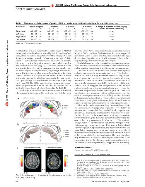
ief communications© 2001 Nature Publish<strong>in</strong>g Group http://neurosci.nature.com© 2001 Nature Publish<strong>in</strong>g Group http://neurosci.nature.comTable 1. Time course <strong>of</strong> the center <strong>of</strong> gravity <strong>of</strong> M1 activations for the hand and elbow, for the different exams.Movement Before surgery 2 months 4 months 6 months Change <strong>in</strong> distance (before surgeryx y z x y z x y z x y z versus 6 months <strong>after</strong>ward)Right hand –42 –22 40 –40 –25 46 –39 –25 45 –39 –27 48 10 mmLeft hand 36 –18 46 33 –23 49 38 –21 48 33 –22 49 6 mmRight elbow –34 –29 49 –26 –33 54 –30 –31 52 –28 –32 54 8 mmLeft elbow 31 –23 47 29 –28 51 30 –26 52 30 –26 54 7 mmValues are <strong>in</strong> Talairach coord<strong>in</strong>ates.<strong>of</strong> either elbow activated a contralateral central region <strong>of</strong> M1 thatcorresponds to the hand <strong>motor</strong> map (Fig. 2a). Six months <strong>after</strong>ward,elbow activations migrated toward the upper part <strong>of</strong> thelimb representation, classically def<strong>in</strong>ed as the arm region 6 . DifferentM1 cortical maps were observed before and six months<strong>after</strong> surgery: before the <strong>graft</strong>, a central region, and <strong>after</strong>ward, anew superior medial area (Fig. 2c). As for hand movements, thenew superior locus <strong>of</strong> activation was apparent at two months (versusbefore surgery; Z > 10; cluster size, 236) for right elbow movements.The signal strength had <strong>in</strong>creased significantly at 4 months(versus 2 months; Z = 5.2; cluster size, 26) but did not <strong>in</strong>creasefurther at 6 months. For left elbow movements, the most significantsignal changes occurred between 4 and 6 months (Z = 6.9;cluster size, 67). The COGs along the four exam<strong>in</strong>ations underwenta progressive shift from the central to the superior part <strong>of</strong>M1 (right elbow, 8 mm; left elbow, 7 mm; Fig. 2b; Table 1).The changes observed with<strong>in</strong> the <strong>motor</strong> <strong>cortex</strong> for hand andelbow representations seemed to be strongly correlated <strong>in</strong> <strong>both</strong>abtime and space. Across the different exam<strong>in</strong>ations, the distancebetween COGs rema<strong>in</strong>ed fairly constant for the two types <strong>of</strong>movements (Table 1). Hand and elbow activations showed a highdegree <strong>of</strong> overlap, the extent <strong>of</strong> which <strong>in</strong>creased from beforesurgery through the exam<strong>in</strong>ations <strong>after</strong> surgery.Parallel changes were also recorded <strong>in</strong> somatosensory <strong>cortex</strong>.Hand and elbow movements activated S1 <strong>in</strong> all four exam<strong>in</strong>ationsand their pattern was similar to that <strong>of</strong> the <strong>motor</strong> <strong>cortex</strong> activations.These results show that <strong>graft</strong>ed <strong>hands</strong> come to be recognizedand activated normally by sensori<strong>motor</strong> <strong>cortex</strong>. The displacement<strong>of</strong> the cortical activity from lateral to medial along the precentralgyrus is remarkably similar for hand and elbowmovements. These cortical maps covered <strong>in</strong> the same amount <strong>of</strong>time a similar distance, as revealed by the trajectory <strong>of</strong> the activationCOGs. This suggests that new peripheral <strong>in</strong>puts alloweda global remodel<strong>in</strong>g <strong>of</strong> the limb cortical map and reversed thefunctional <strong>reorganization</strong> <strong>in</strong>duced by the amputation. The spatialtrajectory <strong>of</strong> these activations <strong>in</strong> time further <strong>in</strong>dicates that thecortical rearrangement occurs <strong>in</strong> an orderly manner; the handand arm representations tend to return to their orig<strong>in</strong>al corticallocus. Hence, bra<strong>in</strong> plasticity is accomplished with reference toa previous pre-amputation somatotopic body representation.What are the mechanisms underly<strong>in</strong>g this cortical reversibility?In monkeys with amputated segments, severed efferentmotoneurons preserve their functional efficacy by target<strong>in</strong>g newmuscles 7 . As efferent and afferent central pathway neurons survive<strong>after</strong> they are cut, the sensori<strong>motor</strong> circuit may be functionallyready <strong>after</strong> the <strong>graft</strong>; this could expla<strong>in</strong> why activity shifts areobserved as early as two months <strong>after</strong> surgery. The <strong>in</strong>tr<strong>in</strong>sicchanges with<strong>in</strong> M1 may result from a shift <strong>in</strong> the strengths <strong>of</strong> activationsamong exist<strong>in</strong>g connections. If we assume that hand andelbow had preexist<strong>in</strong>g connections, the elbow activation <strong>in</strong> thephase before surgery may emerge as a change <strong>in</strong> the weight <strong>of</strong>these connections; that is, the elbow connection may be enhancedat the expense <strong>of</strong> the deprived hand region. The hand transplantmay have restored the efficacy <strong>of</strong> the orig<strong>in</strong>al connections at theexpense, this time, <strong>of</strong> the elbow representation, thus allow<strong>in</strong>g typicalfeatures <strong>of</strong> cortical organization to reappear <strong>in</strong> the <strong>motor</strong> map.cACKNOWLEDGEMENTSThis work was supported by a CNRS grant to A.S.RECEIVED 5 FEBRUARY; ACCEPTED 30 APRIL 2001Fig. 2. Activation maps <strong>in</strong> M1 obta<strong>in</strong>ed <strong>in</strong> the elbow movement condition.(a–c) Same as <strong>in</strong> Fig.1.1. Kaas, J. H. <strong>in</strong> The Organization <strong>of</strong> the Cerebral Cortex (eds. Schmitt, F. O.,Worden, F. G., Adelman, G. & Dennis, S. G.) 223–236 (MIT Press,Cambridge, Massachusetts, 1981).2. Roricht S., Meyer, B. U., Niehaus, L. & Brandt, S. A. Neurology 53, 106–111(1999).3. Dubernard J. M. et al. Lancet 353, 1315–1320 (1999).4. Ramachandran, V. S., Rogers-Ramachandran, D. & Stewart, M. Science 258,1159–1160 (1992).5. Yousry, T. A. et al. Bra<strong>in</strong> 120, 141–157 (1997).6. Penfield W. The Cerebral Cortex <strong>of</strong> the Man (MacMillan, New York, 1950).7. Wu, C. W. & Kaas, J. H. Neuron 28, 967–978 (2000).692 nature neuroscience • volume 4 no 7 • july 2001