

Buccinator myomucosal flap - Vula - University of Cape Town
Buccinator myomucosal flap - Vula - University of Cape Town
Buccinator myomucosal flap - Vula - University of Cape Town

You also want an ePaper? Increase the reach of your titles
YUMPU automatically turns print PDFs into web optimized ePapers that Google loves.
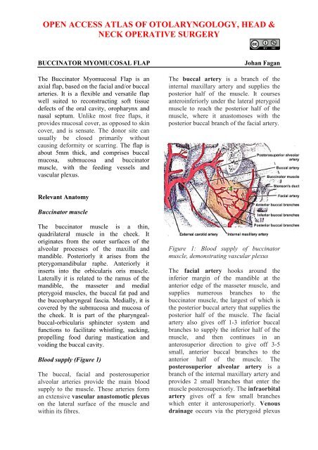
OPEN ACCESS ATLAS OF OTOLARYNGOLOGY, HEAD &NECK OPERATIVE SURGERYBUCCINATOR MYOMUCOSAL FLAPThe <strong>Buccinator</strong> Myomucosal Flap is anaxial <strong>flap</strong>, based on the facial and/or buccalarteries. It is a flexible and versatile <strong>flap</strong>well suited to reconstructing s<strong>of</strong>t tissuedefects <strong>of</strong> the oral cavity, oropharynx andnasal septum. Unlike most free <strong>flap</strong>s, itprovides mucosal cover, as opposed to skincover, and is sensate. The donor site canusually be closed primarily withoutcausing deformity or scarring. The <strong>flap</strong> isabout 5mm thick, and comprises buccalmucosa, submucosa and buccinatormuscle, with the feeding vessels andvascular plexus.Johan FaganThe buccal artery is a branch <strong>of</strong> theinternal maxillary artery and supplies theposterior half <strong>of</strong> the muscle. It coursesanteroinferiorly under the lateral pterygoidmuscle to reach the posterior half <strong>of</strong> themuscle, where it anastomoses with theposterior buccal branch <strong>of</strong> the facial artery.Relevant Anatomy<strong>Buccinator</strong> muscleThe buccinator muscle is a thin,quadrilateral muscle in the cheek. Itoriginates from the outer surfaces <strong>of</strong> thealveolar processes <strong>of</strong> the maxilla andmandible. Posteriorly it arises from thepterygomandibular raphe. Anteriorly itinserts into the orbicularis oris muscle.Laterally it is related to the ramus <strong>of</strong> themandible, the masseter and medialpterygoid muscles, the buccal fat pad andthe buccopharyngeal fascia. Medially, it iscovered by the submucosa and mucosa <strong>of</strong>the cheek. It is part <strong>of</strong> the pharyngealbuccal-orbicularissphincter system andfunctions to facilitate whistling, sucking,propelling food during mastication andvoiding the buccal cavity.Blood supply (Figure 1)The buccal, facial and posterosuperioralveolar arteries provide the main bloodsupply to the muscle. These arteries forman extensive vascular anastomotic plexuson the lateral surface <strong>of</strong> the muscle andwithin its fibres.Figure 1: Blood supply <strong>of</strong> buccinatormuscle, demonstrating vascular plexusThe facial artery hooks around theinferior margin <strong>of</strong> the mandible at theanterior edge <strong>of</strong> the masseter muscle, andsupplies numerous branches to thebuccinator muscle, the largest <strong>of</strong> which isthe posterior buccal artery that supplies theposterior half <strong>of</strong> the muscle. The facialartery also gives <strong>of</strong>f 1-3 inferior buccalbranches to supply the inferior half <strong>of</strong> themuscle, and then continues in ananterosuperior direction to give <strong>of</strong>f 3-5small, anterior buccal branches to theanterior half <strong>of</strong> the muscle. Theposterosuperior alveolar artery is abranch <strong>of</strong> the internal maxillary artery andprovides 2 small branches that enter themuscle posterosuperiorly. The infraorbitalartery gives <strong>of</strong>f a few small brancheswhich enter it anterosuperiorly. Venousdrainage occurs via the pterygoid plexus
and internal maxillary vein. It liesposterior, superior and superficial to thebuccinator muscle and drains into thebuccal vein via the deep facial vein.Anteriorly, the deep facial vein drains intothe facial vein proper.elevated, generally in an anterior toposterior direction. The <strong>flap</strong> is elevated inthe loose areolar plane between thebuccinator muscle and thebuccopharyngeal fascia.InnervationMucosal sensory innervation is by thelong buccal nerve, a branch <strong>of</strong> themaxillary division <strong>of</strong> the trigeminal nerve,which courses with the buccal branch <strong>of</strong>the internal maxillary artery. Motorinnervation <strong>of</strong> the buccinator muscle isvia the temporal and cervical divisions <strong>of</strong>the facial nerve laterally in the buccal fatpad.Parotid ductThe duct pierces the buccinator muscleopposite the 2 nd upper molar, slightlyabove the center <strong>of</strong> the muscle, and shouldbe identified and preserved when raisingthe <strong>flap</strong>.Elevation <strong>of</strong> <strong>flap</strong>A <strong>flap</strong> as large as 7x5cm may be raised.The extent is limited by the parotid ductposterosuperiorly, the oral commissureanteriorly and the pterygomandibular rapheposteriorly. It may be based posteriorly(buccal artery), or anteriorly (facialartery) or superiorly (facial artery).Although not essential, a handheld Dopplercan be used to map out the facial arteryand the buccal artery before raising the<strong>flap</strong> (Figure 2). If a Doppler is not used,then care has to be taken to place the initialincisions so as not to separate the vesselsfrom the <strong>flap</strong>.The buccal mucosa and the buccinatormuscle are incised to the level <strong>of</strong> thebuccopharyngeal fascia, and the <strong>flap</strong>Figure 2: Vessels mapped out withhandheld Doppler probeThe artery that the <strong>flap</strong> will be based on isidentified, and dissection proceedsbetween the artery and thebuccopharyngeal fascia, directed towardsthe origin <strong>of</strong> the vascular pedicle, keepingthe vessel in full view, using dissectingscissors (Figures 3, 4). Preservation <strong>of</strong> thebuccopharyngeal fascia prevents herniation<strong>of</strong> buccal fat and avoids injury to branches<strong>of</strong> the facial nerve. Minor bleeding vesselsare coagulated with bipolar electrocautery.Figure 3: Flap elevation commences afteridentifying artery (Black arrow)2
is ligated and divided superiorly. Thedissection continues in a plane lateral tothe vessels, as the <strong>flap</strong> is raised from frontto-back,and from superiorly-to-inferiorly.Superiorly Based <strong>Buccinator</strong> FlapThis is a reversed-flow <strong>flap</strong> based on thefacial artery and its anterior buccalbranches (Figure 9).Figure 6: Flap for anterior floor <strong>of</strong> mouthFigure 9: Blood supply <strong>of</strong> superiorly basedbuccinator <strong>flap</strong>Figure 7: Blood supply <strong>of</strong> anteriorly basedbuccinator <strong>flap</strong>The <strong>flap</strong> is then elevated in a superiordirection. It can be used for s<strong>of</strong>t tissuedefects <strong>of</strong> the hard palate and superioralveolus (Figure 10), including oroantraland oronasal fistulae, as well as for nasalnasal septal defects.Figure 8: Preserved facial artery in Level1The mucosa and the buccinator muscle areincised just posterior (approx 1cm) to thecommissure <strong>of</strong> the mouth, and the facialartery is identified in the cheek. The arteryFigure 10: Superiorly based <strong>flap</strong> forsuperior alveolectomy defect4
Dissection is commenced at the inferiormargin <strong>of</strong> the <strong>flap</strong>, where the facial arteryis identified and ligated.Benefits <strong>of</strong> buccinator <strong>flap</strong>The buccinator <strong>flap</strong> is a versatile <strong>flap</strong> thatcan be used to reconstruct a variety <strong>of</strong>defects. It is very reliable, simple andquick to raise, replaces mucosa withmucosa, is sensate, and has very littledonor site morbidity. It is remarkablyelastic and malleable, and can be stretchedto conform to complexly shaped defectsand is an excellent alternative to radial freeforearm <strong>flap</strong>s for reconstruction <strong>of</strong> smalland moderate sized defects <strong>of</strong> the oralcavity and oropharynx (Figures 11 a, b, c).Figure 11a: Defect following anterolateralfloor <strong>of</strong> mouth resection andmarginal mandibulectomyUseful referenceVan Lierop A, Fagan JJ. <strong>Buccinator</strong><strong>myomucosal</strong> <strong>flap</strong>: Clinical results andreview <strong>of</strong> anatomy, surgical technique andapplications. J Laryngol Otol, 2008; 122:181-7Author & EditorJohan Fagan MBChB, FCORL, MMedPr<strong>of</strong>essor and ChairmanDivision <strong>of</strong> Otolaryngology<strong>University</strong> <strong>of</strong> <strong>Cape</strong> <strong>Town</strong><strong>Cape</strong> <strong>Town</strong>, South Africajohannes.fagan@uct.ac.zaFigure 11 b: Anteriorly based buccinator<strong>flap</strong>THE OPEN ACCESS ATLAS OFOTOLARYNGOLOGY, HEAD &NECK OPERATIVE SURGERYwww.entdev.uct.ac.za/index_files/Page650.htmThe Open Access Atlas <strong>of</strong> Otolaryngology, Head &Neck Operative Surgery by Johan Fagan (Editor)johannes.fagan@uct.ac.za is licensed under a CreativeCommons Attribution-NonCommercial 3.0 UnportedLicenseFigure 11 c: Inset <strong>of</strong> <strong>flap</strong>5


