Estella Pulse Generators - BIOTRONIK USA - News
Estella Pulse Generators - BIOTRONIK USA - News
Estella Pulse Generators - BIOTRONIK USA - News
Create successful ePaper yourself
Turn your PDF publications into a flip-book with our unique Google optimized e-Paper software.
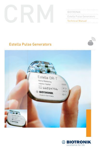
<strong>BIOTRONIK</strong><strong>Estella</strong> <strong>Pulse</strong> <strong>Generators</strong>Technical Manual<strong>Estella</strong> <strong>Pulse</strong> <strong>Generators</strong>
<strong>Estella</strong>Implantable <strong>Pulse</strong> <strong>Generators</strong><strong>Estella</strong> DRX-Ray identification<strong>Estella</strong> DR-TX-Ray identificationRadiopaque IdentificationA radiopaque identification code is visible on standard x-ray, andidentifies the pulse generator:<strong>Estella</strong> DR, DR‐T, SR, and SR-T SFCautionBecause of the numerous available 3.2-mm configurations(e.g., the IS-1 and VS-1 standards), lead/pulse generatorcompatibility should be confirmed with the pulse generatorand/or lead manufacturer prior to the implantation of apacing system.IS-1, wherever stated in this manual, refers to theinternational standard, whereby leads and generators fromdifferent manu facturers are assured a basic fit.[Reference ISO 5841-3:1992(E)].CautionFederal (U.S.A.) law restricts this device to sale by or on theorder of, a physician (or properly licensed practitioner).©2011 <strong>BIOTRONIK</strong>, Inc., all rights reserved.
Contents<strong>Estella</strong> <strong>Pulse</strong> <strong>Generators</strong> Technical Manual i1. Device Description............................................................................. 12. Indications......................................................................................... 33. Contraindications............................................................................... 54. Warnings and Precautions................................................................. 74.1 Medical Therapy................................................................................74.2 Storage and Sterilization..................................................................84.3 Lead Connection and Evaluation......................................................94.4 Programming and Operation..........................................................104.5 Home Monitoring............................................................................124.6 Electromagnetic Interference (EMI)................................................124.6.1 Home and Occupational Environments......................................134.6.2 Cellular Phones..........................................................................144.6.3 Hospital and Medical Environments..........................................154.7 <strong>Pulse</strong> Generator Explant and Disposal...........................................155. Adverse Events................................................................................ 175.1 Observed Adverse Events...............................................................175.1.1 Dromos DR Clinical Study..........................................................175.1.2 PACC Clinical Study....................................................................185.2 Potential Adverse Events................................................................206. Clinical Study................................................................................... 216.1 Dromos DR......................................................................................216.2 Ventricular Capture Control............................................................226.2.1 Primary Objectives.....................................................................226.2.2 Methods......................................................................................226.2.3 Results........................................................................................236.2.4 Clinical Study Conclusions.........................................................276.3 TRUST Clinical Study......................................................................276.3.1 Study Overview...........................................................................276.3.2 Methods......................................................................................276.3.3 Summary of Clinical Results......................................................306.3.4 Conclusions................................................................................346.4 Atrial Capture Control (ACC) and Ventricular Pacing Suppression(V pS).................................................................................................347. Programmable Parameters............................................................. 457.1 Pacing Modes..................................................................................457.1.1 Motion Based Rate-Adaptive Modes..........................................45
ii <strong>Estella</strong> <strong>Pulse</strong> <strong>Generators</strong> Technical Manual7.1.2 Non-Rate-Adaptive Modes.........................................................457.1.3 Mode Switching..........................................................................457.1.4 Pacing Modes with Triggered Response....................................477.2 Rate Related Functions...................................................................477.2.1 Basic Rate...................................................................................487.2.2 Rate Hysteresis...........................................................................487.2.3 Scan Hysteresis..........................................................................497.2.4 Repetitive Hysteresis..................................................................507.2.5 Night Mode.................................................................................517.2.6 Rate Fading.................................................................................517.3 <strong>Pulse</strong> Specific Features..................................................................527.3.1 <strong>Pulse</strong> Amplitude.........................................................................527.3.2 <strong>Pulse</strong> Width.................................................................................537.4 Automatic Sensitivity Control (ASC)................................................537.5 Timing Features..............................................................................547.5.1 Refractory Periods.....................................................................547.5.2 PVARP.........................................................................................547.5.3 AV Delay......................................................................................567.5.4 Ventricular Blanking Period.......................................................597.5.5 Atrial Blanking Period................................................................597.5.6 Far-Field Protection...................................................................597.5.7 Safety AV Delay...........................................................................597.5.8 Upper Rate and UTR Response..................................................607.6 Lead Polarity...................................................................................607.7 Parameters for Rate-Adaptive Pacing............................................617.7.1 Sensor Gain................................................................................617.7.2 Automatic Sensor Gain...............................................................627.7.3 Sensor Threshold.......................................................................637.7.4 Rate Increase..............................................................................637.7.5 Maximum Sensor Rate...............................................................647.7.6 Rate Decrease............................................................................647.8 Management of Specific Scenarios................................................657.8.1 2:1 Lock-In Management...........................................................657.9 Atrial Upper Rate............................................................................667.10 Atrial Overdrive Pacing (Overdrive Mode).....................................667.11 Management of Specific Scenarios..............................................687.11.1 PMT Management....................................................................687.11.2 PMT Protection.........................................................................68
<strong>Estella</strong> <strong>Pulse</strong> <strong>Generators</strong> Technical Manual iii7.12 Adjustment of the PMT Protection Window..................................697.13 Ventricular Capture Control (VCC)................................................707.13.1 Feature Description..................................................................707.13.2 Ventricular Capture Control Programming.............................777.14 Atrial Capture Control (ACC).........................................................777.14.1 Feature Description..................................................................777.15 Ventricular Pace Suppression (V p‐Suppression)..........................787.15.1 Feature Description..................................................................787.15.2 Programmability.......................................................................797.16 Program Consult®........................................................................797.17 Home Monitoring (<strong>Estella</strong> DR-T)...................................................837.17.1 Transmission of Information....................................................847.17.2 Patient Device...........................................................................847.17.3 Transmitting Data.....................................................................847.17.4 Types of Report Transmissions................................................867.17.5 Description of Transmitted Data..............................................878. Statistics.......................................................................................... 918.1 Statistics Overview..........................................................................918.1.1 Timing.........................................................................................918.1.2 Atrial Arrhythmia........................................................................918.1.3 Sensor.........................................................................................918.1.4 Sensing.......................................................................................918.1.5 Ventricular Arrhythmia...............................................................918.1.6 Pacing.........................................................................................928.1.7 General Statistical Information..................................................928.2 Timing Statistics..............................................................................928.2.1 Event Counter.............................................................................928.2.2 Event Episodes...........................................................................938.2.3 Rate Trend 24 Hours...................................................................938.2.4 Rate Trend 240 Days...................................................................938.2.5 Atrial and Ventricular Rate Histogram......................................948.3 Arrhythmia Statistics......................................................................948.3.1 Atrial Burden..............................................................................948.3.2 Time of occurrence.....................................................................948.3.3 Mode Switching..........................................................................948.3.4 Ventricular Arrhythmia...............................................................948.4 Sensor Statistics.............................................................................958.4.1 Sensor Histogram......................................................................95
iv <strong>Estella</strong> <strong>Pulse</strong> <strong>Generators</strong> Technical Manual8.4.2 Activity Report............................................................................968.5 Pacing Statistics..............................................................................968.5.1 Ventricular Pacing Amplitude Histogram..................................978.5.2 V Pacing Threshold Trend..........................................................978.5.3 Capture Control Status...............................................................978.6 Sensing Statistics............................................................................978.7 IEGM Recordings.............................................................................989. Other Functions/Features...............................................................1019.1 Safe Program Settings..................................................................1019.2 Magnet Effect................................................................................1019.3 Temporary Programming..............................................................1029.4 Patient Data Memory....................................................................1039.5 Position Indicator..........................................................................1049.6 Pacing When Exposed to Interference..........................................10410. Product Storage and Handling......................................................10510.1 Sterilization and Storage.............................................................10510.2 Opening the Sterile Container....................................................10610.3 <strong>Pulse</strong> Generator Orientation.......................................................10611. Lead Connection............................................................................10711.1 Auto Initialization........................................................................10912. Follow-up Procedures...................................................................11112.1 General Considerations..............................................................11112.2 Real-time IEGM Transmission ...................................................11112.3 Threshold Test.............................................................................11212.4 P/R Measurement.......................................................................11212.5 Testing for Retrograde Conduction.............................................11312.6 Non‐Invasive Programmed Stimulation (NIPS)..........................11312.6.1 Description.............................................................................11312.6.2 Burst Stimulation...................................................................11412.6.3 Programmed Stimulation.......................................................11412.6.4 Back up Pacing.......................................................................11412.6.5 NIPS Safety Features..............................................................11412.7 Optimizing Rate Adaptation........................................................11512.7.1 Rate/Sensor Trend..................................................................11612.7.2 Adjusting the Sensor Gain......................................................11612.7.3 Adjusting the Sensor Threshold.............................................116
<strong>Estella</strong> <strong>Pulse</strong> <strong>Generators</strong> Technical Manual v13. Elective Replacement Indication (ERI)..........................................11714. Explantation..................................................................................12114.1 Common Reasons to Explant a <strong>Pulse</strong> Generator.......................12115. Technical Data...............................................................................12515.1 Modes..........................................................................................12515.2 <strong>Pulse</strong>- and Control Parameters..................................................12515.2.1 Rate Adaptation......................................................................12915.2.2 Atrial Capture Control (ACC)..................................................12915.2.3 Ventricular Capture Control (VCC).........................................13015.2.4 Home Monitoring Parameters...............................................13015.2.5 Additional Functions...............................................................13115.2.6 NIPS Specifications................................................................13215.3 Programmer................................................................................13215.4 Materials in Contact with Human Tissue....................................13215.5 Electrical Data/Battery................................................................13215.6 Mechanical Data..........................................................................13316. Order Information.........................................................................13517. Appendix A....................................................................................13717.1 Mode-Specific Indications and Contraindications......................13717.1.1 Rate-adaptive Pacing.............................................................13717.1.2 Dual Chamber.........................................................................13817.1.3 Dual Chamber Modes.............................................................13917.1.4 Single Chamber Modes..........................................................14017.1.5 Other Modes...........................................................................14118. Appendix B....................................................................................14318.1 Known Software Anomalies .......................................................143CautionFederal (U.S.A.) law restricts this device to sale by, or on theorder of, a physician (or properly licensed practitioner).
vi <strong>Estella</strong> <strong>Pulse</strong> <strong>Generators</strong> Technical Manual
1. Device Description<strong>Estella</strong> <strong>Pulse</strong> <strong>Generators</strong> Technical Manual 1<strong>Estella</strong> is a multi-programmable, dual chamber pulse generator withrate-adaptive pacing. Rate-adaptation is achieved through motionbasedpacing via a capacitive accelerometer.For standard motion-based rate-adaptation, the <strong>Estella</strong> is equippedwith an accelerometer located within the pulse generator. This sensorproduces an electric signal during physical activity of the patient. If arate-adaptive (R) mode is programmed, then the accelerometer sensorsignal controls the stimulation rate.<strong>Estella</strong> also employs Home Monitoring technology, which is anautomatic, wireless, remote monitoring system for management ofpatients with pulse generators. With Home Monitoring, physicians canreview data about the patient’s cardiac status and pulse generator’sfunctionality between regular follow-up visits, allowing the physician tooptimize the therapy process.<strong>BIOTRONIK</strong> conducted the TRUST study to evaluate the safety andeffectiveness of Home Monitoring. Refer to Section 6.4 for detailsregarding the study design and results. With the TRUST study, <strong>BIOTRONIK</strong>was able to show the following with regards to Home Monitoring:• <strong>BIOTRONIK</strong> Home Monitoring information may be used as areplacement for device interrogation during in-office follow-upvisits.• A strategy of care using <strong>BIOTRONIK</strong> Home Monitoring with officevisits when needed has been shown to extend the time betweenroutine, scheduled in-office follow-ups of <strong>BIOTRONIK</strong> implantabledevices in many patients. Home Monitoring data is helpful indetermining the need for additional in-office follow-up.• <strong>BIOTRONIK</strong> Home Monitoring-patients—who are followed remotelywith office visits when needed—have been shown to have similarnumbers of strokes, invasive procedures and deaths as patientsfollowed with conventional in-office follow‐ups.• <strong>BIOTRONIK</strong> Home Monitoring provides early detection ofarrhythmias.• <strong>BIOTRONIK</strong> Home Monitoring provides early detection of silent,asymptomatic arrhythmias.• Automatic early detection of arrhythmias and device systemanomalies by <strong>BIOTRONIK</strong> Home Monitoring allows for earlierintervention than conventional in-office follow-ups.
2 <strong>Estella</strong> <strong>Pulse</strong> <strong>Generators</strong> Technical Manual• <strong>BIOTRONIK</strong> Home Monitoring allows for improved access to patientdevice data compared to c.onventional in-office follow‐ups sincedevice interrogation is automatically scheduled at regular intervals.<strong>Estella</strong> provides single and dual chamber pacing in a variety of rateadaptiveand non‐rate adaptive pacing modes. Pacing capability issupported by a sophisticated diagnostic set.The device is designed and recommended for use with atrial andventricular unipolar or bipolar leads having IS‐1 compatible connectors.(Note that IS‐1 refers to the International Standard whereby leadsand generators from different manufacturers are assured a basic fit[Reference ISO 5841‐3:1992]).<strong>Estella</strong> is designed to meet all indications for bradycardia therapy asexhibited in a wide variety of patients. The family is comprised of fourpulse generators that are designed to handle a multitude of situations.The four pulse generators include:<strong>Estella</strong> DR<strong>Estella</strong> DR-T<strong>Estella</strong> SR<strong>Estella</strong> SR-TDual chamber, rate-adaptive, unipolar/bipolarDual chamber, rate-adaptive, unipolar/bipolar,with Home MonitoringSingle chamber, rate‐adaptive, unipolar/bipolarSingle chamber, rate‐adaptive, unipolar/bipolar,with Home MonitoringThroughout this manual, specific feature and function descriptions mayonly be applicable to certain pulse generators of the <strong>Estella</strong> family.If specified as dual chamber configurations, the descriptions arespecifically referring to <strong>Estella</strong> DR and <strong>Estella</strong> DR‐T. If specified as singlechamber configurations, the descriptions are specifically referring to<strong>Estella</strong> SR and <strong>Estella</strong> SR-T.
2. Indications<strong>Estella</strong> <strong>Pulse</strong> <strong>Generators</strong> Technical Manual 3Rate-adaptive pacing with <strong>Estella</strong> pulse generators is indicated forpatients exhibiting chronotropic incompetence and who would benefitfrom increased pacing rates concurrent with physical activity.Generally accepted indications for long-term cardiac pacing include,but are not limited to: sick sinus syndrome (i.e. bradycardia-tachycardiasyndrome, sinus arrest, sinus bradycardia), sino-atrial (SA) block,second- and third- degree AV block, and carotid sinus syndrome.Patients who demonstrate hemodynamic benefit through maintenanceof AV synchrony should be considered for one of the dual chamber oratrial pacing modes. Dual chamber modes are specifically indicated fortreatment of conduction disorders that require both restoration of rateand AV synchrony such as AV nodal disease, diminished cardiac outputor congestive heart failure associated with conduction disturbances, andtachyarrhythmias that are suppressed by chronic pacing.
4 <strong>Estella</strong> <strong>Pulse</strong> <strong>Generators</strong> Technical Manual
<strong>Estella</strong> <strong>Pulse</strong> <strong>Generators</strong> Technical Manual 53. ContraindicationsUse of <strong>Estella</strong> pulse generators is contraindicated for the followingpatients:• Unipolar pacing is contraindicated for patients with an implantedcardioverter-defibrillator (ICD) because it may cause unwanteddelivery or inhibition of ICD therapy.• Single chamber atrial pacing is contraindicated for patients withimpaired AV nodal conduction.• Dual chamber and single chamber atrial pacing is contraindicatedfor patients with chronic refractory atrial tachyarrhythmias.For a complete discussion of mode-specific contraindications, pleaserefer to Appendix A of this manual.
6 <strong>Estella</strong> <strong>Pulse</strong> <strong>Generators</strong> Technical Manual
4. Warnings and Precautions<strong>Estella</strong> <strong>Pulse</strong> <strong>Generators</strong> Technical Manual 7Certain therapeutic and diagnostic procedures may cause undetecteddamage to a pulse generator, resulting in malfunction or failure at alater time. Please note the following warnings and precautions:Magnetic Resonance Imaging (MRI)—Avoid use of magnetic resonanceimaging as it has been shown to cause movement of the pulse generatorwithin the subcutaneous pocket and may cause pain and injury to thepatient and damage to the pulse generator. If the procedure must beused, constant monitoring is recommended, including monitoring theperipheral pulse.Rate-Adaptive Pacing—Use rate-adaptive pacing with care in patientsunable to tolerate increased pacing rates.High Output Settings—High output settings combined with extremelylow lead impedance may reduce the life expectancy of the pulsegenerator to less than 1 year. Programming of pulse amplitudes, higherthan 4.8 V, in combination with long pulse widths and/or high pacingrates may lead to premature activation of the replacement indicator.4.1 Medical TherapyBefore applying one of the following procedures, a detailed analysis ofthe advantages and risks should be made. Cardiac activity during oneof these procedures should be confirmed by continuous monitoring ofperipheral pulse or blood pressure. Following the procedures, pulsegenerator function and stimulation threshold must be checked.Therapeutic Diathermy Equipment—Use of therapeutic diathermyequipment is to be avoided for pacemaker patients due to possibleheating effects of the pulse generator and at the implant site. Ifdiathermy therapy must be used, it should not be applied in theimmediate vicinity of the pulse generator/lead. The patient’s peripheralpulse should be monitored continuously during the treatment.Transcutaneous Electrical Nerve Stimulation (TENS)—Transcutaneouselectrical nerve stimulation may interfere with pulse generator function.If necessary, the following measures may reduce the possibility ofinterference:• Place the TENS electrodes as close to each other as possible.• Place the TENS electrodes as far from the pulse generator/leadsystem as possible.• Monitor cardiac activity during TENS use.
8 <strong>Estella</strong> <strong>Pulse</strong> <strong>Generators</strong> Technical ManualDefibrillation—The following precautions are recommended tominimize the inherent risk of pulse generator operation being adverselyaffected by defibrillation:• The paddles should be placed anterior‐posterior or along a lineperpendicular to the axis formed by the pulse generator and theimplanted lead.• The energy setting should not be higher than required to achievedefibrillation.• The distance between the paddles and the pacer/electrode(s) shouldnot be less than 10 cm (4 inches).Radiation—<strong>Pulse</strong> generator electronics may be damaged by exposureto radiation during radiotherapy. To minimize this risk when using suchtherapy, the pulse generator should be protected with local radiationshielding.Lithotripsy—Lithotripsy treatment should be avoided for pacemakerpatients since electrical and/or mechanical interference with the pulsegenerator is possible. If this procedure must be used, the greatestpossible distance from the point of electrical and mechanical strainshould be chosen in order to minimize a potential interference with thepulse generator.Electrocautery—Electrocautery should never be performed within 15 cm(6 inches) of an implanted pulse generator or lead because of the dangerof introducing fibrillatory currents into the heart and/or damaging thepulse generator. Pacing should be asynchronous and above the patient’sintrinsic rate to prevent inhibition by interference signals generated bythe cautery. When possible, a bipolar electrocautery system should beused.For transurethral resection of the prostate, it is recommended thatthe cautery ground plate be placed under the buttocks or around thethigh, but not in the thoracic area where the current pathway could passthrough or near the pacing system.4.2 Storage and SterilizationStorage (temperature)—Recommended storage temperature range is5° to 55°C (41°-131°F). Exposure to temperatures outside this rangemay result in pulse generator malfunction (see Section 10.1).Handling—Do not drop. If an unpackaged pulse generator is droppedonto a hard surface, return it to <strong>BIOTRONIK</strong> (see Section 10.1).FOR SINGLE USE ONLY—Do not resterilize the pulse generator oraccessories packaged with the pulse generator, they are intended forone-time use.
<strong>Estella</strong> <strong>Pulse</strong> <strong>Generators</strong> Technical Manual 9Device Packaging—Do not use the device if the packaging is wet,punctured, opened or damaged because the integrity of the sterilepackaging may be compromised. Return the device to <strong>BIOTRONIK</strong>.Storage (magnets)—Store the device in a clean area, away frommagnets, kits containing magnets, and sources of electromagneticinterference (EMI) to avoid damage to the device.Temperature Stabilization—Allow the device to reach room temperaturebefore programming or implanting the device. Temperature extremesmay affect the initial device function.Use Before Date—Do not implant the device after the USE BEFOREDATE because the device sterility and longevity may be compromised.4.3 Lead Connection and EvaluationThe pulse generator requires atrial and ventricular leads with IS-1compatible connectors. There are no requirements specific to the atriallead. It is required to use a low polarization ventricular lead for activationof Ventricular Capture Control.Lead Check—The <strong>Estella</strong> pulse generators have an automatic lead checkfeature which may switch from bipolar to unipolar pacing and sensingwithout warning. This situation may be inappropriate for patients withan Implantable Cardioverter Defibrillator (ICD).Lead/pulse Generator Compatibility—Because of the numerousavailable 3.2-mm configurations (e.g., the IS-1 and VS‐1 standards),lead/pulse generator compatibility should be confirmed with the pulsegenerator and/or lead manufacturer prior to the implantation of apacing system.IS-1, wherever stated in this manual, refers to the internationalstandard, whereby leads and generators from different manufacturersare assured a basic fit. [Reference ISO 5841-3:1992(E)].Lead Configuration—Lead configuration determines properprogramming of the pulse generator. Pacing will not occur with aunipolar lead if the lead configuration is programmed to bipolar (seeSection 11).Setscrew Adjustment—Back-off the setscrew(s) prior to insertion oflead connector(s) as failure to do so may result in damage to the lead(s),and/or difficulty connecting lead(s).Cross Threading Setscrew(s)—To prevent cross threading thesetscrew(s), do not back the setscrew(s) completely out of the threadedhole. Leave the torque wrench in the slot of the setscrew(s) while thelead is inserted.
10 <strong>Estella</strong> <strong>Pulse</strong> <strong>Generators</strong> Technical ManualTightening Setscrew(s)—Do not overtighten the setscrew(s). Use onlythe <strong>BIOTRONIK</strong> supplied torque wrench.Sealing System—Be sure to properly insert the torque wrench intothe perforation at an angle perpendicular to the connector receptacle.Failure to do so may result in damage to the plug and its self-sealingproperties.4.4 Programming and OperationNegative AV Delay Hysteresis—This feature insures ventricularpacing, a technique which has been used in patients with hypertrophicobstructive cardiomyopathy (HOCM) with normal AV conduction inorder to replace intrinsic ventricular activation. No clinical study wasconducted to evaluate this feature, and there is conflicting evidenceregarding the potential benefit of ventricular pacing therapy for HOCMpatients. In addition, there is evidence with other patient groups tosuggest that inhibiting the intrinsic ventricular activation sequenceby right ventricular pacing may impair hemodynamic function and/orsurvival.Programming VCC—If the SA/CV sequence is not successful, programanother pulse width or test start amplitude. If still unsuccessful,program the pacing pulse amplitude manually.NIPS—Life threatening ventricular arrhythmias can be induced bystimulation in the atrium. Ensure that an external cardiac defibrillator iseasily accessible. Only physicians trained and experienced in tachycardiainduction and reversion protocols should use non-invasive programmedstimulation (NIPS).Unipolar/Bipolar—All <strong>Estella</strong> models can be used with either unipolaror bipolar IS‐1 leads.If the pacing or sensing function is to be programmed to bipolar,it must be verified that bipolar leads have been implanted in thatchamber. If either of the leads is unipolar, unipolar sensing and pacingfunctions must be programmed in that chamber. Failure to program theappropriate lead configuration could result in entrance and/or exit block.Programmers—Use only appropriate <strong>BIOTRONIK</strong> programmersequipped with appropriate software to program <strong>Estella</strong> pulse generators.Do not use programmers from other manufacturers.<strong>Pulse</strong> Amplitude—Programming of pulse amplitudes, higher than 4.8 V,in combination with long pulse widths and/or high pacing rates can leadto premature activation of the replacement indicator.
<strong>Estella</strong> <strong>Pulse</strong> <strong>Generators</strong> Technical Manual 11Pacing thresholds—When decreasing programmed output (pulseamplitude and/or pulse width), the pacing threshold must first beaccurately assessed to provide a 2:1 safety margin. When using theVentricular Capture Control feature, the device will automatically set theoutput to the measured threshold plus the programmed Safety Margin.A new threshold search will occur at scheduled intervals or upon loss ofcapture.EMI—Computerized systems are subject to EMI or “noise”. In thepresence of such interference, telemetry communication may beinterrupted and prevent programming.Programming Modifications—Extreme programming changes shouldonly be made after careful clinical assessment. Clinical judgment shouldbe used when programming permanent pacing rates below 40 ppm orabove 100 ppm.Short Pacing Intervals—Use of short pacing intervals (high pacingrates) with long atrial and/or ventricular refractory periods mayresult in intermittent asynchronous pacing and, therefore, may becontraindicated in some patients.OFF Mode—Use of the OFF mode should be avoided in pacemakerdependent patients. The OFF mode can be transmitted as a temporaryprogram only to permit evaluation of the patient’s spontaneous rhythm.Myopotential Sensing—The filter characteristics of <strong>BIOTRONIK</strong> pulsegenerators have been optimized to sense electrical potentials generatedby cardiac activity and to reduce the possibility of sensing skeletalmyopotentials. However, the risk of pulse generator operation beingaffected by myopotentials cannot be eliminated, particularly in unipolarsystems. Myopotentials may resemble cardiac activity, resulting in pulsegenerator pulse inhibition, triggering and/or emission of asynchronouspacing pulses, depending on the pacing mode and the interferencepattern. Certain follow‐up procedures, such as monitoring pulsegenerator performance while the patient is doing exercises involvingthe use of pectoral muscles, as well as Holter monitoring, have beenrecommended to check for interference caused by myopotentials. Ifsensing of myopotentials is encountered, corrective actions may includeselection of a different pacing mode or sensitivity.Muscle or Nerve Stimulation—Inappropriate muscle or nervestimulation may occur with unipolar pacing when using a non-coatedpulse generator.Programmed to Triggered Modes—When programmed to triggeredmodes, pacing rates up to the programmed upper limit may occur in thepresence of either muscle or external interference.
12 <strong>Estella</strong> <strong>Pulse</strong> <strong>Generators</strong> Technical ManualTriggered Modes—While the triggered modes (DDT, VVT, and AAT) canbe programmed permanently, the use of these modes is intended as atemporary setting in situations where maintaining the programminghead in place would be impossible or impractical (i.e., during exercisetesting or extended Holter monitoring) or as a short term solution topulse generator inhibition by extracardiac interference. To avoid thepotential for early battery depletion, it is important that the triggeredmodes are not used for long term therapy, and that the pulse generatoris returned to a non-triggered permanent program.4.5 Home Monitoring<strong>BIOTRONIK</strong>’s Home Monitoring system is designed to notify clinicians inless than 24 hours of changes to the patient’s condition or status of theimplanted device. Updated data may not be available if:• The patient’s CardioMessenger is off or damaged and is not able toconnect to the Home Monitoring system through an active telephonelink.• The CardioMessenger cannot establish a connection to theimplanted device.• The telephone and/or Internet connection do not operate properly• The Home Monitoring Service Center is off-line (upgrades aretypically completed in less than 24 hours)Patient’s Ability—Use of the Home Monitoring system requires thepatient and/or caregiver to follow the system instructions and cooperatefully when transmitting data.If the patient cannot understand or follow the instructions becauseof physical or mental challenges, another adult who can follow theinstructions will be necessary for proper transmission.Electromagnetic Interference (EMI)—Precautions for EMI interferencewith the <strong>Estella</strong> DR‐T pulse generator are provided in Section 4.6.Sources of EMI including cellular telephones, electronic articlesurveillance systems, and others are discussed therein.Use in Cellular Phone Restricted Areas—The mobile patient device(transmitter/receiver) should not be utilized in areas where cellularphones are restricted or prohibited (i.e., commercial aircraft).4.6 Electromagnetic Interference (EMI)The operation of any implanted pulse generator may be affected bycertain environmental sources generating signals that resemble cardiacactivity. This may result in pulse generator pulse inhibition and/ortriggering or in asynchronous pacing depending on the pacing mode and
<strong>Estella</strong> <strong>Pulse</strong> <strong>Generators</strong> Technical Manual 13the interference pattern. In some cases (i.e., diagnostic or therapeuticmedical procedures), the interference sources may couple sufficientenergy into a pacing system to damage the pulse generator and/orcardiac tissue adjacent to the electrodes.<strong>BIOTRONIK</strong> pulse generators have been designed to significantly reducesusceptibility to electromagnetic interference (EMI). However, due tothe variety and complexity of sources creating interference, there isno absolute protection against EMI. Generally, it is assumed that EMIproduces only minor effects, if any, in pacemaker patients. If the patientpresumably will be exposed to one of the following environmentalconditions, then the patient should be given the appropriate warnings.4.6.1 Home and Occupational EnvironmentsThe following equipment (and similar devices) may affect normal pulsegenerator operation: electric arc welders, electric melting furnaces,radio/television and radar transmitters, power‐generating facilities,high‐voltage transmission lines, electrical ignition systems (alsoof gasoline‐powered devices) if protective hoods, shrouds, etc., areremoved, electrical tools, anti‐theft devices of shopping centers andelectrical appliances, if not in proper condition or not correctly groundedand encased.Patients should exercise reasonable caution in avoidance of deviceswhich generate a strong electric or magnetic field. If EMI inhibitsoperation of a pulse generator or causes it to revert to asynchronousoperation at the programmed pacing rate or at the magnet rate, movingaway from the source or turning it off will allow the pulse generator toreturn to its normal mode of operation. Some potential EMI sourcesinclude:High Voltage Power Transmission Lines—High voltage powertransmission lines may generate enough EMI to interfere with pulsegenerator operation if approached too closely.Home Appliances—Home appliances normally do not affect pulsegenerator operation if the appliances are in proper condition andcorrectly grounded and encased. There are reports of pulse generatordisturbances caused by electrical tools and by electric razors that havetouched the skin directly over the pulse generator.Communication Equipment—Communication equipment such asmicrowave transmitters, linear power amplifiers, or high-poweramateur transmitters may generate enough EMI to interfere with pulsegenerator operation if approached too closely.
14 <strong>Estella</strong> <strong>Pulse</strong> <strong>Generators</strong> Technical ManualCommercial Electrical Equipment—Commercial electrical equipmentsuch as arc welders, induction furnaces, or resistance welders maygenerate enough EMI to interfere with pulse generator operation ifapproached too closely.Electrical Appliances—Electric hand-tools and electric razors (useddirectly over the skin of the pulse generator) have been reported tocause pulse generator disturbances. Home appliances that are in goodworking order and properly grounded do not usually produce enoughEMI to interfere with pulse generator operation.Electronic Article Surveillance (EAS)—Equipment such as retail theftprevention systems may interact with the pulse generators. Patientsshould be advised to walk directly through and not to remain near anEAS system longer than necessary.4.6.2 Cellular PhonesRecent studies have indicated there may be a potential interactionbetween cellular phones and pulse generator operation. Potential effectsmay be due to either the radio frequency signal or the magnet withinthe phone and could include inhibition or asynchronous pacing when thephone is within close proximity (within 6 inches [15 centimeters]) to thepulse generator.Based on testing to date, effects resulting from an interaction betweencellular phones and the implanted pulse generators have beentemporary. Simply moving the phone away from the implanted devicewill return it to its previous state of operation. Because of the greatvariety of cellular phones and the wide variance in patient physiology, anabsolute recommendation to cover all patients cannot be made.Patients having an implanted pulse generator who operate a cellularphone should:• Maintain a minimum separation of 6 inches (15 centimeters)between a hand-held personal cellular phone and the implanteddevice. Portable and mobile cellular phones generally transmit athigher power levels compared to hand held models. For phonestransmitting above 3 watts, maintain a minimum separation of 12inches (30 centimeters) between the antenna and the implanteddevice.• Patients should hold the phone to the ear opposite the side of theimplanted device. Patients should not carry the phone in a breastpocket or on a belt over or within 6 inches (15 centimeters) of theimplanted device as some phones emit signals when they are turnedON but not in use (i.e., in the listen or standby mode). Store thephone in a location opposite the side of implant.
<strong>Estella</strong> <strong>Pulse</strong> <strong>Generators</strong> Technical Manual 154.6.3 Hospital and Medical EnvironmentsElectrosurgical Cautery—Electrosurgical cautery could induceventricular arrhythmias and/or fibrillation, or may cause asynchronousor inhibited pulse generator operation. If use of electrocautery isnecessary, the current path (ground plate) should be kept as far awayfrom the pulse generator and leads as possible.Lithotripsy—Lithotripsy may damage the pulse generator. If lithotripsymust be used, do not focus the beam near the pulse generator.External Defibrillation—External defibrillation may damage the pulsegenerator. Attempt to minimize current flowing through the pulsegenerator and lead system by following the precautions.High Radiation Sources—High radiation sources such as cobalt 60 orgamma radiation should not be directed at the pulse generator. If apatient requires radiation therapy in the vicinity of the pulse generator,place lead shielding over the device to prevent radiation damage.4.7 <strong>Pulse</strong> Generator Explant and DisposalDevice Incineration—Never incinerate a pulse generator. Be sure thepulse generator is explanted before a patient who has died is cremated(see Section 14).Explanted Devices—Return all explanted devices to <strong>BIOTRONIK</strong>.
16 <strong>Estella</strong> <strong>Pulse</strong> <strong>Generators</strong> Technical Manual
5. Adverse EventsNOTE:<strong>Estella</strong> <strong>Pulse</strong> <strong>Generators</strong> Technical Manual 17The <strong>Estella</strong> family of pulse generators is a successor to the<strong>BIOTRONIK</strong>’s Dromos, Philos, Inos, Protos, Cylos, and Evia familiesof pulse generators. Therefore, data from the clinical studies ofthese earlier generations are used to support the safety and efficacyof the <strong>Estella</strong> family of pulse generators.5.1 Observed Adverse Events5.1.1 Dromos DR Clinical StudyThe Dromos DR Clinical Study involved 273 patients with cumulativeimplant duration of 1418 months (mean implant duration 5.2 months).Eleven patients died during the course of the trial; none of the deathswas judged to be device-related. One Dromos DR pulse generator wasexplanted during the trial, secondary to infection.Table 1 reports the adverse events (AE) on a per patient and a perpatient-month basis. The last column gives the expected time (inmonths) between events; i.e., the reciprocal of the AE/patient-monthrate.Table 1: Adverse Events Reported in > 1 PatientCategory# pts(n-273)% ofpatients# ofAEsAE/ptmo(n-1418)Pt-mosbetweenAEsObservations†(total)792* 28.9% 86 0.0606 16Atrial Loss ofSensing10 3.7% 10 0.0071 142Atrial Loss ofCapture8 2.9% 8 0.0056 177PacemakerMediated11 4.0% 12 0.0085 118TachycardiaPremature AVStimulation4 1.5% 4 0.0028 355Arrhythmias 34 12.5% 36 0.0254 39Muscle/DiaphragmaticStimulation3 1.1% 3 0.0021 473
18 <strong>Estella</strong> <strong>Pulse</strong> <strong>Generators</strong> Technical ManualCategoryUnexplainedSyncopeComplications‡(total)Atrial LeadDislodgmentVentricular LeadDislodgment# pts(n-273)% ofpatients# ofAEsAE/ptmo(n-1418)Pt-mosbetweenAEs3 1.1% 3 0.0021 47314* 5.1% 14 0.0099 1016 2.2% 6 0.0042 2364 1.5% 4 0.0028 355All Dromos DR Patients (N-273), Number and % of Patients, Events/Patient Mo., and Pt-Mos. between EventsThe Dromos SR Clinical Study involved 91 patients with a cumulativeimplant duration of 327 months (mean implant duration 3.6 months).Three patients died during the course of the trial; none of the deathswas judged to be device-related. During this clinical study, there were3 ventricular lead dislodgments requiring invasive lead repositioningresulting in 0.0092 AE/patient-month and a mean patient-monthbetween adverse events of 109. There were 2 observations having onlyone occurrence each.NOTE:The Dromos family of pulse generators is an earlier generation of<strong>BIOTRONIK</strong> devices. The <strong>Estella</strong> family of pulse generators is basedupon the Dromos pulse generators.5.1.2 PACC Clinical StudyThe multi-center Philos DR ACC Clinical Study involved 152 devicesin 151 patients with a cumulative implant duration of 764.1 months(average implant duration of 5.1 ± 0.3 months). A total of 109 patientshad an implant duration of greater than 90 days.* Observations are adverse events, which are correctable by non-invasivemeasures, e.g., reprogramming.† Not included in the Table are 6 observations and 4 complications each havingonly one occurrence.‡ Complications are adverse events requiring invasive measures to correct, e.g.,surgical intervention.
<strong>Estella</strong> <strong>Pulse</strong> <strong>Generators</strong> Technical Manual 19There were two patient deaths reported. Both deaths were notpacemaker-related. Two pulse generators were explanted. One explantwas due to a pocket infection and the second explant was due toinfection and sepsis. The second patient was subsequently implantedwith another Philos DR ACC device.Table 2 provides a summary of adverse events that were reported duringthe clinical study regardless of whether or not the events were relatedto the pacemaker system. A complication was defined as a clinicalevent that resulted in additional invasive intervention. An observationwas defined as a clinical event that did not result in additional invasiveintervention. Note that the number of patients and events in eachindividual category are not mutually exclusive; certain patients mayhave had more than one event reported within a category.Table 2: Adverse EventsCategory# ofPatientswith AEs% ofPatientswith AEs# ofAEsAEs /pt-yrComplications—Total 14 9.3% 16 0.25Lead Repositioning 11 7.3% 12 0.19Medical 3 2.0% 4 0.06Device-Related Events 0 0.0% 0 0.00Observations—Total 42 27.8% 54 0.85Sensing & Pacing 17 11.3% 20 0.31Holter Evaluation 15 9.9% 15 0.23Medical 11 7.3% 12 0.19Arrhythmias 4 2.6% 4 0.06B-KAC.V.U Software 3 2.0% 3 0.05Number of Patients=151, Number of Patient-Years=63.7
20 <strong>Estella</strong> <strong>Pulse</strong> <strong>Generators</strong> Technical Manual5.2 Potential Adverse EventsThe following possible adverse events may occur with this type of devicebased on implant experience including:• Cardiac tamponade• Cardiac perforation• Air embolism• Pocket erosion• Infection• Lead fracture/ insulationdamage• Lead dislodgment• Lead-related thrombosis• Body rejection phenomena• Muscle or nerve stimulation• Elevated pacing thresholds• Pocket hematoma• Myopotential sensing• Local tissue reaction/fibrotictissue formation• <strong>Pulse</strong> generator migration• Pacemaker-mediatedtachycardia (dual chambermodes only)• Undersensing of intrinsicsignals
<strong>Estella</strong> <strong>Pulse</strong> <strong>Generators</strong> Technical Manual 216. Clinical Study6.1 Dromos DRPrimary Objectives: To evaluate the safety and effectiveness of theDromos DR pulse generator and the utility of the DDDR pacing modein patients with chronotropic incompetence (CI) in a crossover, doubleblindtrial. CI was defined as the inability to achieve a heart rate of a)60% of their age predicted maximum (220-age), or b) 100 bpm.Patients, Methods and Results: A total of 273 patients were implantedwith the Dromos DR pulse generator between July 21, 1995 and July 31,1996, at 34 investigational centers (32 in the US, 1 France, and 1Mexico). Mean patient age was 71 years with a range of 31 to 95, and145 of 273 (53%) were male. Pre-implantation clinical symptomologywas: bradycardia in 44% of the patients, dizziness in 31%, syncope in25%, ECG indications were: Sick Sinus Syndrome in 46%, heart blockin 40%, and atrial fibrillation/atrial flutter in 13% of the patients. Themean implant duration was 5.2 months (range = 0 to 16 months) with atotal implant experience of 1418 months. At the one-month follow‐up,212 patients (91%) were programmed to a rate-adaptive mode accordingto the sensor parameter optimization procedure. Of the 63 patientscompleting a DDD exercise test (CAEP protocol) at one‐month, 25were found to be CI, and 21 completed the paired exercise testing atsix‐weeks. Patients performed the exercise tests, including metabolicmeasurements, in both the DDD and DDDR modes in randomized order.Table 3: Dromos DR Metabolic Exercise Testing at 6 WeeksEndpointsMaximum VO 2(mL/kg/minute)DDDRModeDDD Mode20.4 ± 8.0 17.8 ± 6.2VO 2@ AT (mL/kg/minute) 14.6 ± 3.6 13.1 ± 4.0Total exercise time(minutes)Exercise time to AT(minutes)9.2 ± 3.0 8.2 ± 3.36.3 ± 2.4 5.7 ± 2.8Heart rate @AT (bpm) 113 ± 16 84 ± 16.5Difference (CI)2.67* ± 2.77[1.5, 3.8]1.5* ± 2.71[0.33, 2.6]0.92* ± 1.08[0.45,1.4]0.69* ± 1.43[0.04, 1.3]29* ± 18[21,37]All chronotropically incompetent patients tested, n =21, Mean ±SD and [95% confidenceinterval] 95% confidence interval = mean difference ± 1.96 SEM* Difference statistically significant, p
22 <strong>Estella</strong> <strong>Pulse</strong> <strong>Generators</strong> Technical ManualThere were no pulse generator-related deaths or unusual rates ofobservations or complications (see Section 5, Adverse Events).Conclusions: No unusual safety concerns were raised by the results ofthe clinical study. The accelerometer-based motion sensor provided thepatients with appropriate rate-adaptation when programmed accordingto the sensor parameter optimization procedure. Additionally, theDDDR mode provided statistically significant improvement in metabolicmeasures during paired exercise testing of CI patients at 6 weeks.6.2 Ventricular Capture ControlAll references to Active Capture Control feature are now synonymouswith Ventricular Capture Control (VCC) in the <strong>Estella</strong> devices. The clinicalstudy involved 151 patients, of which 72 were male (47.7%) and 79 werefemale (52.3%) with a mean age of 72 years (range: 30-93 years). Themajority of patients presented with an abnormal sino-atrial node (85%)and an abnormal conduction system (57%) at implant.6.2.1 Primary ObjectivesThe multi-center, non-randomized clinical investigation was designedto demonstrate the safety and effectiveness of the Philos DR ActiveCapture Control pulse generator in patients with standard pacemakerindications. The specific predefined objectives of the investigationincluded 3 primary endpoints.1. Appropriate ACC Performance—Safety2. ACC Algorithm Performance—Effectiveness3. ACC Threshold Comparison—Effectiveness6.2.2 MethodsThe prospective, multi-center, controlled Philos DR ACC Clinical Studyinvolved 151 patients with a cumulative implant duration of 764.1months (average implant duration of 5.1 ± 0.3 months). The investigationwas conducted at 14 centers.The patients selected for participation were from the investigator’sgeneral patient population meeting the indications for use of the PhilosDR ACC pulse generator. The average patient was a 72 year-old female,with indications for a pacemaker of Sinus Bradycardia.Each patient was followed at hospital discharge and at one, three andsix month post-implant and every 6 months thereafter. 24 hour Holterrecordings were performed at the one month follow-up.
<strong>Estella</strong> <strong>Pulse</strong> <strong>Generators</strong> Technical Manual 25ACC pacing thresholds at a single follow-up. One discrepancy occurredat implant and three others occurred at pre-discharge follow-up (within2 days of implant). All subsequent follow-ups (after lead maturation) forthese four patients showed a difference of less than 0.5 volts betweenthe ACC and manual pacing threshold. All differences of 0.5 volts orhigher were recorded at instances where the ACC threshold was higherthan the manual threshold. There is little risk of non-capture or safetyconcerns because the ACC programmed output would be set to theACC threshold plus the safety margin (0.5V), providing a much highereffective safety margin. Also, it is important to note that 96.4 % of thepatients enrolled in the PACC study had an absolute difference lowerthan the actual 0.5 volt safety margin. There were no ACC thresholdsmore than 0.4 volts lower than the manual threshold. Therefore, the useof a nominal safety margin of 0.5 volts is adequate to provide patientsafety.Figure 1 below provides a distribution of the mean absolute differencesper patient.Figure 1: Absolute Ventricular Threshold Comparison50%47.1%45%40%Percentage of Patients35%30%25%20%15%31.4%10%5%0%8.6%5.0% 4.3%1.4% 2.1%0.0%0
26 <strong>Estella</strong> <strong>Pulse</strong> <strong>Generators</strong> Technical ManualIt is concluded that the automatic ventricular pacing threshold isequivalent within 0.2 volts to the manual determination. The thresholdmeasurement analysis clearly demonstrates the ACC algorithm isable to accurately perform threshold measurements in both acute andchronic conditions.Additional ResultsThe study evaluated the evolution of the successful activation of the ACCfeature at the scheduled follow-ups.Figure 2 provides a comparison of the ACC activation rates at thepre‐discharge and three‐month follow-ups. The reasons for failed ACCactivation are non-capture or high polarization artifact.Figure 2: CC Activation Rates100%95%90%85%80%89.1%Pre-discharge96.3%3-MonthDuring the one-month follow-up, the ACC algorithm was tested todetermine the highest maximum ACC amplitude setting (from 2.4Vto 6.4V) for which ACC can be successfully activated. Table 5 providesthe distribution of the highest maximum ACC amplitudes successfullyactivated at the one-month follow-up visit. A high percentage (88.9%) ofpatients can be safely programmed at or above 4.8V where ACC remainsactivated.Table 5: Highest Maximum ACC Amplitude SettingTesting of Maximum ACC AmplitudeNumber of tests completedHighest Functional Maximum ACC Amplitude2.4 Volts3.6 Volts4.8 Volts6.4 VoltsResult994 (4.0%)7 (7.1%)15 (15.2%)73 (73.7%)
6.2.4 Clinical Study Conclusions<strong>Estella</strong> <strong>Pulse</strong> <strong>Generators</strong> Technical Manual 27The clinical data support the following conclusions regarding the safetyand efficacy of the ACC feature.The Philos DR ACC Pacing System is safe and effective for use in patientsthat are indicated for pacing therapy. The Philos DR ACC Clinical Studyfulfilled the predefined primary safety and efficacy endpoints. Theseendpoints included safety and effectiveness of the ACC feature.The gender distribution in this clinical investigation is consistent withother clinical studies and includes a representative proportion of femaleparticipants. There were also no significant differences found betweenhigh and low volume implant centers in either the safety or effectivenessendpoints.In accordance with the above conclusions, the clinical data providesassurance that the ACC feature is safe and effective for the treatmentof conditions requiring chronic cardiac pacing as specified in Section 2,Indications for Use.6.3 TRUST Clinical Study6.3.1 Study OverviewThe TRUST study is a multi-center, prospective and randomized trial.The purpose of the study was to demonstrate that the use of the<strong>BIOTRONIK</strong> Home Monitoring system (HM) can safely reduce thenumber of regularly scheduled office follow up visits, compared tothe conventional method of ICD follow-up. The assessment consistsof comparing the number of in-office follow-ups for patients with HM(HM group) versus patients without HM (Control group). With the use ofHM, the number of in-office follow up visits per year could be reducedfrom an expected five scheduled office follow up visits (3, 6, 9, 12 and15 months) to two visits (3 and 15 months). Additionally, the time fromonset to evaluation of arrhythmias in both groups was compared. It wasexpected that evaluation of cardiac events in the HM arm would occurearlier than those in the Control group.6.3.2 MethodsAll enrolled patients received a <strong>BIOTRONIK</strong> ICD with Home Monitoring/IEGM-OnlineÒ technology and were randomized to either Group 1 (HomeMonitoring (HM)) or Group 2 (No Home Monitoring (Control)) using arandomization ratio of 2:1.
28 <strong>Estella</strong> <strong>Pulse</strong> <strong>Generators</strong> Technical ManualGroup 1 (HM)Device evaluations for scheduled follow-ups, patient-initiated inquiriesand event triggered notifications were performed with HM/IEGM Online.Patients were scheduled for office device interrogations only at the 3month and 15 month follow-up points (following the HM online check). At6, 9 and 12 months, a HM check was performed first. Investigators maythen elect to perform an office device interrogation if they determinethat it is necessary after reviewing the HM data.Group 2 (Control)Patients were evaluated using conventional, calendar-based office visitsat 3, 6, 9, 12 and 15 months post-implant. Interim visits were madeaccording to physician discretion (e.g. following any ICD discharges orsymptoms). Home Monitoring wasbe programmed OFF for the durationof the study.HM Event Triggered Device EvaluationsInvestigators with patients in Group 1 (HM) may receive HM notificationsin response to pre-programmed events such as VT1 detected and SVTdetected. Upon the receipt of a HM Event Notification, investigatorsreviewed the notification and the associated information on the HM/IEGM-Online website and recorded the type of event and what type ofaction, if any, was taken as a result of this notification.Patient-Initiated Device EvaluationsInvestigators may be contacted by the patient for device/arrhythmiarelatedcare (e.g. perceived device discharge, symptoms). For patientsin Group 1 (HM), investigators triaged the complaint using the HomeMonitoring website. Investigators recorded if the information fromHome Monitoring was sufficient. For patients in Group 2 (Control),the complaint was assessed per standard of care or normal clinicprocedures.Primary EndpointsThe purpose of primary endpoint 1 (HM efficacy) was to compare thenumber of in-office ICD follow-ups for patients in Group 1 (HM) to theconventional, calendar-based method of ICD follow-up as in Group 2(Control).The purpose of the primary endpoint 2 (safety) was to compare theSafety Event Rate (SER), which includes death, incidence of strokesand events requiring surgical interventions (e.g. device explants or leadrevision) between the two groups.
<strong>Estella</strong> <strong>Pulse</strong> <strong>Generators</strong> Technical Manual 29Secondary EndpointsThe purpose of secondary endpoint 1 was to compare AF, VT and VFevents between Group 1 and Group 2 in terms of the number, categories,and detection time relative to onset.Inclusion CriteriaTo support the objectives of this investigation, the inclusion criteria atthe time of patient enrollment for this investigational study included thefollowing requirements:• Implanted within the last 45 days or being considered for implantwith a <strong>BIOTRONIK</strong> ICD with Home Monitoring/IEGM-Onlinetechnology• Able to utilize the HM system throughout the study• Ability to give informed consent• Geographically stable and able to return for regular follow-ups forfifteen (15) months• At least 18 years oldExclusion CriteriaTo support the objectives of this investigation, the exclusion criteria atthe time of patient enrollment included the following requirements:• Patients who do not fulfill all inclusion criteria• Patients who are pacemaker dependent• Currently enrolled in any other cardiac clinical investigation.Clinical Events CommitteeThe Clinical Events Committee (CEC) is an advisory review boardcomprised of three physicians that are not participating in the TRUSTStudy who reviewed and adjudicated all deaths, strokes, surgicalinterventions, and cardiac adverse events that occur during the study.The CEC also reviewed all divergent classifications of actionable vs. nonactionableoffice follow up visits between the physician and <strong>BIOTRONIK</strong>,and reviewed a random sampling of 1% of office follow up visits in whichthere is no disputed classification.
30 <strong>Estella</strong> <strong>Pulse</strong> <strong>Generators</strong> Technical Manual6.3.3 Summary of Clinical ResultsThe study involved 1443 patients (1038 males, 71.9%), with a meanage of 63.5 years (range: 20-95). The cumulative enrollment durationis 18,367 months with mean enrollment duration of 12.7 months. Thepatient follow-up compliance rate for all enrolled patients is 87.5% inGroup 1 and 78.8% in Group 2.6.3.3.1 Primary Endpoint 1: Home MonitoringEffectivenessThe purpose of primary endpoint 1 (HM efficacy) was to compare thenumber of in-office ICD follow-ups for patients in Group 1 (HM) to theconventional, calendar-based method of ICD follow‐up as in Group 2(Control).Detailed primary endpoint 1 results are presented in Table 6.Table 6: Primary Endpoint Group 1 vs. Group 2Group 1(HM)Group 2(Control)No. ofPts†898414Office Follow-up Visits*Scheduled Unscheduled Totaln = 9911.3 ± 1.0 perpt yr13.1%actionableN = 11103.0 ± 1.1 perpt yr10.7%actionablen = 4010.6 ± 1.7 perpt yr29.7%actionableN = 1170.4 ± 1.4 perpt yr29.1%actionable1.9 ±1.9per ptyr3.4 ±1.7per ptyrp value < 0.001 0.032 < 0.001AnalysisThe comparison of the number of 3, 6, 9, and 12 month and unscheduledoffice follow-up visits in Group 1 versus Group 2 showed that there wasan average number of 1.9 office follow-up visits on a per year basis inGroup 1 (HM) and an average number of 3.4 office follow-up visits on aper year basis in Group 2 (Control). Therefore, the null hypothesis (HØ)* Up to and including 12 month follow-up data† Number of patients that have contributed at least 1 follow-up
<strong>Estella</strong> <strong>Pulse</strong> <strong>Generators</strong> Technical Manual 31can be rejected, indicating that the average number of office visits peryear is statistical significantly less in the HM group than in the Controlgroup (p < 0.001). The primary effectiveness endpoint was met.6.3.3.2 Primary Endpoint 2: Safety Event RateThe purpose of the primary endpoint 2 was to compare the Safety EventRate (SER), which includes death, incidence of strokes and eventsrequiring surgical interventions (e.g. device explants or lead revision)between the two groups.Table 7 summarizes the Safety Event Rate for the study patients for 12months post-enrollment. Figure 3 shows these data in a Kaplan-Meieranalysis.Table 7: Safety Event Rate ComparisonSafety Event Rate* Group 1 Group 2 p value†Type of EventDeathStrokeSurgical interventionAny Event36 / 608 (5.9%)2 / 574 (0.3%)57 / 605 (9.4%)95 / 643(14.8%)Figure 3: Safety Event Rate Kaplan Meier18 / 245 (7.3%)3 / 227(1.3%)22 / 239 (9.2%)42 / 256(16.4%)0.4400.1411.0000.539* Only includes events occurring within 12 months of enrollment† 2-sided Fisher Exact test
32 <strong>Estella</strong> <strong>Pulse</strong> <strong>Generators</strong> Technical ManualAnalysisThe safety event rate for a 12-month duration was 14.8% for Group 1(HM) and 16.4% for Group 2 (Control), with a non‐inferiority p-value of0.005. Therefore, the safety event rate for HM Group was non-inferiorto the safety event rate for the Control Group within 5%. The upper,one‐sided 95% confidence bound for the difference was 2.7%.A rejection of the null hypothesis indicates that the safety event rate forGroup 1 (HM) is equivalent (non-inferior) to that of Group 2 (Control).6.3.3.3 Secondary Endpoint 1: Early Detection of CardiacEvents (AF, VT & VF)The purpose of secondary endpoint 1 was to compare AF, VT and VFevents between Group 1 and Group 2 in terms of the number, categories,and detection time relative to onset.Table 8 compares the time from onset to evaluation of the first AF, VTand VF events for each patient that have occurred in each group, as wellas the first of any type of event for each patient in each group. Figure 4illustrates the time from onset to evaluation of arrhythmic events in aboxplot graph.Table 8: Time from First Event Onset to EvaluationTime from Event Onset toEvaluation of First Event/PatientMedianMean ± SD (days)MinMax# of patients with eventsMedianMean ± SD (days)MinMax# of patients with eventsGroup 1N=972AF5.025.2 +/- 34.2017173 (7.5%)VT1 & VT22.012.9 +/- 33.80256149 (15.3%)Group 2N=47139.546.8 +/- 33.7111428 (5.9%)32.046.6 +/- 46.9024553 (11.2%)p valuep < 0.001p = 0.005p < 0.001p < 0.001
<strong>Estella</strong> <strong>Pulse</strong> <strong>Generators</strong> Technical Manual 33Time from Event Onset toEvaluation of First Event/PatientGroup 1N=972Group 2N=471p valueVF1.010.5 +/- 22.20145236 (24.3%)SVT2.016.6 ± 27.4010894 (9.7%)MedianMean ± SD (days)MinMax# of patients with events35.545.0 +/- 47.0028792 (19.5%)p < 0.001p < 0.001MedianMean ± SD (days)MinMax# of patients with events39.042.1 ± 35.6015735 (7.4%)p < 0.001p < 0.001Figure 4: Median Time from Onset to Evaluation of Arrhythmic EventsAnalysisThe mean time from onset to evaluation of first AF, VT, and VF events inGroup 2 is greater than the mean time from onset to evaluation of firstAF, VT, or VF events in Group 1. A rejection of the null hypothesis forAF, VT and VF event types indicates that the mean time from onset toevaluation of the first AF, VT and VF events in Group 1 is significantly lessthan the mean time from onset to evaluation of the first AF, VT and VFevents in Group 2. P-values are =0.005,
34 <strong>Estella</strong> <strong>Pulse</strong> <strong>Generators</strong> Technical Manual6.3.4 Conclusions• Use of HM in Group 1 resulted in an average of 1.9 office visits perpatient year in the 12 months post-implant, versus an average of3.4 office visits per patient year in Group 2, a 44% reduction in officevisits. The average number of office visits is significantly less in theHM group than in the Control group (p < 0.001).• The safety event rate for a 12 month duration for Group 1 (HM) wasnon-inferior to the safety event rate for Group 2 (Control) within5% (p = 0.005). The upper, one-sided 95% confidence bound for thedifference was 2.7%.• The mean time from onset to evaluation of AF, VT and VF eventsindicates that those events for Group 1 patients are evaluated insignificantly less time when compared to Group 2 patients (AFp = 0.005, VT p < 0.001, VF p < 0.001).6.4 Atrial Capture Control (ACC) and Ventricular PacingSuppression (V pS)The Evia Master Study was used to support the safety and effectivenessdetermination of the ACC and V pSuppression features for the Evia familyof pulse generators6.4.4.1 Primary ObjectivesThe clinical study included evaluation of the safety and effectiveness ofthe ACC feature to demonstrate that the feature functions appropriately.6.4.4.2 MethodsA total of 175 patients were implanted with Evia pulse generators andwere enrolled in a controlled, prospective study. The investigation wasconducted at 34 centers. The average patient was a 74 year-old (52.6%male, 47.4% female), having an intermittent or complete AV block(48.6%) and/or symptomatic bradycardia (41.7%).Primary EndpointThe purpose of primary endpoint 1 (ACC efficacy) was to comparethe mean automatic atrial pacing threshold measurements of theACC algorithm to the mean manual right atrial pacing thresholdmeasurement at one-month follow-up. The associated hypothesisis evaluated based on the difference in the mean ACC and manualthresholds as smaller than 0.2 V and greater than -0.2 V.
<strong>Estella</strong> <strong>Pulse</strong> <strong>Generators</strong> Technical Manual 35Secondary EndpointsThe purpose of secondary endpoint 1 was to evaluate the safety of theEvia devices by requiring the investigator to record any complicationpossibly related to the implanted pacemaker during the entire course ofthe study.The purpose of secondary endpoint 2 was to evaluate the ACCfeature through an analysis of the rate of loss of capture caused byinappropriate functioning of the ACC feature in patients with Evia DR/DR-T pacemakers. This evaluation was based on the occurrence ofinappropriate loss of capture observed at one and three month follow-upright after interrogation of the pacemaker.Additional Data of InterestIn addition to the data collected in order to support the predefinedendpoints, <strong>BIOTRONIK</strong> also collected information about othermeasurements associated with Evia devices. The additional informationcollected included:• The Home Monitoring function of the Evia DR-T/SR-T devices wasinvestigated using the information provided by the HMSC 3.0. Theinvestigator was asked to evaluate if the Cardio Report with theperiodic IEGM provides them with sufficient information about thetechnical functionality for the pacemaker.• The ACC and V pSuppression features were investigated through ananalysis of 24 hour Holter recordings in a subgroup of patients withEvia DR/DR‐T devices between the discharge and the three-monthfollow-up.• Adverse events and adverse device effects that may occur during thecourse of the study were analyzed.6.4.4.3 Summary of Clinical ResultsThe study involved 175 patients (92 males and 83 females), which wereimplanted with the following: 121 Evia DR-T, 20 Evia DR, 27 Evia SR-T,and 7 Evia SR devices. The mean follow-up time was 5.7 ± 1.2 monthswith a median of 6.0 months. To address the difference in the numberof enrolled patients versus the number of paired measurements forACC, a flowchart is provided in Figure 5. The flowchart specifies thenumber of patients at pre-discharge, number of patients with pairedmeasurements and the reasons why patients were excluded from themeasurements.
36 <strong>Estella</strong> <strong>Pulse</strong> <strong>Generators</strong> Technical ManualFigure 5: ACC Flowchart of Paired MeasurementsThe review of the reasons for missing data showed the following mostcommon reasons:1. 8 subjects had rate fluctuations (as defined by the algorithm)2. 8 subjects had atrial fibrillation or atrial flutter3. 6 subjects did not have the manual test performed
<strong>Estella</strong> <strong>Pulse</strong> <strong>Generators</strong> Technical Manual 37To address the difference in the number of enrolled patients versus thenumber of patients with V pSuppression activated, a flowchart is providedin Figure 6.Figure 6: V pSuppression Flowchart of Ventricular Pacing PercentageData
38 <strong>Estella</strong> <strong>Pulse</strong> <strong>Generators</strong> Technical ManualThe review of the reasons for missing data showed the following mostcommon reasons as being:1. 23 patients were programmed with V pS, but 11 of these subjectsdid not have electronic data available.2. 63 subjects were programmed with AV-delay with Hysteresis, but25 of these subjects did not have electronic data available.3. 7 subjects were programmed with no Hysteresis, but 1 subject didnot have electronic data available.Review of the missing data revealed that this data would not have alteredthe main study results and did not raise additional concerns about theeffectiveness of the ACC feature.6.4.4.4 Primary Endpoint 1: Comparison of AutomaticAtrial Threshold Testing vs. Manual MeasurementIn total 93 pairs of measurements at the one month follow-upwere available with a mean ± standard deviation = 0.01 V ± 0.14 V,minimum = -0.7 V, maximum = 0.6 V. The two‐sided 95% confidenceinterval for the mean calculates to [-0.03 V, 0.02 V]. Since this interval isentirely included in the equivalence-defining interval [-0.2 V, 0.2 V] it canbe concluded that the two measurements can be regarded as equivalent,so the alternative hypothesis can be accepted.In 19 out of a total of 384 threshold tests (4.9%) the difference betweenthe automatic and manual atrial threshold was greater than ± 0.2 V.Detailed primary endpoint 1 results are presented in Table 9.Table 9: Mean Difference Between Triggered Automatic and ManualThreshold TestDifference inAtrium [V]AutomaticmanualItem Visit Type N Mean StdDev Min Median MaxPredischarge95 -0.01 0.11 -0.4 0.0 0.3One Month 93 -0.01 0.14 -0.7 0.0 0.6Three Month 103 -0.01 0.13 -0.5 0.0 0.7Six Month 93 -0.02 0.09 -0.4 0.0 0.3Total 384 -0.01 0.12 -0.7 0.0 0.7
<strong>Estella</strong> <strong>Pulse</strong> <strong>Generators</strong> Technical Manual 39The low number of pacing threshold measurements that differedsignificantly between ACC and manual methods was consistent withacceptable ACC performance.6.4.4.5 Secondary Endpoint 1: Complication Free RateThe purpose of the secondary endpoint 1 was to evaluate the safety ofthe Evia (SR, SR-T, DR, DR-T) pacemaker by asking the investigator torecord any complication possibly related to the implanted pacemakerduring the entire course of the study.Complications related to the implanted pacemaker have been reportedin two patients. Hence a complication free rate of 173/175 = 98.9%results. The exact two-sided 95% confidence interval is [95.9%, 99.9%],p = 0.007. Therefore the null-hypothesis can be rejected and it can beconcluded that the complication free rate is statistically significantgreater than 95%.One of the two complications was caused by a damage of the sealingplugs, very likely caused by strong forces during the introduction of thetorque wrench at implantation, but there were no signs of a material ormanufacturing problem. The second complication was a repositioningof the pacemaker due to pain in the circumference of the device, butthere were no signs of infection, and the examinations of the bloodsamples were normal. The low rate of complications was consistentwith acceptable, high overall device performance.6.4.4.6 Secondary Endpoint 2: Appropriate Atrial CaptureControl Performance Based on Follow-UpMeasurementsThe purpose of secondary endpoint 2 was to evaluate the ACCfeature through an analysis of the rate of loss of capture caused byinappropriate functioning of the ACC feature in patients with Evia DRand DR-T pacemakers. This evaluation was based on the occurrence ofinappropriate loss of capture observed at one and three month followupsafter interrogation of the device.In total, atrial capture control performance was evaluated in 156 casesin 92 patients at one and three month follow-up. In 155 cases the atrialstimulation was successful and classified as capture. Once, intermittentatrial fibrillation was the reason for non-capture.The proportion of successful atrial capture control performance was155/156 = 99.4%, two-sided 95% confidence interval: [96.5%, 100.0%],p = 0.006 (one-tailed). Since the events are correlated within patients,a GEE-correction was carried out, yielding a two-sided (GEE-corrected)
40 <strong>Estella</strong> <strong>Pulse</strong> <strong>Generators</strong> Technical Manual95% confidence interval of [99.4%, 100.0%]. Therefore the nullhypothesiscan be rejected and it can be concluded that the rate ofsuccessful atrial capture is statistically significant greater than 95%.Table 10 lists the distribution as to whether the pacemaker deliversstimuli to the atrium at device interrogation. If the device deliveredstimuli, the atrial pacing performance was retrieved (capture/noncapture)as summarized in Table 11.Table 10: At Device Interrogation the Pacemaker Delivers Stimuli tothe Atrium, N(%)Visit Type No Yes TotalOne Month 61 (44.5) 76 (55.5) 137 (100.0)Three Month 49 (37.1) 83 (62.9) 132 (100.0)Six Month* 55 (41.4) 78 (58.6) 133 (100.0)Total 165 (41.0) 237 (59.0) 402 (100.0)Table 11: Atrial Pacing Performance at Device Interrogation, N (%)Visit TypeCaptureNon-CaptureNotAssessableTotalOne Month 75 (98.7) 0 (0.0) 1 (1.3) 76 (100.0)ThreeMonth80 (96.4) 1 (1.2) 2 (2.4) 83 (100.0)Six Month* 77 (100.0) 0 (0.0) 0 (0.0) 77 (100.0)Total 232 (98.3) 1 (0.4) 3 (1.3)236(100.0)The rate of successful atrial capture was calculated by (1-(number offollow-ups where inappropriate loss of capture is observed divided by thenumber of analyzable assessments)). However, the atrial capture controlfeature cannot prevent loss of capture under certain circumstances.For example, lead dislodgement could results in an elevated pacingthreshold beyond the programmed maximum pacing output. Therefore,events where the true measured pacing output is above the maximumprogrammed ACC output are not counted against the ACC feature.Patients who had the ACC feature disabled at the previous followupdue to insufficient signal quality were not included in the analysisof the endpoint. The high rate of pacing capture with the ACC featureactivated was consistent with acceptable, high overall performance ofthe algorithm.* not endpoint related
6.4.4.7 Additional Study Results<strong>Estella</strong> <strong>Pulse</strong> <strong>Generators</strong> Technical Manual 41For ACC, the core laboratory analyzed 21 Holter ECG recordings with aduration of 24 hours, and revealed short episodes of atrial non-capturein 3 Holter ECGs. In one patient 3 consecutive beats, in another 9 beatswithin 1 minute, and in a third patient atrial non-capture was recorded,but not linked to the ACC feature.In total, 98 adverse events were reported, where two were classified aspacemaker-related complications. The per-patient complication-freerate is 173/175 = 98.9%, 95% confidence interval [95.9%, 99.9%].6.4.4.8 V pSuppression ResultsFor V pS, the corelab and in-house group analyzed 17 Holter ECGrecordings for patients who had completed the 1 and 3 month followupsand had the feature enabled. The purpose of the analysis was toidentify sequences that were indicative of pauses (≥2 seconds) relatedto the feature. In all of the patients, the analysis did not find a persistentproblem with respect to the V pS behavior.For V pS, an analysis of 1 month follow-up data for 132 patients in theMaster Study was conducted to demonstrate how the percentage ofventricular pacing varied between the following patient groups:• Patients with their device programmed to V pSuppression (Group 1)• Patients with their device programmed to AV-delay with Hysteresis(Group 2)• Patients with their device programmed to normal AV-delay (Group 3)The overall percentage of ventricular pacing between patientsprogrammed with V pS versus the patients programmed with AV-delaywith Hysteresis or the patients programmed with normal AV-delaydemonstrated that V pS resulted in the lowest percentage of ventricularpacing as shown in Table 12 and Table 13.
42 <strong>Estella</strong> <strong>Pulse</strong> <strong>Generators</strong> Technical ManualTable 12: Percentage of Ventricular PacingFUInterval1 Month3MonthsV pS(Group 1)Mean[95% CI](N)16% [-5,36] (12)17% [2,31] (13)Hysteresis(Group 2)Mean [95%CI](N)33% [23,44] (38)37% [27,47] (42)NoHysteresis(Group 3)Mean [95%CI](N)95% [85,106] (6)91% [75,107] (7)%-pacing V pSversus %-pacingHysteresis,%‐pacing V pSversus %-pacingNo HysteresisS, SS, SS=SignificantNS=NotSignificantTable 13: Mean Percentage of Ventricular Pacing for Patients withoutAV-BlockIndication% VentricularPacing@ 1 month% VentricularPacing@ 3 monthsSSS, Brady-Tachy 0 N/ASSS, SA exit block 0 0Syncopeunknown originN/A 0SSS unspecified N/A 0SSS, Brady-Tachy 1 3SSS, Bradycardia N/A 12SSS, Brady-Tachy 0 N/ASSS, Brady-Tachy 56 27SSS, Brady-Tachy 3 3SSS, Brady-Tachy 6 N/ASSS, Brady-Tachy 2 N/ATotal Number patients 11Total Mean %VentricularPacing11.3 %
<strong>Estella</strong> <strong>Pulse</strong> <strong>Generators</strong> Technical Manual 43Overall, the V pS group (Group 1) had a much lower percentage ofventricular pacing compared to the patient group with Hysteresisenabled (Group 2) as well as compared to the patient group with normalAV‐delays (Group 3).6.4.4.9 ConclusionAll study endpoints were achieved successfully. The final analysisshowed that the ACC feature worked as expected. The mean differenceand standard deviation for automatic versus manual threshold testwas ‐0.01 V ± 0.14 V. A similar result of ‐0.1 V ± 0.12 V was achievedwhen including all 384 available paired measurements of all the followups.The triggered automatic atrial threshold test was successfullycompleted in 80.4%, and accordingly 19.6% were correctly aborted by thepacemaker due to high atrial rates > 108 bpm (37.0%), rate fluctuation(26.0%) or other reasons.Atrial capture performance was evaluated at the beginning of eachfollow-up. Only one out of 236 evaluations atrial stimulation was notsuccessful due to atrial fibrillation. Other than this exception, theautomatically adjusted pacing output always led to successful atrialstimulation.Over the course of the study, 21 Holter ECGs were recorded and analyzedwith respect to the atrial pacing performance with ACC activated. In 3Holter ECGs the core laboratory revealed atrial non-capture events (only3 beats and 9 beats within 24 hours). These atrial non-capture eventsmight have been avoided with higher programmed safety margin.The pacemaker related complication free rate showed the expectedappropriate behavior of Evia. In total 98 adverse events were reported,where two were classified as endpoint related. One complication wascaused by damage to the sealing plugs, very likely due to strong forcesduring the introduction of the torque wrench during implantation, butthere were no signs of a material or manufacturing problem. The secondwas a repositioning of the pacemaker due to pain in the circumference,but there were no signs of infection.The V pSuppression feature was safe as there were no issues causedby the V pS feature reported over the course of the Evia Master Study orduring analysis of seventeen 24 hour Holter recordings; the feature waseffective in demonstrating a significant reduction in the percentage ofventricular pacing compared to patients programmed to AV-delay withHysteresis or patients programmed to normal AV-delay. Analysis of thereduction in percentage of ventricular pacing in patients without AVblock and the V pSuppression feature activated showed that most hadmeaningful reductions as intended for this feature.
44 <strong>Estella</strong> <strong>Pulse</strong> <strong>Generators</strong> Technical Manual
7. Programmable Parameters<strong>Estella</strong> <strong>Pulse</strong> <strong>Generators</strong> Technical Manual 45For a complete list of programmable parameters and the availablesettings, see Section 15.7.1 Pacing ModesFor a complete list of pacing modes available in each <strong>Estella</strong>configuration, see Section 15.1.NOTE:Ventricular Capture Control is only available with the followingpacing modes: DDDR, VDDR, VVIR, DDD, VDD, VVI, DDD-ADI andDDDR-ADIR.7.1.1 Motion Based Rate-Adaptive ModesThe motion based rate-adaptive modes are designated with a “R” in thefourth position of the NBG pacemaker code on the programmer screen.The rate-adaptive modes function identically to the correspondingnon‐rate-adaptive modes, except that the basic rate increases whenphysical activity is detected by the motion sensor.In demand modes (DDDR, DDIR, DVIR, VDDR, VVIR, AATR, VVTR, VDIR,AAIR), it is possible that the atrial and/or ventricular refractory periodcan comprise a major portion of the basic interval at high sensormodulatedrates. This may limit the detection of spontaneous eventsor even exclude their recognition altogether. Further details of thispotential occurrence are provided in Section 7.5.1.CautionRate-Adaptive Pacing—Use rate-adaptive pacing with care inpatients unable to tolerate increased pacing rates.7.1.2 Non-Rate-Adaptive ModesNon-rate-adaptive modes that are programmable with the <strong>Estella</strong>pacemaker perform similarly to earlier generations of <strong>BIOTRONIK</strong> pulsegenerators (i.e., Philos II DR and Dromos DR).7.1.3 Mode Switching<strong>Estella</strong> provides Mode Switching to change pacing modes as a result ofatrial tachycardias. Mode Switching is designed to avoid tracking of nonphysiologicatrial rates due to paroxysmal atrial tachycardias (PATs).Mode Switching is only available in atrial tracking modes DDD(R),VDD(R), and DDDR-ADIR.
46 <strong>Estella</strong> <strong>Pulse</strong> <strong>Generators</strong> Technical ManualMode SwitchingThe Mode Switching algorithm causes the pulse generator to changepacing modes when a programmed number of atrial intervals (X) outof 8 consecutive atrial intervals (p-p) are faster than the programmedmode switch intervention rate (X out of 8). X is programmable from3 to 8. The rate at which an atrial interval is determined to signify anatrial tachyarrhythmia is called the mode switch intervention rate. Themode switch intervention rate is programmable from 100 to 250 bpm. AMode Switch Basic Rate can be programmed to allow for a higher basicrate during an active Mode Switch, in order to diminish undesirablehemodynamic behaviors.The mode switch occurs from atrial tracking to non-atrial trackingpacing modes (e.g. DDDR to DDIR) as described in Table 14.Reversion back to the programmed pacing mode occurs in a similarlyprogrammable manner. If a programmable number of atrial intervals (Z)out of 8 consecutive atrial intervals (p-p) are slower than the programmedmode switch intervention rate (Z out of 8), the device will revert backto the permanently programmed parameters. Z is programmable from3 to 8. The device will also revert back to the permanent program if noatrial paced or sensed events have occurred for at least 2 seconds.Additionally, during DDI(R), the AV‐delay is set to 100 ms.Mode Switch Events are recorded in memory and are available to the userthrough the following diagnostics:• IEGM Recordings Found in the Holter Tab• Mode Switch Counter• Total Mode Switch DurationMode Switching is available during magnet application after 10 cycles ofASYNC pacing and during ERI. Mode Switching occurs as described inTable 14.Table 14: Programmable Mode SwitchesProgrammed Pacing Programmable Mode Switch Pacing ModesModeRate-Adaptive Non-Rate-AdaptiveDDDR and DDDR-ADIR DDIR N/AVDDR VDIR N/ADDD and DDD-ADI DDIR DDIVDD VDIR VDI
<strong>Estella</strong> <strong>Pulse</strong> <strong>Generators</strong> Technical Manual 477.1.4 Pacing Modes with Triggered ResponsePacing modes with triggered response correspond to their respectivedemand pacing modes, except that a sensed event will not inhibit butwill rather trigger a pacing pulse, simultaneously with the sensed event,into the same chamber where sensing has occurred. The demand andcorresponding triggered pacing modes are:Demand: DDD VVI AAITriggered: DDT VVT AATThe triggered pacing mode fixes the AV delay to 150 ms and does notprovide a safety AV delay.Pacing modes with triggered response may be indicated in the presenceof interference signals to prevent inappropriate pulse inhibition. Theymay also have diagnostic application for ECG identification of senseevents as an alternative to marker signals. Triggered pacing may alsobe used for hemodynamic as well as electrophysiologic studies andfor termination of tachycardias by non-invasive triggering of pulsegenerator pulses with chest wall stimuli generated by an external pulsegenerator.CautionProgrammed to Triggered Modes—When programmed totriggered modes, pacing rates up to the programmed upperlimit may occur in the presence of either muscle or externalinterference.Triggered Modes—While the triggered modes (DDT, VVT,and AAT) can be programmed permanently, the use of thesemodes is intended as a temporary setting in situations wheremaintaining the programming head in place would be impossibleor impractical (i.e., during exercise testing or extended Holtermonitoring) or as a short term solution to pulse generatorinhibition by extracardiac interference. To avoid the potential forearly battery depletion, it is important that the triggered modesare not used for long term therapy, and that the pulse generatoris returned to a non-triggered permanent program.7.2 Rate Related FunctionsThe availability of parameters and parameter values is determined bythe software used for programming/ interrogating the pulse generator.
48 <strong>Estella</strong> <strong>Pulse</strong> <strong>Generators</strong> Technical Manual7.2.1 Basic RateThe basic rate is the pacing rate in the absence of a spontaneous rhythmand is programmable up to 200 ppm.CautionProgramming Modifications—Extreme programming changesshould only be made after careful clinical assessment. Clinicaljudgment should be used when programming permanent pacingrates below 40 ppm or above 100 ppm.7.2.2 Rate HysteresisHysteresis can be programmed OFF or to values as low as ‐5 bpm andas high as -90 bpm. The Hysteresis rate is based on the lower rate andthe value of the programmable parameter. Hysteresis is initiated by asensed event. The resulting Hysteresis rate is always less than the lowerrate. A conflict symbol will appear and transmission will be prohibitedfor Hysteresis rates which are less than 30 bpm. The ability to decreasethe effective lower rate through Hysteresis is intended to preserve aspontaneous rhythm. The pulse generator operates by waiting for asensed event throughout the effective lower rate interval (Hysteresisinterval). If no sensed event occurs, a pacing pulse is emitted followingthe Hysteresis interval.NOTE:If rate adaptation is active, the Hysteresis rate is based on thecurrent sensor-indicated rate and the value of the programmableparameter.Hysteresis is not available in DVI, and DVIR modes.If Hysteresis is used in the DDI mode, the AV delay must beprogrammed shorter than the spontaneous AV conduction time.Otherwise, stimulation in the absence of spontaneous activity occursat the hysteresis rate instead of the lower rate.During night mode the rate will not fall below the programmed nightrate even if Hysteresis can take it to a lower rate. Programmingconflicts arise when the total decrease in rate is below 30 ppm. Careshould be exercised to avoid programming a Night Mode rate andhysteresis that is below what is appropriate and may be tolerated bythe individual patient.
7.2.3 Scan Hysteresis<strong>Estella</strong> <strong>Pulse</strong> <strong>Generators</strong> Technical Manual 49Scan hysteresis is expanded programmability of the Hysteresis feature.Scan hysteresis searches for an underlying intrinsic cardiac rhythm,which may exist slightly below the programmed lower rate (or sensorindicatedrate) of the pulse generator. Following 180 consecutive pacedevents, the stimulation rate is temporarily decreased to the hysteresisrate for a programmed number of beats. If a cardiac rhythm is notdetected within the programmed number of beats at the hysteresis rate,the stimulation rate returns back to the original lower rate (or sensorindicatedrate). Several programmable beat intervals are available toallow a greater probability of detecting a spontaneous rhythm.If an intrinsic cardiac rhythm is detected within the programmed numberof beats between the hysteresis rate and the lower rate, the intrinsicrhythm is allowed and the pulse generator inhibits.Figure 7: Scan HysteresisScan hysteresis has been incorporated to promote intrinsic cardiacrhythm and may reduce pulse generator energy consumption.NOTE:Scan Hysteresis can be used during night mode, but it will not takethe rate below the programmed night rate.Scan Hysteresis is only available when Hysteresis is selected on.After the ASYNC effect following magnet application, hysteresis isavailable.
50 <strong>Estella</strong> <strong>Pulse</strong> <strong>Generators</strong> Technical Manual7.2.4 Repetitive HysteresisRepetitive hysteresis is expanded programmability of the Hysteresisfeature. Repetitive hysteresis searches for an underlying intrinsiccardiac rhythm, which may exist slightly below the programmed lowerrate (or sensor-indicated rate) of the patient. Following 180 consecutivesensed events, this feature allows the intrinsic rhythm to drop to orbelow the hysteresis rate. During the time when the intrinsic rate isat or below the hysteresis rate, pacing occurs at the hysteresis ratefor the programmed number of beats (up to 15). Should the numberof programmed beats be exceeded, the stimulation rate returns to thelower rate (or sensor-indicated rate).If an intrinsic cardiac rhythm is detected within the programmed numberof beats between the hysteresis rate and the lower rate, the intrinsicrhythm is allowed and inhibits the pulse generator.Figure 8: Repetitive HysteresisRepetitive hysteresis has been incorporated to promote spontaneouscardiac rhythm and may reduce pulse generator energy consumption.NOTE:Repetitive Hysteresis can be used during night mode but it will nottake the rate below the programmed night rateRepetitive Hysteresis is only available when Hysteresis is selectedon.There is one Standard Hysteresis interval which occurs before theprogrammable number of Repetitive Hysteresis occur.
<strong>Estella</strong> <strong>Pulse</strong> <strong>Generators</strong> Technical Manual 517.2.5 Night ModeProgrammable Night Time Begin and End in 10 minute steps.The Night Mode feature allows a temporary reduction of the base rateduring normal sleeping hours. If selected, the base rate is gradually andtemporarily reduced to the programmed night pacing rate. At the end ofnight mode, the base rate gradually returns to the original values.The Night Mode feature has been incorporated to allow the patient’sspontaneous night rhythm and may reduce pulse generator energyconsumption.NOTE:When Night Mode and Ventricular Capture Control are programmedON simultaneously in VVI(R), VCC will not take the rate below theprogrammed night rateOver time, the pulse generator’s internal time-of-day clock willexhibit a discrepancy with the actual time (less than 1 hour peryear). This will cause a corresponding discrepancy between theprogrammed bed and wake times and the actual times that thesystem changes the rate.The programmer automatically updates the pulse generator timeof‐dayclock each time the pulse generator is programmed.The actual time when the respective increase or decrease in rateoccurs may begin up to 4 minutes after the programmed timebecause of internal pulse generator timing.7.2.6 Rate FadingRate Fading is intended to prevent a sudden drop in heart rate when thepulse generator transitions from tracking an intrinsic rhythm to pacingdue to an abrupt decrease in the intrinsic rate, in order to preventpotential reactions such as dizziness, light headedness, lack of energyand fainting.With Rate Fading enabled, the pulse generator calculates the FadingRate, which is a four beat average of the intrinsic rate reduced by 10ppm. When the intrinsic rate drops considerably (below the Fading Rate),the pacing rate begins at the RF rate and then decreases gradually bythe programmable Decay Rate to the Sensor Indicated Rate or BasicRate. This behavior is illustrated in Figure 9.NOTE:The Fading Rate cannot exceed the programmed MaximumActivity Rate and cannot increase faster than the RF Rate decrease(programmable in ppm/cycle).
52 <strong>Estella</strong> <strong>Pulse</strong> <strong>Generators</strong> Technical ManualFigure 9: Rate FadingThe Rate Fading feature is available after 10 ASYNC while in magnetmode and disabled at ERI and in backup mode.7.3 <strong>Pulse</strong> Specific FeaturesFeatures related to the pacing pulse.7.3.1 <strong>Pulse</strong> AmplitudeThe programmed pulse amplitude determines the voltage applied to theheart during each pacing pulse. The pulse amplitude is independentlyprogrammable for the atrial and ventricular channels up to 7.5 volts.The pulse amplitude remains consistent throughout the service life ofthe pulse generator. The pacing safety margin is therefore not reducedby a decrease in the pulse generator’s battery voltage.NOTE:When VCC is programmed to ON or ATM, the pulse amplitude cannotbe programmed by the user to a value higher than the programmedMaximum VCC Amplitude (Max. Ampl.). When VCC is programmedto ON, the pulse amplitude will be set by the device to the thresholdplus the programmed Safety Margin, but never lower than theMinimum VCC Amplitude (fixed at 0.7V.)
<strong>Estella</strong> <strong>Pulse</strong> <strong>Generators</strong> Technical Manual 53Caution<strong>Pulse</strong> Amplitude—Programming of pulse amplitudes, higherthan 4.8 V, in combination with long pulse widths and/or highpacing rates can lead to premature activation of the replacementindicator. If a pulse amplitude of 7.0 V or higher is programmedand high pacing rates are reached, output amplitudes may differfrom programmed values.Programming Modifications—Extreme programming changesshould only be made after careful clinical assessment. Clinicaljudgment should be used when programming permanent pacingrates below 40 ppm or above 100 ppm.7.3.2 <strong>Pulse</strong> WidthThe selected pulse width determines the duration for which theprogrammed pulse amplitude will be applied to the heart. The pulsewidth is independently programmable (up to 1.5 ms) for the atrial andventricular channels. <strong>Pulse</strong> width remains constant throughout theservice life of the pulse generator.NOTE:When VCC is programmed to ON or ATM, the pulse width cannot beprogrammed to a value higher than 0.4 ms.7.4 Automatic Sensitivity Control (ASC)The parameter “sensitivity” is used to set the pulse generator’sthreshold for detecting intracardiac signals. The lower the programmedsensitivity values the higher the device’s sensitivity.If intracardiac signals are of low amplitude, a change to a highersensitivity (lower value) may be indicated. Conversely, if the sensingamplifier is responding to extraneous signals, such as artifacts orinterference, a change to a lower sensitivity (higher value) may resolvethe difficulty. The sensitivity values for the atrial and ventricular channelsare independently programmable or automatic sensing control can beutilized. With Unipolar programming, the highest possible sensitivitysetting is 0.5 mV.The Automatic Sensitivity Control (ASC) feature allows the pulsegenerator to automatically measure the peak amplitude of a sensedevent and adapts the sensing threshold accordingly. After each senseddetection (electrogram is above actual threshold for 2 consecutivesamples) the ASC is starting the detection hold-off period of 121 ms inthe ventricle (atrium 101 ms) and detects within the first 80 ms of this
54 <strong>Estella</strong> <strong>Pulse</strong> <strong>Generators</strong> Technical Manualinterval the highest amplitude of the peak. After this initial period, thesensing threshold is first set to 50% of the measured peak amplitude.After the step duration of 125 ms the threshold is set to 25% of the peakamplitude (atrium 82 ms) but never below the minimum threshold of 2.0mV in the ventricle (atrium 0.2 mV bipolar and 0.5 mV unipolar).7.5 Timing FeaturesFeatures related to pulse generator timing cycles.7.5.1 Refractory PeriodsImmediately upon sensing or pacing, the pulse generator starts arefractory period in the same channel. During the refractory period,intracardiac signals are ignored. This prevents the pulse generatorfrom responding to the depolarization signal or the repolarization signal(T-wave) that might otherwise result in inappropriate inhibition ortriggering.If the pulse generator is programmed to dual chamber sensing, therefractory periods are independently programmable for each sensingchannel. There are two refractory periods in the atrium:Regular atrial refractory period, which is automatically adjusted by theAuto Aref functionalityPVARP is started with each ventricular pace outside of the AV delay. InDDI mode, PVARP is also started with a regular ventricular sensePVARP after PVC is started after a premature ventricular sensed event(PVC)Far-field protection is activated in the atrium for ventricular sensedand paced events. The pulse generator's ventricular refractory period isalways initiated by a sensed or paced ventricular event.CautionShort Pacing Intervals—Use of short pacing intervals (highpacing rates) with long atrial and/or ventricular refractoryperiods may result in intermittent asynchronous pacing and,therefore, may be contraindicated in some patients.7.5.2 PVARPThe Post Ventricular Atrial Refractory Period (PVARP) is a function inthe pacemaker to help prevent Pacemaker Mediated Tachycardia (PMT),by preventing false classification of a retrograde conduction as an atrialevent.
<strong>Estella</strong> <strong>Pulse</strong> <strong>Generators</strong> Technical Manual 55There are 2 different behavior modes of PVARP based on programmedmode, which is described below.• In P-synchronous modes(e.g. DDD), the PVARP timer is startedafter: V p, V p(WKB), V p(SW), V p(BU)• In R-synchronous modes (e.g. DDI), the PVARP timer is startedafter: V p, V p(SW), V p(BU), VES, V sand V s(AVC).After a VES, the parameter PVARP after PVC is automatically extendedto PVARP + 150ms, up to a maximum of the user programmable “PMTVA Criterion limit” + 50 ms. This parameter was formerly known as PMTprotection after PVC.This behavior is demonstrated in Figure 10.Figure 10: PVARP TimingTable 15 shows the PVARP parameter values and ranges.Table 15: PVARP Parameter Values and RangesStandardParameter Name RangeValueUnitsPVARP AUTO, 175..(5)..600 AUTO msPVARP after PVC PVARP + 150ms 400ms ms7.5.2.1 AUTO PVARPIf the PVARP feature is set to AUTO, the algorithm optimizes the PVARPto reduce the incidence of PMT. The nominal values of PVARP value is setto 250ms, and PVARP after PVC is set to 400ms. Once a PMT is detected,the algorithm automatically extends the PVARP and PVARP after PVC by50ms, up to the maximum value of 600ms.
56 <strong>Estella</strong> <strong>Pulse</strong> <strong>Generators</strong> Technical ManualIf there is no PMT detected by the algorithm, both the PVARP and PVARPafter PVC is reduced by 50ms every 7 days, but no less than the minimumvalue of 175ms.7.5.3 AV Delay7.5.3.1 Dynamic AV DelayThe AV delay defines the interval between an atrial paced or sensedevent and the ventricular pacing pulse. If the pulse generator isprogrammed to a dual chamber sensing mode, an intrinsic ventricularevent falling within the AV delay will inhibit the ventricular pacing pulse.If not contraindicated, a longer AV delay can be selected to increase theprobability of ventricular output pulse inhibition. Short AV delays areavailable for testing purposes or if ventricular pre-excitation is desired(i.e., hemodynamic considerations).Dynamic AV Delay provides independent selection of AV Delays fromfive rate ranges at pre-set AV Delay values. In addition, the AV Delayafter atrial pace events can be differentiated from the AV interval afteratrial sense events for dual chamber pacing modes. Dynamic AV Delayis programmable within the following atrial rate ranges at the valuesspecified in Table 16.Table 16: Dynamic AV delay settingsRate Rangesbelow 70 bpm70—90 bpm91—110 bpm111—130 bpmabove 130 bpmLOW180 ms170 ms160 ms150 ms140 msIn addition the Dynamic AV Delays may be programmed individually foreach rate range or a fixed AV delay may be programmed for all ranges.The AV Delay feature includes an AV shortening option (sensedcompensation) for dual chamber pacing modes. The sense compensationcan be programmed to OFF and –10 … (-5) … -120 ms. When selected,the AV delay after an atrial sense event is the AV delay after an atrialpace minus the sense compensation.The Dynamic AV Delay is intended to mimic the physiologic,catecholamine-induced shortening of the AV Delay with increasing rate.
7.5.3.2 AV Hysteresis<strong>Estella</strong> <strong>Pulse</strong> <strong>Generators</strong> Technical Manual 57AV Hysteresis allows a user-programmable change in AV delay that isdesigned to encourage normal conduction of intrinsic signals from theatrium into the ventricles. An AV hysteresis interval can be programmedto low, middle, or high setting as defined in Table 17 to promote intrinsicAV conduction. With AV hysteresis enabled, the AV delay is extendedby a defined time value after sensing a ventricular event. The long AVinterval is used as long as intrinsic ventricular activity is detected. Theprogrammed short AV delay interval resumes after a ventricular pacedevent.Table 17: AV Hysteresis SettingsAV Setting Low Medium High15ms 85ms 125ms 165ms50ms 120ms 160ms 200ms75ms 145ms 185ms 225ms100ms 170ms 210ms 250ms120ms 190ms 230ms 270ms130ms 200ms 240ms 280ms140ms 210ms 250ms 290ms150ms 220ms 260ms 300ms160ms 230ms 270ms 310ms170ms 240ms 280ms 320ms180ms 250ms 290ms 330ms190ms 260ms 300ms 340ms200ms 270ms 310ms 350ms225ms 295ms 335ms 375ms250ms 320ms 360ms 400ms300ms 370ms 410ms 450ms350ms 420ms 450ms 450ms7.5.3.3 AV Repetitive HysteresisWith AV Repetitive Hysteresis, the AV delay is extended by a definedhysteresis value after sensing an intrinsic ventricular event. Whena ventricular stimulated event occurs, a long AV delay is used for theprogrammed number of cycles. (1 … 10). If an intrinsic rhythm occurs
58 <strong>Estella</strong> <strong>Pulse</strong> <strong>Generators</strong> Technical Manualduring one of the repetitive cycles, the long duration AV delay intervalremains in effect. If an intrinsic rhythm does not occur during therepetitive cycles, the original AV delay interval resumes.7.5.3.4 AV Scan HysteresisWith AV Scan Hysteresis enabled, after 180 consecutive pacing cycles,the AV delay is extended for the programmed number of pacing cycles. (1… 10). If an intrinsic rhythm is detected within the extended AV delay, thelonger AV delay remains in effect. If an intrinsic rhythm is not detectedwithin the number of scan cycles, the original AV delay value resumes.7.5.3.5 Negative AV Delay HysteresisWith Negative AV Delay Hysteresis, the AV delay is decreased by adefined value after a ventricular event is sensed, thereby promotingventricular pacing. The Negative AV Delay Hysteresis value correspondsto the programmed AV delay, multiplied by approximately 2/3, limited toa minimum of 15 ms.The normal AV delay resumes after the programmed number ofconsecutive ventricular paced events (Repetitive Negative AV DelayHysteresis) elapses.CautionNegative AV Delay Hysteresis—This feature insures ventricularpacing, a technique which has been used in patients withhypertrophic obstructive cardiomyopathy (HOCM) with normalAV conduction in order to replace intrinsic ventricular activation.No clinical study was conducted to evaluate this feature, andthere is conflicting evidence regarding the potential benefit ofventricular pacing therapy for HOCM patients. In addition, thereis evidence with other patient groups to suggest that inhibitingthe intrinsic ventricular activation sequence by right ventricularpacing may impair hemodynamic function and/or survival.7.5.3.6 I-OptThe I-Opt function serves to support the intrinsic rhythm of the heart. Itactivates all of the AV hysteresis function parameters in a single step.
<strong>Estella</strong> <strong>Pulse</strong> <strong>Generators</strong> Technical Manual 59Subsequent to switching I-Opt on, the range of values is pre-configuredas follows:Table 18: I-Opt ParametersFunctionI-OptStandardProgramAV Hysteresis 400 ms OFFAV Scan Hysteresis 5Repetitive AV Hysteresis 5NOTE:Activate PMT Protection to prevent pacemaker mediated tachycardia.7.5.4 Ventricular Blanking PeriodThe ventricular blanking time is the period after an atrial pacing pulseduring which ventricular sensing is deactivated. It is intended to preventventricular sensing of the atrial pacing pulse (“crosstalk”).The blanking time shall be as short as possible in order to provideventricular sensing when a ventricular depolarization could occur.Crosstalk may be encountered if a shorter blanking time, unipolarventricular sensing, a higher ventricular sensitivity (lower value) and/ora high atrial pulse amplitude and pulse width are programmed.7.5.5 Atrial Blanking PeriodThe atrial blanking period is the time after a ventricular pacing pulseis delivered during which atrial sensing is deactivated. It is intended toprevent atrial sensing of the ventricular pacing pulse (“crosstalk”).The blanking time shall be as short as possible in order to provide atrialsensing when an atrial depolarization could occur.7.5.6 Far-Field ProtectionFar-field protection affects the atrial channel for both sensed and pacedevents to prevent inappropriate mode switching. The blanking intervalbegins with a ventricular paced / sensed event. The device recognizesthe paced / sensed far-field event on the atrial channel but does notuse the event in the mode switch criterion or use the event to restartpacemaker timing cycles.7.5.7 Safety AV DelayThe safety AV delay (set at 100 ms) applies to the pacing modes DDD(R),DVI(R), DDI(R), and DDD(R)-ADI(R).
60 <strong>Estella</strong> <strong>Pulse</strong> <strong>Generators</strong> Technical ManualTo prevent ventricular pulse inhibition in the presence of crosstalk,a ventricular pulse will be emitted at the end of the safety AV delay(Figure 12). When pacing is AV sequential at the pre-set safety AVdelay, the presence of crosstalk should be considered and appropriatereprogramming performed (lengthen the ventricular blanking time,lower ventricular sensitivity, bipolar configuration, and/or lower atrialpulse energy).Figure 11: Ventricular blanking time and safety AV delay (Dualchamber)7.5.8 Upper Rate and UTR ResponseThe upper rate is programmable (up to 200 ppm) for the dual chambersensing modes [DDD(R) and VDD(R)], and for all triggered modes (singleand dual chamber). The ventricular pacing rate will never exceed theprogrammed upper rate regardless of the patient’s atrial rate.7.6 Lead PolarityThe programmed lead polarity determines whether the pulse generatorsenses or paces in a unipolar or bipolar configuration. Lead polarity canbe programmed separately for sensing and pacing in both chambers.If a bipolar lead is connected to the pulse generator, unipolar or bipolarconfiguration can be programmed for pacing and sensing. As comparedto bipolar pacing, the unipolar pacing pulse has the advantage of beingclearly identifiable on the ECG. Unipolar pacing occasionally results inmuscle stimulation in the pulse generator pocket or diaphragm.Bipolar sensing offers an improved “signal-to-noise” ratio due tothe decreased susceptibility to interference signals like skeletalmyopotentials or EMI, and, therefore, permits programming of highersensitivities (lower values).
<strong>Estella</strong> <strong>Pulse</strong> <strong>Generators</strong> Technical Manual 61WarningUnipolar/Bipolar—All <strong>Estella</strong> models can be used witheither unipolar or bipolar IS‐1 leads.7.7 Parameters for Rate-Adaptive PacingThe <strong>Estella</strong> pulse generator achieves rate adaption through programmingstandard motion-based pacing via a capacitive accelerometer.For standard motion-based rate adaption, the pulse generator isequipped with an accelerometer located on the hybrid circuit of thepulse generator. This sensor produces an electric signal during physicalactivity of the patient. If a rate-adaptive mode is programmed, thenthe sensor signal controls the stimulation rate. Sensing and inhibitionremains in effect during sensor controlled operation. In the case of highpacing rates, however, the refractory periods may cover a majority of thelower rate interval, resulting in asynchronous operation.For a complete list of rate-adaptive pacing modes available in the<strong>Estella</strong>, see Section 15.1.The following functions are available for tailoring the motion based rateadaptation to the individual patient.7.7.1 Sensor GainThe sensor gain defines the slope of the linear function betweenexertion and pacing rate. It designates a factor by which the electricsignal of the sensor is amplified prior to the signal processing stages.The programmable amplification permits adaptation of the individuallyprogrammed sensor gain to the desired rate response. The optimumsetting is achieved when the desired maximum pacing rate duringexertion is reached during maximum exercise levels. The rate increase,rate decrease and maximum sensor rate settings must be checked fortheir suitability with respect to the individual patient before adjustingthe sensor gain.If the sensor-driven rate is not sufficient at high levels of exertion thesensor gain setting should be increased. The sensor gain should bereduced if high pacing rates are obtained at low levels of exertion.
62 <strong>Estella</strong> <strong>Pulse</strong> <strong>Generators</strong> Technical ManualFigure 12: Influence of sensor gain on the rate response.7.7.2 Automatic Sensor Gain<strong>Estella</strong> pulse generators offer an Automatic Sensor Gain setting, whichallows the physician to have the Sensor Gain parameter adjustedautomatically.When the Automatic Sensor Gain is activated, the pulse generatorsamples the sensor-indicated rate. The sensor gain will be increasedby 10%, if the activity rate does not reach or exceed the programmedactivity rate (fixed to 90% of maximum sensor rate) for 30 minuteseach day over 7 consecutive days. An increase in gain cannot occurmore often than every 7 days. If, during the 24 hour period beginning atmidnight, the activity rate reaches or exceeds the programmed activityrate (90% of maximum sensor rate) for one hour, the sensor gain settingis reduced by 10%. A change in the sensor gain only occurs at midnight.NOTE:If the reed switch is closed, the accumulated time at maximumsensor rate is reset to zero, and the sensor indicated ratemeasurement resumes for the period that remains in that day.The Automatic Sensor Gain function is primarily influenced by theMaximum Sensor Rate setting. Therefore the Maximum Sensor Ratemust be appropriately selected.
Figure 13: Automatic Sensor Gain<strong>Estella</strong> <strong>Pulse</strong> <strong>Generators</strong> Technical Manual 637.7.3 Sensor ThresholdThe effects of rate adaptation are limited to sensor signals exceeding theprogrammable sensor threshold. Sensor signals below this thresholddo not affect rate response (Figure 14). The programmable sensorthreshold ensures that a stable rate at rest can be achieved by ignoringsensor signals of low amplitude that are not related to exertion.If the pacing rate at rest is unstable, or tends to stay above the lowerrate without activity, the sensor threshold should be increased. Thesensor threshold should be reduced if a sufficient rate increase is notobserved at a given level of exertion.Figure 14: Effect of sensor threshold7.7.4 Rate IncreaseThe rate increase parameter determines the maximum rate of changein the pacing rate if the sensor signal indicates increasing exertion(Table 19). In DDDR, VDDR, DOOR, VVIR, VOOR, AAIR and AOOR, a rateincrease setting of 2 ppm per second increase in pacing rate would take45 seconds to change from a pacing rate of 60 ppm to 150 ppm.
64 <strong>Estella</strong> <strong>Pulse</strong> <strong>Generators</strong> Technical ManualTable 19: Rate IncreaseIncrease inRate (ppm/s)Time to IncreaseRate (seconds)1 902 454 2310 9In DDIR and DVIR, the rate increase is slightly slower than indicatedhere (depending on the programmed AV interval). The programmedrate increase setting applies only to the increase in pacing rate duringsensor-driven operation and does not affect the pacing rate during atrialtriggered ventricular pacing.7.7.5 Maximum Sensor RateRegardless of the sensor signal strength, the pacing rate during sensordrivenoperation will never exceed the programmed maximum sensorrate. The maximum sensor rate only limits the pacing rate duringsensor-driven operation and is independent of the rate limit. Themaximum sensor rate (programmable up to 160 ppm) must be less thanor equal to the programmed UTR.NOTE:In the DDIR and DVIR modes, the sensor rates control the ventricularpacing rates independent of the AV Delay.CautionProgramming Modifications—Extreme programming changesshould only be made after careful clinical assessment. Clinicaljudgment should be used when programming permanent pacingrates below 40 ppm or above 100 ppm.7.7.6 Rate DecreaseThe rate decrease parameter determines the maximum rate of changein the pacing rate, if the sensor signal indicates decreasing exertion. InDDDR, VDDR, DOOR, VVIR, VOOR, AAIR, and AOOR, the rate decreasesetting of 0.5 ppm per second decrease in pacing rate would take180 seconds to change from 150 ppm to 60 ppm.
Table 20: Rate DecreaseDecrease inRate (ppm/s)<strong>Estella</strong> <strong>Pulse</strong> <strong>Generators</strong> Technical Manual 65Time to DecreaseRate (seconds)0.1 9000.2 4500.5 1801.0 90The programmed rate decrease setting applies only to the decreasein pacing rate during sensor-driven operation in the primary chamberbeing paced.7.8 Management of Specific Scenarios7.8.1 2:1 Lock-In Management2:1 Lock-In Management is an expansion to the Mode Switch feature. Ifthe AV delay and far-field protection intervals are programmed such thatevery second intrinsic atrial event falls within the blanking period andthe pulse generator detects an atrial rate that is half of the actual rate,the pulse generator does not Mode Switch during an atrial tachycardiaas programmed. With 2:1 Lock-In Management, the tachycardia isdetected and confirmed, thereby triggering a Mode Switch.The 2:1 Lock-In Management feature consists of suspicion, confirmationand termination phases, which are described below:SuspicionThe pulse generator suspects 2:1 Lock-In when the following criteria aremet:• 8 successive V-pace—A-sense (V pA s) sequences have occurred withan average length shorter than the 2:1 Lock-In VA Length Criterion.This VA Length Criterion is based on the AV delay (A sV p) and far-fieldprotection intervals (FFB).• The mean deviation of these 8 V pA sintervals is less than the 2:1Lock-In Stability Criterion, defined as 50ms.
66 <strong>Estella</strong> <strong>Pulse</strong> <strong>Generators</strong> Technical ManualConfirmationWhen the suspicion criteria have been met, the AV delay is increased bythe programmed far-field protection interval (up to a maximum of 300ms. If an atrial event is detected within the AV delay and the detectedatrial rate is less than the programmed Mode Switch Detection rate, a2:1 Lock-In is confirmed.Otherwise, 2:1 Lock-In is not confirmed, and the AV delay is graduallydecreased to the programmed value. The 2:1 Lock-In Managementfeature is suspended for 120 seconds.TerminationThe pulse generator will immediately mode switch to a non-atrialtracking mode (e.g., DDDR to DDIR) when the confirmation criteria havebeen met, and then the 2:1 Lock-In Management feature is suspendeduntil the pulse generator mode switches back to the programmedpacing mode.In order to optimize the programmability of the 2:1 Lock-In Managementfeature, the far field protection period is programmable to allow thephysician to ensure that the protection period is sufficient to recognizecases where a 2:1 lock-in is likely to occur. Refer to Section 7.5.5 forrecommendations for programming the far field protection period inconjunction with the 2:1 Lock-In Management feature.7.9 Atrial Upper RateThe atrial upper rate (AUR) prevents atrial pacing from occurring in thevulnerable phase after an atrial sensed event during the PMT protectioninterval, and ensures that the next atrial paced event occurs after theheart’s natural atrial refractory period.To avoid this, an atrial upper rate of 240 ppm (atrial upper interval (AUI),250 ms) is started after a PMT-As.The next Ap can only be emitted after the expiration of the AUI. Whenthere are high sensor rates, the atrial pacing is shifted.NOTE:Right atrial pacing does not occur when mode switching is activated,and when the atrial upper rate is activated in DDI mode at the end ofthe sensor or basic interval.7.10 Atrial Overdrive Pacing (Overdrive Mode)The atrial pacing rate increases after each atrial sensed event that isnot classified as an atrial extrasystole, in an attempt to suppress atrialtachyarrhythmias. The overdrive algorithm triggers atrial overdrive
<strong>Estella</strong> <strong>Pulse</strong> <strong>Generators</strong> Technical Manual 67pacing and guarantees that pacing occurs at a rate slightly above theintrinsic sinus rate. Atrial overdrive pacing thereby minimizes thenumber of atrial sensed events. The overdrive mode is available inDDD(R).The features of Atrial Overdrive pacing include:After every atrial sensed event (non-AES), the pacing rate is increased bya fixed rate increase (8 ppm) above the last P-P interval. If the intrinsicrate does not continue to rise after the programmable number of cycles(overdrive pacing plateau), the overdrive pacing rate is reduced in stepsof 1 ppm. In each instance, the rate drop occurs after a fixed 20 cycleshas been completed.The pacing rate is reduced until an atrial event is again sensed.Afterwards, the overdrive pacing cycle begins again at an increased rate.Protection Function of the AlgorithmAtrial overdrive pacing (Overdrive Mode) consists of different functionsthat become effective at high atrial rates:• When the maximum activity rate (MAR, standard setting 120 ppm,(90… (5)…160 ppm) is exceeded as with atrial tachycardias, thealgorithm is automatically deactivated. If the rate falls below theMAR, the overdrive algorithm is reactivated.• The function is deactivated when the mean of the atrial rate over aperiod of twelve hours exceeds the average safety rate (“overdriveaverage rate limit = OAR”). The average safety rate is determinedindirectly from the maximum overdrive pacing rate (MOR minus10ppm). If the average safety rate is exceeded, the pacing rate isincrementally reduced to the basic rate. If the average atrial heartrate falls below the average safety rate, the preventive overdrivepacing is reactivated (activation/deactivation only in a 12 hourrhythm).• If the function is deactivated for a third time because the averagesafety rate has been exceeded, overdrive pacing remains OFFpermanently. The overdrive mode can not be reactivated until afterthe pacemaker has been programmed.
68 <strong>Estella</strong> <strong>Pulse</strong> <strong>Generators</strong> Technical ManualCautionOverdrive Pacing Mode—When programming the overdrivepacing mode, check whether the selected program can causePMT, and whether atrial over drive pacing would result.Corresponding to the measured retrograde conduction time, thePMT protection interval must be programmed to a correct value.7.11 Management of Specific Scenarios7.11.1 PMT ManagementA PMT is defined as a tachycardia caused by inadvertently trackingretrograde P‐waves. The PMT management feature includes PMTProtection/Termination and a programmable PMT detection andtermination algorithm.7.11.2 PMT ProtectionPacemaker‐mediated tachycardia (PMT) is normally triggered byventricular depolarizations that are not synchronized with atrialdepolarizations (e.g., VES). The tachycardia is maintained in a retrogradedirection by intrinsic VA conduction of the stimulated ventriculardepolarization and in an antegrade direction by ventricular pacing of thepacemaker that is triggered by P-waves. It is the objective of the atrialPMT protection interval to not use retrogradely conducted atrial sensedevents for pacemaker timing, but only to statistically evaluate them fordetection of atrial tachycardia incidents.To prevent occurrence of a PMT, <strong>Estella</strong> pacemakers start an atrial PMTprotection interval after each ventricular paced event. If an atrial eventis sensed within this PMT protection interval, this will neither start anAV delay nor a basic interval.The length of the PMT protection can be set to automatic (Auto). In thiscase, the PMT protection window can be automatically extended afterthe PMT is detected and terminated.NOTE:The initial values of the PMT protection interval in the automaticsetting at 250 ms after a V p, and 400 ms after PVC.
7.11.2.1 PMT Detection and Termination<strong>Estella</strong> <strong>Pulse</strong> <strong>Generators</strong> Technical Manual 69In addition to PMT prevention, <strong>Estella</strong> contains a programmable PMTdetection and termination algorithm. The termination feature will takeaction in case the prevention was not effective and a PMT is detected.The PMT detection constantly monitors for the presence of a PMT.The <strong>Estella</strong> PMT detection/termination algorithm consists of suspicion,confirmation and termination components and is described as follows.SuspicionA PMT is suspected when two criteria are met:• 8 successive V pace-A sense (V p-A s) sequences have occurredwith a length shorter than the VA criterion. This VA criterion isprogrammable between 250 and 500 ms.• The mean deviation of these 8 V p-A sintervals is less than the Stabilitycriterion parameter, defined as less than 25 ms.ConfirmationWhen the suspicion criterion has been met, the <strong>Estella</strong> slightly modifiesthe AV delay interval (+ or—50 ms) or the upper tracking rate (+50ms)for one cardiac cycle. If the V p-A sinterval remains stable, a PMT isconfirmed. Otherwise, a PMT is not confirmed and the algorithmrestarts. Once the PMT algorithm has confirmed a PMT, the cycle isbroken as follows:Termination<strong>Estella</strong> extends PVARP (Post Ventricular Atrial Refractory Period) to theVA criterion value + 50 ms7.12 Adjustment of the PMT Protection WindowThe PMT protection window can be automatically adjusted. Thisautomatic adjustment functions in the following manner:When the PMT is detected and terminated, the PMT protection intervalis extended by 50 ms. If no additional PMTs arise within seven days, thelength of the PMT protection interval is reduced by 50 ms. The initialvalues of the PMT protection interval in the automatic setting at 250 msafter V pand 400 ms after a PVC.
70 <strong>Estella</strong> <strong>Pulse</strong> <strong>Generators</strong> Technical Manual7.13 Ventricular Capture Control (VCC)7.13.1 Feature DescriptionThe VCC feature periodically measures the capture threshold, andautomatically adjusts the pacing output (with a programmable safetymargin). Additionally, the feature continuously assesses ventricularpacing capture on a beat-to-beat basis and responds to any loss ofcapture with a safety back-up pulse. During the clinical evaluation of theVentricular Capture Control algorithm, it was demonstrated that use ofVentricular Capture Control can increase device longevity (as comparedto standard programming).Differences in the signal morphology between the polarization artifactand the evoked response signal are used to distinguish capture eventsfrom non-capture events. The polarization artifact is the signal causedby the pacing pulse between the pacing electrode and the cardiac tissue.The evoked response signal is the intracardiac signal measured duringelectrical activation of the myocardium.Figure 15 shows an example of an evoked response signal and apolarization artifact. After an effective blanking period of 20 ms, thesignal is evaluated over the next 60 ms. Several characteristics of thesignal falling into this window are evaluated in order to discriminatethe evoked response (capture) from polarization artifact (possible noncapture).Figure 15: Example of a Capture and a Non-Capture beatTable 21 contains a list and description of the acronyms and termspertaining to the VCC feature used in this manual.
Table 21: Acronyms and TermsCVATMTermEvokedResponseLOCMaxVCCAmpNCPolarizationArtifactSafety MarginSA<strong>Estella</strong> <strong>Pulse</strong> <strong>Generators</strong> Technical Manual 71DefinitionCapture Verification—A component of the VCCfeature that provides beat-to-beat classification ofcapture and non-capture.Automatic Threshold Measurement—A componentof the VCC feature that periodically measures theventricular pacing threshold. ATM can only occurafter a successful CV.The intracardiac signal measured during electricalactivation of the cardiac tissue.Loss-of-Capture—The VCC feature classifies loss ofcapture when a series of ventricular pacing pulses atvarying AV delays did not capture (with a maximumof 3 consecutive NC’s).Threshold test start—This programmable parameteris the maximum voltage setting that VCC will setafter a successful CV.Non-Capture—The VCC feature identifies a noncaptureas a single ventricular pacing pulse withoutcapture.The signal or noise caused by the pacing pulsebetween the pacing electrode and the cardiac tissue.The safety margin is the difference between thepacing threshold and the programmed pacingamplitude.Signal Analysis—A component of the VCC featurethat periodically determines whether evokedresponses are appropriately detected and thatpacing artifact is sufficiently small in amplitude. Ifthe SA determines that the signal is not acceptable,then the other portions of the VCC feature cannot beactivated.
72 <strong>Estella</strong> <strong>Pulse</strong> <strong>Generators</strong> Technical ManualFigure 16: VCC flow chartThe feature includes three primary components: Signal Analysis,Capture Threshold Search, and Capture Verification. The followingparagraphs describe the operation of these three components (SA, ATM,
<strong>Estella</strong> <strong>Pulse</strong> <strong>Generators</strong> Technical Manual 73and CV) of VCC while active in DDD(R) pacing mode. In the other modes(VDD(R) and VVI(R)), there are slight variations in the operation of VCC.Figure 16 provides a flow chart of the VCC operation.Signal Analysis (SA)A polarization artifact that is too large may disturb the cardiac signalfollowing the pacing pulse and result in misclassification of the event.Conversely, the evoked response signal may be too small or may notmeet the capture criteria, which again may lead to misclassificationof the event. Therefore, the SA analyzes the evoked response and theamplitude of the polarization artifact. A successful SA must always becompleted before the Capture Threshold Search or the activation ofCapture Verification.1. SA is performed in two separate phases. In both phases, the AVdelay is shortened to 50 ms after a paced atrial event and to 15ms after a sensed atrial event to ensure ventricular pacing. First,five ventricular pacing pulses are delivered at MaxVCCAmp, whichis the programmable maximum voltage setting (2.4, 3.0, 3.6, 4.2,4.8V). If non-capture is detected at the maximum voltage setting,the second phase of the SA is aborted and the test is classified asunsuccessful. In the next phase, five “double” pacing pulses (onepacing pulse followed by another pacing pulse 100 ms later, in theabsolute refractory period) are delivered. These pulses are used toverify that the polarization artifact is small enough to distinguishcapture from non-capture. If the artifact following the secondpacing pulse is higher than a certain limit, then SA is classifiedas unsuccessful. If necessary, this test can be repeated at a lowermaximum voltage setting.2. If the first SA after activating VCC is not completed successfully,VCC is immediately disabled, and the pacing amplitude isprogrammed to MaxVCCAmp. VCC then requires manualreactivation of the feature with the programmer.3. If the first SA after activating VCC is completed successfully,but subsequent SA’s are not completed successfully, then VCCis suspended, and the pacing amplitude is programmed to thelast measured threshold plus the safety margin. The SA will beattempted up to three times. After the 3rd failure, VCC is disabled,and the pacing amplitude is programmed to the MaxVCCAmp plusthe Safety Margin of 1.2V. VCC then requires manual reactivationof the feature with the programmer.
74 <strong>Estella</strong> <strong>Pulse</strong> <strong>Generators</strong> Technical ManualAutomatic Threshold Measurement1. The Automatic Threshold Measurement is the component of theVCC feature that measures the ventricular pacing threshold bystepping down the output until non-capture occurs.2. The Automatic Threshold Measurement occurs over a series ofcardiac cycles and begins at the threshold test start that decreasesuntil capture is lost. AV delay is shortened to 50 ms after a pacedatrial event and to 15 ms after a sensed atrial event to ensureventricular pacing.3. The pacing amplitude decrements with every paced beat by 0.6V,until the first non-captured beat. The algorithm then decrementsby smaller increments of 0.1V until the first failed capture, atwhich point it determines that the capture threshold is precedingvalue. .4. If two out of three non-capture events are detected, the pacingamplitude is set to the sum of the pacing threshold voltage andthe programmed safety margin. If two non-captures are detectedwhen the voltage decrements are greater than 0.1 V, the pacingamplitude is set to the previous amplitude and then the amplitudedecrements by 0.1 V until the pacing threshold is determined.When another two non-capture are detected, the previous voltagesetting is the pacing threshold.5. The pacing amplitude is then set to the pacing threshold plus aprogrammable safety margin (0.5 V through 1.2 V in steps of 0.1 V).6. In addition to performing the threshold search after a loss ofcapture, the search is also conducted at a programmable intervalor a specific time during the day to provide an accurate safetymargin even with gradual changes in the pacing threshold. Notethat a CV initiated by spontaneous loss of capture will reset thetimer that triggers the next periodic measurement if the search isprogrammed to interval.Capture Verification (CV)The Capture Verification function is the component of the VCC featurethat provides beat-to-beat capture verification. (Not available in the ATMmode).1. If VCC determines that capture has been maintained, then thepulse amplitude remains at that current setting and no action isrequired.
<strong>Estella</strong> <strong>Pulse</strong> <strong>Generators</strong> Technical Manual 752. If VCC determines that non-capture (NC) occurred, then a safetyback-up pacing pulse is delivered at an increased energy (sameoutput voltage and pulse width extended to 1.0 ms) within 130 msafter the non-captured pacing pulse.3. If a series of ventricular pacing pulses at varying AV delays resultin loss-of-capture, a Signal Analysis (SA) and Automatic ThresholdSearch are initiated to determine the current pacing threshold.4. The algorithm is designed to respond appropriately to fusionbeats. In order to discriminate non-capture from fusion, a captureconfirmation algorithm varies the AV delay after detection ofnon-capture in the dual chamber pacing modes. First, fusion isdiminished by extending the AV delay. If a second non-capture isdetected, the AV delay is returned to the programmed AV delay.If a third consecutive non-capture is detected, loss of capture isconfirmed and a Signal Analysis and Automatic Threshold Searchare initiated. If the first event was truly fusion, the extended AVdelay could allow intrinsic conduction. The AV delay will notreturn to the normal programmed value until ventricular pacingis required. In case the capture confirmation continues to detectnon‐captures without detecting 3 consecutive non‐captures, thealgorithm shortens the AV delay (50 ms after an atrial paced eventand 15 ms after an atrial sensed event for up to 2 paced cycles) toconfirm the occurrence of non-capture.7.13.1.1 Algorithm Suspension, Abort and DisablingThe VCC feature is inactive until the first SA and CV after programmingis completed successfully. The SA/CV sequence is unsuccessful whenthe SA or CV fail or are aborted. In addition, an SA/CV sequence can bepostponed and CV can be temporarily suspended.The SA/CV sequence will fail when the following events occur:• Non-capture during SA: non-capture occurs twice in the secondthrough fifth cycles of the SA (at the maximum voltage amplitudesetting).• High polarization artifact during SA: the polarization artifactmeasured during the SA is too high.• No non-capture during CV: the threshold search decreases theoutput to 0.1 V without detecting non-capture.
76 <strong>Estella</strong> <strong>Pulse</strong> <strong>Generators</strong> Technical ManualAn ongoing SA/CV sequence will abort when the following events occur:• Mode Switch: Mode switch has a higher priority than the SA/CV. IfMode switch aborts an SA/CV, the device will be set to high output.After reversion back to the programmed mode, a new SA/CV will beinitiated.• Programmer Wand application: An ongoing SA/CV aborts when amagnet (programmer wand) is applied and the device is then set tohigh output. After removal of the magnet, a new SA/CV is started.• Search timer expiration (120 seconds): if the SA/CV takes longerthan 120 seconds to be completed (e.g. due to sensing), the SA/CVwill be aborted and the device will be set to high output.• Noise detection: if excessive noise is detected, the initial SA/CV willbe aborted and the device will be set to high output.A SA/CV sequence will be postponed when the following events occur:• Mode switch is ongoing: If Mode switch is ongoing when an SA/CV isscheduled, the SA/CV will be postponed until reversion back to theprogrammed mode.• The presence of a magnet is detected: A scheduled SA/CV ispostponed when a magnet (programmer wand) is detected. Afterremoval of the magnet, the SA/CV is started.• The ventricular rate is higher than 110 bpm: A scheduled SA/CV ispostponed when the ventricular rate is higher than 100 bpm. Whenthe ventricular rate drops below 10 bpm, the SA/CV is started.The continuous capture control (CV) will be suspended and the ventricularpacing amplitude is set to high output when the following events occur:• The ventricular rate is higher than 110 bpm: CV is suspended whilethe ventricular rate is higher than 110 bpm. When the ventricularrate drops below 110 bpm, CV is resumed.• Mode Switch: CV is suspended and the device is set to high outputuntil reversion back to the programmed mode.The VCC feature will be automatically turned OFF when the followingevents occur:• The initial SA/CV sequence after activating VCC failed• Three subsequent and consecutive failed SA attempts• The occurrence of 25 Losses of Capture between two consecutivedays• ERI: the device will be set to ERI mode with VCC OFF• Unipolar lead failure is detected
<strong>Estella</strong> <strong>Pulse</strong> <strong>Generators</strong> Technical Manual 77The occurrence of these unsuccessful, aborted or postponed SA/CVsequences and disabling of the VCC feature are reported in the Statuslog in the VCC statistics.7.13.2 Ventricular Capture Control ProgrammingVCC is programmable to ON, Active Threshold Monitoring (ATM) modes,and OFF. Table 21 provides details about the VCC programmability.Table 22: VCC ProgrammabilityProgrammable Modes for VCCVCC Components ON ATM<strong>Pulse</strong> widthprogrammabilityless than or equal to 0.4 msSA and CaptureThreshold searchesYesYesSet output to threshold +safety marginYesNoCapture Verification (CV) Yes NoWhen programmed to ON, the device will continuously monitor thecardiac signal following the pacing pulse to verify that a depolarizationoccurred as a result of the pulse. Upon detection of non-capture,the device will issue a back-up pulse at a higher output (pulse widthincreased to 1ms) within 130 ms. If loss-of-capture (LOC) occurs, thedevice will initiate an SA/CV. Additionally, the device will perform regularthreshold searches at the programmed time each day. After a successfulthreshold search, the device will program the pulse amplitude to thethreshold plus the programmed Safety Margin. The device will never setthe amplitude lower than the fixed minimum VCC Amplitude (Min Ampl.)of 0.7V.NOTE:After programming the Ventricular Capture Control feature to ONor ATM, the device will perform a Signal Analysis and a capturethreshold search. This sequence can take up to 2 minutes.7.14 Atrial Capture Control (ACC)7.14.1 Feature DescriptionAutomatic atrial threshold measurement can be performed duringfollow-up using the programmer. The ACC feature periodicallymeasures the pacing threshold and amplitude adjustment in the atrium.
78 <strong>Estella</strong> <strong>Pulse</strong> <strong>Generators</strong> Technical ManualThe standard setting is one threshold measurement per day, but theuser may choose another frequency of measurement. The thresholdsearch is based on the presence or absence of atrial sensing markersgenerated by the device. The atrium is stimulated at a pacing rate higherthen the intrinsic rate to suppress atrial intrinsic events. As soon as thepacing output is lower then the atrial threshold, sensed atrial eventwill be detected either due to the emerging intrinsic rhythm or due toretrograde conducted events caused by ventricular paces. The detectionof sensed atrial activity is used to discriminate between atrial captureand non-capture. The atrial capture control is performed in four steps:1. Setup-Phase: The device monitors the rate and rhythm conditionand determines the actual rate in the atrium immediately before itstarts the threshold search. Automatic measurements are allowedif the atrial and ventricular rate is below 110 ppm and no modeswitching is active. If these conditions are met, the activation ofthe capture control algorithm causes a mode switch to DDI with anatrial overdrive pacing of +20 % of the actual determined rate. TheA p-V pinterval will be programmed to 50 ms, to avoid retrogradeconduction from the ventricle.2. Threshold search: The threshold is determined by decreasingthe amplitude stepwise at a programmed pulse duration untilloss of capture occurs. Loss of capture for one test amplitude isdeclared if in a test window of five cardiac cycles (5 A p-V pintervals)two or more intrinsic atrial events are sensed which indicatesunsuccessful pacing.3. Confirmation phase: The pacing threshold is considered to beconfirmed if capture is determined with the first step and loss ofcapture is confirmed with the second step.4. Amplitude adjustment: The pacing amplitude is defined by addingthe programmed safety margin to the determined threshold.7.15 Ventricular Pace Suppression (V p‐Suppression)7.15.1 Feature DescriptionV p-Suppression is a feature that is available in DDD(R)-ADI(R) mode.This feature promotes the intrinsic AV conduction by only pacing theventricle when intrinsic conduction becomes unstable or disappears.Depending on the presence or absence of AV conduction, the feature isimplemented either in the ventricular pacing suppression state ADI(R),which promotes the intrinsic conduction, or in the DDD(R) ventricularpacing state V pDDD(R), which provides ventricular pacing. Automaticswitching capabilities between those two states promote the intrinsicconduction as much as possible without harming the patient. Scheduled
<strong>Estella</strong> <strong>Pulse</strong> <strong>Generators</strong> Technical Manual 79V ssearching tests look for intrinsic conduction using an extended AVdelay of 450ms. In order to protect the patient from high ventricularrates, the feature provides Mode Switching independent of the presentstate of the algorithm. The feature itself becomes suspended for thetime of Mode Switching. When V p-Suppression becomes enabled, thedevice starts in the V p-DDD(R) state and looks for intrinsic conductionby starting a V s-searching. Following any suspension (e.g. Mode Switch),the V pSuppression feature will resume in V p-DDD(R) state. The featureprovides user programmability with respect to the switching criteria inorder to support intrinsic conduction.7.15.2 ProgrammabilityThe V p-Suppression feature will provide the following programmability:Parameter Range DefaultV p-Suppression feature On, Off OffV pS to DDD(R) x/8 cycles withoutV s1,2,3,4 3DDD(R) to V pS x consecutive V s1,2,3,4,5,6,7,8 6V pSuppression works only if DDD(R)-ADI(R) mode is selected.7.16 Program Consult®ProgramConsult® provides clinicians with the option to increaseprogramming efficiency by providing programming suggestions forfrequent pacemaker patient conditions and having the capability to storeindividual programming for future usage (can store up to three individualparameter sets for future programming).Program Consult is not an algorithm but uses preset settings basedon the recommendations of the ACC/AHA/HRS guidelines for devicebased therapy*. Program Consult only shows recommendations for* ACC/AHA/HRS 2008 Guidelines for Device-Based Therapy of Cardiac RhythmAbnormalities: Executive Summary: A Report of the American College of Cardiology/American Heart Association Task Force on Practice Guidelines (Writing Committee to Revisethe ACC/AHA/NASPE 2002 Guideline Update for Implantation of Cardiac Pacemakers andAntiarrhythmia Devices) Developed in Collaboration With the American Association forThoracic Surgery and Society of Thoracic SurgeonsAndrew E. Epstein, John P. DiMarco, Kenneth A. Ellenbogen, N.A. Mark Estes, III, Roger A.Freedman, Leonard S. Gettes, A. Marc Gillinov, Gabriel Gregoratos, Stephen C. Hammill,David L. Hayes, Mark A. Hlatky, L. Kristin Newby, Richard L. Page, Mark H. Schoenfeld,Michael J. Silka, Lynne Warner Stevenson, and Michael O. Sweeney
80 <strong>Estella</strong> <strong>Pulse</strong> <strong>Generators</strong> Technical Manualspecific parameter settings to the user and clearly highlights them asmodifications to the active permanent program prior to making anychanges to device programming.This feature is part of the ICS parameter section and is located on themain <strong>Estella</strong> programmer screen under the selection Program Sets.Upon selection of Program Consult, a subwindow opens with multipleselections depending on the underlying disease. Upon selection of aparticular option, the parameter page displays the suggested parametervalues in blue color. This indicates the recommended setting changes tothe user (from the current settings) so that they can confirm them priorto activation. If desired, modifications can be made prior to transmittingthe new program to the pulse generator as an updated permanentprogram. The following tables compare the suggested Program Consultand standard parameters for different patient conditions:Table 23: SSS including Brady-Tachy Syndrome and SSS withChronotropic CompetenceStandardProgramSSS includingBrady-TachySyndromeSSS withChronotropicCompetenceValid for: DR(-T) DR(-T) DR(-T)ParametersMode DDD-R DDD DDD-ADIRate Hysteresis (ppm) OFF --- OFFAV Hysteresis OFF IOPT ---Holter settingsHigh atrial rate AT ModeSw ModeSwHVR limit (bpm) 180 160 160Table 24: Permanent High-Degree AV Block and ParoxysmalHigh‐Degree AV BlockStandardProgramPermanentHigh-DegreeAV BlockParoxysmalHigh-DegreeAV BlockValid for: DR(-T) DR(-T) DR(-T)ParametersMode DDD-R DDD DDDRate Hysteresis (ppm) OFF -10 -10
<strong>Estella</strong> <strong>Pulse</strong> <strong>Generators</strong> Technical Manual 81StandardProgramPermanentHigh-DegreeAV BlockRate Hysteresis Rep.Cycles--- 5 5Rate Hysteresis ScanCycles--- 5 5AV Hysteresis OFF OFF ---Holter settingsHigh atrial rate AT OFF OFFHigh ven. Rate ON OFF OFFPre-trigger recording (%) 75 --- ---IEGM signal Filtered --- ---HAR limit (bpm) 200 --- ---HVR limit (bpm) 180 --- ---HVR counter 8 --- ---ParoxysmalHigh-DegreeAV BlockTable 25: Bradycardia with Permanent Atrial Fibrillation and DualNode Disease + Permanent AV BlockStandardProgramBradycardiawith PermanentAtrialFibrillationDual nodeDisease +PermanentAV BlockValid for: DR(-T) DR(-T), SR(-T) DR(-T)ParametersMode DDD-R VVI DDDRate Hysteresis (ppm) OFF --- ---AV Hysteresis OFF --- OFF2:1 Lock-in Protection ON --- ONMode switch ON --- ONPMT ON --- ONHolter settingsHigh atrial rate AT OFF ModeSwHigh ven. Rate ON ON ON
82 <strong>Estella</strong> <strong>Pulse</strong> <strong>Generators</strong> Technical ManualStandardProgramBradycardiawith PermanentAtrialFibrillationHAR limit (bpm) 200 --- 200HVR limit (bpm) 180 160 160HVR counter 8 8 8Dual nodeDisease +PermanentAV BlockTable 26: Dual Node Disease + Paroxysmal AV Block and VasovagalSyncopeStandardProgramDual NodeDisease +Paroxysmal AVBlockVasovagalSyncopeValid for: DR(-T) DR(-T) DR(-T)ParametersMode DDD-R DDD DDDRate Hysteresis (ppm) OFF --- ---AV Hysteresis OFF IOPT IOPTHolter settingsHigh atrial rate AT ModeSw OFFHigh ven. Rate ON ON OFFPre-trigger recording (%) 75 75 ---IEGM signal Filtered Filtered ---HAR limit (bpm) 200 200 ---HVR limit (bpm) 180 160 ---HVR counter 8 8 ---
<strong>Estella</strong> <strong>Pulse</strong> <strong>Generators</strong> Technical Manual 83Table 27: Symptomatic First-Degree AV Block andInfrequent Paroxysmal Pauses (< 5%)StandardProgramSymptomaticFirst-DegreeAV BlockInfrequentParoxysmalPauses(< 5%)Valid for: DR(-T) DR(-T) SR(-T)ParametersMode DDD-R DDD VVIRate Hysteresis (ppm) OFF OFF -10Rate Hysteresis Rep.Cycles--- --- 5Rate Hysteresis ScanCycles--- --- 5AV Hysteresis OFF OFF ---2:1 Lock-in Protection ON ON ---Mode switch ON ON ---PMT ON ON ---Holter settingsHigh atrial rate AT OFF OFFHigh ven. Rate ON OFF OFFPre-trigger recording (%) 75 --- ---IEGM signal Filtered --- ---HAR limit (bpm) 200 --- ---HVR limit (bpm) 180 --- ---HVR counter 8 --- ---7.17 Home Monitoring (<strong>Estella</strong> DR-T)Home Monitoring enables the exchange of information about a patient’scardiac status from the implant to the physician. Home Monitoring canbe used to provide the physician with advance reports from the implantand can process them into graphical and tabular format called a CardioReport. This information helps the physician optimize the therapyprocess, as it allows the patient to be scheduled for additional clinicalappointments between regular follow-up visits if necessary.
84 <strong>Estella</strong> <strong>Pulse</strong> <strong>Generators</strong> Technical ManualThe implant’s Home Monitoring function can be used for the entireoperational life of the implant (prior to ERI) or for shorter periods, suchas several weeks or months.Home Monitoring can be utilized as a functional replacement for inofficefollow-up visits and allows the time between routine, scheduled,in-office follow-ups of <strong>BIOTRONIK</strong> implantable devices to be extendedto twelve months or more. Home Monitoring evaluation of implanteddevices and patient status is as safe as conventional in-office followups.<strong>BIOTRONIK</strong>’s Home Monitoring system provides early detectionof arrhythmic events and of silent, asymptomatic events. Automaticearly detection of clinical events by Home Monitoring leads to earlierintervention than conventional in-office follow-ups and improvesadherence to scheduled follow-ups.NOTE:When ERI mode is reached, this status is transmitted. Furthermeasurements and transmissions of Home Monitoring data are nolonger possible.7.17.1 Transmission of InformationThe implant transmits information with a small transmitter, which hasa range of about 6 feet (2 meters). The patient’s implant data are sent tothe corresponding patient device in configurable periodic intervals.The minimal distance between the implant and the patient device mustbe 6 inches (15 cm).7.17.2 Patient DeviceThe patient device (Figure 17) is designed for use in or away fromthe home and is comprised of the mobile unit and the associatedcharging station. The patient can carry the mobile unit during his or heroccupational and leisure activities. The patient device is rechargeable,allowing for an approximate operational time of 24 hours. It receivesinformation from the implant and forwards it via the cellular mobilenetwork or the standard telephone system to a <strong>BIOTRONIK</strong> ServiceCenter.For additional information about the patient device, please refer to itsmanual.7.17.3 Transmitting DataThe implant’s information is digitally formatted by the <strong>BIOTRONIK</strong>Service Center and processed into a concise report called a CardioReport. The Cardio Report, which is adjusted to the individual needsof the patient, contains current and previous implant data. The Cardio
<strong>Estella</strong> <strong>Pulse</strong> <strong>Generators</strong> Technical Manual 85Report is sent to the attending physician via fax or is available on theInternet, which is selected during registration of the patient. Formore information on registering for Home Monitoring, contact your<strong>BIOTRONIK</strong> sales representative.The password protected <strong>BIOTRONIK</strong> Home Monitoring website can beaccessed at the following URL:www.biotronik-homemonitoring.comAn online help menu is available in order to assist with the use of theHome Monitoring website.Use of the Internet for reviewing Home Monitoring data must be inconjunction with the system requirements listed in Table 28Table 28: System Requirements / RecommendationsSystemRequirementsSystemRecommendations (forOptimal Usage)Screen Resolution 1024 x 768 ≥ 1280 x 1024Internet Bandwidth 56 kB/sec≥ 128 kB/sec(DSL, cable modem)PC800 MHz Pentiumprocessor, 128 MBRAMN/AInternet BrowserMS InternetExplorer 5.5≥ MS Internet Explorer 5.5- or -≥ Mozilla 1.8 (Firefox ≥2.0)Acrobat Reader Version 6.1 Version 6.1 or higherCommunicationChannelFax (G3) or e-mailFax (G3), e-mail or mobilephoneAdditionally, the attending physician may register to be informed ofthe occurrence of an Event Triggered Message through email or SMS(i.e., mobile phone) with a brief text message. If registered for Internetavailability, the patient’s detailed implant data can then be viewed bylogging onto the Home Monitoring website.
86 <strong>Estella</strong> <strong>Pulse</strong> <strong>Generators</strong> Technical ManualFigure 17: Example of Patient Device with Charging Stand7.17.4 Types of Report TransmissionsWhen the Home Monitoring function is activated, the transmission of areport (Cardio Report) from the implant can be triggered as follows:• Trend report—the time period (daily) initiates the report• Event report—the pulse generator detects certain events, whichinitiate a report7.17.4.1 Trend ReportThe time of the report transmission is programmable. For periodicmessages, the time can be set anywhere between 0:00 and 23:59 hours.It is recommended to select a time between 0:00 and 4:00.The length of the time interval (monitoring interval) is preset to “daily”.For each monitoring interval, a data set is generated in the implant andthe transmission is initiated at the designated time.7.17.4.2 Event ReportWhen certain cardiac and technical events are detected by the implant,a report transmission is automatically triggered. This is described as an“event message” as part of the daily transmission.
<strong>Estella</strong> <strong>Pulse</strong> <strong>Generators</strong> Technical Manual 87The following clinical and technical events initiate a Home Monitoringmessage transmission:• Atrial Lead Check < 100+/-50 Ohm and > 2500+/-500 Ohm• Ventricular Lead Check < 100+/-50 Ohm and > 2500+/-500• VCC Disabled• ERI detected• Atrial Capture Control• High Ventricular rate• Atrial tachyarrhythmia persisting beyond a programmable time limitor• Mode Switch episode persisting beyond a programmable time limitNOTE:The attending physician must notify the <strong>BIOTRONIK</strong> Service Centerabout which of these events he/she wishes to be informed.7.17.5 Description of Transmitted DataThe following data are transmitted by the Home Monitoring system,when activated. In addition to the medical data, the serial number of theimplant is also transmitted.The Monitoring IntervalThe monitoring interval is considered the time period since the lastperiodic message was transmitted. In a periodic report, the monitoringinterval since the previous periodic report would be 24 hours.The following data are transmitted for the Cardio Report by the HomeMonitoring system, when activated. In addition to the medical data, theserial number of the implant is also transmitted.Device Status & Home Monitoring SettingsContaining device and message identifying values that pertain to theimplant and Home Monitoring:• Implantation Date• Device Status• Remaining capacity for ERI calculation (done by the Service Center)• Last Follow-up• Message Creation Date/Time• Device Serial Number
88 <strong>Estella</strong> <strong>Pulse</strong> <strong>Generators</strong> Technical ManualLeads• Automatic Threshold Monitoring--Measured RV pacing threshold--RV enabled/disabled--RA enabled/disabled--Date/time of ATM measurement--RA threshold• Pacing Impedance (RA, RV)• Sensing Amplitude (RA, RV)Pacing Counters (Brady)• AV-Sequences--Intrinsic Rhythm (A sV s)--Conducted Rhythm (A sV p)--Atrial Paced Rhythm (A pV s)--Complete Paced Rhythm (A pV p)Atrial Arrhythmia• Atrial Tachy Episodes (36 out of 48 criteria)--Counter on AT/AF detections per day--Atrial Burden per day°--Ongoing Atrial Episode Time (programmable for 6, 12 or 18 hrs)• Mode Switching--Number of Mode Switches--Duration of Mode SwitchesVentricular Arrhythmia• High Ventricular Rate Counters
<strong>Estella</strong> <strong>Pulse</strong> <strong>Generators</strong> Technical Manual 89Transmitted Device SettingsThe primary programmed parameters for the following are sent in thedata package:• Leads—(e.g., Pacing Output, Configuration)• Brady—(e.g., Basic Rate, UTR, AV-Delays, RV Sensitivity)• IOPT—(ON/OFF)• HM Settings—(e.g., ON/OFF, transmission time (daily), IEGMtransmissions ON/OFF, periodic IEGM, ongoing atrial episode,statistics, holter)• Miscellaneous other information7.17.5.1 IEGM Online HDsThe <strong>Estella</strong> provides the ability to transmit periodic IEGM Online HD(IEGM and marker data) from the periodic follow-ups as an addition tothe current messages.An IEGM with up to 2 channels (RV and/or RA) are sent in one message,depending on the number of IEGM channels programmed in the Holterconfiguration.The following markers are also transmitted: A S(including A rs), A S(PMT),A P, V S(including V rs), V P(atrial and refractory sensed events includedwith sensed events).The <strong>Estella</strong> includes a programmable parameter to disable or enablethe IEGM transmission.
90 <strong>Estella</strong> <strong>Pulse</strong> <strong>Generators</strong> Technical Manual
<strong>Estella</strong> <strong>Pulse</strong> <strong>Generators</strong> Technical Manual 918. Statistics8.1 Statistics Overview<strong>Estella</strong> pulse generators can store a variety of statistical information.The various statistics consist of such features as rate histograms, eventcounters, sensor trends, VES statistics, and activity reports, which aredescribed in the following sections.8.1.1 Timing• Event Counters• Event Episodes• Rate Trend 24 hours• Rate Trend 240 days• Rate Histograms8.1.2 Atrial Arrhythmia• Atrial Burden• Time of Occurrence• Mode Switching8.1.3 Sensor• Sensor Histogram• Activity Report8.1.4 Sensing• P/R -wave Trends• Far Field Histogram• A s-V sInterval Distribution Curve• A p-V sInterval Distribution Curve8.1.5 Ventricular Arrhythmia• PVC• Couplets• Triplets• Runs 4…8• Runs > 8• High Ventricular Rate Episode Count
92 <strong>Estella</strong> <strong>Pulse</strong> <strong>Generators</strong> Technical Manual8.1.6 Pacing• Lead Impedance Trends• <strong>Pulse</strong> amplitude / Threshold Trend• <strong>Pulse</strong> Amplitude Histogram• Capture Control Status8.1.7 General Statistical Information• The <strong>Estella</strong> pulse generators statistics modes are always inoperation and cannot be selected OFF.• The counters within the statistic features do not operate when amagnet is applied to the pulse generator.• The counters within the statistic features are reset each time thepulse generator is permanently programmed.• The histogram bars are standardized to a rate class width of 10 ppmto avoid distortion of the rate distribution that would be caused byvarying rate class widths. The formula is:Bar Length =percentage of total events occurring represented bythe events counted in this class x 10rate width of this class8.2 Timing Statistics8.2.1 Event CounterThe event counter totals all of the sensed and paced events. With theevent counter, the following events and event sequences can be registeredover several years:• Atrial:--A s, atrial sensed events--A s(PVARP), atrial events sensed in the PVARP window--A rs, atrial events sensed in the ARP--A s(FFP), atrial events sensed in the far field protection period--A p, atrial paced events
<strong>Estella</strong> <strong>Pulse</strong> <strong>Generators</strong> Technical Manual 93• Ventricular:NOTE:--V s, ventricular sensed events--PVC, ventricular sensed event not preceded by an atrial sensedevent.--V rs, ventricular sensed events that occur within the ventricularrefractory period.--V p, ventricular paced eventsAll event counter data are transmitted to the programmer andevaluated there, but not all events are displayed in detail on theprogrammer.8.2.2 Event EpisodesIn contrast to the event counter, it is not the individual events, but ratherthe event sequences that are counted:• A sfollowed by V s• A sfollowed by V p• A pfollowed by V s• A pfollowed by V p• V xfollowed by V xThe event sequence V-V means two consecutive ventricular events(sensing or pacing) without a previous atrial event.8.2.3 Rate Trend 24 HoursThe rate trend is displayed as a trend chart and consists of the heartrate trend and the pacing rate trend. The atrial and ventricular eventsrecorded at a set time. In the rate trend, the heart rate in pulses perminute (ppm) is recorded in the upper rate chart, and the percentage ofpacing is shown in the lower chart. Please note that a gap in the trendwill be displayed for the duration of an asynchronous magnet programor temporary program.8.2.4 Rate Trend 240 DaysThe rate trend is displayed as a trend chart and consists of the heartrate trend and the pacing rate trend recorded over 240 days. The atrialand ventricular events recorded at a set time. In the rate trend, the heartrate in pulses per minute (ppm) is recorded in the upper rate chart, andthe percentage of pacing is shown in the lower chart.
94 <strong>Estella</strong> <strong>Pulse</strong> <strong>Generators</strong> Technical Manual8.2.5 Atrial and Ventricular Rate HistogramThe <strong>Estella</strong> pacemakers are provided with separate atrial and ventricularhistograms. A bar chart displays the heart rate as a percentage andcorresponding absolute value. The number of times in which the heartrate occurs in specific ranges is recorded separately according tosensing and pacing. The rate range between 380 ppm is dividedinto 10 ppm increments along a rate measuring axis. The distribution ofdistribution of the heart rates can be displayed on the programmer as adiagram during follow-up examinations.NOTE:The bars of the histogram are standardized to a rate class width of10 ppm to avoid distortion of the rate distribution.8.3 Arrhythmia Statistics8.3.1 Atrial BurdenAtrial Burden is the time that the patient is in an atrial tachycardia duringthe day. The graphs are divided into the number of episodes which occurduring the day and the duration the atrial tachycardia is present duringa 240 day period.8.3.2 Time of occurrenceTime of occurrence is the time of day the atrial tachycardia begins. Thetotal number of events are displayed at the bottom of the graph. Eachevent is counted in the time bins and the percentage of events in eachtime bin (24h) is calculated and displayed.8.3.3 Mode SwitchingMode Switching shows the total number Modes switches that haveoccurred along with the total time of Mode switching since the lastfollow-up. The intervention rate is also displayed.8.3.4 Ventricular ArrhythmiaThis function enables long-term recording and analysis of prematureventricular contraction (PVC events). PVC events are defined asventricular events that have not been preceded by an atrial sensed orpacing event.
<strong>Estella</strong> <strong>Pulse</strong> <strong>Generators</strong> Technical Manual 95PVC ClassificationPVC Sequence• Single PVC V- PVC• CoupletsV-PVC-PVC• TripletsV-PVC-PVC-PVC• Runs (4…8) V-PVC-PVC-…• #VT Episodes More than 8 consecutive PVCThe High Ventricular Rate count is also displayed.NOTE:If in DDD(R) and atrial undersensing occurs, spontaneouslyconducted ventricular events are evaluated as PVC events. For thisreason, we recommend using the PVC analysis in DDD(R) mode onlyin conjunction with bipolar sensing and an appropriately high atrialsensitivity.The interval between two consecutive PVC events must be shorterthan 500 ms (i.e., over 120 ppm) for them to be counted. Otherwise, thesecond PVC will be ignored and the sequence interpreted or terminated.This predominantly eliminates the possibility of PVC events beingmiscounted as a result of atrial undersensing.8.4 Sensor Statistics8.4.1 Sensor HistogramThis function records how often the sensor rate is within certain ranges.The rate range is subdivided into 16 rate classes going from 40 to 180,including bins for rates < 40 bpm and rates >180 bpm. The percentageand total number of sensed and paced events occurring within a rateclass is displayed.Sensor rate recording is independent of the effectiveness of therespective pacing rate, and it is not influenced by inhibition of pacingdue to spontaneous events. Rate data are also recorded in non-rateadaptivemodes.Recording stops when the memory available for recording the sensorrates is full. Recordings can be stored for several years. The frequencydistribution of the sensor rates can be displayed as a diagram duringfollow-up examinations.
96 <strong>Estella</strong> <strong>Pulse</strong> <strong>Generators</strong> Technical ManualNOTE:When Event Counters exceed 8 digits, they are presented inexponential form. Heart Rate and Sensor Rate Histograms willswitch to exponential form when the Counters exceed 6 digits (e.g.,1,000,000 events will appear as 1.0E + 06).8.4.2 Activity ReportThis feature operates by recording characteristic pulse generator datarelated to patient activity.• No Activity• Activity• Maximum Sensor RateThis data can assist in the analysis of heart and sensor activity. Forexample, a high value for the activity may indicate that the sensor gain isset too high. In contrast, an extremely low value for activity may indicatethat the sensor gain is too low.8.5 Pacing StatisticsLead Impedance Trends with Lead Check<strong>Estella</strong> pulse generators can perform lead impedance measurementsfor both atrial and ventricular leads. These measurements are storedin memory for use in lead impedance trend data as a function of time.The pace current and voltage is measured in order to determine the leadimpedance.Every 30 seconds, the lead impedance measurements are taken and areavailable for diagnostic trend display. The programmer will display along-term trend of 240 days.Impedance trends are always recorded. The lead impedancemeasurements are used to determine if a lead failure has occurred. Therange for normal lead impedance is from 100 to 2500 ohms.If the <strong>Estella</strong> pulse generator detects a bipolar lead failure, polarityfor the respective lead will automatically be changed to unipolarconfiguration. A bipolar lead failure is verified if the lead impedancemeasurement falls outside of the acceptable range for three consecutivereadings. When a lead failure has been detected, a message is displayedon the programmer screen at the next follow-up visit in order to notifythe physician of the change.Lead Check is temporarily suspended during magnet application and isinactive during ERI.
<strong>Estella</strong> <strong>Pulse</strong> <strong>Generators</strong> Technical Manual 978.5.1 Ventricular Pacing Amplitude HistogramThis function records how often the ventricular pulse amplitude is withinspecific ranges. The rate range is subdivided into categories rangingfrom 0.1 V to >6.0 V. The ventricular pacing amplitude is sampled at 2second intervals and entered in the histogram. The percentage and totalnumber of 2 second intervals occurring within an amplitude class isdisplayed.Recording stops when VCC is disabled between follow-ups or if thememory available for recording the ventricular amplitude is full. Datamay be recorded for several years. The frequency distribution of thesensor rates can also be displayed as a diagram during follow-upexaminations.8.5.2 V Pacing Threshold TrendThis trend records the ventricular pacing threshold measured duringSA/CV sequences. A threshold sample is measured every 24 hours. Themaximum trend duration is approximately 240 days with a samplinginterval of approximately 24 hours.The pacing threshold sampled is always the most recent measuredthreshold. In other words, the logged pacing threshold is unaffected byVCC algorithm failures or aborts.8.5.3 Capture Control StatusThe Capture Control Status displays the status, threshold last value(including time and date), current pacing amplitude, and reason for ACCand VCC being disabled or suspended (if applicable).8.6 Sensing StatisticsP- and R-wave Trends<strong>Estella</strong> pulse generators periodically perform P- and R‐wave amplitudemeasurements to be displayed later as trend data. A P- and R-wavelong-term trend of up to 240 days is available. After the initial timeframehas elapsed, the first data stored is overwritten with new data; therefore,the most recent data are available for review.Far-field HistogramThe Far-field Histogram provides information related to cross-talkfollowing V pand V sevents. The range is 220 ms for each typeof event. The display provides the percentage of far-field events at eachvalue.
98 <strong>Estella</strong> <strong>Pulse</strong> <strong>Generators</strong> Technical ManualA p-V sinterval distribution curveThis graph provides information related to the amount of V sresponseto atrial paced events. The information is divided into 5 rate bins andprovides the minimum, mean and maximum A p-V sintervals for each ratebin. The programmed AV Delay is also shown.The data is also displayed on a graph with the Y axis showing the rangeof programmable AV Delay options and the X axis graph showing heartrate.The number of successful AV hysteresis scans is provided on this graph.A v-V sinterval distribution curveThis graph provides information related to the amount of V sresponseto atrial sensed events. The information is divided into 5 rate bins andprovides the minimum, mean and maximum A p-V sintervals for each ratebin.The data is also displayed on a graph with the Y axis showing the rangeof programmable AV Delay options and the X axis graph showing heartrate.The number of successful AV hysteresis scans is provided on this graph.V pSuppressionThis section provides information related to the number and amountof V psuppression that has occurred. Data in this section includes thenumber of V psuppression switches and the number of V ssearches forV psuppression. The graph shows the percentage of V psuppression foreach day.8.7 IEGM Recordings<strong>Estella</strong> pulse generators can provide IEGM Recordings, which are storedintracardiac events based on programmable triggers for later displayand review via the programmer screen. The intracardiac events arerepresented on the programmer screen by event markers. Recordingsmay be triggered by the following events:• High atrial rates• High ventricular rates• Patient activation (by applying a magnet)• Mode Switches
<strong>Estella</strong> <strong>Pulse</strong> <strong>Generators</strong> Technical Manual 99<strong>Estella</strong> pulse generators can be programmed to store an IEGM on anyor all of the events listed above. However, the programmability of theHigh Atrial Rate and Mode Switch triggers are linked such that only onetrigger can be activated at a time.By applying a magnet over the pulse generator for approximately2 seconds, the current heart rhythm will be instantly recorded. However,<strong>Estella</strong> commits the recording to memory only when the magnet hasbeen removed.The following intracardiac events are stored with each IEGM:• Type of IEGM snapshot• Date and time of IEGM snapshot• Duration of episode (for Mode Switch and High ventricular ratesonly)• Maximum ventricular rate during episode• Atrial IEGM markers (i.e., atrial paced events, atrial sensed events,atrial unused or refractory sensed events)• Ventricular IEGM markers (i.e., ventricular paced events, ventricularsensed events, ventricular unused or refractory sensed events)• Atrial IEGM• Ventricular IEGM<strong>Estella</strong> pulse generators allow a maximum of twelve separate IEGMrecordings that are approximately 10 seconds per event.Upon interrogation of the <strong>Estella</strong> pulse generator containing storedIEGMs, a list of the stored IEGMs (with date and time stamp) is displayedunder the Holter tab. If the number of events triggering a snapshotis greater than the available memory, the IEGMs will be overwrittenaccording to an internal priority list.An IEGM is not recorded when the programming wand is placed over thepulse generator.
100 <strong>Estella</strong> <strong>Pulse</strong> <strong>Generators</strong> Technical Manual
9. Other Functions/Features<strong>Estella</strong> <strong>Pulse</strong> <strong>Generators</strong> Technical Manual 101<strong>Estella</strong> pulse generators offer many additional functions and features toassist the physician in the care of the pacemaker patient.9.1 Safe Program SettingsActivating the preset values for the Safe Program is a quick andconvenient way to provide VVI pacing at a high output setting in urgentsituations. Listed in Table 29 are the Safe Program settings for <strong>Estella</strong>pulse generators.Table 29: Safe Program SettingsParameter Dual chamber Single chamberMode VVI VVIPacing Rate 70 ppm 70 ppmAmplitude 4.8 V (ventricle) 4.8 V<strong>Pulse</strong> Width 1.0 ms 1.0 msSensitivity 2.5 mV 2.5 mV (ventricle)VentricularRefractory Period300 ms 300 msPacing Polarity Unipolar UnipolarSingle ChamberHysteresisOFFOFF9.2 Magnet EffectAutomatic Magnet Effect:After magnet application the pulse generator paces at 90 ppm for10 cycles asynchronously. Thereafter, the pulse generator pacessynchronously at the programmed basic rate. During asynchronouspacing, the AV interval is reduced to 100 ms.Asynchronous Magnet Effect:When programmed to asynchronous operation, magnet applicationresults in asynchronous pacing. The pulse generator pacesasynchronously at 90 ppm as long as the magnet is over the pulsegenerator. Upon magnet removal, the current basic interval is completedbefore the pulse generator reverts to its original operating mode.
102 <strong>Estella</strong> <strong>Pulse</strong> <strong>Generators</strong> Technical ManualIf the magnet effect is set to asynchronous, the AV delay is reduced to100 ms (or the programmed AV delay, whichever is shorter). Shorteningof the AV delay to 100 ms during asynchronous AV sequential stimulationis provided to avoid ventricular fusion beats in the presence of intactAV conduction. This allows efficient diagnosis of ventricular capture orfailure to capture.Synchronous Magnet Effect:If the magnet effect is programmed to synchronous operation, magnetapplication does not affect timing and sensing behavior of the pulsegenerator. Synchronous operation is of particular importance duringfollow-up, if sensing and inhibition functions are desired during magnetapplication.Trend monitor and event counter operation is interrupted during anymagnet application.9.3 Temporary ProgrammingCautionOFF Mode—Use of the OFF mode should be avoided inpacemaker dependent patients. The OFF mode can betransmitted as a temporary program only to permit evaluationof the patient’s spontaneous rhythm.A temporary program is a pacing program which remains activated whilethe programming head is positioned over the pulse generator. Uponremoval of the programming head (at least 15 cm away from the pulsegenerator), the temporary program will be automatically deactivatedand the permanent program will again be in effect.Generally, every pacing program displayed on the programmer screenmay be transmitted as a temporary program by pressing the keydesignated on the programmer keyboard. With few exceptions, thisalso applies to pacing programs containing a parameter conflict,which cannot be programmed as permanent programs. Temporaryprogramming facilitates follow‐up and enhances patient safety. Testprograms affecting patient safety, like pacing threshold measurementsin a pacemaker‐dependent patient, should be activated as a temporaryprogram only.When interrogating the pulse generator, the permanent program willalways be displayed and documented, even though a temporary programwas activated during the interrogation.
<strong>Estella</strong> <strong>Pulse</strong> <strong>Generators</strong> Technical Manual 103During temporary program activation, the rate adaptation, trend monitor,and the event counter are always inactive.9.4 Patient Data MemoryIndividual patient data can be stored in the pulse generator’s memory.The stored data is automatically displayed upon each interrogation. Theamount of data stored is determined by the software version being used.The patient data memory contains the following data categories:• Patient Index (Code)• Patient Name• Date of Birth• Gender• Symptom• Etiology• ECG Indication• Physician• Implantation Date• Lead Polarity (A / V)• Lead Type• Lead Manufacturer• Lead Position• NYHA Class• LVEF• Hospital• CityWarningUnipolar/Bipolar—All <strong>Estella</strong> models can be used witheither unipolar or bipolar IS‐1 leads.If the pacing or sensing function is to be programmed tobipolar, it must be verified that bipolar leads have beenimplanted in that chamber. If either of the leads is unipolar,unipolar sensing and pacing functions must be programmedin that chamber. Failure to program the appropriate leadconfiguration could result in entrance and/or exit block.Symptom, etiology and ECG indication are specified using the EuropeanPASSPORT code system. The PASSPORT code is an identification systemof two character codes that represent specific conditions. A listing ofthe codes available with definitions is displayed on the screen of theprogrammer when patient data is selected. When the patient datascreen is entered symptom, etiology, or ECG indication may be entered,and can be accessed following interrogation to check code definition.When the patient data screen is printed, the date of last follow-up isautomatically given on the print-out.
104 <strong>Estella</strong> <strong>Pulse</strong> <strong>Generators</strong> Technical Manual9.5 Position IndicatorThe position indicator facilitates positioning of the programmer head.The programmer optically and acoustically indicates whether theprogrammer head is in communication with the pulse generator.9.6 Pacing When Exposed to InterferenceCautionEMI—Computerized systems are subject to EMI or “noise”. Inthe presence of such interference, telemetry communicationmay be interrupted and prevent programming.A sensed event occurring during the interference interval willcontinuously reset that interval for the corresponding chamber withoutresetting the basic interval. Depending upon whether the interference(electromagnetic interference, muscle potentials, etc.) is detected by theatrial and/or ventricular channel, atrial and/or ventricular asynchronouspacing at the programmed timing intervals will result for the duration ofthe interference. The interference interval has a duration of 51 ms.Depending on the programmed pacing mode and the channel in whichelectromagnetic interference (EMI) occurs, Table 30 details the resultingpacing modes for the duration of exposure to EMI.Table 30: Response to EMIMODE EMI* (A) EMI* (V) EMI* (A+V)DDD(R)DDI(R)DVI(R)VDD(R)VVI(R)AAI(R)DDTVDIVVTAATDVDR---DVD(R)DVI(R)---VVI(R)---AOO(R)VVTVVT---AOODADRVOORDAD(R)DAI(R)DOO(R)VAT(R)VOO(R)---VATVOOVOO---DOOR---DOO(R)DOO(R)---VOO(R)------VOOVOO------* EMI = Electromagnetic Interference
10. Product Storage and Handling10.1 Sterilization and Storage<strong>Estella</strong> <strong>Pulse</strong> <strong>Generators</strong> Technical Manual 105The pulse generator is shipped in a cardboard box, equipped with aquality control seal and product information label. The label containsthe model specifications, technical data, serial number, expiration date,and sterilization and storage information of the pulse generator. Thebox contains a double container with the pulse generator and productdocumentation.The pulse generator and its accessories have been sealed in a containerand gas sterilized with ethylene oxide. To assure sterility, the containershould be checked for integrity prior to opening. If a breach of sterility issuspected, return the pulse generator to <strong>BIOTRONIK</strong>.CautionStorage (temperature)—Recommended storage temperaturerange is 5° to 55°C (41°-131°F). Exposure to temperaturesoutside this range may result in pulse generator malfunction.Handling—Do not drop. If an unpackaged pulse generator isdropped onto a hard surface, return it to <strong>BIOTRONIK</strong>.CautionFOR SINGLE USE ONLY—Do not resterilize the pulse generatoror accessories packaged with the pulse generator, they areintended for one-time use.Device Packaging—Do not use the device if the packaging iswet, punctured, opened or damaged because the integrity of thesterile packaging may be compromised. Return the device to<strong>BIOTRONIK</strong>.Storage (magnets)—Store the device in a clean area, awayfrom magnets, kits containing magnets, and sources ofelectromagnetic interference (EMI) to avoid damage to thedevice.Use Before Date—Do not implant the device after the USEBEFORE DATE because the device may have reduced longevity.If a replacement pulse generator is needed, contact your local<strong>BIOTRONIK</strong> representative.
106 <strong>Estella</strong> <strong>Pulse</strong> <strong>Generators</strong> Technical Manual10.2 Opening the Sterile ContainerThe pulse generator is packaged in two plastic containers, one withinthe other. Each is individually sealed and then sterilized with ethyleneoxide. Due to the double packing, the outside of the inner container issterile and can be removed using standard aseptic technique and placedon the sterile field.Peel off the sealing paper of the outercontainer as indicated by the arrow.Take out the inner sterile container by thegripping tab and open it by peeling thesealing paper as indicated by the arrow.A torque wrench is included within the blister package of each <strong>Estella</strong>pulse generator.10.3 <strong>Pulse</strong> Generator OrientationThe pulse generator may be used in either the left or right side pectoralimplants. Either side of the pulse generator can face the skin to facilitateexcess lead wrap.
11. Lead Connection<strong>Estella</strong> <strong>Pulse</strong> <strong>Generators</strong> Technical Manual 107<strong>Estella</strong> pulse generators have been designed and are recommended foruse with bipolar or unipolar leads having an IS‐1 connector. The IS‐1configured leads may be placed in one or both chambers of the heart,depending upon model selected.WarningUnipolar/Bipolar—All <strong>Estella</strong> models can be used witheither unipolar or bipolar IS‐1 leads.If the pacing or sensing function is to be programmed tobipolar, it must be verified that bipolar leads have beenimplanted in that chamber. If either of the leads is unipolar,unipolar sensing and pacing functions must be programmedin that chamber. Failure to program the appropriate leadconfiguration could result in entrance and/or exit block.NOTE:Connecting systems with a 3.2 mm configuration that do notexpressly claim to agree with the IS-1 dimensions generally haveto be regarded as incompatible with IS‐1 connectors and can onlybe used with <strong>BIOTRONIK</strong> products together with an appropriateadapter. For questions regarding lead-generator compatibility,consult your <strong>BIOTRONIK</strong> representative.In case of pulse generator replacement, make sure that the existinglead connector and lead are not damaged.CautionLead/pulse Generator Compatibility—Because of thenumerous available 3.2-mm configurations (e.g., the IS-1 andVS-1 standards), lead/pulse generator compatibility should beconfirmed with the pulse generator and/or lead manufacturerprior to the implantation of a pacing system.IS-1, wherever stated in this manual, refers to the internationalstandard, whereby leads and generators from differentmanufacturers are assured a basic fit. [Reference ISO 5841-3:1992(E)].
108 <strong>Estella</strong> <strong>Pulse</strong> <strong>Generators</strong> Technical Manual<strong>BIOTRONIK</strong> recommends the use of bipolar pacing leads with newimplants so that all of the programmable parameters of <strong>Estella</strong> pulsegenerators are available for use. <strong>Estella</strong> pulse generators have a selfsealingheader. Refer to the following steps when connecting a lead(s) tothe pulse generator.First, confirm that the setscrew(s) is not protruding into the connectorreceptacle. To retract a setscrew, insert the enclosed torque wrenchthrough the perforation in the self-sealing plug at an angle perpendicularto the lead connector until it is firmly placed in the setscrew.CautionSetscrew Adjustment—Back-off the setscrew(s) prior toinsertion of lead connector(s) as failure to do so may result indamage to the lead(s), and/or difficulty connecting lead(s).Cross Threading Setscrew(s)—To prevent cross threadingthe setscrew(s), do not back the setscrew(s) completely out ofthe threaded hole. Leave the torque wrench in the slot of thesetscrew(s) while the lead is inserted.Rotate the wrench counterclockwise until the receptacle is clear ofobstruction. Then connect the pacing leads as described below.Insert the lead connector pin into the connector receptacle of the pulsegenerator without bending the lead until the connector pin becomesvisible behind the setscrew. Hold the connector in this position.1. Insert the enclosed torque wrench through the perforation in theself-sealing plug at an angle perpendicular to the lead connectoruntil it is firmly placed in the setscrew.CautionTightening Setscrew(s)—Do not overtighten the setscrew(s).Use only the <strong>BIOTRONIK</strong> supplied torque wrench.Sealing System—Be sure to properly insert the torque wrenchinto the perforation at an angle perpendicular to the connectorreceptacle. Failure to do so may result in damage to the plugand its self-sealing properties.2. Securely tighten the setscrew of the connector clockwise with thetorque wrench until torque transmission is limited by the wrench.
<strong>Estella</strong> <strong>Pulse</strong> <strong>Generators</strong> Technical Manual 1093. After retracting the torque wrench, the perforation will selfseal.The proximal electrode of bipolar leads is automaticallyconnected. Connect the second lead as described above.4. Pass non-absorbable ligature through the hole in the connectorreceptacle to secure the pulse generator in the pocket.NOTE:Do not lubricate the grommets.11.1 Auto InitializationAuto Initialization detects when a pacing lead is connected to the pulsegenerator at implantation as well as the polarity of the lead is detected.Upon successful detection, the pulse generator automatically initiatesseveral key features. Auto Initialization consists of four phases, whichare described below.1. Lead detectionIn order to detect a lead, the <strong>Estella</strong> pulse generator continuouslydelivers sub-threshold paces in both the ventricular and the atrialchannel to measure the lead impedance. The <strong>Estella</strong> pulse generatorconsiders a lead “detected” when the measured impedance is within100 and 2500 Ohms.2. Detection and Configuration of the Lead PolarityThe <strong>Estella</strong> pulse generator switches the lead polarity to bipolarimmediately after a bipolar lead is detected. When the leadimpedance is between 100 and 2500 Ohms, the lead connected isclassified as bipolar and the sense and pace polarities are setappropriately. The device switches back to unipolar if the leadimpedance falls outside this range.3. 10-Minute Confirmation PhaseA 10-minute confirmation phase is initiated after detection of thelead. The lead impedance measurement is performed alternatingbetween the atrial and ventricular channel. Additional impedancemeasurements at the end of this phase confirm the lead detectionand lead polarity. The impedance measurements need to fall withinthe range of 100 to 2500 Ohms.A lead impedance measurement outside this range restarts theconfirmation process.Interrogation of the pulse generator during the confirmation phaseresults in a message that implant detection is activated. Theconfirmation phase is terminated and the pulse generator featuresare activated if it is reprogrammed during this time.
110 <strong>Estella</strong> <strong>Pulse</strong> <strong>Generators</strong> Technical Manual4. Activation of <strong>Pulse</strong> Generator FeaturesThe following pulse generator features are activated after theconfirmation phase has been successfully completed:--Auto Lead Check--Capture Control--Statistics--Rate response--PMT Management--Auto PVARP--2:1 Lock-In protection
12. Follow-up Procedures12.1 General Considerations<strong>Estella</strong> <strong>Pulse</strong> <strong>Generators</strong> Technical Manual 111The pacemaker follow‐up serves to verify appropriate function of thepacing system, and to optimize the parameter settings.In most instances, pacing system malfunction attributed to causes suchas chronic threshold can be corrected by reprogramming the pulsegenerator. The follow‐up intervals are, therefore, primarily determinedby medical judgment, taking possible pacemaker dependency intoconsideration.The following notes are meant to stress certain product features,which are of importance for follow-up visit. For detailed information onfollow-up procedures and medical aspects, please refer to the pertinentmedical literature.NOTE:In order to enable full device functionality, including statisticsfunctions and ERI detection, transmit a permanent program afterimplantation by pressing the [Transmit/Program] button.CautionProgramming Modifications—Extreme programming changesshould only be made after careful clinical assessment. Clinicaljudgment should be used when programming permanent pacingrates below 40 ppm or above 100 ppm.12.2 Real-time IEGM TransmissionThe pulse generators provide real time transmission of the intracardiacelectrogram (IEGM) to the programmer. During dual chamber operation,IEGMs from the atrium and ventricle can be simultaneously recorded.The IEGMs may be transmitted to the programmer via the programminghead positioned over the implanted pulse generator. They are thendisplayed together with surface ECG and markers on the programmerscreen and printed on the ECG recorder. Likewise, intracardiac signalsand markers identifying atrial/ventricular paced and sensed eventsare received via the programming head, and may be displayed on theprogrammer screen and printed on the ECG recorder.To determine the amplitudes of intracardiac signals (P-/R-waves) theautomatic P/R-wave measurement function may be used.
112 <strong>Estella</strong> <strong>Pulse</strong> <strong>Generators</strong> Technical Manual12.3 Threshold TestThe pulse generator models are equipped with a high-precisionthreshold test with a resolution of 0.1 V ranging from 0.1 V to 7.5 V.The threshold test is activated as a temporary program whose specificoperation is defined by the applicable software version. The threshold isdetermined by observing the ECG or IEGM. Likewise, all determinationsof threshold or threshold margin, by any means, should only beperformed by use of temporary programming to permit immediatereactivation of the permanent program in case of loss of capture.Removal of the programmer head immediately stops the test andreactivates the permanent program.The threshold test should be performed with the pulse widthprogrammed to the same value as that selected for the permanentprogram. To ensure pacing, the pacing rate of the threshold test programshould exceed the patient’s intrinsic rate.To determine the threshold, the ECG or IEGM must be observedcontinuously. Based on the measured threshold, the pulse amplitudefor the permanent program should be adjusted. Please consult thepertinent medical literature for specific recommendations regardingnecessary safety margins.In addition to the manual ventricular threshold test, the thresholdtest can be performed automatically, requiring no user interaction.The automatic threshold test uses the VCC function to determine thethreshold. Once the threshold is determined, VCC is deactivated if thefeature is not programmed ON in the permanent program.12.4 P/R MeasurementThe pulse generators provide a P-/R-wave test for measuring theamplitude of intrinsic events during follow-up examination. The testdetermines the minimum, mean and maximum amplitude values over aprogrammable period of time. In addition, these values may be printedout.To permit evaluation of the sensing function, the pacing rate must belower than the patient’s intrinsic rate. In demand pacing, the propersensing function can be recognized if the interval between intrinsicevents and the following pacing pulse equals the basic interval (if noHysteresis is programmed).For evaluation of the sensing function, the pulse generator features anintracardiac electrogram (IEGM) with marker signals to indicate sensedand paced events. In addition, triggered pacing modes can be selected,which synchronously to the detection of an intrinsic event, emit a pacingpulse and mark the sensed event and its timing on the ECG.
<strong>Estella</strong> <strong>Pulse</strong> <strong>Generators</strong> Technical Manual 113Especially with unipolar sensing functions, the selected sensitivitylevel should be checked for possible interference from skeletalmyopotentials. If oversensing is observed, the programming of a lowersensitivity (higher value), or bipolar sensing function, if the implantedlead is bipolar, should be evaluated.12.5 Testing for Retrograde ConductionRetrograde conduction from the ventricle to the atrium can be assumedwhen a 1:1 relationship between the ventricular stimulation and atrialdepolarization has been obtained with a constant coupling intervalduring ventricular stimulation. The pulse generator features a test formeasuring retrograde conduction time. During operation of this test,the patient is paced at an increased ventricular rate over several cycleswhile the retrograde conduction time is measured.Both the programmer display and printout provide measured retrogradeconduction times (minimum, mean and maximum). The duration of timethat the test is conducted may be selected.To prevent retrograde P-waves from triggering ventricular pulses,thereby mediating a “re-entry” tachycardia (pacemaker mediatedtachycardia, PMT), the programmed post-ventricular atrial refractoryperiod must be longer than the retrograde conduction time.12.6 Non‐Invasive Programmed Stimulation (NIPS)WarningNIPS—Life threatening ventricular arrhythmias can beinduced by stimulation in the atrium. Ensure that an externalcardiac defibrillator is easily accessible. Only physicianstrained and experienced in tachycardia induction andreversion protocols should use non-invasive programmedstimulation (NIPS).12.6.1 DescriptionThe implanted pulse generator/lead system may be used in conjunctionwith the programmer to generate externally controlled pacing pulses.Burst Stimulation or Programmed Stimulation may be selected with upto four extra stimuli at pacing rates to 800 ppm.
114 <strong>Estella</strong> <strong>Pulse</strong> <strong>Generators</strong> Technical Manual12.6.2 Burst StimulationBurst Stimulation offers a burst of pacing pulses to the atrium whenthe programming wand is placed directly over the pulse generator. Theduration of the burst is as long as the burst key on the programmer istouched. When the burst key is no longer touched, the program reverts tothe backup program. Should the wand be removed, the pulse generatorreverts to the permanent program.Burst Stimulation may be stepped up or down from the nominal valueto user-defined high or low limits as long as the selection is touchedon the touchscreen. When the Step Up or Step Down key is touched,NIPS is invoked starting at the nominal burst rate and then steps up ordown respectively in 10 ms steps. As soon as the step up or step downkey is released, NIPS terminates. Subsequent inductions resume at theinitially programmed burst rate.12.6.3 Programmed StimulationProgrammed Stimulation offers burst pacing at specifically definedintervals that are user defined. Programmed stimulation offers S1‐S1,S1‐S2, S2‐S3, S3‐S4, S4‐S5 individual intervals. In addition, up to10 cycles are available containing a programmable pause of up to50 seconds. The last selected interval decrements in 0 to 100 ms steps.As with Burst Stimulation, the pacing mode switches to the permanentprogram when the wand is removed.12.6.4 Back up PacingThe back up pacing program remains active once NIPS has been selectedand remains active during burst or programmed burst stimulation andwithin this menu. This program remains active until the Stop touchkeyis pressed.CautionShort Pacing Intervals—Use of short pacing intervals (highpacing rates) with long atrial and/or ventricular refractoryperiods may result in intermittent asynchronous pacing and,therefore, may be contraindicated in some patients.12.6.5 NIPS Safety FeaturesThe <strong>BIOTRONIK</strong> offers the following safety features during NIPS sessions.• When the battery voltage has reached the Elective ReplacementIndicator point (ERI), the NIPS feature is no longer available.
<strong>Estella</strong> <strong>Pulse</strong> <strong>Generators</strong> Technical Manual 115• Atrial pacing support is available to pacemaker dependent patientsduring burst or programmed burst stimulation through the backup pacing program as long as the wand is within 15 cm of thepulse generator. Removing the programmer wand or placement todistance greater than 15 cm from the pulse generator returns thepulse generator to its permanent program.• NIPS may only be programmed temporarily.NOTE:High pacing rates and pulse amplitudes together with wide pulsewidths may temporarily decrease the amplitude of the pacing pulse.The pacing pulse must be continuously verified with an ECG toassure effectiveness.To perform NIPS function, the programmer wand must be placeddirectly over the pulse generator to enable continuous telemetry.12.7 Optimizing Rate AdaptationIt is recommended to check the parameters controlling rate adaptationduring each follow-up for their individual therapeutic suitability. Anyintermediate change in the patient’s general well being and cardiacperformance since the last follow-up should be taken into consideration.It must be assured that in all cases, the settings for sensor gain,maximum sensor rate, rate increase and rate decrease are welltolerated by the patient.WarningRate-Adaptive Pacing—Use rate-adaptive pacing with carein patients unable to tolerate increased pacing rates.Use of the diagnostic functions for recording the pacing and/or intrinsicrate during follow-up and during daily activities facilitates evaluation ofthe parameter settings for rate adaptation. The rolling mode of the A/Vrate trend is particularly useful during follow-up since the time periodimmediately preceding the follow‐up may be evaluated.When in doubt about the suitability of particular sensor settings for acertain patient, the sensor rate forecast can be utilized to observe thesensor response without the sensor actually controlling the pacing rate.The simulation of the sensor activity can be recorded using the sensoroptimization feature.
116 <strong>Estella</strong> <strong>Pulse</strong> <strong>Generators</strong> Technical Manual12.7.1 Rate/Sensor TrendThe sensor rate forecast may be used to optimize the rate adaptationparameters without repeated exercise tests. The pulse generatorrecords the sensor rate over a period of 12 minutes. During this time,the pulse generator develops a sensor rate curve. This curve is usedto forecast optimal parameters such as the sensor gain, threshold, andmaximum sensor rate.12.7.2 Adjusting the Sensor GainThe sensor gain controls the change in stimulation rate for a certainchange in workload detected by the sensor. An exercise test isrecommended in order to achieve a rate response proportional to workload by optimizing the sensor gain. If the pacing rate tends to be too highfor the specific amount of work load or if the selected maximum sensorrate is achieved at too low of an exercise level, the sensor gain shouldbe reduced by selecting a lower gain setting. If, on the other hand, rateadaptation is insufficient for a specific amount of workload, selection ofa higher gain setting may be indicated. The memory functions can beused to record the pacing rate during exercise.The sensor rate forecast function facilitates optimization of the rateadaptiveparameters.12.7.3 Adjusting the Sensor ThresholdThe sensor threshold controls the (motion) signal level that has to beexceeded to cause a rate increase. This parameter is meant to assurea stable pacing rate at rest and to prevent rate increases at signallevels not consistent with physical exertion. The sensor gain should beoptimized prior to adjusting the sensor threshold. Otherwise, changingof the gain setting will cause changes in the effective threshold.If rate increase is caused by low level activities, when no rate‐adaptionis desired, the sensor threshold setting should be increased by selectingthe next higher setting (e.g., low to mean). If, on the other hand, thepulse generator tends to respond only at higher levels of work, areduction of the sensor threshold may be indicated (e.g., high reducedto mean). It may be useful to record a sensor test trend for evaluation ofsensor response. The sensor rate forecast may also be used to tailor thesensor threshold to the patient.
<strong>Estella</strong> <strong>Pulse</strong> <strong>Generators</strong> Technical Manual 11713. Elective Replacement Indication (ERI)The service time of <strong>Estella</strong> pulse generators vary based on severalfactors, including battery properties, storage time, lead systemimpedance, programmed parameters, amount of pacing and sensingrequired, and circuit operating characteristics. Service time is the timefrom beginning of service (BOS) to the end of service (EOS). To assistthe physician in determining the optimum time for pulse generatorreplacement, an elective replacement indicator is provided that isactivated when the battery cell capacity drops to a predetermined level.The following table defines the different service cycles (at standardsettings, 37°C, and with a lead impedance of 500 ohms). The beginningof the replacement cycle is displayed on the programmer after pulsegenerator interrogation and appears on the printout. Table 31 shows theservice cycle definitions.Table 31: Service Cycle DefinitionsAbbreviation Service Cycle DefinitionBOSERIEOSBeginning ofServiceElectiveReplacementIndicationEnd ofServiceNormal service cycle; battery ingood conditionIdentifies the time of electivereplacement indication. Therate occurring at ERI dependsupon the programmed mode andmagnet application.Identifies the end of the electivereplacement indication period.The pulse generator indicates the need for replacement by a defineddecrease in the programmed rate without a magnet applied. The ratechange is dependent on the programmed pacing mode.The pacing rate decreases by 11% when programmed to DDD(R), DDT(R),D00(R), VDD(R), VDI(R), VDT(R), VVI(R), VVT(R), AAI(R), AAT(R), or A00(R).In DDI(R) and DVI(R) modes, only the V‐A delay is extended by 11%. Thisreduces the pacing rate by 4.5‐11%, depending on the programmed AVdelay.The pulse generator indicates the need for replacement by a defineddecrease of its rate after magnet application and the programmerdisplays it upon interrogation of the pulse generator programmedparameters. The magnet rate in all modes decreases as shown inTable 32.
118 <strong>Estella</strong> <strong>Pulse</strong> <strong>Generators</strong> Technical ManualTable 32: <strong>Pulse</strong> Generator Behavior after Reaching ERIMagnet ModeAutomaticAsynchronousSynchronousCycles 1-10 aftermagnet applicationAsynchronous, basicrate at 80 ppmAsynchronous, basicrate at 80 ppmSynchronized withbasic rate reduced by4.5—11%After Cycle 10Synchronized withbasic rate reduced by4.5—11%Asynchronous withbasic rate at 80Synchronized withbasic rate reduced by4.5—11%If the pulse generator is programmed to dual chamber pacing, itwill switch to single chamber pacing when it reaches the electivereplacement indication. The “ERI mode” varies according to theprogrammed pacing mode and is indicated by the pulse generator.CautionHigh output settings combined with very low lead impedance mayreduce the life expectancy of the pulse generator to less than1 year. Programming of pulse amplitudes, higher than 4.8 V, incombination with long pulse widths and/or high pacing rates canlead to premature activation of the replacement indicatorTable 33 shows the expected longevity (in years) from BOS to ERI for<strong>Estella</strong> pulse generators. The programmer software for the <strong>Estella</strong>pulse generators provides an estimated time to ERI in months and yearsthat is updated each time the device is reprogrammed. This estimationallows the physician to understand the longevity effects of modifyingprogrammed parameters.
Table 33: Nominal <strong>Pulse</strong> Generator LongevityAmplitudeImpedance(Ohms)<strong>Estella</strong> <strong>Pulse</strong> <strong>Generators</strong> Technical Manual 119Pacing10% 50% 100%2.5 V 500DR(-T)SR(-T)DR(-T)SR(-T)DR(-T)SR(-T)12.1 15.0 10.7 14.1 8.2 11.9The mean* expected time intervals from ERI to EOS at standardprogram for <strong>Estella</strong> pulse generators is 6 months. All service intervals,including the above-cited nominal pulse generator longevity, arebased on considerations that consider the battery discharge behaviorand the hybrid circuit properties including current consumption andreplacement indicator. The statistical calculations are based on a basicrate of 60 ppm, pulse width of 0.4 ms, shelf life of 1.5 years, and datasupplied by the battery manufacturer.* 50% of all pacemakers reach or exceed the given value
120 <strong>Estella</strong> <strong>Pulse</strong> <strong>Generators</strong> Technical Manual
14. Explantation<strong>Estella</strong> <strong>Pulse</strong> <strong>Generators</strong> Technical Manual 121Explanted pulse generators or explanted accessories may not be reused.Explanted pulse generators can be sent either to the local <strong>BIOTRONIK</strong>representative or the <strong>BIOTRONIK</strong> home office for expert disposal.If possible, the explanted pulse generator should be cleaned with asodium‐hyperchlorine solution of at least 1% chlorine and, thereafter,washed with water prior to shipping.The pulse generator should be explanted before the cremation of adeceased patient.CautionDevice Incineration—Never incinerate a pulse generator. Besure the pulse generator is explanted before a patient who hasdied is cremated.Explanted Devices—Return all explanted devices to <strong>BIOTRONIK</strong>.14.1 Common Reasons to Explant a <strong>Pulse</strong> GeneratorA pulse generator may be explanted emergently or at a physician’sdiscretion at any time subsequent to an implant procedure. Reasonsfor explant include, but are not limited to: patient death; no output/intermittent output; loss of capture/ sensing; inability to program/interrogate the pulse generator; infection, EOS (normal or premature);system upgrade; physician preference for another pulse generatormodel; and/or other reason(s) which may or may not be known to thepulse generator manufacturer. Complications related to other portionsof the pacing system (i.e., lead, patient) may also result in pulsegenerator explant. Table 34 summarizes some of the more commonreasons for pulse generator explant.
122 <strong>Estella</strong> <strong>Pulse</strong> <strong>Generators</strong> Technical ManualTable 34: Common reasons to explant a pulse generatorSource Cause Possible EffectBatteryCircuitryConnector,Setscrew,etc.Prematuredepletion due tohigh programmedoutput or othercause(s) resultingin excessivebattery currentdrain.Electricalparameterchanges due toshorts, opens,or componentparametric drift.ElectromagneticInterference (EMI)from large powertools, industrialequipment,electrocautery,defibrillation,radiation therapy,RF ablationtherapy, etc.Poor connection,intrusion of bodyfluid.Output voltage decrease; ratedecrease; loss of capture;in¬creased pulse width; inabilityto program/interrogate; sensingdifficulty.No output; rate increase,rate decrease; reversion toasyn¬chronous mode; lossof capture and/or sensing.Permanent or temporary lossof output; output inhibition;reversion to asynchronousmode with rate change orinstability; pacing synchronizedto interference; reversion to“Elective Replacement” orelectrical reset parameters;inability to program/interrogate; other damage tocircuit components resultingin permanent or temporaryparameter changes.Excessive current drain; earlybattery depletion; intermittent orcontinuous loss of capture and/or sensing.
<strong>Estella</strong> <strong>Pulse</strong> <strong>Generators</strong> Technical Manual 123Source Cause Possible EffectLeadsPatientMisc.Displacement,fracture, lossof insulationintegrity.CardiacperforationMyocardialirritability attime of insertion,e.g., from anacute myocardialinfarctionThresholdElevationNormal medicalcomplicationBody rejectionphenomenaUnipolar pacingsystemsPhysicianpreferenceIntroducer causedIntermittent or continuous loss ofcapture and/or sensing; excessivecurrent drain; early batterydepletion.The above plus cardiac tamponade;muscle or nerve stimulation.FibrillationLoss of capture and/or sensing.InfectionFluid accumulation; migration;erosion.Inhibition of pulse generator dueto sensing of skeletal muscleactivity.Upgrade to bipolar, dual chamber,rate-adaptive pulse generator,etc.Air embolism or pneumothorax.
124 <strong>Estella</strong> <strong>Pulse</strong> <strong>Generators</strong> Technical Manual
<strong>Estella</strong> <strong>Pulse</strong> <strong>Generators</strong> Technical Manual 12515. Technical Data15.1 ModesModesDRDR-T*SRSR-TModesDRDR-T*SRSR-TDDD(R)-ADI(R) X XDDDR X X VVI X X X XDDIR X X VDI X XDVIR X X AAI X X X XVDDR X X DOO X XVVIR X X X X VOO X X X XAAIR X X X X AOO X X X XVDIR X X DDT X XVVTR X X X X VVT X X X XDDD-ADI X X AAT X X X XDDDR-ADIR X X OFF X X X XAATR X X X X DDD X XDOOR X XVOOR X X X XAOOR X X X XNOTE:Programmability dependent on programmer software utilized.Bold parameters indicate factory settings.Parameters specified at 37º C, with a lead impedance of 500 ohms.15.2 <strong>Pulse</strong>- and Control ParametersBasic rate30...(1)...60...(1)...88...(2)...122...(3)...140...(5)...200 ppm* The Home Monitoring function is available for the following pacing modes. WithHome Monitoring deactivated, all <strong>Estella</strong> DR(-T) pacing modes are available.
126 <strong>Estella</strong> <strong>Pulse</strong> <strong>Generators</strong> Technical ManualNight rateOff, 30...(1)...60...(1)...88...(2)...122...(3)...140...(5)...200 ppmRate HysteresisOff; -5...(5)...-90 bpmRepetitive HysteresisOff; 1…(1)…15Scan HysteresisOff: 1…(1)…15Upper Rate90…(10)…130…(10)…200 ppmV pSuppressionOff; OnUTR ResponseWRL (automatic selection)Rate Limitation*,†,‡190…220 ppmDynamic AV Delay (Dual chamber only)Off; low; medium; high, (individual, or fixed), I-OptAV Delay Values (Dual chamber only)15…(5)…180…(5)…350 ms(Programmable in 5 ranges)AV Delay Hysteresis (Dual chamber only)Off; low; medium; high; negative, IOPT* The corresponding intervals t correlate with the rates f by the formula t = 60.000/ f (t in ms, f in ppm).† In the event of electronic defect.‡ Rate Limitation changes as the Pacemaker approaches End of Service. TheRate Limitation is nominally 190 ppm at Beginning of Service (BOS) and can reach220 ppm at End of Service (EOS) due to battery depletion.
AV Repetitive Hysteresis (Dual chamber only)Off; 1…(1)…10<strong>Estella</strong> <strong>Pulse</strong> <strong>Generators</strong> Technical Manual 127AV Scan Hysteresis (Dual chamber only)Off; 1…(1)…10Repetitive AV Delay Hysteresis (Negative AV Hysteresis)Off, 1…(1)…10…(5)…100…(10)…180AV safety delay (Dual chamber only)100 msSense CompensationOff; -10...(5)…45…(5)...-120 msFar Field after V s100…(10)…220 msFar Field after V p100…(10)…150…(10)…220 msVentricular Blanking after A p30…(5)…70 msMagnet effectAutomatic; asynchronous; synchronousAsynchronous Magnet Effect: paces at 90 ppm.Automatic Magnet Effect; 10 cycles at 90 ppm asynchronous; thereaftersynchronous with the programmed basic rateSynchronous Magnet Effect; synchronous with programmed basic rate<strong>Pulse</strong> amplitudeAV0.2...(0.1)...3.0...(0.1)...6.0...(0.5)...7.5 V0.2...(0.1)...3.0...(0.1)...6.0...(0.5)...7.5 V
128 <strong>Estella</strong> <strong>Pulse</strong> <strong>Generators</strong> Technical Manual<strong>Pulse</strong> widthA 0.1; 0.2; 0.3; 0.4; 0.5; 0.75; 1.0; 1.25; 1.5 msV 0.1; 0.2; 0.3; 0.4; 0.5; 0.75; 1.0; 1.25; 1.5 msSensitivityA AUTO, 0.1...(0.1)...1.5...(0.5)...7.5 mVV AUTO, 0.5...(0.5)...2.5...(0.5)...7.5 mVRefractory periodA AUTOV 200…(25)…250…(25)…500 msPVARPAUTO, 175…(5)…250…(5)…600 msAutomatic Lead CheckOff; OnMode Switch (X out of Y)Off; OnX = 3...(1)...8Z= 3...(1)...8Intervention Rate100, 110...(10)...160…(10)…250 ppmMode Switch Basic RateOff, +5…(5)…+10…(5)…+302:1 Lock-In ProtectionOff; On
<strong>Estella</strong> <strong>Pulse</strong> <strong>Generators</strong> Technical Manual 129Lead PolarityPace: A unipolar; bipolarVSense: AVunipolar; bipolarunipolar; bipolarunipolar; bipolar15.2.1 Rate AdaptationSensor gain1.0, 1.1, 1.3, 1.4, 1.6, 1.8, 2.0, 2.3, 2.6, 3.0, 3.3, 3.7, 4.0, 4.5, 5.0, 6.0, 7.0,8.0, 8.5, 10, 11,13, 14, 16, 18, 20, 23Sensor thresholdvery low; low; medium; high; very highRate increase1…(1)…4…(1)…10 ppm/cycleMaximum sensor rate80...(5)…120…(5)...160 ppmRate decrease0.1, 0.2, 0.5, 1.0 ppm/cycleAutomatic Sensor GainOff; On15.2.2 Atrial Capture Control (ACC)Atrial Capture ControlOFF;ON; ATM (monitoring only)Minimum Amplitude0.5...(0.1)...1.0...4.8VSafety Margin0.3...(0.1)...1.0...1.2V
130 <strong>Estella</strong> <strong>Pulse</strong> <strong>Generators</strong> Technical ManualSearch SchedulingInterval, Time of DayInterval0.1, 0.3, 1, 3, 6, 12, 24 hoursTime of Day00:00...23:50 in 10 minute increments, nominal 02:0015.2.3 Ventricular Capture Control (VCC)Ventricular Capture ControlOff, On, ATMMaximum VCC Amplitude2.4, 3.0, 3.6, 4.2 4.8 VSafety MarginOFF, 0.3…(0.1)…0.5…(0.1)… 1.2VSearch SchedulingInterval, Time of DayInterval0.1, 0.3, 1, 3, 6, 12, 24 hoursTime of Day00:00 … 23:50 in 10 minute increments, nominal 02:0015.2.4 Home Monitoring ParametersHome MonitoringOff, OnMonitoring Interval1 dayTime of the Trend Report TransmissionAuto, 00:00...(30)...23:30 hours
Periodic TransmissionOff, 30, 60, 90, 120, 180 daysOngoing Atrial Episode6, 12, 18 hoursEvent ReportOff, OnPatient ReportOff, On<strong>Estella</strong> <strong>Pulse</strong> <strong>Generators</strong> Technical Manual 13115.2.5 Additional FunctionsNOTE:Availability of the following functions is dependent upon pulsegenerator configuration.• Temporary ProgramActivation• High Precision Thresholdtest in the range up to 7.5V with 0.1 V resolution• PAC (pulse amplitudecontrol) system producesconsistent pulses• Two channel Real TimeIEGM Transmission withmarkers• Patient Data Memory• Sensor Simulation• Position Indicator for theprogrammer head• 24-hour Trend• Heart Rate Histogram• Sensor Rate Histogram• Sensor Test Trend withcomplete Rate Forecast• Automatic Sensor Gain withTrend Monitor• VES Analysis• Retrograde Conduction Test• Mode Switching• Activity Report• Event counter• P-/R-wave Tests with TrendData• External <strong>Pulse</strong> Control up to800 ppm• Night Program• IEGM Recordings• Lead Impedance Trends• Automatic Lead Check• Rate Fading
132 <strong>Estella</strong> <strong>Pulse</strong> <strong>Generators</strong> Technical Manual15.2.6 NIPS SpecificationsBurst Mode Burst Chamber AtriumCoupling Interval / ms None… 2000Burst stimulationBurst TypePushbutton, RampA. OnlyBurst Range / ppm 125…800S1‐S1S1‐S2, S2‐S3,S3‐S4, S4‐S5Programmed Cycles 0…10StimulationPause / ms Stop… 50A. OnlyNo. of intervals 4Decrement ms 0…100ModesVOO,VVIRate / ppm 30…200Back-up PacingAmplitude / V0.2…7.5<strong>Pulse</strong> width / ms0.1, 0.2, 0.3, 0.4, 0.5,0.75, 1.0, 1.25, 1.5Pace PolarityBipolar, Unipolar15.3 ProgrammerICS 300015.4 Materials in Contact with Human TissueHousing: TitaniumConnector receptacle: Epoxy resinSealing Plugs: Silicone Rubber15.5 Electrical Data/BatteryNOTE:At 37º C, with pacing impedance of 500 Ohms.
Parameter<strong>Estella</strong> <strong>Pulse</strong> <strong>Generators</strong> Technical Manual 133<strong>Estella</strong> DR<strong>Estella</strong>DR-T<strong>Estella</strong>SR<strong>Estella</strong>SR-TPaceUnipolar/bipolarrsame same same<strong>Pulse</strong> formBiphasic,asymmetricsame same samePolarity Cathodic same same sameInputimpedancePower sourceBattery voltageat BOSConductingsurface(uncoated)Conductingsurface (coated)ConductingShape(coated)(uncoated)>10 kW (A);>10 kW (V)LiJ>10 kW (A);>10 kW (V)Ag/SVO/CFx(QMR),MDX>10 kW >10 kWSameas<strong>Estella</strong>DRSame as<strong>Estella</strong>DR-T2.8 V 3.0 V 2.8 V 3.0 V33 cm2 same same same7 cm2 same same sameEllipsoidalFlattenedellipsoidal15.6 Mechanical DatasamesamesamesamesamesameModel Leads Size Mass Volume<strong>Estella</strong> DR IS-1 6.5 x 43 x 53 mm 26 g 11 cc<strong>Estella</strong> DR-T IS-1 6.5 x 44.5 x 53 mm 25 g 12 cc<strong>Estella</strong> SR IS-1 6.5 x 39 x 53 mm 25 g 10 cc<strong>Estella</strong> SR-T IS-1 6.5 x 39 x 53 mm 24 g 11 cc
134 <strong>Estella</strong> <strong>Pulse</strong> <strong>Generators</strong> Technical Manual
16. Order Information<strong>Pulse</strong> Generator Type<strong>Estella</strong> <strong>Pulse</strong> <strong>Generators</strong> Technical Manual 135<strong>Estella</strong> DR 371 205<strong>Estella</strong> DR-T 371 207<strong>Estella</strong> SR 371 209<strong>Estella</strong> SR-T 371 200Order NumberFCC Statement: (FCC ID: QRIPRIMUS): This transmitter is authorized byrule under the Medical Device Radiocommunication Service (in part 95of the FCC Rules) and must not cause harmful interference to stationsoperating in the 400.150-406.000 MHz band in the MeteorologicalAids (i.e., transmitters and receivers used to communicate weatherdata), the Meteorological Satellite, or the Earth Exploration SatelliteServices and must accept interference that may be caused by suchstations, including interference that may cause undesired operation.This transmitter shall be used only in accordance with the FCC Rulesgoverning the Medical Device Radiocommunication Service. Analog anddigital voice communications are prohibited. Although this transmitterhas been approved by the Federal Communications Commission, thereis no guarantee that it will not receive interference or that any particulartransmission from this transmitter will be free from interference.This device complies with part 15 of the FCC Rules. Operation is subjectto the following two conditions:1 This device may not cause harmful interference, and2 this device must accept any interference received, includinginterference that may cause undesired operation
136 <strong>Estella</strong> <strong>Pulse</strong> <strong>Generators</strong> Technical Manual
<strong>Estella</strong> <strong>Pulse</strong> <strong>Generators</strong> Technical Manual 13717. Appendix A17.1 Mode-Specific Indications and Contraindications17.1.1 Rate-adaptive Pacing<strong>Estella</strong> DR and <strong>Estella</strong> DR-T:DDDR, DDDR-ADIR, DDD-ADI, DDIR, DVIR, VDDR, VVIR, AAIR, VVTR,DOOR, VOOR, AOOR, AATR, VDIR<strong>Estella</strong> SR and <strong>Estella</strong> SR-T:VVIR, VOOR, VVTR, AATR, AOOR, AAIRNOTE:For indications specific to the VDDR mode, see Indications for Use(Section 2).Indications for rate-adaptive pacing may include but are not limitedto the following:--Patients with chronotropic incompetence who have ananticipated moderate or high level of activity and in whom thereis a stable atrial rhythm, and for whom DDD, DDI, DVI, VDD, orVVI pacing is also indicated.--Patients who have persistent VA conduction (dual chambermodes).These indications include but are not limited to sick sinus syndromeand AV block.The rate-adaptive modes of the <strong>Estella</strong> family of pulse generatorsare contraindicated for patients who are known to develop angina orischemia at accelerated pacing rates. In addition, the rate-adaptivemodes are contraindicated in circumstances where the applicablenon-rate-adaptive mode is noted as contraindicated in the followingtext.
138 <strong>Estella</strong> <strong>Pulse</strong> <strong>Generators</strong> Technical Manual17.1.2 Dual Chamber17.1.2.1 DDDThe DDD mode is clearly indicated if• AV synchrony is needed over a broad range of rates, such as--active or young patients with an adequate increase in atrial rate,and/or--significant hemodynamic indication, and/or--previous occurrence of pacemaker syn drome or of a re ductionin systolic blood pressure of more than 20 mm Hg under ventricularpacing with pulse generator implantation (regardless ofany evidence of retrograde VA conduction).The DDD mode is conditionally indicated in the case of• a complete AV block or of sick sinus syndrome and stable atrial rate,and/or• proof that simultaneously setting the atrial and ventricular ratescan inhibit tachyarrhythmia or if the pulse generator can be set to apacing mode suited for interrupting arrhythmia.The DDD mode is contraindicated in the case of• frequent or persistent supraventricular tachyarrhythmia, includingatrial fibrillation or flutter, and/or• inadequate intra-atrial complexes that do not permit safe sensing,and/or• angina pectoris which would be aggravated by increased heartrates.*17.1.2.2 DDIThe DDI mode is useful in all cases in which dual chamber pacing isnecessary, but where intermittent supraventricular arrhythmiasfrequently occur.* The ACC/AHA Guidelines cannot replace a study of the relevant specializedliterature, especially since the indications and contraindications for usingparticular pacing modes are subject to constant advances in medicalknowledge.
<strong>Estella</strong> <strong>Pulse</strong> <strong>Generators</strong> Technical Manual 13917.1.2.3 DVIThe DVI mode is clearly indicated if• AV sequential contraction is necessary due to symptomaticbradycardia and slow atrial rate, and/or• a pacemaker syndrome has already been documented.The DVI mode is conditionally indicated for• frequent supraventricular arrhythmia in which a combination ofpacing and medication has proved therapeutically effective, and/or• the presence of a bradycardia-tachycardia syndrome, presumingthat setting the atrial rate and the AV interval with or withoutaccompanying medication stops or prevents supraventriculararrhythmia.The DVI mode is contraindicated for• frequent or persistent supraventricular tachyarrhythmia, includingatrial fibrillation or flutter.17.1.3 Dual Chamber Modes17.1.3.1 VDDThe VDD mode is clearly indicated for• ventricular pacing when adequate atrial rates and adequateintracavitary complexes are present. The indication includes thepresence of complete AV block when--the atrial contribution is necessary for hemodynamicoptimization, and/or--a pacemaker syndrome has already occurred or is expected.The VDD mode is conditionally indicated for• patients with normal sinus rhythms and normal AV conduction, butwho intermittently need ventricular pacing.The VDD mode is contraindicated for• frequent or persistent supraventricular tachyarrhythmia, includingatrial fibrillation or flutter, and/or• inadequate intra-atrial complexes that do not permit safe sensing,and/or• intact retrograde conduction.
140 <strong>Estella</strong> <strong>Pulse</strong> <strong>Generators</strong> Technical Manual17.1.4 Single Chamber Modes17.1.4.1 VVIThe VVI mode is clearly indicated for• all symptomatic bradyarrhythmias, but particularly if--the atrium does not significantly contribute to the hemodynamics(persistent or paroxysmal atrial flutter or fibrillation,dilated atria).• there are no grounds for development of pacemaker syndromethrough loss of the atrial contribution or through negative atrialcontribution.The VVI mode is conditionally indicated for• symptomatic bradycardia when the simplicity of the pacing systemis of crucial significance due to--senility (for the sole purpose of prolonging life).--incurable illness.--great distance from the follow-up care center to the patient’shome.--absence of retrograde VA conduction.The VVI mode is contraindicated if• a pacemaker syndrome is known to exist or if the patient developsparticular symptoms during temporary pacing or pulse generatorimplantation, and/or• there is a need to maximize the atrial contribution due to--congestive heart failure, and/or--a specific need for ventricular rate adaptation.17.1.4.2 AAIThe AAI mode is clearly indicated for• symptomatic sino-atrial node dysfunction (sick sinus syndrome),given that adequate AV conduction has been established by anappropriate examination.
The AAI mode is conditionally indicated if<strong>Estella</strong> <strong>Pulse</strong> <strong>Generators</strong> Technical Manual 141• the hemodynamics of patients with bradycardia and symptomaticallyreduced cardiac output can be improved by raising the heart rate,given that adequate AV conduction has been established by anappropriate diagnostic examination.The AAI mode is contraindicated for• previously established AV conduction delay or AV block or ifdiminishing AV conduction has been determined by appropriatetests, and/or• inadequate intra-atrial complexes that do not permit safe sensing.17.1.5 Other ModesIn addition to the ACC/AHA guidelines, the modes listed above mayhave further indications due to medical/technical complications such aselectromagnetic interference, sensing defects, fracture of the lead(s),detection of myopotentials, muscle stimulation, etc. The same appliesto the asynchronous DOO(R), AOO(R) and VOO(R) pacing modes derivedfrom the above by restricting the sensing functions [SOO(R) modeavailable with <strong>Estella</strong> SR models]. The triggered DDT, AAT and VVTpacing modes and the VDI and OFF modes are indicated for diagnosticpurposes to assess intrinsic cardiac activity. Use of the OFF mode iscontraindicated in pacemaker dependent patients.
142 <strong>Estella</strong> <strong>Pulse</strong> <strong>Generators</strong> Technical Manual
<strong>Estella</strong> <strong>Pulse</strong> <strong>Generators</strong> Technical Manual 14318. Appendix B18.1 Known Software AnomaliesAnomalyData Export (via .pdf file) cannotbe performed if insufficientprogrammer memory is available.The programmer correctly abortsthe data export process butincorrectly displays the message:“Data Export successful”ICS 3000 programmer may notrecharge battery when device is inhibernation shut down mode (OFFbutton quickly pressed).GENERAL PROGRAMMER ISSUESIf an Operation Module carryinga new but highly depleted battery(less than 10% capacity) whendetached from the docking stationan incorrect warning message maybe displayed “”The battery urgentlyneeds to be replaced,”Display and activation of the“Implantation Module” button onthe Operation Module (OM) screenmay be delayed for 30 sec afterthe OM has been reattached to thedocking station and placed in tohibernation mode. The button willappear light grey until it becomesactive.Possible Effect on Patient orImplant ProcedureNo effect on patient,inconvenience to clinicalpersonnel. No incorrectdata is exported, and theinformation is available on theprintout and the programmerdisplay.No effect on patient, If batteryhas depleted to a low level,system boot may not bepossible. The system willneed to be connected to awall electrical outlet. Minorinconvenience to field clinicalpersonnel.No effect on patient, but theICS 3000 may be returnedunnecessarily for batteryreplacement. The depletionstatus of the battery is clearlydiscernible to the user(battery LEDs).No effect on patient, but thedelay in activating the buttonmay cause user confusion.
144 <strong>Estella</strong> <strong>Pulse</strong> <strong>Generators</strong> Technical ManualAnomaly<strong>Estella</strong> Software ApplicationIf the sensing test fails duringautomatic follow-up test sequence,the error messages are displayedtoo quickly to read (less than1 second).<strong>Estella</strong> PacemakersApplication of the programmingwand in a narrow time window mayresult in a missing data point on thetrend graph. In very rare instances,this error can lead to statistics notbeing displayed until statistics arerestarted.Possible Effect on Patient orImplant ProcedureNo effect on patient, the testwill need to be re-run as noresult will be displayed.Diagnostic data may not beavailable for a short amountof time. The situation istemporary, and there is nopatient risk involved.
Manufactured by:<strong>BIOTRONIK</strong> SE & Co. KGWoermannkehre 112359 Berlin GermanyDistributed By;<strong>BIOTRONIK</strong>, Inc.6024 Jean RoadLake Oswego, OR 97035‐5369(800) 547‐0394 (24‐hour)(800) 291‐0470 (fax)www.biotronik.com© 2011 <strong>BIOTRONIK</strong>, Inc.All rights reserved. M4136-A 02/11


