Resistive Vertebral Artery Doppler Waveform on Carotid Duplex ...
Resistive Vertebral Artery Doppler Waveform on Carotid Duplex ...
Resistive Vertebral Artery Doppler Waveform on Carotid Duplex ...

You also want an ePaper? Increase the reach of your titles
YUMPU automatically turns print PDFs into web optimized ePapers that Google loves.
Radiologic Importance of a High-<br />
<str<strong>on</strong>g>Resistive</str<strong>on</strong>g> <str<strong>on</strong>g>Vertebral</str<strong>on</strong>g> <str<strong>on</strong>g>Artery</str<strong>on</strong>g> <str<strong>on</strong>g>Doppler</str<strong>on</strong>g><br />
<str<strong>on</strong>g>Waveform</str<strong>on</strong>g> <strong>on</strong> <strong>Carotid</strong> <strong>Duplex</strong><br />
Ultras<strong>on</strong>ography<br />
Abbreviati<strong>on</strong>s<br />
BA, basilar artery; CDU, carotid duplex ultras<strong>on</strong>ography;<br />
CTA, computed tomographic angiography; EDV, enddiastolic<br />
velocity; HR, high-resistive; LR, low-resistive;<br />
MRA, magnetic res<strong>on</strong>ance angiography; PSV, peak systolic<br />
velocity; RI, resistive index; VA, vertebral artery<br />
Received February 8, 2010, from the N<strong>on</strong>invasive<br />
Vascular Laboratory and Department of Cardiovascular<br />
Medicine, Secti<strong>on</strong> of Vascular Medicine, Cleveland<br />
Clinic, Cleveland, Ohio USA. Revisi<strong>on</strong> requested March<br />
3, 2010. Revised manuscript accepted for publicati<strong>on</strong><br />
March 17, 2010.<br />
Address corresp<strong>on</strong>dence to Esther S. H. Kim, MD,<br />
MPH, Department of Cardiovascular Medicine, Desk J3-<br />
5, Cleveland Clinic, 9500 Euclid Ave, Cleveland, OH<br />
44195 USA.<br />
E-mail: kims@ccf.org<br />
CME<br />
Article includes CME test<br />
CME<br />
Esther S. H. Kim, MD, MPH, Megan Thomps<strong>on</strong>, Kristine M. Naci<strong>on</strong>, BA,<br />
Carmel Celestin, MD, Alejandro Perez, MD, Heather L. Gornik, MD, MHS<br />
Objective. The appearance of the vertebral artery (VA) waveform <strong>on</strong> a pulsed <str<strong>on</strong>g>Doppler</str<strong>on</strong>g> examinati<strong>on</strong><br />
performed during standard carotid duplex ultras<strong>on</strong>ography (CDU) may suggest vertebrobasilar disease.<br />
We sought to determine the radiographic importance of high-resistive (HR) pulsed <str<strong>on</strong>g>Doppler</str<strong>on</strong>g> VA waveforms<br />
seen <strong>on</strong> CDU. Methods. The N<strong>on</strong>invasive Vascular Laboratory database was queried for CDU<br />
studies noting the HR VA <str<strong>on</strong>g>Doppler</str<strong>on</strong>g> signal. Studies with unilateral or bilateral HR and antegrade VA<br />
waveforms with correlative neuroimaging studies within 60 days were included. Imaging reports were<br />
reviewed to determine the following: (1) a normal VA; (2) at least moderate distal VA or basilar artery<br />
(BA) stenosis, occlusi<strong>on</strong>, or dissecti<strong>on</strong>; (3) a c<strong>on</strong>genitally diminutive VA; or (4) other abnormalities.<br />
Results. Of 1338 studies with 1 or more HR VA waveforms, 79 studies met all inclusi<strong>on</strong> criteria (n =<br />
157 arteries) and had adequate correlative neuroimaging. There were 90 HR VAs, and HR waveforms<br />
were equally distributed between right and left sides. The mean peak systolic velocity of HR versus lowresistive<br />
(LR) VAs was 51.7 versus 63.6 cm/s (P = .04); the mean end-diastolic velocity of HR versus LR<br />
VAs was 4.6 versus 17.3 cm/s (P < .001); and the resistive index of HR versus LR VAs was 0.92 versus<br />
0.73 (P < .001). Of all HR VAs, 18.9% were normal; 38.9% had distal vertebrobasilar stenosis or occlusi<strong>on</strong>;<br />
35.6% were c<strong>on</strong>genitally diminutive; and 6.7% had other abnormalities (proximal stenosis,<br />
excessive tortuosity, fibromuscular dysplasia, and BA hypoplasia). C<strong>on</strong>clusi<strong>on</strong>s. The finding of an HR<br />
spectral <str<strong>on</strong>g>Doppler</str<strong>on</strong>g> signal in the VA was associated with major vertebrobasilar disease (46% of cases) and<br />
should prompt additi<strong>on</strong>al neuroimaging in the appropriate clinical situati<strong>on</strong>. Key words: angiography;<br />
duplex ultras<strong>on</strong>ography; vertebral artery.<br />
A<br />
Article<br />
lthough standard duplex ultras<strong>on</strong>ography is not<br />
a comprehensive modality for assessment of the<br />
vertebral arteries (VAs), useful informati<strong>on</strong> can<br />
be obtained with limited interrogati<strong>on</strong> of the<br />
VAs during carotid duplex ultras<strong>on</strong>ography (CDU).<br />
Determinati<strong>on</strong> of the directi<strong>on</strong> of blood flow (ie, antegrade<br />
or retrograde) in the VAs using color and pulsed <str<strong>on</strong>g>Doppler</str<strong>on</strong>g><br />
waveforms can be especially helpful in assessment of<br />
severe subclavian stenosis with the vertebral steal phenomen<strong>on</strong>.<br />
In additi<strong>on</strong> to directi<strong>on</strong>al informati<strong>on</strong>, CDU of<br />
the VAs can provide blood flow velocities and allow for<br />
qualitative analysis of <str<strong>on</strong>g>Doppler</str<strong>on</strong>g> waveform characteristics.<br />
The normal VA has substantial pandiastolic flow as it feeds<br />
the low-resistance vascular bed of the circle of Willis and<br />
the cerebral vasculature. A high-resistive (HR) vertebral<br />
signal from the cervical VA with loss of diastolic flow may<br />
indicate more cephalad vertebrobasilar disease (Figure 1). 1<br />
© 2010 by the American Institute of Ultrasound in Medicine J Ultrasound Med 2010; 29:1161–1165 0278-4297/10/$3.50
High-<str<strong>on</strong>g>Resistive</str<strong>on</strong>g> <str<strong>on</strong>g>Vertebral</str<strong>on</strong>g> <str<strong>on</strong>g>Artery</str<strong>on</strong>g> <str<strong>on</strong>g>Doppler</str<strong>on</strong>g> <str<strong>on</strong>g>Waveform</str<strong>on</strong>g><br />
1162<br />
Limited pulsed <str<strong>on</strong>g>Doppler</str<strong>on</strong>g> interrogati<strong>on</strong> of the<br />
VAs is a standard comp<strong>on</strong>ent of the CDU examinati<strong>on</strong>.<br />
Complete evaluati<strong>on</strong> of the VAs with<br />
CDU is limited by its intraforaminal course with<br />
c<strong>on</strong>siderable acoustic shadowing and dropout of<br />
the <str<strong>on</strong>g>Doppler</str<strong>on</strong>g> signal. Based <strong>on</strong> the Intersocietal<br />
Commissi<strong>on</strong> for the Accreditati<strong>on</strong> of Vascular<br />
Laboratories standards for extracranial cerebrovascular<br />
testing, in additi<strong>on</strong> to the evaluati<strong>on</strong><br />
of the comm<strong>on</strong>, internal, and external carotid<br />
arteries, bilateral VAs are identified and, at minimum,<br />
a single midartery <str<strong>on</strong>g>Doppler</str<strong>on</strong>g> signal is<br />
obtained for each cervical VA. 2 Although an HR<br />
<str<strong>on</strong>g>Doppler</str<strong>on</strong>g> signal in the cervical VA may indicate<br />
disease located cephalad to the ultrasound<br />
probe, ultras<strong>on</strong>ographic-angiographic correlati<strong>on</strong><br />
studies have been limited by small numbers<br />
of angiographic correlati<strong>on</strong>s 1,3 or by preselecti<strong>on</strong><br />
of patients with neurologic symptoms. 4 Thus, the<br />
clinical importance of an HR pulsed <str<strong>on</strong>g>Doppler</str<strong>on</strong>g><br />
VA waveform in an unselected patient populati<strong>on</strong><br />
presenting for routine carotid ultras<strong>on</strong>ography<br />
remains uncertain.<br />
Materials and Methods<br />
Our instituti<strong>on</strong>al N<strong>on</strong>invasive Vascular Laboratory<br />
database was queried for sequential CDU studies<br />
performed between January 1, 2002, and December<br />
31, 2007, regardless of the indicati<strong>on</strong> for ordering<br />
the study. Study reports that noted antegrade HR<br />
(decreased or absent end-diastolic velocity)<br />
pulsed wave <str<strong>on</strong>g>Doppler</str<strong>on</strong>g> VA waveforms in <strong>on</strong>e or both<br />
VAs were identified. The HR appearance of the VA<br />
waveform was entered into the study report and<br />
database at the time of the ultras<strong>on</strong>ographic<br />
examinati<strong>on</strong> and was based <strong>on</strong> qualitative<br />
interpretati<strong>on</strong> by the vascular technologist and<br />
interpreting physician. Those studies that had<br />
correlative neuroimaging performed within 60<br />
days of ultras<strong>on</strong>ography were included in the<br />
analysis. Acceptable neuroimaging studies were<br />
catheter-based angiography, computed tomographic<br />
angiography (CTA), and magnetic res<strong>on</strong>ance<br />
angiography (MRA). Studies were excluded<br />
if there was a documented neurologic event<br />
between the dates of the CDU and correlative<br />
neuroimaging studies.<br />
Based <strong>on</strong> the Intersocietal Commissi<strong>on</strong> for the<br />
Accreditati<strong>on</strong> of Vascular Laboratories standards,<br />
2 the complete CDU protocol in our laboratory<br />
includes spectral <str<strong>on</strong>g>Doppler</str<strong>on</strong>g> waveforms<br />
taken from the proximal, mid, and distal porti<strong>on</strong>s<br />
of bilateral comm<strong>on</strong> and internal carotid arteries<br />
and limited <str<strong>on</strong>g>Doppler</str<strong>on</strong>g> assessment of bilateral<br />
innominate, subclavian, external carotid, and<br />
vertebral arteries. The VA is imaged in the midcervical<br />
segment (V2) with at least 1 pulsed<br />
<str<strong>on</strong>g>Doppler</str<strong>on</strong>g> waveform recorded from each side.<br />
A <str<strong>on</strong>g>Doppler</str<strong>on</strong>g> angle of 60° or less with respect to the<br />
directi<strong>on</strong> of blood flow is maintained during<br />
the examinati<strong>on</strong>.<br />
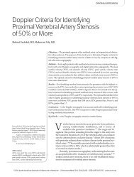
Figure 1. Left panel, Typical pulsed wave <str<strong>on</strong>g>Doppler</str<strong>on</strong>g> spectrum of a normal VA showing a systolic peak with c<strong>on</strong>tinuous flow in diastole.<br />
The end-diastolic velocity is 12.7 cm/s. This is a typical <str<strong>on</strong>g>Doppler</str<strong>on</strong>g> waveform of an arterial vessel supplying a low-resistance bed, such as<br />
the brain. Right panel, Abnormal HR VA. There is a rapid rise to a sharp systolic peak followed by a rapid descent in flow velocity back<br />
to baseline. There is no antegrade flow at end diastole (end-diastolic velocity is 0). Although this is a more typical <str<strong>on</strong>g>Doppler</str<strong>on</strong>g> waveform<br />
of an artery supplying a high-resistance vascular bed, such as the resting limbs, it is a pathologic finding in the evaluati<strong>on</strong> of the VA<br />
and may indicate cephalad vertebrobasilar occlusive disease.<br />
J Ultrasound Med 2010; 29:1161–1165
Two investigators independently reviewed the<br />
final reports of the correlative neuroimaging<br />
studies to categorize the vertebrobasilar system<br />
into <strong>on</strong>e of the following: (1) a normal VA; (2) at<br />
least moderate distal VA or basilar artery (BA)<br />
stenosis, occlusi<strong>on</strong>, or dissecti<strong>on</strong>; (3) a c<strong>on</strong>genitally<br />
diminutive VA; or (4) other vertebrobasilar<br />
abnormalities (proximal stenosis, excessive<br />
tortuosity, fibromuscular dysplasia, and BA<br />
hypoplasia). In cases of disagreement, a third<br />
investigator independently reviewed the neuroimaging<br />
reports and acted as a tie breaker.<br />
All statistics were performed using Stata software<br />
(StataCorp LP, College Stati<strong>on</strong>, TX). 5<br />
<str<strong>on</strong>g>Resistive</str<strong>on</strong>g> indices (RIs) of HR and low-resistive<br />
(LR) VA waveforms were calculated by dividing<br />
the difference in peak systolic velocity (PSV)<br />
and end-diastolic velocity (EDV) by the PSV<br />
[(PSV – EDV)/PSV]. 6 Mean velocities and RIs<br />
between HR and LR VA waveforms were compared<br />
using the Student t test. Analysis of radiographic<br />
correlati<strong>on</strong>s according to the EDV of the<br />
<str<strong>on</strong>g>Doppler</str<strong>on</strong>g> waveforms (EDV of 0 or EDV >0) was<br />
d<strong>on</strong>e using χ 2 testing. All statistical testing was<br />
performed with a significance level of P = .05.<br />
Results<br />
There were 1338 studies with HR VA waveforms<br />
noted. Of these, 86 studies met all inclusi<strong>on</strong> criteria.<br />
Of the 172 possible VAs to be analyzed in these<br />
86 studies, 4 VAs were not able to be visualized<br />
during the ultras<strong>on</strong>ographic examinati<strong>on</strong>s, and<br />
11 VAs were not adequately assessed by neuroimaging,<br />
resulting in 79 complete studies (n =<br />
157 VAs). Of the 157 VA waveforms with correlative<br />
neuroimaging, n<strong>on</strong>e were repeated studies in<br />
the same patient. The mean age of the patients<br />
was 69.1 years, and 66.6% were male. Correlative<br />
neuroimaging included catheter-based angiography<br />
(23.3%), CTA (16.3%), MRA (46.5%), or more<br />
than 1 modality (14.0%).<br />
Table 1. Velocities in the VAs by HR and LR <str<strong>on</strong>g>Waveform</str<strong>on</strong>g>s<br />
Of the 157 VA waveforms in the 79 studies, 90<br />
VA waveforms were HR; 13.9% of patients (n = 11)<br />
had bilateral HR waveforms. Ultras<strong>on</strong>ographic<br />
findings are shown in Table 1. High-resistive VA<br />
waveforms were equally distributed between the<br />
right and left sides (53.3% versus 46.7%, respectively;<br />
P not significant). The mean EDV am<strong>on</strong>g<br />
HR VA waveforms was 4.6 cm/s (range, 0–89.0<br />
cm/s). High-resistive VA waveforms had significantly<br />
lower mean PSVs and mean EDVs when<br />
compared with LR VA waveforms. As expected,<br />
the mean RI of HR VAs was significantly higher<br />
than that of those with normal <str<strong>on</strong>g>Doppler</str<strong>on</strong>g> waveforms<br />
(0.92 versus 0.73; P < .001).<br />
Of all HR VAs with correlative neuroimaging<br />
studies, 18.9% were normal (false-positive HR<br />
waveforms); 38.9% had at least moderate distal<br />
vertebrobasilar disease; 35.6% had c<strong>on</strong>genitally<br />
diminutive VAS; and 6.7% had other abnormalities<br />
(proximal VA stenosis, excessive tortuosity,<br />
fibromuscular dysplasia, or BA hypoplasia).<br />
When categorized by EDV of 0 versus greater<br />
than 0 (mean EDV 10.8 cm/s), HR VAs with 0<br />
end-diastolic flow were no more likely to be<br />
reflective of vertebrobasilar disease <strong>on</strong> neuroimaging<br />
(Table 2); however, the sample size for<br />
this determinati<strong>on</strong> was low.<br />
Discussi<strong>on</strong><br />
Although c<strong>on</strong>venti<strong>on</strong>al angiography remains the<br />
reference standard for assessing the vertebrobasilar<br />
circulati<strong>on</strong>, it is invasive and reserved<br />
for patients for whom there is a str<strong>on</strong>g neurologic<br />
indicati<strong>on</strong>. Although previous studies have<br />
shown a correlati<strong>on</strong> between <str<strong>on</strong>g>Doppler</str<strong>on</strong>g> ultras<strong>on</strong>ography<br />
of the VAs and angiographic findings,<br />
1,3,4 to our knowledge, ours is the largest to<br />
investigate the correlati<strong>on</strong> between a single<br />
<str<strong>on</strong>g>Doppler</str<strong>on</strong>g> waveform of the V2 segment of the VA<br />
and correlative neuroimaging in an unselected<br />
patient populati<strong>on</strong> referred for CDU.<br />
Parameter HR (n = 90) LR (n = 67) P<br />
Mean PSV (range), cm/s 51.7 (15–180) 63.6 (19–276) .04<br />
Mean EDV (range), cm/s 4.6 (0–89) 17.3 (0–120)
High-<str<strong>on</strong>g>Resistive</str<strong>on</strong>g> <str<strong>on</strong>g>Vertebral</str<strong>on</strong>g> <str<strong>on</strong>g>Artery</str<strong>on</strong>g> <str<strong>on</strong>g>Doppler</str<strong>on</strong>g> <str<strong>on</strong>g>Waveform</str<strong>on</strong>g><br />
1164<br />
Because of the differing modes of neuroimaging<br />
we used as the comparator studies (CTA,<br />
MRA, and angiography), calculati<strong>on</strong> of the sensitivity<br />
or specificity of a single VA waveform to<br />
detect vertebrobasilar disease was not performed.<br />
A previously published ultras<strong>on</strong>ographicangiographic<br />
correlati<strong>on</strong> study of 58 patients<br />
(116 VAs) by Nicolau et al 4 found that <str<strong>on</strong>g>Doppler</str<strong>on</strong>g><br />
ultras<strong>on</strong>ography of the intertransverse V2 segment<br />
of the VA had sensitivity of 90%, specificity<br />
of 100%, a positive predictive value of 100%, and<br />
a negative predictive value of 95% for the detecti<strong>on</strong><br />
of disease at any level of the vertebrobasilar<br />
circulati<strong>on</strong>. This study, however, was limited to<br />
ultras<strong>on</strong>ographic investigati<strong>on</strong> of patients with<br />
stroke symptoms who were referred by general<br />
or stroke neurologists for evaluati<strong>on</strong> of the<br />
extracranial circulati<strong>on</strong>. Although our study did<br />
not include as detailed an ultras<strong>on</strong>ographic<br />
investigati<strong>on</strong> of the VAs as the study by Nicolau et<br />
al 4 (a spectral <str<strong>on</strong>g>Doppler</str<strong>on</strong>g> waveform obtained at <strong>on</strong>e<br />
site in the VA in our study compared with investigati<strong>on</strong><br />
of the course of the VA in the V2 segment<br />
by taking <str<strong>on</strong>g>Doppler</str<strong>on</strong>g> readings in several vertebral<br />
interspaces in the study by Nicolau et al), we did<br />
find that an HR appearance of a single VA<br />
<str<strong>on</strong>g>Doppler</str<strong>on</strong>g> waveform in an unselected populati<strong>on</strong><br />
was associated with documented vascular<br />
abnormalities <strong>on</strong> correlative neuroimaging in a<br />
significant proporti<strong>on</strong> of cases (46%). Although it<br />
is not a pathologic finding, c<strong>on</strong>genital n<strong>on</strong>dominance<br />
accounted for most of the remaining<br />
waveforms.<br />
Table 2. Radiographic Correlati<strong>on</strong>s of HR VA <str<strong>on</strong>g>Doppler</str<strong>on</strong>g> Signals<br />
The high false-positive rate of 18.9% may have<br />
been due to several factors, including the qualitative<br />
assessment of VA <str<strong>on</strong>g>Doppler</str<strong>on</strong>g> waveforms into HR<br />
and LR categories and the lack of a single reference<br />
standard neuroimaging comparator (angiography).<br />
Additi<strong>on</strong>al analyses could be performed<br />
using the RI as the discriminator between HR and<br />
LR waveforms, but our laboratory does not have<br />
validated RI standards for assessing the VA. Future<br />
correlati<strong>on</strong> studies may also c<strong>on</strong>sider stratifying<br />
patients according to the indicati<strong>on</strong> for ordering<br />
the ultras<strong>on</strong>ographic examinati<strong>on</strong> (neurologic<br />
symptoms versus other reas<strong>on</strong>s) and using a<br />
quantitative definiti<strong>on</strong> of HR and LR waveforms<br />
in additi<strong>on</strong> to qualitative assessment in an effort<br />
to increase the performance characteristics of VA<br />
<str<strong>on</strong>g>Doppler</str<strong>on</strong>g> waveform analysis to detect vertebrobasilar<br />
disease.<br />
Notable weaknesses in our study were the<br />
review of the neuroimaging study reports, rather<br />
than independent review of the actual images,<br />
and assessment of VA <str<strong>on</strong>g>Doppler</str<strong>on</strong>g> waveforms <strong>on</strong>ly<br />
by the performing technologist and interpreting<br />
clinician before being entered into the clinical<br />
database without an independent sec<strong>on</strong>d physician<br />
review.<br />
Despite these limitati<strong>on</strong>s, we found that a significant<br />
proporti<strong>on</strong> of VAs with an HR <str<strong>on</strong>g>Doppler</str<strong>on</strong>g><br />
waveform <strong>on</strong> CDU were associated with vertebrobasilar<br />
disease, and when accompanied by<br />
clinical findings, the appearance of an HR VA<br />
<str<strong>on</strong>g>Doppler</str<strong>on</strong>g> waveform <strong>on</strong> CDU may warrant additi<strong>on</strong>al<br />
neuroimaging. These data emphasize the<br />
importance of careful review of the VA <str<strong>on</strong>g>Doppler</str<strong>on</strong>g><br />
waveform for its morphologic characteristics as<br />
well as the directi<strong>on</strong>ality of blood flow.<br />
Vertebrobasilar<br />
C<strong>on</strong>genitally Occlusive Other<br />
Parameter Normal, n (%) Diminutive, n (%) Disease, n (%) a Abnormalities, n (%) b Overall P<br />
LR (n = 67) 46 (68.7) 3 (4.5) 10 (14.9) 8 (11.9) 0 cm/s (n = 38) 8 (21.1) 14 (36.8) 11 (29.0) 5 (13.2)<br />
Findings of vertebrobasilar neuroimaging by EDV <strong>on</strong> CDU. High-resistive VAs with an EDV of 0 (no flow at end diastole) were no more likely<br />
to have significant vertebrobasilar disease than those with an EDV of greater than 0 cm/s (mean EDV for HR VAs with an EDV >0 cm/s,<br />
10.8 cm/s; P not significant).<br />
a Includes at least moderate distal VA or BA stenosis, occlusi<strong>on</strong>, or dissecti<strong>on</strong>.<br />
b Includes proximal stenosis, excessive tortuosity, fibromuscular dysplasia, and BA hypoplasia.<br />
J Ultrasound Med 2010; 29:1161–1165
References<br />
1. Biedert S, Winter R, Staudacher T, Betz H, Reuther R. <str<strong>on</strong>g>Doppler</str<strong>on</strong>g><br />
s<strong>on</strong>ography in basilar artery occlusi<strong>on</strong>. Neuroradiology 1985;<br />
27:430–433.<br />
2. Intersocietal Commissi<strong>on</strong> for the Accreditati<strong>on</strong> of Vascular<br />
Laboratories. ICAVL standards for accreditati<strong>on</strong> in n<strong>on</strong>invasive<br />
vascular testing, part II. Vascular laboratory operati<strong>on</strong>s:<br />
extracranial cerebrovascular testing. Intersocietal Commissi<strong>on</strong><br />
for the Accreditati<strong>on</strong> of Vascular Laboratories website;<br />
2007. http://www.icavl.org/icavl/pdfs/extracranial2007.pdf.<br />
3. Vis<strong>on</strong>à A, Lusiani L, Castellani V, R<strong>on</strong>sisvalle G, B<strong>on</strong>anome<br />
A, Pagnan A. The echo-<str<strong>on</strong>g>Doppler</str<strong>on</strong>g> (duplex) system for the<br />
detecti<strong>on</strong> of vertebral artery occlusive disease: comparis<strong>on</strong><br />
with angiography. J Ultrasound Med 1986; 5:247–250.<br />
4. Nicolau C, Gilabert R, Chamorro A, Vázquez F, Bargalló N,<br />
Brú C. <str<strong>on</strong>g>Doppler</str<strong>on</strong>g> s<strong>on</strong>ography of the intertransverse segment<br />
of the vertebral artery. J Ultrasound Med 2000; 19:47–53.<br />
5. StataCorp LP. Intercooled Stata 9.0 for Windows. College<br />
Stati<strong>on</strong>, TX: StataCorp LP; 2005.<br />
6. Pourcelot L. Applicati<strong>on</strong>s cliniques de l’examen <str<strong>on</strong>g>Doppler</str<strong>on</strong>g><br />
transcutane. In: Per<strong>on</strong>neau P (ed). Velocimetrie Ultras<strong>on</strong>ore<br />
<str<strong>on</strong>g>Doppler</str<strong>on</strong>g>. Vol 34. Paris, France; Institut Nati<strong>on</strong>al de la Santé<br />
et de la Recherche Médicale; 1974:780–785.<br />
Kim et al<br />
J Ultrasound Med 2010; 29:1161–1165 1165