Inhibition of Contractile Vacuole Function by ... - Universität zu Köln
Inhibition of Contractile Vacuole Function by ... - Universität zu Köln
Inhibition of Contractile Vacuole Function by ... - Universität zu Köln

You also want an ePaper? Increase the reach of your titles
YUMPU automatically turns print PDFs into web optimized ePapers that Google loves.
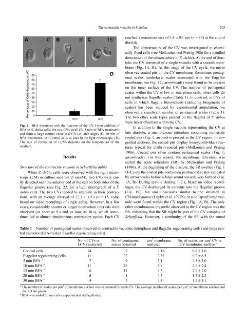
Fig. 2 BFA interferes with the function <strong>of</strong> the CV. Upon addition <strong>of</strong><br />
BFA to S. dubia cells, the two CVs swell (B, 5 min <strong>of</strong> BFA treatment)<br />
and form a large central vacuole (LCV) at later stages (C, 10 min <strong>of</strong><br />
BFA treatment). (A) Control cells as seen in the light microscope. (D)<br />
The rate <strong>of</strong> formation <strong>of</strong> LCVs depends on the temperature <strong>of</strong> the<br />
medium.<br />
Results<br />
Structure <strong>of</strong> the contractile vacuole in Scherffelia dubia<br />
When S. dubia cells were observed with the light microscope<br />
(LM) in culture medium (5 mosM), two CVs were easily<br />
detected near the anterior end <strong>of</strong> the cell on both sides <strong>of</strong> the<br />
flagellar groove (see Fig. 2A for a light micrograph <strong>of</strong> a S.<br />
dubia cell). The two CVs tended to alternate in their contractions,<br />
with an average interval <strong>of</strong> 22.3 ± 3.5 s (n = 15, value<br />
based on video recordings <strong>of</strong> single cells). However, in a few<br />
cases, considerably shorter or longer contraction intervals were<br />
observed (as short as 9 s and as long as 30 s), which sometimes<br />
led to almost simultaneous contraction cycles. Each CV<br />
The contractile vacuole <strong>of</strong> S. dubia 203<br />
reached a maximum size <strong>of</strong> 1.4 ± 0.1 µm (n = 15) at the end <strong>of</strong><br />
diastole.<br />
The ultrastructure <strong>of</strong> the CV was investigated in chemically<br />
fixed cells (see Melkonian and Preisig 1986 for a detailed<br />
description <strong>of</strong> the ultrastructure <strong>of</strong> S. dubia). At the end <strong>of</strong> diastole,<br />
the CV consisted <strong>of</strong> a single vacuole with a smooth membrane<br />
(Fig. 1A, B). At this stage <strong>of</strong> the CV cycle, we never<br />
observed coated pits on the CV membrane. Sometimes pentagonal<br />
scales (underlayer scales associated with the flagellar<br />
membrane, see Fig. 1C, arrowheads) were found to be present<br />
on the inner surface <strong>of</strong> the CV. The number <strong>of</strong> pentagonal<br />
scales within the CV is low in interphase cells, when cells do<br />
not synthesize flagellar scales (Table 1). In contrast, in CVs <strong>of</strong><br />
cells in which flagella biosynthesis (including biogenesis <strong>of</strong><br />
scales) has been induced <strong>by</strong> experimental amputation, we<br />
observed a significant number <strong>of</strong> pentagonal scales (Table 1).<br />
The two other scale types present on the flagella <strong>of</strong> S. dubia<br />
were never observed within the CV.<br />
In addition to the single vacuole representing the CV at<br />
late diastole, a membranous reticulum containing numerous<br />
coated pits (Fig. 1, arrows) is present in the CV region. In tangential<br />
sections, the coated pits display honeycomb-like structures<br />
typical for clathrin-coated pits (Melkonian and Preisig<br />
1986). Coated pits <strong>of</strong>ten contain pentagonal scales (Fig. 1,<br />
arrowheads). For this reason, the membrane reticulum was<br />
called the scale reticulum (SR) <strong>by</strong> Melkonian and Preisig<br />
(1986). At the beginning <strong>of</strong> the diastole, the SR swelled (Fig. 1<br />
D–I; note the coated pits containing pentagonal scales indicated<br />
<strong>by</strong> arrowheads) before a large round vacuole was formed (Fig.<br />
1A, B). During systole (lasting 1–2 s, based on video recordings),<br />
the CV discharged its contents into the flagellar groove<br />
(Fig. 1K). No small vacuoles similar to the situation in<br />
Chlamydomonas (Luykx et al. 1997b), or a collapsed large vacuole<br />
were found within the CV region (Fig. 1A, B). The only<br />
other membranous organelle observed in the CV region was the<br />
SR, indicating that the SR might be part <strong>of</strong> the CV complex <strong>of</strong><br />
Scherffelia. However, a continuity <strong>of</strong> the SR with the round<br />
Table 1 Number <strong>of</strong> pentagonal scales observed in contractile vacuoles (interphase and flagellar regenerating cells) and large central<br />
vacuoles (BFA-treated flagellar regenerating cells).<br />
No. <strong>of</strong> CVs or<br />
LCVs analysed<br />
No. <strong>of</strong> pentagonal<br />
scales observed<br />
µm 2 membrane<br />
analysed<br />
No. <strong>of</strong> scales per µm 2 CV or<br />
LCV membrane surface a<br />
Control cells 14 2 3.18 0.6 ± 1.6<br />
Flagellar regenerating cells 11 22 2.33 9.2 ± 6.5<br />
5min BFA b 7 9 5.1 4.8 ± 3.0<br />
10 min BFA b 11 25 6.9 3.6 ± 2.4<br />
15 min BFA b 6 11 4.3 2.9 ± 2.0<br />
20 min BFA b 6 8 4.5 1.5 ± 2.3<br />
30 min BFA b 4 5 3.3 1.5 ± 1.1<br />
a 2 2<br />
The number <strong>of</strong> scales per µm <strong>of</strong> membrane surface was calculated for each CV. The average number <strong>of</strong> scales per µm <strong>of</strong> membrane surface and<br />
the SD are given.<br />
b<br />
BFA was added 20 min after experimental deflagellation.